Leukemia: AML, CML, ALL and CLL - WWW.RN.ORG
←
→
Page content transcription
If your browser does not render page correctly, please read the page content below
Leukemia: AML, CML, ALL and CLL
                         WWW.RN.ORG®
      Reviewed October, 2019, Expires October, 2021
Provider Information and Specifics available on our Website
            Unauthorized Distribution Prohibited
         ©2019 RN.ORG®, S.A., RN.ORG®, LLC
                  By Wanda Lockwood, RN, BA, MA

Purpose
The purpose of this course is to define leukemia, describe current
treatment, and describe the characteristics, symptoms, diagnosis, and
treatment for the 4 primary types of leukemia: AML, CML, ALL, and
CLL.

Goals
Upon    completion of this course, the healthcare provider should be able
to:
    •   Define leukemia.
    •   Discuss hematopoiesis of leukemia.
    •   Differentiate between acute and chronic forms.
    •   Describe at least 8 common symptoms of leukemia.
    •   Discuss normal and abnormal laboratory tests.
    •   Explain how to calculate the absolute neutrophil count.
    •   Discuss the implications of neutropenia.
    •   Discuss the normal platelet count and the implications of
        thrombocytopenia.
  •     Discuss the 3 common stages of chemotherapy.
  •     Explain the cell cycle and the difference between cell cycle-
        specific agents and cell cycle-non-specific agents.
  •     Discuss 10 different types of chemotherapeutic agents.
  •     Explain 3 types of radiation commonly used for leukemia.
  •     Discuss hematopoietic stem cell transplantation.
  •     Discuss acute myelogenous leukemia (AML), including
        characteristics, symptoms, diagnostic findings, and treatment
        considerations.
  •     Discuss subtypes of AML.
  •     Discuss chronic myelogenous leukemia (CML), including
        characteristics, symptoms, diagnostic findings, and treatment
        considerations.
  •     Discuss the 3 different phases of CML.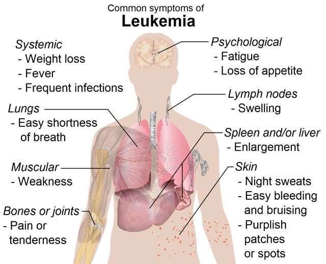
•   Discuss acute lymphocytic leukemia (ALL), including
       characteristics, symptoms, diagnostic findings, and treatment
       considerations.
   •   Discuss subtypes of ALL.
   •   Discuss chronic lymphocytic leukemia (CLL), including
       characteristics, symptoms, diagnostic findings, and treatment
       considerations.
   •   Discuss two classification systems for CLL.

Introduction
Leukemia is a group of malignant disorders affecting the blood and
blood-forming tissues in the bone marrow, lymphatic system, and
spleen. The word leukemia literally means “white blood” because it is a
neoplastic proliferation of one type of blood cell, typically a leukocyte
or white blood cell. Leukocytosis, an increased white blood cell count,
is a normal response to infection, but when leukocytosis becomes
chronic or progressively elevates without obvious cause, then it may
indicate malignancy.

In 2010, approximately 43,050 men and women (24,690 men and
21,840 women) in the United States were diagnosed with leukemia,
and 21,840 died of the disease. Leukemia accounts for 33% of
cancers in children and 1340 deaths yearly, so it is often thought of as
a disease of childhood. In children, the highest incidence is between 1
to 4 and the highest death rate between 10 and 19. However,
incidence is 10 times higher in adults. The median age for diagnosis is
66, and median age of death from leukemia is 74.

Despite much research into leukemia, the cause is often elusive.
Leukemia appears to result from a combination of factors, which can
include genetic predisposition, chromosomal changes, chemical agents
(benzene), chemotherapeutic agents, radiation, immunocompromise,
and viruses. Although viruses have been tied to leukemias in animals,
only adult T cell leukemia is known to result from a virus, human T cell
leukemia virus type 1 (HTLV-1).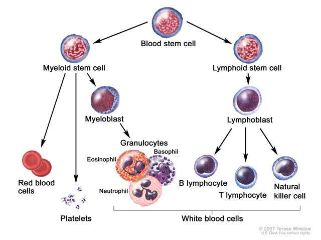
Regardless of the type of leukemia, the abnormal cells in the bone
marrow depress production of other cells, resulting in a number of
adverse effects:
  • Anemia occurs as erythrocyte (red blood cell) production falls.
  • Risk of infection occurs if neutrophil count decreases.
  • Clotting factors decrease, increasing risk of bleeding as
     thrombocytopenia (reduced platelet count) occurs.
  • Risk of physiological fracture increases as periosteum weakens
     because of proliferation of cells in the bone marrow.
  • Hypertrophy and fibrosis may occur in other organs, such as the
     liver, spleen, and lymph glands because of infiltration of
     malignant cells.
  • Increased intracranial pressure, ventricular dilation, and irritation
     of the meniges with resultant headache, vomiting, papilledema,
     nuchal rigidity, coma, and death can occur from infiltration of
     malignant cells into the central nervous system.
  • Hypermetabolic state deprives cells of nutrients and causes loss
     of appetite, weight loss, general fatigue, and muscle atrophy.

Classification
Hematopoiesis, the process by which blood cells are formed, involves
production of specific cells from stem cell precursors according to body
needs. In leukemia, a defect occurs in the myeloid or the lymphoid
stem cell. The most common feature of all types of leukemia is
unregulated proliferation of leukocytes in the bone marrow.

Leukemias are classified as lymphoid or myeloid, depending on the
affected stem cell type. Usually leukemias classified as blast cell or
stem cell refer to lymphoid defects. Additionally, leukemias are
classified as acute or chronic.

The 4 primary types of leukemia are acute lymphocytic/lymphoblastic
leukemia (ALL), acute myelogenous leukemia (AML) or acute
nonlymphoblastic leukemia (ANLL), chronic myelogenous leukemia
(CML), and chronic lymphocytic leukemia (CLL) with a number of
subtypes. Leukemias are often referred to by a number of different
names, depending on the involved cell, and this can be confusing at
times.

Acute and chronic forms differ in cell maturity and onset:
  • Acute forms: Onset is often abrupt, within weeks, and death
      may occur within weeks to months without treatment.
      Proliferation of abnormal cells leaves little room for normal cell
      production. Cells may proliferate in the liver and spleen as well,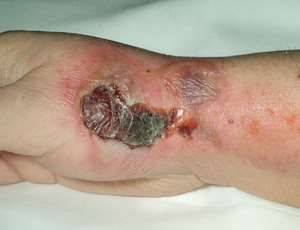
and they may infiltrate other organs, such as the meninges,
        gums, lymph nodes, and skin.

        Typically, leukocyte development halts at the blast phase, so
        most affected leukocytes are undifferentiated or blasts. With
        acute leukemia, the white blood count may remain low because
        the cells are halted at the blast stage. Acute forms may occur in
        both adults and children.

   •    Chronic forms: Onset is much slower, often over months or
        years. Normal cell production may occur as well for a long period
        of time, but in late stages of chronic disease, the abnormal cells
        interfere with normal cell production. The majority of leukocytes
        are mature. Chronic forms are rare in children.

Laboratory tests
Leukocytes are white blood cells (WBCs). Normal total values vary
according to age. The white count usually increases with infection and
returns to normal when infection subsides.

Leukocyte count
Total       1-3 years: 6000 to 17,500
            8-13 years: 4500 to 13,500
            Adult:      4500 to 11,000

The differential is the percentage of each type of leukocyte out of the
total. An increase in the white blood cell count is usually related to an
increase in one type and often an increase in immature neutrophils,
known as bands, referred to as a “shift to the left,” generally an
indication of an infectious process.
   • Normal WBC for adults: 4,500-11,000.
   • Acute infection: 11,000+, 30,000 indicates a severe infection.
   • Viral infection: 4,000 and below.

Because the proportion of most leukocytes is small, only an increase in
neutrophils or lymphocytes is usually significant enough to result in
increased total white count with leukemia.

Differential
Cell type                  1 yr            10 yrs             Adult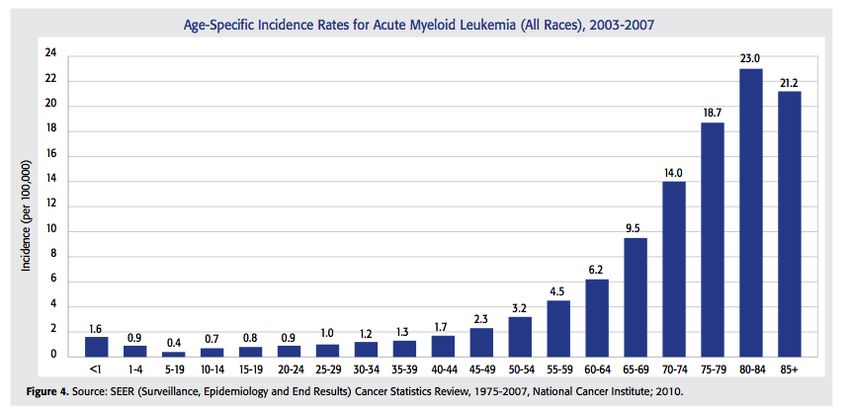
Neutrophils (Total)       31%             54%                59%
Neutrophils (bands)       3.1%            3%                 3%
Neutrophils               28%             51%                58%
(segments)
Absolute neutrophil       1500 to 8500    1800 to 8000       1800 to
count (ANC)                                                  7700
Lymphocytes               61%             38%                34%
Monocytes                 4.8%            4.3%               4%
Eosinophils               2.6%            2.4%               2.7%
Basophils                 0.4%            0.5%               0.5%

Absolute neutrophil count (ANC) is an important measure used to
determine the degree of neuotropenia. The total ANC should be about
1800 to 2000 mmm3 or higher. Risk of infection is significant if the
level falls to 1000 and severe at 500. ANC is not calculated directly but
is determined from the total white count and the percentages of
neutrophils and bands:

   •   ANC = Total WBC X (% neutrophils + % bands/100

Using this formula, if the white blood count is 3200 with 70%
neutrophils and 3% bands, the calculation would indicate that despite
the low WBC count, the patient does not have neutropenia:
   • ANC = 3000 X 73/100 = 2190

If the white count is 5300 with 10% neutrophils and 1% bands,
neutropenia is evident despite the normal WBC:
    • ANC = 5300 X 11/100 = 583

Why is neutropenia important?
The ANC falls with increased destruction of neutrophils or decreased
production. With leukemia, neutropenia is a direct result of both the
leukemic disease process and chemotherapy used to treat it, so it is a
primary concern during therapy.

Neutropenia increases the risk of both exogenous (environmental) and
endogenous (GI tract, skin) infection, depending on the severity and
duration. A short-lasting severe neutropenia may pose less actual risk
than a longer mild neutropenia.

There are no specific symptoms related to neutropenia, so without
regular blood testing, it may go undiagnosed until infection occurs. In
some cases, chemotherapy is withheld or reduced until the ANC rises,
but this puts the patient at increased risk from the malignancy,
especially if the treatment has a potential for cure. In that case,
growth factors (G-CSF or CM-CSF) may be administered to stimulate
production. As the bone marrow recovers, neutrophil production may
increase again.
Patients who exhibit both neutropenia and fever usually have an
infection and are hospitalized for broad-spectrum intravenous
antibiotic therapy. Cultures of blood, urine, and sputum and chest x-
ray are done to try to determine the site of infection and the
pathogenic agent. When cultures and sensitivities are completed, the
antibiotic regimen may be modified. Patients receiving antibiotics must
be carefully monitored for superinfection, especially fungal.
Oral moniliasis
Maintaining adequate nutrition is necessary as decreased protein
stores impair the immune system. Invasive procedures, such as IVs,
increase risk of infection.
Measures to prevent infection in those with neutropenia include:
• Monitor blood count daily and temperature every 4 hours,
notifying physician if temperature >38C (101F).
• Prohibit visitors with illness, including cold or sore throat.
• Provide private room if ANC• Provide oral hygiene after meals and every 4 hours during
daytime, avoiding lemon glycerine swabs, mouthwash, and
hydrogen peroxide.
• Avoid plastic cannulas for peripheral IVs if ANC• Reduced platelet production (which can occur with acute
leukemias and chemotherapy): Platelet transfusions may
increase platelet count.
• Increased platelet destruction (which can occur with CLL):
The bone marrow shows increased megakaryocytes (cells from
which platelets develop) and normal or increased production of
platelets as the body attempts to compensate. If platelets are
being destroyed, platelet transfusions provide little relief as the
transfused platelets are also destroyed. In this case, treatment
is usually similar to that for idiopathic thrombocytopenia
purpura: Immunosuppressive agents, such as prednisone,
cyclophosphamide, azathioprine, and dexamethasone.
Standard treatment protocol
The protocol for treatment varies depending on
the type of leukemia. Generally, a combination
of drugs is given as this approach is more
effective than monotherapy. There are generally
3 stages to chemotherapy:
• Induction
• Consolidation
• Maintenance
Induction Patients are usually hospitalized for 4 to 6 weeks
during initial treatment. The purpose is to induce
remission with bone marrow clear of disease and bloodcounts within normal limit. During this time, chemotherapy eradicates
both leukemic cells and normal myeloid cells, so the person becomes
severely neutropenic, anemic, and thrombocytopenic, putting patients
at risk for severe infections and bleeding. Patients may develop
bacterial, fungal, and viral infections, and severe mucositis, which
causes diarrhea and impairs nutritional absorption.
Supportive care includes administering blood products, such as packed
red blood cells and platelets, providing antibiotics to treat infections,
Granulocytic growth factors (G-CSF or GM-CSF) may shorten the
period of neutropenia by stimulating the bone marrow to increase
production of leukocytes.
Consolidation After the patient has recovered from the effects of
induction, consolidation treatment is provided over
4 to 8 months, often with the same
chemotherapeutic agents used during induction but at lower dosages
in order to kill any remaining malignant cells. Intrathecal
chemotherapy may be administered concurrently as a prophylaxis to
prevent CNS involvement. This treatment may be done on an
outpatient basis with multiple treatment cycles.
Maintenance Continued treatment may be provided for up to 3
years with some types of leukemia but with less
intense chemotherapy in order to retain remission.
The patient is monitored closely for both progress and side effects with
weekly blood counts.
Relapse Sometimes people relapse after completing the 3 stages
of chemotherapy. When that occurs, re-induction may be
carried out, especially with children, usually using a
different protocol of drugs. Many drugs currently used to treat
leukemia, especially for relapses, are those in clinical trials.
Other treatments Additional treatments may be used, depending
on the severity of the disease and degree of
infiltration:
• Intrathecal chemotherapy is administered into the spinal fluid for
treatment of infiltration of the central nervous system.
• Radiation to the brain may be indicated in addition to intrathecal
chemotherapy with severe disease, especially in children when
infiltration poses danger to brain development.
• Bone marrow transplant or peripheral blood stem cell transplant
with donor stem cells or the patient’s stem cells.• Blood transfusions: packed red blood cells, platelets.
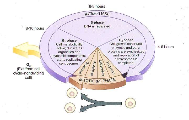
The cell cycle and chemotherapy
The purpose of chemotherapy is primarily to prevent replication of
malignant cells and to treat systemic disease, such as leukemia or
other metastasized cancers. Chemotherapy is used to cure, control, or
provide palliation, so medications are chosen based on the realistic
goal for the individual patient.
Cells that are actively proliferating are the most sensitive to
chemotherapy. Cells that are not dividing but have the potential for
proliferation are not destroyed by chemotherapy, so repeated cycles of
therapy are required in order to kill these cells as they become
activated. Cells go though predictable cell cycle patterns in which one
cell divides to become two daughter cells.
Cell phases:
• G1: RNA and protein synthesis occur.
• S: DNA synthesis occurs.
• G2: DNA synthesis is complete, mitotic spindle forms.
• M: Mitosis occurs with cell division.
• G0: Resting (inactive phase).
Classification Chemotherapeutic agents are classified by
relationship to the cell phase and by chemicalgroup.
Chemotherapeutic agents are classified by whether or not they target
a particular cell phase:
• Cell cycle-specific agents: Some chemotherapeutic agents are
classified according to which part of the cell cycle they target.
These drugs are referred to as cell cycle-specific agents because
they destroy cells that are actively reproducing by interfering
with this process. Most target S phase or M phase.
• Cell cycle-nonspecific agents: Some chemotherapeutic agents
act independent of the cell phase and have a longer effect,
resulting in damage or death of the malignant cell.
Chemotherapeutic agents are also classified according to their
chemical group, with each group providing a different mechanism of
action. There are many different agents in most groups (some
examples listed below).
Alkylating agents Cell cycle- Alter DNA structure, causing
(Busulfan, carboplatin, nonspecific breaks.
chlorambucil, cisplatin
dacarbazine, melamine,
ifosfamide)
Antimetabolites Cell cycle- Interfere with metabolites
(Methotrexate AKA MTX, specific (S) needed for RNA and DNA
cytarabine AKA synthesis.
arabinosylcytosine or
Ara-C, 5-fluoracil AKA 5-
FU, gemcitabine,
fludarabine, L-
asparaginase)
Antitumor antibiotics Cell cycle- Modify DNA function and
Anthracycline, non-specific interfere with RNA so cells
doxorubicin, idarubicin, die when they try to divide.
daunorubicin, mitomycin,
mitoxantrone,
dactinomycin, plicamycin,
mitomycin, bleomycin)
Corticosteroids Cell cycle- Disrupt cell membrane.
(Cortisone, non-specific Decrease circulating
hydrocortisone, lymphocytes.
dexamethasone) Depress immune system.
Hormonal agents Cell cycle Stimulate cellular
(Androgens, anastrozole, non-specific differentiation.raloxifene) Inhibit enzyme needed for
estrogen synthesis.
Estrogen antagonist
Miscellaneous agents Varies, Varies, depending on agent.
(L-asparaginase, depending
tamoxifen, procarbazine, on agent
arsenic trioxide, all-trans
retinoic acid AKA ATRA,
hydroxyurea)
Mitotic spindle poisons Cell cycle- Interfere with cellular
• Plant alkaloids specific (M) replication.
(epipodophyllotoxin,
etoposide*,
teniposide,
vinblastine,
vincristine)
• Taxanes (paclitaxel,
docetaxel)
Nitrosoureas Cell cycle- Alter DNA structure, causing
(Carmustine, lomustine, nonspecific breaks (like alkylating
semustine, streptozocin) agents) and able to cross
blood-brain barrier.
Topoisomerase Cell cycle- Cause breaks in DNA strand.
inhibitors specific (S)
• Type I (Irinotecan,
topotecan)
• Type II (Amsacrine,
etoposide, etoposide
phosphate, teniposide)
*Also classified as a Topoisomerase inhibitor
Targeted therapy Targeted therapy is a new approach to
chemotherapy in which characteristics that
differentiate cancer cells from normal cells are
targeted. There are 3 categories of targeted therapy:
• Varied targeted therapy: Small molecules enter cells and
disrupt cell function, causing the cells to die:
o Signal transduction inhibitors: Imatinib mesylate, gefitinib,
cetuximab, lapatinib.
o Biologic response modifier agent: Denileukin, Diftitox.
o Proteasome inhibitor: Bortezomib.
• Monoclonal antibodies: Target specific antigens (proteins) on
B or T-lymphocyte cancer cells so immune system can destroythem. Agents used for leukemia include rituximab, alemtuzumab
and ofatumumab.
• Angiogenesis inhibitors: Blood vessels that supply nutrients to
the cell are targeted in order to starve the cells.
Protocol In designing a treatment protocol, often combinations of
drugs are used. For example, a protocol may call for the
following:
• Antimetabolite: Cytarabine (cell cycle-specific targeting S
phase).
• Antitumor antibiotic: Daunorubicin (cell cycle-non-specific).
• Mitotic spindle poison: Etoposide (cell cycle-specific targeting M
phase).
Most chemotherapeutic agents have adverse effects, often severe,
depending upon the particular agent. Common adverse effects include:
• Bone marrow suppression.
• Nausea and vomiting.
• Thrombocytopenia.
• Neutropenia.
• Neuropathies.
• Anorexia.
• Hepatic, renal, and/or cardiac toxicity.
• Hair loss.
• Stomatitis.
• Renal toxicity.
• Diarrhea.
ALERT
Healthcare providers should always be aware of specific guidelines
for the administration of chemotherapeutic agents as they are often
highly toxic and may be absorbed through inhalation or contact with
skin. OSHA provides guidelines for proper handling of
chemotherapeutic agents.
Extravasation Chemotherapy for leukemia is usually
administered intravenously although some types
may be given orally. Patients receiving intravenous
chemotherapy must be monitored carefully as the agents may irritate
vessel walls, and extravasation (infiltration) may result in severe painand local tissue damage, as many agents are vesicants that can cause necrosis. Early signs of extravasation include swelling, redness, itching, and vesicles on the skin. Most patients experience pain initially, but some do not. If the agent is a vesicant, the tissue begins to ulcerate within a few days and may create a large open ulcer that requires skin grafting, so monitoring the IV and instructing patients about the signs of extravasation are critical. If extravasation does occur, the medication should be stopped immediately. The site should be assessed and measured. Photographing the site is also helpful for reference if the tissue deteriorates. Local cooling is used for DNA-binding agents (anthracyclines such as doxorubicin, daunorubicin, epirubicin, idarubicin) to constrict blood vessels while local heat is used for non- DNA binding agents (vinca alkaloids such as vincristine, vinblastine) to increase blood flow. Specific antidotes may be available for some chemotherapeutic agents. A vascular access device may be used for administration of chemotherapeutic agents, especially with combination therapies, but these pose an increased risk of systemic infection, especially if neutropenia occurs. Commonly used types of vascular access devices are the silastic right atrial catheter, peripherally inserted central venous catheter (PICC), midline catheter (MLC), implanted infusion ports, and infusion pumps (external or implanted). Radiotherapy and HCST
Radiotherapy       Radiation is used for all types of leukemia.
                    Radiotherapy is usually used in conjunction with
chemotherapy, but it may also be used as CNS prophylaxis to prevent
spread of acute leukemias to the brain and spinal cord. Cranial
irradiation lowers the risk of relapse in acute leukemias. Radiotherapy
may be used to relieve pain from hepatomegaly and splenomegaly.

Three types of radiation are commonly used with leukemia patients:
   • External beam radiation: EBRT is commonly used with CML
     and helps decrease swelling of liver, spleen, and lymph nodes.
   • Total body irradiation: A linear accelerator irradiates the
     entire body in preparation for chemotherapy and stem cell
     transplant.
   • Total marrow irradiation: This form of radiation targets major
     marrow sites and reduces radiation exposure to internal organs.
     TMI is used as preparation for stem cell transplant.

Stem cell transplantation
Hematopoietic stem cell transplantation (HSCT) comprises bone
marrow transplant (BMT), peripheral blood stem cell transplant
(PBSTC, and umbilical cord blood stem cell transplant (UCBSTC).

Sources of stem cells include:
  • Syngenic: From identical twin.
  • Autologous: From the patient. Autologous purged transplant
     requires treatment ex vivo to remove malignant cells prior to
     transplantation.
  • Allogenic: From a family member, usually a sibling. In some
     cases, partially-matched related donor may be used. Allogenic
     donors are usually the first choice.
  • Matched unrelated donor (MUD): Donors are usually matched
     through a donor registry. In some cases, partially matched cord
     blood stem cells may be used.

HCST allows for higher doses of chemotherapeutic agents because
there is less concern about marrow toxicity. Transplantation is usually
done after chemotherapy and radiation has ablated the patient’s bone
marrow. The transplanted cells then begin to produce normal blood
cells.
    • BMT is an invasive procedure that requires bone marrow to be
       extracted from the donor, usually from the pelvic bones. The
       extraction is done as an out-patient procedure under general or
regional anesthesia.
• PBSCT is less invasive for the donor, but stem cells are sparse
in the peripheral blood so it may be difficult to extract enough
from the bloodstream; therefore, the donor must take
medication (granulocyte colony-stimulating factor—G-CSF) for a
few days prior to donation to increase the number of stems cells
released from the bone marrow into the bloodstream.
• UCBSCT is not as readily available, but this type of transplant is
less prone to rejection and has lower incidence of graft vs host
disease. Only about 50 mL of cord blood is obtained with each
donation, so this amount is usually suitable only for
transplantation in small children. However, ex vivo expansion
techniques and combining cord donations from two donors are
being explored to allow UCBSCT for adults.
While HSCT may be lifesaving, it is not a benign procedure.
Complications of HSCT include:
• Toxicity from radiation and chemotherapy.
• Complications related to pancytopenia (bleeding, infection,
anemia).
• Immunological disorders (graft vs host disease, graft rejection,
immunodeficiency).
• Endocrine and growth abnormalities.
• Decreased fertility.
• Cataracts.
• Learning disabilities.
• Secondary malignancy.
Acute myelogenous leukemia (AML)
Acute myelogenous leukemia is also referred to a granulocytic,
myelocytic, myeloid, monocytic, myeloid, monoblastic,
monomyeloblastic, and acute nonlymphoblastic leukemia (ANLL). AML
is characterized by uncontrolled proliferation of myeloblasts, which are
precursors of granulocytes. AML is the second most common leukemia
in children, causing 17%, usually thosedisorder, familial and acquired monosomy 7, and severe aplastic
anemia.
AML is the most common adult leukemia, causing 85% of leukemias.
In adults, onset is usually after age 60, especially in males, and may
be associated with a history of smoking, previous radiation, and/or
chemotherapy. AML may result from childhood treatment of ALL or
other cancers. Most people who develop secondary cancers related to
treatment develop AML rather than other forms of leukemia.
Prognosis varies depending on the subtype, but 5-year survival rates
for children 60.
Symptoms Onset of symptoms is usually quite abrupt and may
include severe infection and abnormal bleeding.
Symptoms most often result from decreased
production of other blood cells because of the large number of
leukemic myeloblasts in the bone marrow although other organs may
become infiltrated. Neutropenia and thrombocytopenia are common.
Typical signs and symptoms include:
• Weakness, lethargy.
• Increased bruising, petechiae.
• Abnormal bleeding, nosebleeds.
• Fever.
• Anorexia, weight loss.
• Anemia.• Increased infection.
• Mouth sores.
With infiltration of the central nervous system through the blood or
lymphoid system, symptoms may include headache, vomiting, and,
papilledema, Sixth cranial nerve palsy results from masses of leukemic
cells putting pressure on the nerves, preventing the eye from moving
laterally. Other common sites for infiltration include the spinal cord
and testicles, which painlessly enlarge. The liver may become enlarged
and painful. Hyperplasia of the gums may occur. Proliferation in the
bone may cause bone pain as the bone marrow expands.
Complications can include bleeding and infection. Major hemorrhage
occurs when platelet count drops to 30%.
Treatment considerations Usually a two-phase approach to
chemotherapy is used with induction
and consolidation but not
maintenance. Treatment usually involves combination chemotherapy.
A common approach to adult induction is referred to as “3 and 7.”
• 3 days of 15 to 30 minutes of infusions with idarubicin or
daunorubicin or mitoxantrone followed by arabinosylcytosine
(ara-C) in 24-hour infusions daily for 7 days.
This is usually followed by consolidation treatment with high-dose ara-
C.
However, this treatment protocol may vary according to the patient’s
age and condition. In many cases, patients are enrolled in clinical
trials, especially with recurrent disease. Some may receive bone
marrow transplant (BMT) or peripheral blood stem cell transplants
(PBSCT).Treatment approaches are similar for all subtypes except acute
promyelocytic leukemia (M3), which is usually treated with arsenic
trioxide and all-trans retinoic acid (ATRA).
Subtypes Subtypes are classified with two different systems:
French, American, British (FAB) and World Health
Organization (WHO).
French, American, British (FAB): This is commonly used and is
based on the type of blood cell the leukemia originates from and the
maturity of leukemic cells. Using the FAB classification system, there
are 8 primary subtypes:
Subtype Name Discussion
M0 Undifferentiated acute Minimally-differentiated AML.
5% myeloblastic Prognosis is poor.
leukemia.
M1 Acute myeloblastic Dominant leukemic cells in the
15% leukemia with minimalmarrow at diagnosis are
maturation myeloblasts.
Prognosis is average.
M2 Acute myeloblastic Many myeloblasts evident but
25% leukemia with some are maturing into normal
maturation cells.
Prognosis is better than average.
M3 Acute promyelocytic Leukemic cells show translocation
10% leukemia between chromosomes 15 and 17.
Most common in middle-aged
adults and may cause bleeding
and blood clotting, so identifying
APL and using correct treatment is
critical.
Prognosis is best of AML subtypes.
M4 Acute myelomonocytic Leukemic cells often have
20% leukemia inversion of chromosome 16.
Prognosis is average.
M4 eos Acute myelomonocytic Prognosis is better than average.
5% leukemia with
eosinophilia
M5 Acute monocytic Leukemic cells have features of
10% leukemia developing monocytes.
Prognosis is average.
M6 Acute erythroid Leukemic cells have features of5% leukemia developing red blood cells.
Acute erythroid leukemia.
Prognosis is poor.
M7 Acute megakaryocytic Leukemic cells have features of
5% leukemia developing platelets.
Prognosis is poor.
World Health Organization (WHO): This newer classification divides
subtypes into broad groups based on expected outcomes.
• AML with certain genetic abnormalities:
o AML with a translocation between chromosomes 8 and 21
o AML with a translocation or inversion in chromosome 16
o AML with changes in chromosome 11
o APL (M3), which usually has translocation between
chromosomes 15 and 17
• AML with multi-lineage dysplasia (more than one abnormal
cell).
• Therapy-related AML.
• AML not otherwise specified:
o undifferentiated AML (M0)
o AML with minimal maturation (M1)
o AML with maturation (M2)
o acute myelomonocytic leukemia (M4)
o acute monocytic leukemia (M5)
o acute erythroid leukemia (M6)
o acute megakaryoblastic leukemia (M7)
o acute basophilic leukemia
o acute panmyelosis with fibrosis
o myeloid sarcoma (also known as granulocytic sarcoma or
chloroma)
• Undifferentiated or biphenotypic acute leukemias (having
both lymphocytic and myeloid features).
Chronic myelogenous leukemia (CML)
Chronic myelogenous leukemia is also referred to as chronic myeloid
leukemia, chronic myelocytic, and chronic granulocytic leukemia. CML
results from mutation of the myeloid stem cells and resultant
proliferation of mature neoplastic granulocytes and blast forms in the
bone marrow although some normal cells are still produced, so a wide
range of cell forms exists.Huge numbers of abnormal cells expand the bone marrow and spread
into the peripheral circulation and eventually infiltrate the liver and
spleen, where more cells are formed in a process referred to as
extramedullary hematopoiesis, causing the organs to enlarge.
In up to 95% of those with CML, these abnormal cells contain a
genetic marker that makes them distinct, the Philadelphia
chromosome (Ph1), a translocation between chromosomes 9 and 22.
The location of these changes is on the BCR gene of chromosome 22
and the ABL gene on chromosome 9. The two genes fuse and produce
an abnormal protein that increases the rate that leukocytes divide.
This BCR-ABL gene is present is almost everyone with CML.
Currently about 20,000 people in the United States have CML with
4870 people diagnosed in 2010 and 440 deaths in 2010. CML is rare in
people 1 year with treatment. In addition to the symptoms
experienced during the chronic phase, the patient mayexperience fever, night sweats, weight loss, anemia, and
dyspnea.
• Blast crisis: Blast cells begin to proliferate in large number and
production of other blood cells, including erythrocytes and
platelets, so the patient can experience bruising, bleeding, and
infection. During this phase the disease resembles AML in most
patients and ALL in a few.
Diagnostic findings CML is usually diagnosed with blood studies
that show a white count of 20,000 to 60,000
with increase in mature granulocytes.
Basophils and eosinophils show a mild increase. The mature
granulocytes exhibit a delay in normal apoptosis (programmed cell
death), so they live longer and begin to accumulate. These cells have
no or low levels of alkaline phosphatase, so stains show a low score.
Mild to moderate normochromic, normocytic anemia is usually present.
Platelet counts may vary from low to high. Bone marrow biopsy with
cytogenic studies provides definitive diagnosis, especially with a
finding of the Philadelphia (Ph1) chromosome.
Treatment considerations Treatment introduced during the
chronic phase of CML is more effective
than treatment started in more
advanced phases. The current drugs of choice include imatinib
mesylate (Gleevec), which promotes apoptosis and inhibits tyrosine
kinase activity in cells positive for BCR-ABL (Philadelphia). Remission
rates are 70% and survival of 94% at 3 years for those treated in the
chronic phase. Complete remission rates drop to 28% if treatment is
given during acceleration phase and 4% if during blast crisis.
Two newer generation drugs, nilotinib and dasatinib, shower higher
rates of remission but are associated with more side effects and are
more expensive. In up to 30% of patients, imatinib is not effective in
bringing about complete remission. In these cases, further treatment
with nilotinib or dasatinib may be used. Myelosuppressive therapy,
which was used before FDA approval of imatinib, is rarely used
currently. HCST may be considered in patientsthat differentiate into lymphocytes, preventing development of other types of cell. The cell of origin is the precursor to B lymphocytes in 75% and to T lymphocytes in 25%. This is the most common type of childhood leukemia (85%), peaking between ages 2 to 5 and rare after age 15 with incidence in males higher than females. Down syndrome increases risk. ALL accounts for only 15% of adult leukemias, often after age 70, especially in those with history of chemotherapy or radiation exposure. Five-year survival rates are 90.8% for children
Diagnostic findings     Because production of normal blood cells is
                         inhibited, laboratory testing usually shows
                         decreased numbers of leukocytes,
erythrocytes, and platelets. In some cases the leukocyte count is low
but with a high proportion of immature cells.

 LDH and uric acid levels are usually elevated. If DIC is present,
laboratory studies show an elevated prothrombin time, decreased
fibrinogen levels, and the presence of fibrin split products.

Chest x-ray may show pneumonia and mediastinal mass in those with
T lymphocyte ALL, and CT scan shows the extent of lymphadenopathy.
A bone marrow biopsy provides the definitive diagnosis.

 Treatment considerations      Because only about 20% of adults are
                               cured with standard chemotherapy,
                               many are entered into clinical trials.
The standard adult treatment protocol comprises 4 phases:

  •   Induction: A 4-drug regimen of vincristine, prednisone,
      anthracycline, and cyclophosphamide or L -asparaginase or a 5-
      drug regimen of vincristine, prednisone, anthracycline,
      cyclophosphamide, and L -asparaginase is given over the course
      of 4-6 weeks.
  •   Consolidation: Studies show varying results from consolidation
      treatment, but most studies show benefit. Regimens using a
      standard 4- to 5-drug induction usually include Ara-C in
      combination with an anthracycline or epipodophyllotoxin.
  •   Maintenance: While studies show that adults on maintenance
      have longer periods of remission, the definitive protocol has not
      been developed, so various treatment protocols are used,
      including a 4-drug regimen for 12 months.
  •   CNS prophylaxis: Meningeal infiltration is common with relapse,
      so intrathecal chemotherapy is necessary.

The pediatric treatment protocol comprises 5 phases:

  •   Induction: 3-4 drugs, which may include a glucocorticoid,
      vincristine, asparaginase, and possibly an anthracycline.
  •   Consolidation: drugs are given at doses higher than those used
      during induction or the patient is given different drugs (ie, high-
      dose MTX and 6-mercaptopurine [6-MP]), epipodophyllotoxins
      with Cytarabine (Ara-C), or multiagent combination therapy
• Interim maintenance: oral medications are administered to
maintain remission and allow the bone marrow to recover for 4
weeks.
• Delayed intensification: Intensified treatment for any remaining
leukemic cells that were resistant to previous phases.
• Maintenance: Intrathecal MTX every 3 months, monthly
vincristine, daily 6-MP and weekly MTX.
The duration of pediatric treatment varies depending on the type of
ALL. B-cell acute lymphoblastic leukemia is usually treated with two
months to 8 months of intensive therapy. However, those with B-
precursor and T-cell acute lymphoblastic leukemia require
approximately 2 to 2.5 years of continuation therapy to prevent high
relapse rates. In current acute lymphoblastic leukemia clinical trials,
the total duration of therapy for girls is 2 years from the start of
interim maintenance and for boys is 3 years from the start of interim
maintenance.
Subtypes The FAB classification identifies 3 subtypes of ALL,
based on the way the cells look:
Subtype Percentage Description
L1 30% of Small cells with homogeneous
adult cases chromatin, regular nuclear shape,
small or absent nucleolus, and scanty
85% of cytoplasm. Corresponds to T-cell or
pediatric pre-B cell.
L2 65% of Large and heterogeneous cells,
adult cases heterogeneous chromatin, irregular
nuclear shape, and nucleolus often
14% of large. Corresponds to T-cell or pre-B
pediatric cell.
L3 5% of adult Large and homogeneous cells with
cases multiple nucleoli, moderate deep blue
1% of cytoplasm, and cytoplasmic
pediatric vacuolization that often overlies the
nucleus (most prominent feature).
Corresponds to B cell.
Prognosis is poor.
The WHO classification bases subtypes on certain cell features and
maturity (adult percentages):
Subtype Description
B-cell • Early precursor-B ALL (also called pro-B ALL) -- about10% of cases.
• Common ALL - about 50% of cases
• Precursor-B ALL - about 10% of cases
• Mature B-cell ALL (Burkitt leukemia) -- about 4% of
cases
T-cell • Precursor-T ALL - about 5% to 10% of cases
• Mature T-cell ALL - about 15% to 20% of cases
Prognosis is usually better for T-cell ALL than B-cell. Mature B cell has
the worst prognosis. Other B-cell subtypes fall in the middle. Younger
patients have a better prognosis than older. People with a lower white
blood count (< 30,000 for B-cell ALL and 55. CLL is rareapoptosis so that they don’t die but accumulate in the marrow and
blood.
CLL is more aggressive in some patients, and these may live only 2 to
3 years after diagnosis, but most people live 5 to 10 years.
Thrombocytopenia and anemia are important negative variables,
suggesting a more aggressive course of the disease.
CLL is classified in 2 different ways, and treatment and prognosis
depends on the classification:
Rai Classification Binet Classification
0 Absolute lymphocytosis A No anemia or
3
(>15,000/mm ) without thrombocytopenia and
adenopathy, fewer than three areas
hepatosplenomegaly, anemia, or of lymphoid involvement
thrombocytopenia. (Rai stages 0, I, and II).
I Absolute lymphocytosis with B No anemia or
lymphadenopathy without thrombocytopenia with
hepatosplenomegaly, anemia, or three or more areas of
thrombocytopenia. lymphoid involvement
(Rai stages I and II).
II Absolute lymphocytosis with C Anemia and/or
either hepatomegaly or thrombocytopenia
splenomegaly with or without regardless of the
lymphadenopathy. number of areas of
lymphoid enlargement
(Rai stages III and IV).
III Absolute lymphocytosis and
anemia (hemoglobinin humor and cell-mediated immune systems. Infections, such as
herpes zoster, may become widespread. Patients may develop a group
of symptoms referred to as “B symptoms”:
• Fever.
• Diaphoresis, especially night sweats.
• Weight loss.
Lymph nodes typically are enlarged and painful as lymphocytes
become trapped in the nodes. Hepatomegaly and splenomegaly also
develop. Patients may complain of abdominal fullness and discomfort
and early satiety because of splenomegaly. Skin lesions may occur
with T-cell CLL.
Autoimmune complications are common and can occur at any stage of
the disease. About 10% of those with CLL present with autoimmune
hemolytic anemia. Some develop idiopathic thrombocytopenia purpura
(ITP). In these autoimmune disorders, the reticuloendothelial system
(RES) destroys the body’s erythrocytes or platelets.
Frequent infections occur, including herpes zoster, Pneumocystis
jiroveci, and Candid albicans. In some cases, CLL may transform to
diffuse large cell lymphoma (Richter syndrome), which carries a poor
prognosis.
Diagnostic findings The total WBC count increases and the
lymphocyte count may exceed 100,000.
Despite these numbers, because lymphocytes
are small, they are able travel through the capillaries so pulmonary
and cerebral complications that occur with myelogenous leukemias do
not usually occur with CLL. In later disease stages, reduced
erythrocyte count and thrombocytopenia occur. An antigen, CD52,
which is usually present on T-cells, is present on the surface of many
leukemic B cells.
Zeta-chain-associated protein kinase 70 (ZAP-70) is often used to
determine the need for treatment. A positive ZAP-70 finding in
asymptomatic patients (>30%) is associated with median survival of 6
to 10 years while a negative ZAP-70 is associated with median survival
of >15 years.
The immunoglobulin variable region heavy chain gene (IgVH) mutation
in significant numbers is associated with a median survival in excess of
20 to 25 years. The absence of mutations is associated with a median
survival of 8 to 10 years.Treatment considerations Because some types of CLL have a
long period with slow progression,
some older adults may fare better
with monitoring than with aggressive
treatment. CLL is usually not curable, so patients may be treated
symptomatically for complications, such as hemolytic anemia,
infections, or ITP.
Typically, patients with low risk or Binet A classification are simply
monitored because early chemotherapy has not been associated with
increased survival. Corticosteroids are commonly used to treat
autoimmune hemolytic anemia and thrombocytopenia.
Many chemotherapeutic agents, such as chlorambucil, have been used
to treat CLL and some clinical trials are in place for more aggressive
forms of CLL. The first line agent is fludarabine, which is either given
alone or in combination with cyclophosphamide and/or rituximab (a
monoclonal antibody). Alemtuzumab (a monoclonal antibody directed
at CD52) is approved for use in CLL as both a first-line agent and for
salvage in patients with fludarabine-refractory disease. Allogenic stem
cell transplant is the only know curative treatment.
Summary
Leukemia is a group of malignant disorders affecting the blood and
blood-forming tissues in the bone marrow, lymphatic system, and
spleen. Leukemias are classified as lymphoid or myeloid, depending on
the affected stem cell type, and may be acute or chronic. The 4
primary types of leukemia are acute lymphocytic/lymphoblastic
leukemia (ALL), acute myelogenous leukemia (AML) or acute
nonlymphoblastic leukemia (ANLL), chronic myelogenous leukemia
(CML), and chronic lymphocytic leukemia (CLL) with a number of
subtypes. Two primary concerns with leukemia are neutropenia, which
increases risk of infection, and thrombocytopenia, which increases risk
of bleeding. Treatment options include chemotherapy, radiotherapy,
and HSCT.
References
• Acute adult myeloid leukemia treatment. (2011, February 14).
National Cancer Institute. Retrieved March 25, 2011, from
http://www.cancer.gov/cancertopics/pdq/treatment/adultAML/he
althprofessional• Besa, EC. (2010, December 22). Chronic myelogenous leukemia.
eMedicine. Retrieved March 25, 2011, from
http://emedicine.medscape.com/article/199425-overview
• Blood and bone marrow donation. (2009, May 27). Mayo Clinic.
Retrieved March 25, 2010, from
http://www.mayoclinic.com/health/bone-marrow/MY00525
• Bone Marrow Transplantation and Peripheral Blood Stem Cell
Transplantation. (2010, September 24). National Cancer
Institute. Retrieved March 25, 2010, from
http://www.cancer.gov/cancertopics/factsheet/Therapy/bone-
marrow-transplant
• Childhood acute lymphoblastic leukemia treatment PDQ®).
(2011, March 4). National Cancer Institute. Retrieved March 25,
2011, from
http://www.cancer.gov/cancertopics/pdq/treatment/childALL/He
althProfessional/page1
• Childhood acute myeloid leukemia/Other myeloid malignancies
treatment. (PDQ®). (2010, October 20). National Cancer
Institute. Retrieved March 25, 2011, from
http://www.cancer.gov/cancertopics/pdq/treatment/childAML/He
althProfessional/page12
• Chronic lymphocytic leukemia treatment (PDQ®). (2010, May
18). National Cancer Institute. Retrieved March 25, 2010, from
http://www.cancer.gov/cancertopics/pdq/treatment/CLL/Patient/
page1
• Chronic myelogenous leukemia treatment (PDQ®). (2011,
February 28). National Cancer Institute. Retrieved March 25,
2011, from
http://www.cancer.gov/cancertopics/pdq/treatment/CML/Health
Professional
• Delong, L et al. (2011, February 11). Chronic lymphocytic
leukemia. eMedicine. Retrieved March 25, 2011, from
http://emedicine.medscape.com/article/199313-overview
• Schulmeister, L. (2008, January). Chemotherapy Extravasation.
AHRQ. Retrieved March 25, 2011, from
http://www.webmm.ahrq.gov/case.aspx?caseID=169
• Seiter, K. (2011, March 16). Acute lymphoblastic leukemia.
eMedicine. Retrieved March 25, 2011, from
http://emedicine.medscape.com/article/207631-overview
• Seiter, K. (2011, February 15). Acute myelogenous leukemia.
eMedicine. Retrieved March 25, 2011, from
http://emedicine.medscape.com/article/197802-overview• Smeltzer, SC, Bare, BG, Hinkle, JL, & Cheever, KH. (2008).
Brunner & Suddarth’s Textbook of Medical-Surgical Nursing, 11
ed., Philadelphia: Wolters Kluwer/Lippincott, Williams, & Wilkins.
• What is chemotherapy? (2005). Chemocare. Retrieved March 25,
2011, from http://www.chemocare.com/WHATIS/index.aspYou can also read


