Peptide Szeto Schiller 31 ameliorates doxorubicin induced cardiotoxicity by inhibiting the activation of the p38 MAPK signaling pathway
←
→
Page content transcription
If your browser does not render page correctly, please read the page content below
INTERNATIONAL JOURNAL OF MOlecular medicine 47: 63, 2021

             Peptide Szeto‑Schiller 31 ameliorates doxorubicin‑induced
                    cardiotoxicity by inhibiting the activation of
                         the p38 MAPK signaling pathway
                             Li Zhang1,2*, Mengwen Feng2*, Xuejun Wang2*, Hao Zhang3,
                                   Jingjing Ding4, Zijie Cheng2 and Lingmei Qian1,2

        1
            Department of Cardiology, Tongren Hospital, Shanghai Jiao Tong University School of Medicine, Shanghai 200336;
            2
              Department of Cardiology, The First Affiliated Hospital of Nanjing Medical University, Nanjing, Jiangsu 210029;
              3
               Department of Internal Medicine, The Affiliated Drum Tower Hospital of Nanjing University Medical School,
               Nanjing, Jiangsu 210008; 4Department of General Practice, Tongren Hospital, Shanghai Jiao Tong University
                                           School of Medicine, Shanghai 200336, P.R. China

                                        Received October 23, 2020; Accepted February 8, 2021

                                                     DOI: 10.3892/ijmm.2021.4896

Abstract. Oxidative stress serves a key role in doxorubicin          of SS31. In addition, P79350, a selective agonist of p38 MAPK,
(DOX)‑induced cardiotoxicity. The peptide Szeto‑Schiller             reversed the protective effects of SS31. Taken together, these
(SS)31 is an efficacious antioxidant with the capacity to reduce     results demonstrated the effects of SS31 on ameliorating
mitochondrial reactive oxygen species (ROS) levels and scavenge      DOX‑induced cardiotoxicity and indicated its potential as a drug
free radicals. Although SS31 is involved in the pathophysiolog‑      for the treatment of DOX‑induced cardiotoxicity.
ical process of various cardiovascular diseases, the role of SS31
in DOX‑induced cardiotoxicity remains unclear. To explore            Introduction
the effects of SS31 in DOX‑induced cardiotoxicity, the present
study first constructed DOX‑induced cardiotoxicity models, in        Doxorubicin is an anthracycline anticancer drug that is clini‑
which H9c2 cells were incubated with 1 µM DOX for 24 h and           cally used to treat several types of hematological cancer and
C57BL/6 mice were administered DOX (20 mg/kg cumulative              solid tumors, including leukemia, breast, endometrial and
dose). The results of various assays in these models demonstrated    bladder cancer (1). However, the strong anticancer effect of
that SS31 exhibited a cardioprotective effect in vitro and in vivo   DOX is often accompanied by a range of side effects, such
by attenuating the level of ROS, stabilizing the mitochondrial       as bone marrow suppression, stomatitis, fatigue and alopecia;
membrane potential and ameliorating myocardial apoptosis as          the most severe side effect is dose‑dependent cardiotoxicity,
well as fibrosis following treatment with DOX. Mechanistically,      which limits the clinical use of DOX (2,3). A previous study
the results of the present study revealed that the p38 MAPK          has reported that patients treated with a cumulative dose
signaling pathway was inhibited by SS31 in DOX‑treated H9c2          of 400 mg/m 2 DOX present an increased risk of cardio‑
cells, which was associated with the cardioprotective function       toxicity; this risk is increased by 26 and 48% at 550 and
                                                                     700 mg/m 2 compared with that before starting the DOX
                                                                     treatment, respectively (4). Myocardial oxidative stress, mito‑
                                                                     chondrial impairment, intracellular calcium dysregulation
                                                                     and extracellular matrix remodeling are the main molecular
Correspondence to: Professor Lingmei Qian, Department of             mechanisms underlying DOX‑induced cardiotoxicity (5).
Cardiology, Tongren Hospital, Shanghai Jiao Tong University School
                                                                     Reactive oxygen species (ROS) produced by oxidative stress is
of Medicine, 1111 Xianxia Road, Shanghai 200336, P.R. China
E‑mail: lmqian@njmu.edu.cn                                           a major mechanism among those that have been elucidated to
                                                                     date, accumulating in the myocardium, causing apoptosis and
Dr Zijie Cheng, Department of Cardiology, The First Affiliated       further leading to cardiac dysfunction and eventually heart
Hospital of Nanjing Medical University, 300 Guangzhou Road,
                                                                     failure (6). The development of drugs to treat and prevent the
Nanjing, Jiangsu 210029, P.R. China
                                                                     cardiotoxicity induced by DOX has recently attracted atten‑
E‑mail: zjcheng@njmu.edu.cn
                                                                     tion (7). Dexrazoxane, the most effective agent for alleviating
*
    Contributed equally                                              the DOX‑induced cardiotoxicity, is converted into a chelating
                                                                     agent in cells and interferes with the formation of free radi‑
Key words: doxorubicin, cardiotoxicity, Szeto‑Schiller 31 peptide,   cals mediated by iron; however, dexrazoxane may diminish
reactive oxygen species, p38 MAPK                                    the anticancer effects and potentiate certain side effects of
                                                                     DOX (8). Therefore, it is necessary to find a more effective
                                                                     strategy to treat DOX‑induced cardiotoxicity, for which oxida‑
                                                                     tive stress may be a potential target.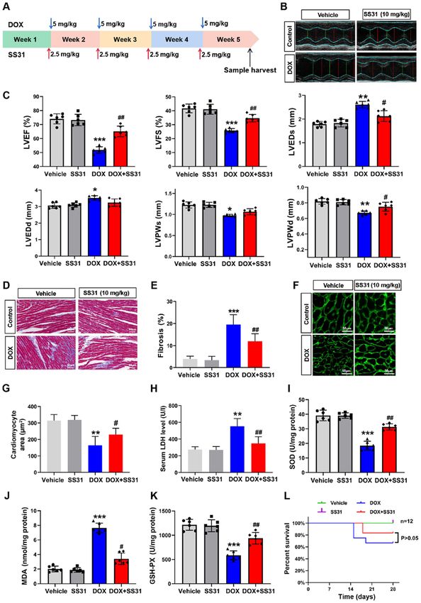
2                           ZHANG et al: SS31 ameliorates doxorubicin-induced cardiotoxicity

    The peptide Szeto‑Schiller (SS)‑31 (SS31; H‑D‑Arg‑Dmt-          SS31 peptide was added to the culture supernatant 2 h prior
Lys‑Phe‑NH2) is an efficacious antioxidant that has the capacity    to DOX treatment. A selective agonist of p38 MAPK P79350
to reduce mitochondrial ROS and scavenge free radicals (9). It      (50 µM; Invitrogen; Thermo Fisher Scientific, Inc.) was used
has been reported that SS31 is involved in the pathophysiolog‑      to activate the p38 signaling pathway. P79350 was added to the
ical processes of a variety of cardiovascular diseases, including   cell culture medium for 24 h at 37˚C.
protection of the myocardium from ischemia‑reperfusion (I/R)
injury by reducing inflammation, oxidative stress, apoptosis        Crystal violet staining assay. Following treatment, H9c2 cells
and fibrosis (10), reduction of proteomic alterations in heart      were seeded in 6‑well plates at 2x10⁵ cells/well, washed with
failure by preserving mitochondrial function (11), prevention       PBS twice to remove dead cells and fixed with 4% paraformal‑
of sepsis‑induced cardiac damage by suppressing the inflam‑         dehyde at room temperature (RT) for 30 min. Subsequently,
matory response and maintaining mitochondrial membrane              the cells were washed again with PBS and stained with 0.1%
potential (12), and amelioration of angiotensin II‑induced          crystal violet (Beyotime Institute of Biotechnology) solution
cardiomyopathy by decreasing the level of ROS (13). However,        for 30 min at RT in the dark. The dye was aspirated, and the
whether SS31 serves a protective role in DOX‑induced cardio‑        cells were washed twice with PBS and air‑dried naturally
toxicity remains to be elucidated.                                  at RT for image capture under light microscopy (x200).
    The mitogen‑activated protein kinase (MAPK) path‑               Subsequently, the dye was solubilized with 33% acetic acid
ways serve a crucial role in the regulation of apoptosis.           solution for 10 min at RT, and the absorbance was quantified
Extracellular‑regulated kinase (ERK) 1/2, p38 MAPK and              at 570 nm using a microplate reader.
c‑Jun N‑terminal kinase (JNK) are three major members of
MAPKs and exert antiapoptotic or proapoptotic effects in            Cell survival analysis. H9c2 cells were seeded in 6‑well plates
different cell types and contexts (14). For example, MAPK           at 2x10⁵ cells/well. Following DOX and peptide treatment,
serves a protective role in I/R‑induced cardiac myocyte             the cells were digested with trypsin‑EDTA solution for 3 min
apoptosis and in isolated perfused hearts that have undergone       at 37˚C and pipetting. Trypan blue staining was used to assess
reperfusion injury, whereas p38 and JNK promote apoptosis           the cell survival rates. Briefly, the cells were resuspended with
in cardiomyocytes subjected to I/R (15). Fasudil suppresses         1 ml PBS, and 100 µl of the cell suspension was added to 100 µl
isoproterenol‑induced heart failure by inhibiting the activation    trypan blue solution and stained for 3 min. The number of cells
of JNK and the nuclear translocation of MAPK (16). Therefore,       was counted in the four squares of the hemocytometer under
the present study focused on the development of strategies to       a Zeiss light microscope (magnification, x100). The following
improve DOX‑induced cardiotoxicity with MAPK as a target.           formula was used for the cell survival rate: Cell survival rate
    The present aimed to determine the therapeutic effects of       (%)=(no. of living cells/no. of total cells) x100%.
SS31 on DOX‑induced cardiotoxicity, and we hypothesized
that the administration of SS31 may provide a new insight           Analysis of cell viability. Cell viability was assessed using
into therapeutic strategies for the treatment of DOX‑induced        the CCK‑8 Assay kit (Beyotime Institute of Biotechnology)
cardiotoxicity.                                                     according to the manufacturer's instructions. H9c2 cells were
                                                                    seeded in 96‑well plates at 1x10 4 cells/well and cultured to
Materials and methods                                               adherence, followed by SS31 and DOX treatment as afore‑
                                                                    mentioned. A total of 10 µl CCK‑8 reagent was supplemented
Peptide synthesis and administration. The peptide SS31              into each well, and the cells were incubated in the dark for
(H‑D‑Arg‑Dmt‑Lys‑Phe‑NH2) was synthesized by Shanghai               2 h at 37˚C. The absorbance was measured using a microplate
Science Peptide Biological Technology Co., Ltd. The peptide         reader at 450 nm, and the cell viability was calculated based
crystal was dissolved in sterile double‑distilled water and         on the absorbance.
diluted to 10, 20 and 50 µmol/l.
                                                                    Lactate dehydrogenase (LDH) determination. The levels of
Cell culture and treatment. H9c2, a rat cardiomyocyte‑derived       LDH were detected by the LDH Release Assay kit (Beyotime
cell line, was acquired from The Cell Bank of Type Culture          Institute of Biotechnology). The reaction solution was prepared
Collection of The Chinese Academy of Sciences. H9c2                 according to the manufacturer's instructions. The H9c2 cell
cells were cultured in Dulbecco's modified Eagle's medium           supernatant (120 µl/well) was collected by centrifugation at
(DMEM; Gibco; Thermo Fisher Scientific, Inc.) supplemented          400 x g for 5 min at RT and mixed with the reaction solution
with 10% fetal bovine serum (Gibco; Thermo Fisher Scientific,       (60 µl/well), and the mixtures were added into 96‑well plates.
Inc.) and 1% penicillin and streptomycin (Wisent, Inc.) in a        The plates were wrapped in tin foil and incubated for 30 min
95% air and 5% CO2 atmosphere at 37˚C.                              at RT on the shaker. Finally, the absorbance was detected with
    The H9c2 cell toxicity model was induced by DOX (cat.           a microplate reader at 490 nm wavelength.
no. CAS 25316‑40‑9; Target Molecule Corp.). The H9c2 cells
at 80% confluence were preincubated with the SS31 peptide or        Analysis of ROS production. The level of intracellular total ROS
vehicle for 2 h at 37˚C, followed by the addition of 1 µM DOX       was assessed by 2',7'‑dichlorofluorescin diacetate (DCFH‑DA)
to the medium for 24 h at 37˚C (17). Cells were designated into     using a Reactive Oxygen Species Assay kit (Beyotime Institute
six groups: i) Control group; ii) SS31 (50 µmol/l) treatment        of Biotechnology). Briefly, H9c2 cells were seeded in 6‑well
group; iii) DOX treatment group; iv) DOX and SS31 (10 µmol/l)       plates at 2x10⁵ cells/well and treated as aforementioned when
cotreatment group; v) DOX and SS31 (20 µmol/l) cotreatment          the cells had grown to 80% confluence. DCFH‑DA was diluted
group; vi) DOX and SS31 (50 µmol/l) cotreatment group. The          to 10 µM in serum‑free DMEM and added into the medium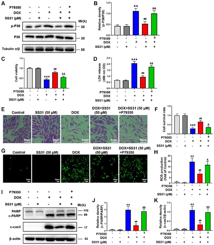
INTERNATIONAL JOURNAL OF MOlecular medicine 47: 63, 2021                                           3

(1 ml/well), and the cells were incubated at 37˚C away from          and iv) DOX + SS31 treated animals. DOX was administrated
light for 20 min. Subsequently, the cells were washed with           by intraperitoneal injection at 5 mg/kg weekly, and the final
PBS thrice to remove the residual DCFH‑DA and examined in            cumulative dose was 20 mg/kg (21). SS31 (2.5 mg/kg) was
at ≥3 fields per sample under a laser scanning confocal micro‑       injected into the tail vein weekly, and the final cumulative dose
scope (magnification, x100). The density of ROS fluorescence         was 10 mg/kg (22). The mice that were not treated with SS31
was examined by ImageJ software 1.26 (National Institutes of         or DOX received the equal volumes of saline. Following the
Health).                                                             treatment, the mice were maintained alive for one week. The
                                                                     mice were anesthetized with 1.5% isoflurane inhalation; the
JC‑1 mitochondrial membrane potential determination. The             depth of anesthesia was evaluated by the immobility and the
JC‑1 Mitochondrial Membrane Potential Assay kit (Beyotime            absence of righting reflex, and echocardiography was used to
Institute of Biotechnology) was used to analyze mitochondrial        detect mouse cardiac function. Following euthanasia by carbon
injury according to the manufacturer's instructions. Briefly,        dioxide asphyxia (30% chamber volume/min), the hearts were
H9c2 cells were seeded in 6‑well plates at 2x10⁵ cells/well,         removed rapidly and harvested to prepare paraffin sections
washed with PBS and incubated with JC‑1 solution for 10 min          for Masson and wheat germ agglutinin (WGA) staining or to
at 37˚C. The cells were washed with the dilution buffer and          isolate the mitochondria. The blood (1 ml) was collected from
analyzed in ≥3 fields per sample under a laser scanning              the abdominal aorta, and the serum was obtained by centrifu‑
confocal microscope (magnification, x100).The density of             gation (1,200 x g, 20 min, 4˚C).
JC‑1 fluorescence was examined by ImageJ software.
                                                                     Echocardiography analysis. For the evaluation of cardiac
Western blotting. H9c2 cells were treated as aforementioned          function, the mice were anesthetized with 1.5% isoflurane,
and lysed by RIPA protein lysis buffer (Beyotime Institute           and echocardiography was performed using a Vevo 2100
of Biotechnology) and 1% PMSF (Beyotime Institute of                 High Resolution Imaging System (VisualSonics, Inc.).
Biotechnology) to extract the total protein. The protein             Cardiac contractile function was examined by echocar‑
concentrations were determined using the Bicinchoninic Acid          diography in conscious, gently restrained mice using a
Protein Assay kit (Beyotime Institute of Biotechnology). The         Vevo 2100 system (MS400C probe). The main measured
proteins (20 µg/lane) were separated by 10% SDS‑PAGE                 indicators included ejection fraction (EF) and fractional
and transferred to PVDF membranes (MilliporeSigma).                  shortening (FS). Other echocardiographic parameters
The membranes were blocked with skimmed milk (5%) for                included left ventricular end‑systolic diameter (LVEDs), left
2 h at RT and incubated with the primary antibodies against          ventricular end‑diastolic diameter (LVEDd), left ventric‑
PARP (1:1,000; cat. no. 9542), cleaved caspase‑3 (1:1,000;           ular end‑systolic volume (LVESV) and left ventricular
cat. no. 9661), bax (1:1,000; cat. no. 2772), bcl‑2 (1:1,000; cat.   end‑diastolic volume (LVEDV). FS was calculated as follows:
no. 4223), phosphorylated (p‑)p38 (1:1,000; cat. no. 4511), p38      FS (%)=[(LVEDd‑LVEDs)/LVEDd] x 100; EF was calcu‑
(1:1,000; cat. no. 8690), JNK (1:1,000; cat. no. 9255), p‑JNK        lated as follows: EF (%)=[(LVEDV‑LVESV)/LVEDV] x 100,
(1:1,000; cat. no. 9251), p‑ERK (1:1,000; cat. no. 4376), ERK        where LVEDV=7 x LVEDd 3/(2.4 + LVEDd) and LVESV=
(1:1,000; cat. no. 4695), α/β‑Tubulin (1:2,000; cat. no. 2148),      7 x LVEDs3/(2.4 + LVEDs).
β ‑actin (1:2,000; cat. no. 4970) and GAPDH (1:2,000;
cat. no. 2118) (all Cell Signaling Technology, Inc.) at 4˚C          WGA and Masson staining. The hearts were harvested and fixed
overnight. Notably, the internal control antibody against            in 4% buffered formaldehyde for 48 h at RT. After embedding
α /β ‑tubulin produced a nonspecific faint band (18,19).             in paraffin and sectioning, 5‑µm sections were stained with
Subsequently, PVDF membranes were washed three times for             fluorescein isothiocyanate‑conjugated WGA (cat. no. L4895;
10 min each time with TBS with 0.1% Tween‑20 buffer and              Sigma‑Aldrich; Merck KGaA) staining according to the
incubated with horseradish peroxidase‑conjugated anti‑rabbit         manufacturer's instructions. Digital images (≥3 fields) were
secondary antibodies (1:3,000; cat. no. 7074; Cell Signaling         captured using a laser scanning confocal microscope (magni‑
Technology, Inc.) for 1 h at RT. The immunoreactive protein          fication, x400). A quantitative digital image analysis system
bands were detected using an enhanced chemiluminescent               Image‑Pro Plus 6.0 (Media Cybernetics, Inc.) was used to
substrate (cat. no. SQ101; Epizyme, Inc.). The protein expres‑       measure the cross‑sectional area of cardiomyocytes. To assess
sion levels were quantified according to their grey values           cardiac fibrosis, the 5‑µm sections were stained with Masson's
determined using ImageJ software.                                    trichrome (cat. no. G1340; Beijing Solarbio Science &
                                                                     Technology Co., Ltd.) according to the manufacturer's instruc‑
In vivo experiment. All animal experiments were carried              tions. Each stained section was observed under a microscope
out in accordance with the Guide for the Care and Use of             (magnification, x400), and ImageJ software was used to
Laboratory (20) and approved by the Institutional Animal Care        evaluate histopathological damage.
and Use Committee of Nanjing Medical University (approval
no. IACUC‑1903030; Nanjing, China). A total of 48 male               LDH, superoxide dismutase (SOD), malondialdehyde (MDA)
C57BL/6 mice (6 weeks old; weight, 16‑20 g) were obtained            and glutathione peroxidase (GSH‑PX) measurement. The
from Shanghai SLAC laboratory animal corporation and main‑           serum LDH concentrations were measured using an ELISA
tained under a 12:12‑h light/dark cycle at 22‑26˚C with a relative   kit (cat. no. J2380; Promega Corporation) according the
humidity of 40‑50% and ad libitum food and water. Following          manufacturer's instructions. A total of 40 mg heart tissues
one week of adjustable feeding, the animals were randomly            were harvested to isolate the mitochondria. Briefly, tissues
assigned to the following groups: i) Vehicle; ii) SS31; iii) DOX;    were incised completely in 500 µl PBS and centrifuged at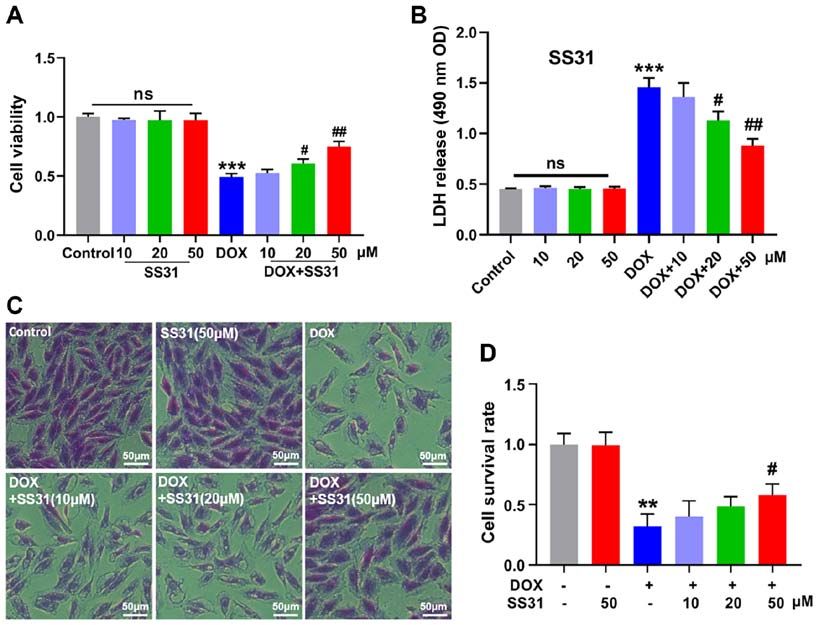
4                               ZHANG et al: SS31 ameliorates doxorubicin-induced cardiotoxicity

Figure 1. SS31 treatment attenuates DOX‑induced inhibition of H9c2 cell survival. (A) The cell viability was determined in H9c2 cells. (B) LDH release
level was measured in H9c2 cells supernatant. (C and D) Crystal violet staining and quantitative analysis of living cells. Magnification, x200. **P
INTERNATIONAL JOURNAL OF MOlecular medicine 47: 63, 2021                                                               5

Figure 2. SS31 attenuates DOX‑induced apoptosis in H9c2 cells. (A) The levels of apoptosis‑related proteins (PARP, cleaved caspase‑3, bax and bcl‑2) were
determined by western blotting. (B‑E) Quantitative analysis of the relative protein levels of (B) c‑PARP, (C) cleaved caspase‑3, (D) bcl‑2 and (E) bax. *P
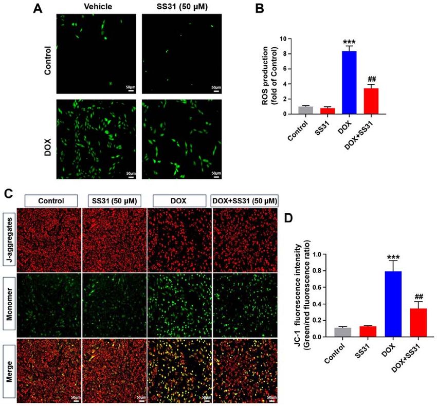
6                                ZHANG et al: SS31 ameliorates doxorubicin-induced cardiotoxicity

Figure 3. SS31 attenuates DOX‑induced mitochondrial oxidative stress injury in H9c2 cells. (A) Representative images of DCFH‑DA (green) staining in H9c2
cells used to detect intracellular ROS following treatment with 50 µM SS31 and DOX. Magnification, x100. (B) Quantitative analysis of ROS. (C) Representative
images of JC‑1 fluorescence representing the mitochondrial membrane potential in H9c2 cells. Magnification, x100. (D) Quantitative analysis of JC‑1 fluores‑
cence intensity. ***P
INTERNATIONAL JOURNAL OF MOlecular medicine 47: 63, 2021                                                                7

Figure 4. Effects of SS31 on DOX‑induced cardiac injury in vivo. (A) Timeline of the experimental procedure for the DOX‑induced mouse cardiotoxicity
model. (B) Representative photographs of the echocardiography analysis. (C) Quantified data of the echocardiography analysis. (D) Masson trichrome staining.
Magnification, x400. (E) Quantitative analysis of fibrosis in the Masson‑stained sections. (F) Representative photographs of wheat germ agglutinin staining.
Magnification, x400. (G) Quantitative analysis of the cardiomyocyte area. (H) Serum LDH levels. n=6 mice/group. (I‑K) SOD, MDA and GSH‑PX levels were
evaluated in mouse heart tissue samples. n=6 mice/group. (L) Survival of mice following DOX‑induced cardiac injury. Day 0 refers to the first DOX injection.
*
 P
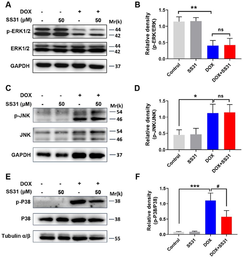
8                                ZHANG et al: SS31 ameliorates doxorubicin-induced cardiotoxicity

Figure 5. SS31 treatment inhibits the activation of p38 MAPK signaling in DOX‑treated H9c2 cells. (A) The protein levels of p‑ERK1/2 and ERK1/2 were
determined by western blotting. (B) Quantitative analysis of the relative protein levels of p‑ERK1/2. (C) The protein levels of p‑JNK and JNK were determined
by western blotting. (D) Quantitative analysis of the relative protein levels of p‑JNK. (E) The protein levels of p‑p38 and p38 were determined by western
blotting. (F) Quantitative analysis of the relative protein levels of p‑p38. *P
INTERNATIONAL JOURNAL OF MOlecular medicine 47: 63, 2021                                                                9

Figure 6. p38 agonist reverses the effects of SS31 in DOX‑treated H9c2 cells. (A) H9c2 cells were treated with P79350 to activate the p38 signaling pathway.
(B) Quantification of the western blot data. (C) H9c2 cell viability was detected following treatment with DOX, SS31 and P79350. (D) LDH release was
detected in H9c2 cells. (E) Crystal violet staining of living cells. Magnification, x200. (F) Quantitative analysis of the crystal violet assay. (G) ROS contents
were detected in H9c2 cells following treatment with DOX, SS31 and P79350. (H) Quantitative analysis of ROS contents. (I) The effects of P79350 on the
levels of apoptosis‑associated proteins in H9c2 cells treated with DOX and SS31. (J) Quantification of the PARP western blot bands. (K) Quantification of the
c‑cas3 western blot bands. **P
10                          ZHANG et al: SS31 ameliorates doxorubicin-induced cardiotoxicity

    The present study further assessed the underlying mecha‑         Acknowledgements
nism of the effects of SS31 in ameliorating DOX‑induced
toxicity in H9c2 cells. As a member of the MAPK family,              Not applicable.
p38 MAPK is susceptible to various stimuli, such as oxidative
stress, inflammatory cytokines, growth factors or high glucose        Funding
levels. The canonical pathway of p38 MAPK activation is
phosphorylation on a threonine and a tyrosine residue of the         This study was supported by grants from the National
activation loop mediated by MAP2K (43). In cardiomyocytes,           Natural Science Foundation of China (grant nos. 81570209
a previous study has demonstrated that the phosphorylation           and 81873540) and The Key Clinical Frontier Technology
of p38 is triggered by hypoxia and exogenous H 2O2 treat‑            Project of Department of Science and Technology of Jiangsu
ment (44). In the present study, DOX treatment significantly         Provincial (grant no. BE2019752).
increased the phosphorylation of p38 MAPK compared with
that in the control cells, whereas administration of SS31            Availability of data and materials
inhibited this trend in H9c2 cells. Notably, SS31 treatment
exerted no significant effects on the expression and phos‑           The datasets used and/or analyzed during the current study are
phorylation levels of ERK1/2 and JNK. A previous study has           available from the corresponding author on reasonable request.
reported that SS31 peptide eliminates high glucose‑induced
mitochondrial oxidative stress by regulating the p38 MAPK            Authors' contributions
signal pathway, which may be a novel therapeutic strategy to
prevent hyperglycemia‑induced neuronal perturbation (45).            LZ and MF performed the experiments and wrote the manuscript.
Another study has demonstrated that SS31 reduces the effects         XW and HZ performed the cell experiments and participated
of sepsis‑induced heart injury by inhibiting the p38 MAPK            in drafting the manuscript. JD performed a part of the in vivo
signaling pathway (12). The results of these studies were            study and participated in drafting the manuscript. LQ and ZC
consistent with those of the present study. Therefore, SS31 may      conceived, designed and supervised the project, and revised the
be a potent antioxidant peptide with a capacity for reducing         manuscript. LZ and MF confirm the authenticity of all the raw
ROS and inhibiting apoptosis, which may be associated with           data. All authors read and approved the final manuscript.
the inactivation of the p38 MAPK signaling pathway.
    A previous study has mainly focused on the mechanism of          Ethics approval and consent to participate
DOX‑induced cardiotoxicity and reported that high levels of
mitochondrial ROS production are required for DOX‑induced            All animal experiments were carried out in accordance with the
cardiac damage, and that the p38 MAPK signaling pathway              Guide for the Care and Use of Laboratory and approved by the
is involved in DOX‑induced cardiotoxicity (46). However, the         Institutional Animal Care and Use Committee of Nanjing Medical
aforementioned study has failed to provide the mechanism by          University (approval no. IACUC‑1903030; Nanjing, China).
which SS31 affects DOX‑induced cardiomyocyte apoptosis.
The present study demonstrated that SS31 may ameliorate              Patient consent for publication
DOX‑induced ROS production and apoptosis in cardiomyo‑
cytes by inhibiting the activation of the p38 MAPK signaling         Not applicable.
pathway. To the best of the author's knowledge, this is the
first report on the effects of SS31 on mitochondrial function        Competing interests
describing the underlying mechanism to date.
    Although the present study illustrated the anticardiotoxic       The authors declare that they have no competing interests.
effects of SS31 in DOX‑induced injury models in vitro and in vivo,
the study had certain limitations. It is necessary to evaluate the   References
function of SS31 in other types of cells derived from heart, such
as primary neonatal rat myocardial cells and AC16 cardiomyo‑
                                                                      1. Martins‑Teixeira MB and Carvalho I: Antitumour anthracyclines:
cytes. In addition, whether different types of modification affect       Progress and perspectives. ChemMedChem 15: 933‑948, 2020.
the function of SS31 remains to be further verified. Therefore,       2. Ji X, Ding W, Xu T, Zheng X, Zhang J, Liu M, Liu G and Wang J:
our future studies will involve evaluating the effects of SS31 in        MicroRNA‑31‑5p attenuates doxorubicin‑induced cardiotoxicity
                                                                         via quaking and circular RNA Pan3. J Mol Cell Cardiol 140:
multiple types of cardiomyocytes and performing modifications            56‑67, 2020.
of SS31 to verify its cardioprotective function.                      3. Kalyanaraman B: Teaching the basics of the mechanism of
    In summary, the results of the present study demonstrated            doxorubicin‑induced cardiotoxicity: Have we been barking up
                                                                         the wrong tree? Redox Biol 29: 101394, 2020.
that the mitochondria‑targeted antioxidant peptide SS31               4. Li DL and Hill JA: Cardiomyocyte autophagy and cancer chemo‑
suppressed the generation of ROS and apoptosis by inhibiting             therapy. J Mol Cell Cardiol 71: 54‑61, 2014.
the phosphorylation of p38 MAPK in DOX‑treated H9c2                   5. Osataphan N, Phrommintikul A, Chattipakorn SC and
                                                                         Chattipakorn N: Effects of doxorubicin‑induced cardiotoxicity
cells. In vivo, cotreatment with SS31 improved cardiac func‑             on cardiac mitochondrial dynamics and mitochondrial function:
tion and suppressed the occurrence of myocardial fibrosis                Insights for future interventions. J Cell Mol Med 24: 6534‑6557,
induced by DOX compared with those in mice treated with                  2020.
DOX alone. Therefore, the results of the present study may            6. Octavia Y, Tocchetti CG, Gabrielson KL, Janssens S, Crijns HJ
                                                                         and Moens AL: Doxorubicin‑induced cardiomyopathy: From
provide a potential candidate molecule for the treatment of              molecular mechanisms to therapeutic strategies. J Mol Cell
DOX‑induced cardiotoxicity.                                              Cardiol 52: 1213‑1225, 2012.
INTERNATIONAL JOURNAL OF MOlecular medicine 47: 63, 2021 11
7. Wang AJ, Zhang J, Xiao M, Wang S, Wang BJ, Guo Y, Tang Y and 28. Kim HS, Lee YS and Kim DK: Doxorubicin exerts cytotoxic
Gu J: Molecular mechanisms of doxorubicin‑induced cardiotox‑ effects through cell cycle arrest and Fas‑mediated cell death.
icity: Novel roles of sirtuin 1‑mediated signaling pathways. Cell Pharmacology 84: 300‑309, 2009.
Mol Life Sci: Jan 13, 2021 (Epub ahead of print). doi: 10.1007/ 29. Yu J, Gao H, Wu C, Xu QM, Lu JJ and Chen X: Diethyl blechnic,
s00018-020-03729-y. a novel natural product isolated from salvia miltiorrhiza bunge,
8. Tahover E, Segal A, Isacson R, Rosengarten O, Grenader T, Gips M, inhibits doxorubicin‑induced apoptosis by inhibiting ROS and
Cherny N, Heching NI, Mesika L, Catane R and Gabizon A: activating JNK1/2. Int J Mol Sci 19: 1809, 2018.
Dexrazoxane added to doxorubicin‑based adjuvant chemotherapy 30. Varela‑Lopez A, Battino M, Navarro‑Hortal MD, Giampieri F,
of breast cancer: A retrospective cohort study with a comparative Forbes‑Hernández TY, Romero‑Márquez JM, Collado R and
analysis of toxicity and survival. Anticancer Drugs 28: 787‑794, 2017. Quiles JL: An update on the mechanisms related to cell death
9. Ajith TA and Jayakumar TG: Mitochondria‑targeted agents: and toxicity of doxorubicin and the protective role of nutrients.
Future perspectives of mitochondrial pharmaceutics in cardio‑ Food Chem Toxicol 134: 110834, 2019.
vascular diseases. World J Cardiol 6: 1091‑1099, 2014. 31. Gorini S, De Angelis A, Berrino L, Malara N, Rosano G
10. Lee FY, Shao PL, Wallace CG, Chua S, Sung PH, Ko SF, Chai HT, and Ferraro E: Chemotherapeutic drugs and mitochondrial
Chung SY, Chen KH, Lu HI, et al: Combined therapy with SS31 dysfunction: Focus on doxorubicin, trastuzumab, and sunitinib.
and mitochondria mitigates myocardial ischemia‑reperfusion Oxid Med Cell Longev 2018: 7582730, 2018.
injury in rats. Int J Mol Sci 19: 2782, 2018. 32. Orrenius S, Gogvadze V and Zhivotovsky B: Mitochondrial
11. Dai DF, Hsieh EJ, Chen T, Menendez LG, Basisty NB, Tsai L, oxidative stress: Implications for cell death. Annu Rev Pharmacol
Beyer RP, Crispin DA, Shulman NJ, Szeto HH, et al: Global Toxicol 47: 143‑183, 2007.
proteomics and pathway analysis of pressure‑overload‑induced 33. Wallace KB, Sardao VA and Oliveira PJ: Mitochondrial deter‑
heart failure and its attenuation by mitochondrial‑targeted minants of doxorubicin‑induced cardiomyopathy. Circ Res 126:
peptides. Circ Heart Fail 6: 1067‑1076, 2013. 926‑941, 2020.
12. Liu Y, Yang W, Sun X, Xie L, Yang Y, Sang M and Jiao R: SS31 34. Mitchell W, Ng EA, Tamucci JD, Boyd KJ, Sathappa M,
ameliorates sepsis‑induced heart injury by inhibiting oxidative Coscia A, Pan M, Han X, Eddy NA, May ER, et al: The mitochon‑
stress and inflammation. Inflammation 42: 2170‑2180, 2019. dria‑targeted peptide SS‑31 binds lipid bilayers and modulates
13. Dai DF, Chen T, Szeto H, Nieves‑Cintrón M, Kutyavin V, surface electrostatics as a key component of its mechanism of
Santana LF and Rabinovitch PS: Mitochondrial targeted antioxi‑ action. J Biol Chem 295: 7452‑7469, 2020.
dant Peptide ameliorates hypertensive cardiomyopathy. J Am Coll 35. Hou Y, Shi Y, Han B, Liu X, Qiao X, Qi Y and Wang L: The
Cardiol 58: 73‑82, 2011. antioxidant peptide SS31 prevents oxidative stress, downregu‑
14. Yue J and Lopez JM: Understanding MAPK signaling pathways lates CD36 and improves renal function in diabetic nephropathy.
in apoptosis. Int J Mol Sci 21: 2346, 2020. Nephrol Dial Transplant 33: 1908‑1918, 2018.
15. Yue TL, Wang C, Gu JL, Ma XL, Kumar S, Lee JC, Feuerstein GZ, 36. Reddy PH, Manczak M, Yin X and Reddy AP: Synergistic
Thomas H, Maleeff B and Ohlstein EH: Inhibition of extracellular protective effects of mitochondrial division inhibitor 1 and mito‑
signal‑regulated kinase enhances ischemia/reoxygenation‑induced chondria‑targeted small peptide SS31 in Alzheimer's disease.
apoptosis in cultured cardiac myocytes and exaggerates reperfu‑ J Alzheimers Dis 62: 1549‑1565, 2018.
sion injury in isolated perfused heart. Circ Res 86: 692‑699, 2000. 37. Zhou J, Li Z, Chen Z and Yang K: Protective effect of mitochon‑
16. Wang N, Guan P, Zhang JP, Li YQ, Chang YZ, Shi ZH, Wang FY dria‑targeted antioxidant SS31 on early brain injury following
and Chu L: Fasudil hydrochloride hydrate, a Rho‑kinase inhibitor, subarachnoid hemorrhage in rats. Zhong Nan Da Xue Xue Bao
suppresses isoproterenol‑induced heart failure in rats via JNK Yi Xue Ban 42: 1003‑1009, 2017 (In Chinese).
and ERK1/2 pathways. J Cell Biochem 112: 1920‑1929, 2011. 38. Yin X, Manczak M and Reddy PH: Mitochondria‑targeted
17. Liu D, Ma Z, Di S, Yang Y, Yang J, Xu L, Reiter RJ, Qiao S and molecules MitoQ and SS31 reduce mutant huntingtin‑induced
Yuan J: AMPK/PGC1α activation by melatonin attenuates acute mitochondrial toxicity and synaptic damage in Huntington's
doxorubicin cardiotoxicity via alleviating mitochondrial oxida‑ disease. Hum Mol Genet 25: 1739‑1753, 2016.
tive damage and apoptosis. Free Radic Biol Med 129: 59‑72, 2018. 39. Ma H, Chen S, Xiong H, Wang M, Hang W, Zhu X, Zheng Y,
18. Krais JJ and Johnson N: Ectopic RNF168 expression promotes Ge B, Li R and Cui H: Astaxanthin from Haematococcus
break‑induced replication‑like DNA synthesis at stalled replica‑ pluvialis ameliorates the chemotherapeutic drug (doxorubicin)
tion forks. Nucleic Acids Res 48: 4298‑4308, 2020. induced liver injury through the Keap1/Nrf2/HO‑1 pathway in
19. Fan J, Shen W, Lee SR, Mathai AE, Zhang R, Xu G and mice. Food Funct 11: 4659‑4671, 2020.
Gillies MC: Targeting the Notch and TGF‑β signaling pathways 40. Vu M, Kassouf N, Ofili R, Lund T, Bell C and Appiah S:
to prevent retinal fibrosis in vitro and in vivo. Theranostics 10: Doxorubicin selectively induces apoptosis through the inhibition of
7956‑7973, 2020. a novel isoform of Bcl2 in acute myeloid leukaemia MOLM13 cells
20. National Research Council (US): Committee for the Update of with reduced Beclin 1 expression. Int J Oncol 57: 113‑121, 2020.
the Guide for the Care and Use of Laboratory Animals: Guide for 41. Faridvand Y, Haddadi P, Vahedian V, Nozari S, Nejabati HR,
the Care and Use of Laboratory Animals. 8th edition. National Pezeshkian M, Afrasiabi A, Safaie N, Jodati A and Nouri M:
Academies Press, Washington, DC, 2011. Human amnion membrane proteins prevent doxorubicin‑ induced
21. Oh J, Lee BS, Lim G, Lim H, Lee CJ, Park S, Lee SH, Chung JH oxidative stress injury and apoptosis in rat H9c2 cardiomyocytes.
and Kang SM: Atorvastatin protects cardiomyocyte from Cardiovasc Toxicol 20: 370‑379, 2020.
doxorubicin toxicity by modulating survivin expression through 42. Wu YZ, Zhang L, Wu ZX, Shan TT and Xiong C: Berberine
FOXO1 inhibition. J Mol Cell Cardiol 138: 244‑255, 2020. ameliorates doxor ubicin‑induced cardiotoxicity via a
22. Zhang L, Wang X, Feng M, Zhang H, Xu J, Ding J, Cheng Z SIRT1/p66Shc‑mediated pathway. Oxid Med Cell Longev 2019:
and Qian L: Peptidomics analysis reveals peptide PDCryab1 2150394, 2019.
inhibits doxorubicin‑induced cardiotoxicity. Oxid Med Cell 43. Cuadrado A and Nebreda AR: Mechanisms and functions of
Longev 2020: 7182428, 2020. p38 MAPK signalling. Biochem J 429: 403‑417, 2010.
23. Liang L, Tu Y, Lu J, Wang P, Guo Z, Wang Q, Guo K, Lan R, Li H 44. Kulisz A, Chen N, Chandel NS, Shao Z and Schumacker PT:
and Liu P: Dkk1 exacerbates doxorubicin‑induced cardiotox‑ Mitochondrial ROS initiate phosphorylation of p38 MAP kinase
icity by inhibiting the Wnt/β‑catenin signaling pathway. J Cell during hypoxia in cardiomyocytes. Am J Physiol Lung Cell Mol
Sci 132: cs228478, 2019. Physiol 282: L1324‑L1329, 2002.
24. Rochette L, Guenancia C, Gudjoncik A, Hachet O, Zeller M, 45. Cao M, Jiang J, Du Y and Yan P: Mitochondria‑targeted anti‑
Cottin Y and Vergely C: Anthracyclines/trastuzumab: New oxidant attenuates high glucose‑induced P38 MAPK pathway
aspects of cardiotoxicity and molecular mechanisms. Trends activation in human neuroblastoma cells. Mol Med Rep 5:
Pharmacol Sci 36: 326‑348, 2015. 929‑934, 2012.
25. Hantson P: Mechanisms of toxic cardiomyopathy. Clin Toxicol 46. Guo Z, Tang N, Liu FY, Yang Z, Ma SQ, An P, Wu HM, Fan D
(Phila) 57: 1‑9, 2019. and Tang QZ: TLR9 deficiency alleviates doxorubicin‑ induced
26. Zhao L, Qi Y, Xu L, Tao X, Han X, Yin L and Peng J: cardiotoxicity via the regulation of autophagy. J Cell Mol
MicroRNA‑140‑5p aggravates doxorubicin‑induced cardiotox‑ Med 24: 10913‑10923, 2020.
icity by promoting myocardial oxidative stress via targeting Nrf2
and Sirt2. Redox Biol 15: 284‑296, 2018. This work is licensed under a Creative Commons
27. Zhang P, Chen Z, Lu D, Wu Y, Fan M, Qian J and Ge J:
Attribution-NonCommercial-NoDerivatives 4.0
Overexpression of COX5A protects H9c2 cells against doxoru‑
bicin‑induced cardiotoxicity. Biochem Biophys Res Commun 524: International (CC BY-NC-ND 4.0) License.
43‑49, 2020.You can also read