Network based transcription factor analysis of regenerating axolotl limbs
←
→
Page content transcription
If your browser does not render page correctly, please read the page content below
Jhamb et al. BMC Bioinformatics 2011, 12:80
http://www.biomedcentral.com/1471-2105/12/80

 RESEARCH ARTICLE                                                                                                                              Open Access

Network based transcription factor analysis of
regenerating axolotl limbs
Deepali Jhamb1, Nandini Rao2,6, Derek J Milner3, Fengyu Song4,5, Jo Ann Cameron3,5, David L Stocum2,5*,
Mathew J Palakal1,5*

  Abstract
  Background: Studies on amphibian limb regeneration began in the early 1700’s but we still do not completely
  understand the cellular and molecular events of this unique process. Understanding a complex biological process
  such as limb regeneration is more complicated than the knowledge of the individual genes or proteins involved.
  Here we followed a systems biology approach in an effort to construct the networks and pathways of protein
  interactions involved in formation of the accumulation blastema in regenerating axolotl limbs.
  Results: We used the human orthologs of proteins previously identified by our research team as bait to identify the
  transcription factor (TF) pathways and networks that regulate blastema formation in amputated axolotl limbs. The
  five most connected factors, c-Myc, SP1, HNF4A, ESR1 and p53 regulate ~50% of the proteins in our data. Among
  these, c-Myc and SP1 regulate 36.2% of the proteins. c-Myc was the most highly connected TF (71 targets). Network
  analysis showed that TGF-b1 and fibronectin (FN) lead to the activation of these TFs. We found that other TFs known
  to be involved in epigenetic reprogramming, such as Klf4, Oct4, and Lin28 are also connected to c-Myc and SP1.
  Conclusions: Our study provides a systems biology approach to how different molecular entities inter-connect
  with each other during the formation of an accumulation blastema in regenerating axolotl limbs. This approach
  provides an in silico methodology to identify proteins that are not detected by experimental methods such as
  proteomics but are potentially important to blastema formation. We found that the TFs, c-Myc and SP1 and their
  target genes could potentially play a central role in limb regeneration. Systems biology has the potential to map
  out numerous other pathways that are crucial to blastema formation in regeneration-competent limbs, to compare
  these to the pathways that characterize regeneration-deficient limbs and finally, to identify stem cell markers in
  regeneration.

Background                                                                           Urodele amphibians (axolotls, salamanders and newts),
Current thinking in regenerative medicine envisions the                              which regenerate amputated limbs perfectly throughout
derivation, from autogeneic somatic cells, of pluripotent                            larval and adult life, provide a research model that lends
cells that can be directed to differentiate into transplanta-                        itself well to furthering our understanding of this process.
ble replacements for cells destroyed by injury or disease                            Two hundred fifty years after Lazzaro Spallanzani first
[1]. Beyond this, however, is another goal: the chemical                             demonstrated the regeneration of amputated newt limbs
induction of regeneration directly at the site of tissue                             [3], we still do not fully understand the mechanisms of
damage [2]. Achievement of this goal will require a deep                             this process. Urodele limbs initiate regeneration through
understanding of the molecular components, networks                                  the formation of a blastema: a limb bud-like structure
and pathways that characterize regenerative competence.                              composed of undifferentiated progenitor cells. Blastema
                                                                                     cells originate by a reverse developmental process in
                                                                                     which the tissue matrix near the amputation plane is
* Correspondence: dstocum@iupui.edu; mpalakal@iupui.edu
1
 School of Informatics, Indiana University-Purdue University Indianapolis,
                                                                                     degraded by proteases, releasing both mature cells that are
Indianapolis, IN, 46202, USA                                                         reprogrammed to a mesenchymal stem cell-like state, and
2
 Department of Biology, Indiana University-Purdue University Indianapolis,           muscle stem cells (satellite cells) [4-7]. Within a few days
Indianapolis, IN, 46202, USA
Full list of author information is available at the end of the article
                                                                                     after amputation, these cells accumulate under the apical

                                        © 2011 Jhamb et al; licensee BioMed Central Ltd. This is an Open Access article distributed under the terms of the Creative Commons
                                        Attribution License (http://creativecommons.org/licenses/by/2.0), which permits unrestricted use, distribution, and reproduction in
                                        any medium, provided the original work is properly cited.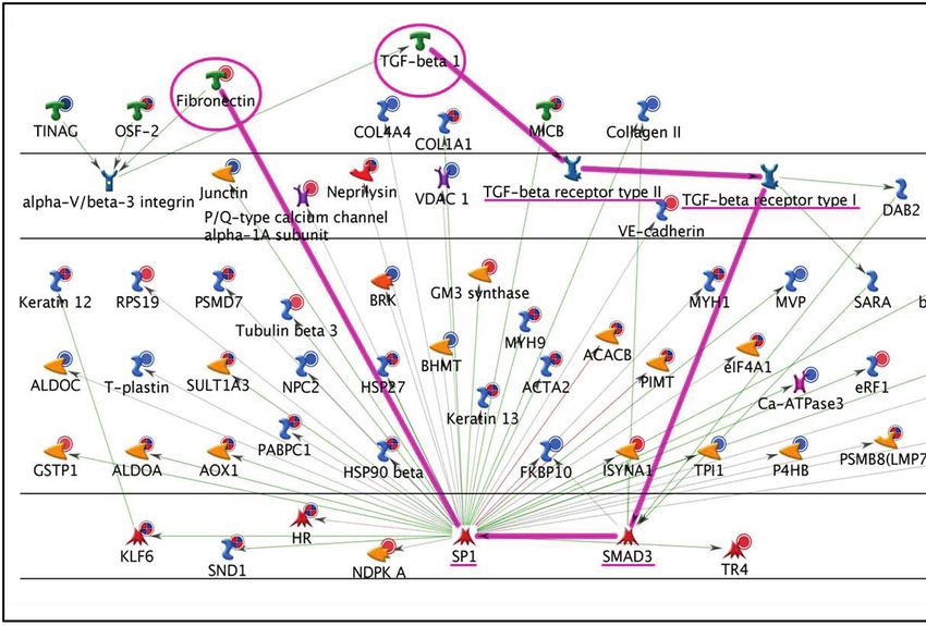
Jhamb et al. BMC Bioinformatics 2011, 12:80                                                                     Page 2 of 12
http://www.biomedcentral.com/1471-2105/12/80

epidermal cap (AEC), where they proliferate and are pat-       limb regeneration. In addition, Klf4 (kruppel-like factor 4),
terned into the missing limb parts.                            Oct4 (octamer-binding protein 4), and Lin28, TFs common
   The ability to form a blastema is what distinguishes        to embryonic stem cells, induced pluripotent cells (iPSCs)
urodele limbs from the limbs of most other tetrapod ver-       and blastema cells [15-18], were also found to be con-
tebrates that do not regenerate or which regenerate            nected to c-Myc and SP1. Our results highlight the utility
poorly. Thus, understanding the mechanisms that lead to        of systems biology for understanding complex processes
blastema formation is crucial to understanding why uro-        such as limb regeneration.
dele limbs regenerate, and why the limbs of other species
do not. In general, the reductionist approach has been to      Methods
study the individual genes or proteins involved in biologi-    Processing of Axolotl Proteomics Data
cal processes. With the development of high throughput         Network and pathway analysis was based on proteomics
technology over the last decade, there has been a shift in     data obtained at 1, 4 and 7 days post-amputation (dpa) in
this approach. The ability to obtain large scale omics data    our study of blastema formation in the hindlimb of the
has led to the development of discovery approaches that        wild-type axolotl, Ambystoma mexicanum [15]. In that
interrelate the elements of biological processes, revealing    study, 309 high confidence peptides were identified with
networks and pathways of organization in a system [8].         significant fold changes relative to control on one or more
Very few studies so far have analyzed global gene or pro-      dpa. Since bioinformatics resources for the axolotl are lim-
tein expression patterns during limb regeneration. In the      ited, we identified human orthologs for these peptides.
axolotl Ambystoma mexicanum, expressed sequence tag            The search was carried out against the Homo sapiens data-
(EST) resources have been developed [9] and transcrip-         base (taxid: 9606) using the BLASTp tool (Basic Local
tion profiles of denervated vs. innervated limbs have been     Alignment Search Tool for proteins) available from NCBI
analyzed [10]. Some global studies have been performed         (National Center for Biotechnology Information) [19]. The
in an anuran amphibian, the frog Xenopus laevis. How-          peptides in the original study were five to thirty three
ever, unlike urodeles, Xenopus possesses the ability to        amino acids long and were aligned throughout their length
regenerate lost limbs in early tadpole stages of develop-      for ortholog identification. A human ortholog was used
ment, but gradually loses the capability for regeneration      only if the percentage identity between an axolotl: human
as development proceeds, until it is lost completely in        peptide was greater than 85%; unmatched peptides were
adults [11]. Xenopus studies have focused on subtractive       excluded from the analysis. Although proteins with lower
hybridization [12]; microarray analysis [13] and proteo-       percentage identities could well be important, LC/MS/MS
mics [14] for molecular screening of limb regeneration.        analysis relies on peptide sequences for alignment, not
   Although extensive research has been carried out to         complete protein sequences, making it essential to set a
understand how the blastema is formed and which mole-          high percentage identity threshold for stringency. The
cular entities are crucial to regeneration, very little is     UniProt database [20] was used to assign the gene name
known about the interactive pathways and networks that         to each of the human orthologs. The Database for Annota-
lead to blastema formation in an amputated limb. Recently,     tion, Visualization and Integrated Discovery (DAVID)
we conducted a proteomic analysis of blastema formation        [21,22] was used for the assessment of biological pro-
in the amputated limbs of Ambystoma mexicanum. Our             cesses. The ortholog data was divided into 6 groups with
analysis revealed a number of significant changes in protein   respect to up- and down-regulated proteins at each time
expression related to cell signaling, transcription, metabo-   point: 1d-, 1d+, 4d-, 4d+, 7d-, and 7d+. Thus, 1d- refers to
lism, cell protection, and cell cycle [15]. We are now         all down-regulated proteins at 1 dpa, and 1d+ refers to all
engaged in a broad, systems level analysis of high through-    up-regulated proteins at 1dpa; all other groups are inter-
put datasets in limb regeneration. Here we focus on the        preted in a similar manner.
networks and pathways regulated by the transcription fac-
tors (TFs) c-Myc (myc proto-oncogene protein) and SP1          Network Analysis
(specificity factor 1), which we found to be connected to      1. TF connectivity map
36.2% of the proteins expressed during axolotl limb regen-     All the human orthologs identified from the axolotl pro-
eration blastema formation. In particular, we found that       teomics data were used as a bait to identify TFs connected
TGF-b1 (transforming growth factor - beta 1) could poten-      to these orthologs. Few proteins had upstream (incoming)
tially lead to the activation of SP1 and then to the expres-   interactions with these TFs, so only downstream (out-
sion of FN (fibronectin), which is produced by the             going) interactions were used to construct a unidirectional
blastema cells and the AEC. In turn, FN activates c-Myc        connectivity map. Transcription factor identification was
via integrins and the Wnt pathway. Within these pathways       done using the Transcription Regulation algorithm from
we identified several TFs such as SMAD3 (mothers against       the commercial software MetaCore™ version 5.4, build
decapentaplegic homolog 3), which may be involved in           19940 (GeneGo, Inc) which is based on manual curation.
Jhamb et al. BMC Bioinformatics 2011, 12:80 Page 3 of 12
http://www.biomedcentral.com/1471-2105/12/80
This algorithm generated sub-networks centered on TFs mentioned above. The significance was calculated on
that have direct links to our bait list data. Transcription the basis of z-score (a built-in feature of MetaCore™).
factors were ranked according to their p-value, based on
hypergeometric distribution [23]. The ranking represents Results
the probability of picking up a TF by chance, considering 1. Temporal and Functional Data Analysis
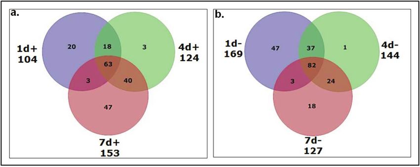
the number of bait list proteins it mapped to from our Figure 1 depicts Venn diagrams for the proteins up regu-
data versus the number of genes in the network within the lated at 1, 4 and 7 dpa (Figure 1a), and down regulated at
full set of all proteins in the networks. That is, the higher 1, 4 and 7 dpa (Figure 1b). Figure 1a shows that 7d+ has
the number of direct interactions for a TF in the given the highest number of differentially regulated proteins
proteomic dataset, the lower is the p-value. A TF connec- (153) followed by 4d+ (124) and 1d+ (104). Interestingly,
tivity map was constructed using the radial tree layout in the 4d+ group had few unique proteins (3) with most of
Cytoscape [24,25]. the proteins (63) up regulated at all three time points,
2. Upstream receptor identification similar to the down regulated group. A functional enrich-
Networks were built to specifically target the upstream ment analysis by the DAVID tool showed that 20 pro-
pathways that activate the TFs c-Myc and SP1. Recep- teins unique to the 1d+ group were enriched in cell cycle
tors of upstream pathways were identified using the processes. Forty-seven proteins unique to the 7d+ group
Analyze Network (Receptor) algorithm from Meta- were enriched in cell structure and motility, and RNA
Core™. This algorithm generates a network for each processing. Forty proteins common to the 4d+ and 7d+
receptor in the input data consisting of the shortest groups showed enrichment in metabolism, cell cycle, and
paths from it to the nearest TF. A similar p-value score, mRNA-related processes. Proteins common to all time
as described above, was used for the statistical evalua- points were enriched in intracellular protein trafficking,
tion of networks. endocytosis, chromatin packaging and neurotransmitter
release (possibly in regenerating nerves). Figure 1b shows
Pathway Analysis that 1d- has the maximum number of statistically signifi-
The target proteins of c-Myc and SP1 in the bait list as cant differentially regulated proteins (169) followed by
well as the rest of the proteins were evaluated for signif- 4d- (144) and 7d-(127). Of these, the majority of proteins
icant pathways with respect to up and downregulated (82) were downregulated at all three time points. Only 1
groups at each time point. Statistical significance was protein was unique to the 4d- group and very few pro-
measured by the number of proteins that map onto a teins (3) were common to the 1d- and 7d- groups. The
given pathway. Hence, this method did not identify functional enrichment analysis revealed that 47 proteins
pathways for each individual protein, but rather those unique to 1d-, 18 proteins unique to 7d-, and 24 proteins
which are more likely to be prevalent in the groups common to 4d- and 7d-groups were enriched in cell
Figure 1 Venn diagram for a) upregulated and b) downregulated groups. The diagram shows the number of up and down regulated proteins in
the axolotl proteomics data at 1, 4 and 7 day post amputation. 1d+ refers to the upregulated proteins and 1d- refers to the downregulated proteins at
day 1. Other time points can be similarly interpreted. The value under each time point shows the total number of proteins up/down regulated at that
time point.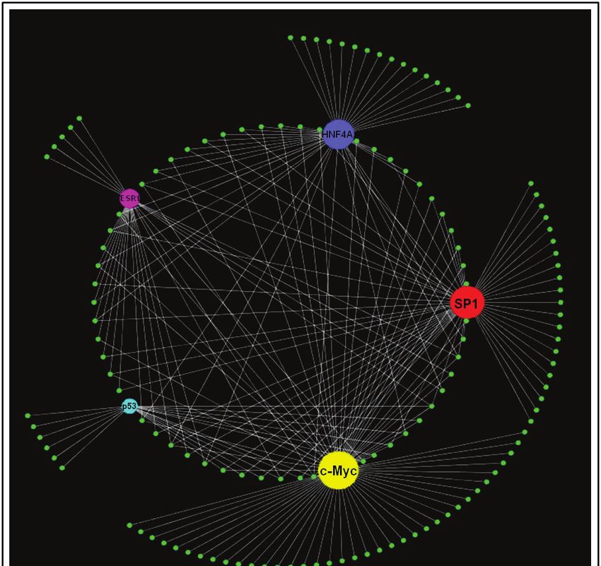
Jhamb et al. BMC Bioinformatics 2011, 12:80 Page 4 of 12
http://www.biomedcentral.com/1471-2105/12/80
cycle related processes but more proteins were involved c-Myc and SP1 were found to regulate 109 unique tar-
than in the 1d+ group. The 82 proteins common to all get proteins from our data. This number was calculated
time points were enriched in biological processes related by removing all overlapping proteins between c-Myc
to general development, cell structure and motility, mus- and SP1. Thus, c-Myc and SP1 alone were responsible
cle carbohydrate metabolism, cell cycle, and mRNA spli- for potentially regulating 36.2% of the target proteins
cing (categories specified by the DAVID tool). A from the bait list. Figures 3 and 4 show the c-Myc and
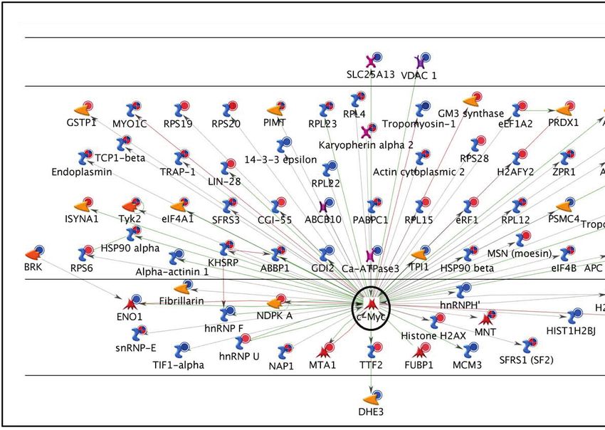
comprehensive list of all 301 proteins from the axolotl SP1 networks, respectively, with all the target proteins
proteomics data with their human orthologs, respective to which they connected.
gene names and fold change difference is provided in the
supplementary material [Additional file 1]. A list of 3. Network Construction and Pathway Analysis
enriched biological processes (obtained using DAVID) Once c-Myc and SP1 were identified as the most signifi-
for each sector in the Venn diagram can also be found in cant TFs, further investigation of the interacting
the supplementary material [Additional file 2]. upstream and downstream elements for these TFs was
carried out. Downstream elements were the target pro-
2. Transcription Factor Analysis teins for these TFs in our data (Figures 3 and 4). These
To understand a complex biological process such as limb networks were statistically found to be the most signifi-
regeneration, it is crucial to elucidate and understand its cant in our data. Many other proteins not identified by
regulatory machinery. One of the limitations of the our proteomics screen but well established in limb
LC/MS/MS method used in our original proteomic ana- regeneration, such as MMPs (matrix metalloproteinases)
lysis of blastema formation in axolotl limbs [15] is that it [28,29] and annexins [14,15] were also present in these
often fails to identify proteins expressed in low amounts networks. This further validates the significance of the
and post translationally modified proteins (PTMs) networks with respect to limb regeneration.
[26,27]. As a result, certain growth factors, signaling We identified several canonical pathways known to be
molecules and TFs have a higher probability of going present in humans or mice [30-32] spread across these net-
undetected. We used the human ortholog bait list to fish works. We found that TGF-b1 activates SP1 through
out the missed TFs by using the manually curated Meta- SMAD proteins. One of the downstream targets of SP1 is
Core™ database. Significance is based on the number of FN (this also is a well established canonical pathway).
proteins to which the TFs connected in our data. Supple- Fibronectin then activates c-Myc via integrins and the Wnt
mentary material contains information about the five pathway. These paths are highlighted in Figures 5 and 6.
most significant TFs in each data group (1d- to 7d+), the The most significant pathways regulated by target pro-
number of target proteins to which each TF linked from teins of c-Myc and SP1 in our data are provided in the
our bait list, a p-value to describe their significance, and supplementary information [Additional file 5]. Supple-
the enriched GO processes [Additional file 3]. mentary information also provides the pathways that
Figure 2 represents the overall connectivity for the five were identified using the remainder of the proteins not
most highly connected TFs from the bait list: (c-Myc, regulated by either c-Myc or SP1 [Additional file 6].
SP1, HNF4A (hepatocyte nuclear factor 4-alpha), ESR1 Among the pathways identified, several are already well
(estrogen receptor1), and cellular tumor antigen p53). established in limb regeneration such as cytoskeleton
The results show that one hundred thirty-nine (46.2% of remodeling, cell adhesion and development related, thus
the total) proteins in our bait list could potentially be validating the approach. Some pathways that have been
regulated by these five TFs (all the overlapping targets of interest recently in limb regeneration such as cell
between different TFs were removed to calculate this cycle, immune response, and metabolism were also
number). c-Myc was found to have the highest connec- identified [15].
tivity (71 targets) as well as the maximum number of
unique targets. SP1 was the second highest connected 4. Stemness in Limb Regeneration
TF (56 targets) and also had more unique targets than Blastema cells express TFs associated with stemness
the other three TFs (excluding c-Myc). Figure 2 also (e.g., Msx-1) [1,4]. Recently, combinations of the TFs
shows that for most of the other three TFs (HNF4A, c-Myc, Oct4, Sox2, Klf4, Lin28, and Nanog have been
ESR1, and p53) there was a high degree of overlap shown to reprogram adult fibroblasts to iPSCs [16,17].
(fewer unique targets) since most of their targets were c-Myc has been shown to enhance the ability of Oct4,
shared by c-Myc and SP1. For this reason, we focused Sox2 and Klf4 to induce pluripotency up to 10 fold [16].
on c-Myc and SP1 in this study. Supplementary table However, high levels of c-Myc are only transiently
lists the details of all target proteins in the bait list for required and sustained levels were found to lead to
each TF [Additional file 4]. tumors [33,34]. c-Myc, Klf4 and Sox2 have been shown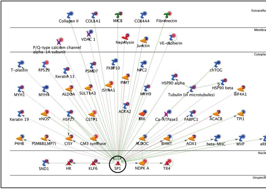
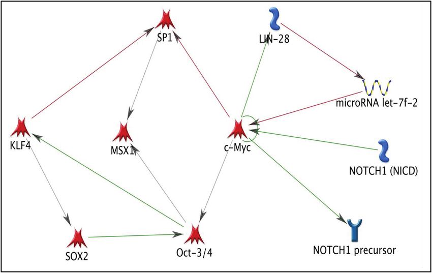
Jhamb et al. BMC Bioinformatics 2011, 12:80                                                                                             Page 5 of 12
http://www.biomedcentral.com/1471-2105/12/80

 Figure 2 Transcription factor connectivity in axolotl proteomics data. The five most highly connected TFs (c-Myc, SP1, HNF4A, ESR1 and
 p53) and their downstream targets from the bait list proteins (represented by small green circles) are shown here. The size of the TF circle
 corresponds to its connectivity; a bigger circle entails higher overall connectivity. The targets in the outer circle are unique targets of each TF
 while those in the inner circle are shared by two or more TFs.

to be expressed in regenerating newt limb tissue, and                       have been described in detail in the supplementary
Lin28 in regenerating axolotl limb tissue [15,18,35,36].                    information [Additional file [7]].
Hence, we constructed a network (Figure 7) that
included all of these TFs to evaluate their significance                    Discussion
for stemness in a mammalian system. This figure shows                       1. Human Ortholog Identification
how mammalian stem cells might be related to urodele                        Our previous proteomics study [15] identified 309 differ-
blastema cells. The various symbols used in the network                     entially regulated proteins at three time points (1, 4, and
Jhamb et al. BMC Bioinformatics 2011, 12:80                                                                                       Page 6 of 12
http://www.biomedcentral.com/1471-2105/12/80

 Figure 3 c-Myc Network. The network shows c-Myc (enclosed by a black circle) with its 71 targets from the bait list. The horizontal lines
 separate the proteins in the network into the following categories: extracellular, membrane, cytoplasm, nucleus, and unspecified. The various
 symbols used in the network have been described in detail in the supplementary information [Additional file 7].

 Figure 4 SP1 Network. The network shows SP1 (enclosed by a black circle) with its 56 targets from the bait list.
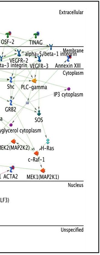
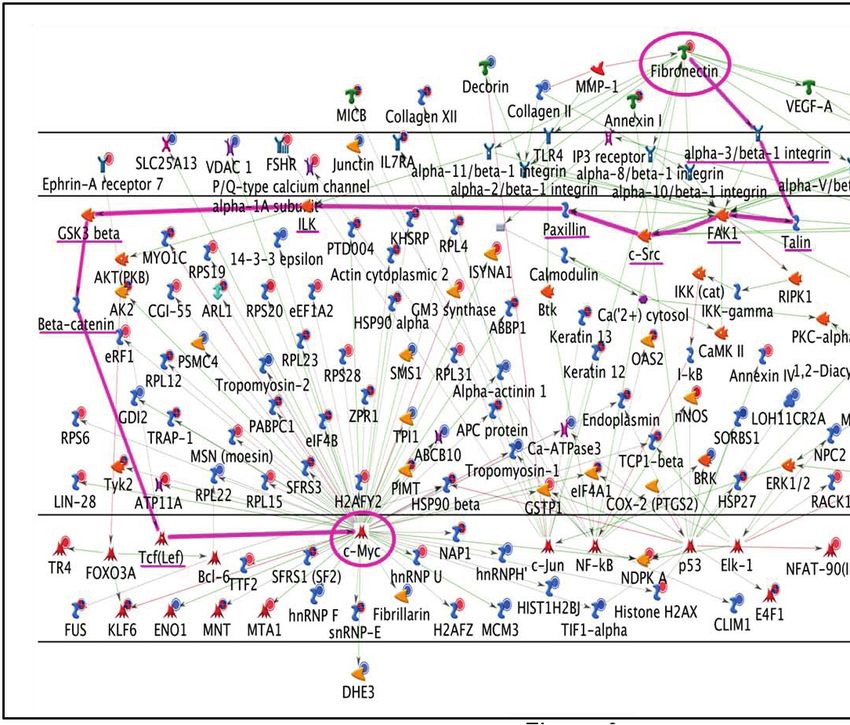
Jhamb et al. BMC Bioinformatics 2011, 12:80                                                                                      Page 7 of 12
http://www.biomedcentral.com/1471-2105/12/80

 Figure 5 TGF-b1, SP1, FN network. The path from TGF-b1 to SP1 that ultimately leads to the activation of FN has been highlighted. The start
 point (TGF-b1) and end -point (FN) are highlighted with a circle and the connecting proteins are underlined.

 Figure 6 Fibronectin - c-Myc network. This network shows that multiple signaling pathways from FN can lead to the activation of c-Myc. The
 pathway highlighted in pink includes proteins involved in canonical Wnt signaling (GSK3beta, beta-catenin, and Tcf(Lef)).
Jhamb et al. BMC Bioinformatics 2011, 12:80                                                                          Page 8 of 12
http://www.biomedcentral.com/1471-2105/12/80

                                                                     regulate 36.2% of the proteins. c-Myc was the most
                                                                     highly connected TF (71 target proteins) and also had
                                                                     the highest number of unique targets (not regulated by
                                                                     other TFs) while SP1 had the next highest TFs. For
                                                                     these reasons, we focused on these two TFs.
                                                                        c-Myc is involved in various biological processes such
                                                                     as proliferation, growth, apoptosis, energy metabolism
                                                                     and differentiation [33,34]. It has been shown to act
                                                                     with b-catenin to inhibit wound healing by interfering
                                                                     with differentiation in chronic ulcers [37] and is
                                                                     expressed in regenerating limb and lens of the newt
                                                                     Notophthalmus viridescens. In the regenerating newt
                                                                     limb, in-situ hybridization has shown that c-Myc is
 Figure 7 Stemness in limb regeneration. This network highlights     localized in both the epidermis and subjacent blastema
 the relationship of known stem cell markers (Oct3/4, Sox2, KLF4,
                                                                     cells. This expression has been correlated with the
 Lin28) with TFs identified by this study (c-Myc and SP1) and
 previously identified TFs in limb regeneration (Msx1 and Notch1).   maintenance of blastema cell proliferation [35,36].
                                                                     Recently, along with other stem cell factors, c-Myc
                                                                     expression in the regenerating newt limb was found to
7 dpa) during blastema formation in regenerating axolotl             be highest during the dedifferentiation phase of blas-
limbs. Here we used the human orthologs of these 309                 tema formation. Expression then decreased at later
proteins as bait to identify potential associations with             stages but still remained higher than the control tissue
other proteins that were not identified in the proteomics            [18]. These studies have related c-Myc to proliferation
screen. We obtained 301 significant human orthologs for              as well as stemness, but the downstream targets of
309 axolotl proteins and used them to construct interac-             c-Myc which result in these effects have not been iden-
tive protein networks. Our focus was on the identifica-              tified. Here, we have identified 71 downstream targets
tion of significant TFs and molecules regulating or                  of c-Myc in our bait list. These targets are involved in
regulated by these TFs that might be key to axolotl limb             various biological processes/pathways related to limb
regeneration.                                                        regeneration. Using this information, hypotheses about
                                                                     the specific role of c-Myc in limb regeneration can be
2. Temporal And Functional Data Analysis                             proposed and tested.
We first separated the orthologs into up and down regu-                 Specificity factor1 was the second highest connected
lated groups for each day post-amputation. In both the               TF (56 target proteins). SP1 is a ubiquitously expressed
up and down regulated groups, very few proteins (3 and               protein and has varied roles in cell growth, differentia-
1 respectively) were unique to day 4. This suggests that             tion, apoptosis, angiogenesis, tumorigenesis and immune
day 4 proteins are involved in carrying out biological pro-          response. It is known to interact with cyclins which pro-
cesses similar to either day 1 or day 7. Most of the pro-            mote the G1/S phase transition, as well as with cyclin-
teins were either up regulated at all time points or down            dependent inhibitors that inhibit progression through
regulated at all time points. Those down regulated at all            the cell cycle. Similarly, its target genes include both
time points were enriched for proteins involved in devel-            pro- and anti-apoptotic genes and pro- and anti-angio-
opment, cell structure and motility, muscle contractile              genic genes. Specificity factor1 is also linked to chroma-
activity, carbohydrate metabolism, cell cycle, and mRNA              tin remodeling through its interaction with p300 and
splicing, whereas those up regulated at all time points              histone deacetylases (HDACs) and is known to interact
were enriched for intracellular protein trafficking, endo-           with several TFs including c-Myc in order to activate
cytosis, chromatin packaging and neurotransmitter                    several downstream target genes. However, SP1 action is
release (possibly in regenerating nerves).                           highly dependent on its interaction with other members
                                                                     of the SP family and extracellular signals [38-40]. This is
3. Transcription Factor Network Analysis                             the first time SP1 has been identified in relation to limb
Next, we derived a TF-centric protein interaction net-               regeneration and no studies have yet been done on the
work using commercially available MetaCore™ software                 specific role it plays in limb regeneration.
to identify all human TFs that were connected to at
least five proteins in the data. The five most highly con-           4. Stem Cell Factors In Limb Regeneration
nected TFs, c-Myc, SP1, HNF4A, ESR1 and p53, were                    A number of TFs associated with stemness are expressed
found to regulate almost half of the proteins (46.2%) in             during blastema formation. Some of these, such as the
our data. Of these, c-Myc and SP1 alone were found to                Hox and Meis genes, regulate pattern formation in the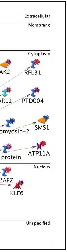
Jhamb et al. BMC Bioinformatics 2011, 12:80                                                                      Page 9 of 12
http://www.biomedcentral.com/1471-2105/12/80

growing blastema [41-46], whereas others such as msx-1,         on FN produced by the eccentric AEC. TGF-b1 is strongly
nrad, Klf4, Oct4, Sox2, and Lin28 are associated with           up regulated during blastema formation in amputated axo-
dedifferentiation and proliferation [15,18,47-51]. With         lotl limbs [52]. FN is a target gene of TGF-b1 that is highly
the exception of Lin28, we did not identify any of these        expressed by basal cells of the wound epidermis during
TFs in our proteomic analysis of blastema formation in          blastema formation [54]. Inhibition of TGF-b1 expression
the axolotl hind limb [15], but Figure 5 demonstrates           with SB-431542, reduces FN expression and results in fail-
that they interact with c-Myc and SP1. This suggests that       ure of blastema formation [52], again suggesting that FN
c- Myc and SP1 are central to a network of TFs that reg-        provided by the AEC provides directional guidance for
ulate mesenchymal stem cell properties of the blastema          blastema cells.
and that play a role in the nuclear reprogramming of dif-         In the present study, we identified a canonical pathway
ferentiated limb cells to blastema cells.                       in which TGF-b1 leads to the activation of SP1 through
                                                                TGF-b receptors and SMAD3. Transforming growth fac-
5. Pathway Analysis                                             tor-b1 is one of the major inducers of epithelial-
Next we mapped out the pathways of two molecules, TGF-          mesenchymal transformation (EMT) via SMAD family
b1 and FN, that connect SP1 and c-Myc, respectively, and        member proteins (SMAD2, SMAD3 and SMAD4)
which are known to be important in mammalian tissue             [58,59]. The epidermal cells that establish the wound epi-
repair and urodele limb regeneration. Within the TGF            dermis in regenerating urodeles limbs take on some of
superfamily, the TGF-b and BMP subfamilies of growth            the characteristics of mesenchymal cells, shedding their
factors are major players in skin wound repair and bone         specialized epithelial junctions and up regulating cytoske-
regeneration. Transforming growth factor beta isoforms          letal components essential for migration. TGF-b1 binds
attract immune cells into skin wounds, and induce the           Type I and type II receptor serine/threonine kinases. The
migration and proliferation of skin fibroblasts to form         receptor type II phosphorylates the receptor type I,
granulation tissue. Transforming growth factor b activates      which activate SMADs [31,32] and SMAD3 then leads to
many target genes in wound healing, including connective        activation of SP1 which is capable of activating FN [30].
tissue growth factor (CTGF) and FN [52].                        Interestingly, there is a non-canonical TGF-b1 pathway
   The wound epidermis covering the amputation surface          in which SMAD 3 can repress c-Myc through a novel
plays a crucial role in blastema formation [11]. Establish-     repressive SMAD binding element within the TGF-b
ment of the wound epidermis after amputation of the             inhibitory element of the c-Myc promoter [60]. Wound
Xenopus tadpole tail requires TGF-b signaling [53]. TGF-        epidermal cells migrating over the amputation surface do
b and SMAD 2 are up regulated early in formation of the         not divide [11]. In this context, SMAD3 could possibly
wound epidermis and later in the tissues of the blastema.       inhibit the division of migrating epidermal cells via this
Inhibition of TGF-b signaling with the inhibitor of SMAD        pathway.
phosphorylation SB-431542 immediately after amputation            Figure 6 illustrates multiple pathways that lead to c-Myc
prevents establishment of the wound epidermis and inhi-         activation from FN. The highlighted pathway is the longest
bits the signaling cascades that initiate blastema formation.   canonical pathway and it involves the cell adhesion pro-
Fibronectin is an important substrate molecule for both         teins talin, FAK1 (focal adhesion kinase1), c-Src, Paxillin,
migrating epidermal cells and fibroblasts of wounds. FN is      ILK (integrin-linked protein kinase) and components of
strongly upregulated during blastema formation in axolotl       the canonical Wnt signaling pathway (GSK3-b-glycogen
limb regeneration [15]. A prerequisite for blastema forma-      synthase kinase-3 beta, b-catenin, and Tcf/Lef (T-cell-
tion and growth in urodele limb regeneration is the thick-      specific transcription factor/lymphoid enhancer-binding
ening of the initial wound epidermis to form the AEC.           factor 1). Wnt signaling is known to control cell prolifera-
Fibronectin is an important component of the blastemal          tion and cell fate determination. Members of the Wnt and
ECM and is highly expressed by the basal layer of the AEC       BMP pathways have been shown to be required in verte-
starting within 24 hrs after amputation, as well as by blas-    brates for normal limb development [61]. Canonical
tema cells themselves [54,55]. The AEC appears to direct        Wnt signaling is also known to keep stem cells in a self-
the migration of blastema cells to form the accumulation        renewing and undifferentiated state[62]. Loss- and gain-of-
blastema beneath it. This was shown by experiments in           function experiments in axolotl, Xenopus, and zebrafish
which shifting the position of the AEC laterally caused a       showed that Wnt signaling is required for limb and fin
corresponding shift in blastema cell accumulation [56],         regeneration [61]. Another study in zebrafish and chick
and transplantation of an additional AEC to the base of         embryos has identified molecular interactions of Wnt2b
the blastema resulted in supernumerary blastema forma-          with Tbx5 that are responsible for limb identity and out-
tion [57]. The redirected accumulation of blastema cells in     growth [63]. These findings indicate that Wnt signaling is
these experiments may be due to the migration of the cells      probably required for the activation of c-Myc.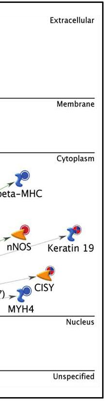
Jhamb et al. BMC Bioinformatics 2011, 12:80                                                                                                Page 10 of 12
http://www.biomedcentral.com/1471-2105/12/80

Conclusions                                                                     Additional file 4: The five most highly connected TFs with their
In the present study we asked whether the use of pro-                           target proteins. The gene symbols (from additional file 1) are defined
teins identified in a proteomic analysis of blastema for-                       for the target proteins of each TF.
mation in amputated axolotl hindlimbs could be used as                          Additional file 5: Pathways for the target proteins of c-Myc and SP1
                                                                                in each up and downregulated group of proteins. The list of
bait to identify transcription factors and their down-                          significant pathway names as obtained from MetaCore with their
stream targets involved in blastema formation, and con-                         respective p-values for the target proteins of c-Myc and SP1 in each up
struct these into interactive protein networks and                              and downregulated group of 1, 4 and 7 dpa. The pathways are arranged
                                                                                in decreasing order of significance within each group.
pathways. We identified multiple targets of c-Myc and
                                                                                Additional file 6: Pathways for those target proteins not regulated
SP1, and also several upstream molecules (TGF-b recep-                          by c-Myc and SP1 in each up and downregulated group of
tors, SMADs, and cell adhesion molecules) that lead to                          proteins. The list of significant pathway names obtained from MetaCore,
the activation of c-Myc and SP1. We conclude that this                          with their respective p-values. The pathways are arranged in decreasing
                                                                                order of significance within each group.
strategy can be successful, not only for transcription fac-
                                                                                Additional file 7: Reference for the symbols used in the
tors and their targets, but for other molecules as well                         construction of networks. Reference guide for various symbols used in
that might be important to regeneration and/or wound                            the networks of Figures 3 to 7.
repair.
  The next step is to construct hypotheses that allow
experimental testing of the roles of the molecules compris-                 Acknowledgements
ing the interactive protein pathways in regeneration-com-                   This work was supported by grants from the W.M. Keck Foundation and the
petent limbs. This will unite the question-driven approach                  US Army Research Office (#W911NF07-10176) to D.L.S.
used here with the hypothesis-driven approach. Both are                     Author details
equally important for analysis and synthesis of data                        1
                                                                             School of Informatics, Indiana University-Purdue University Indianapolis,
derived from complex biological processes. We are cur-                      Indianapolis, IN, 46202, USA. 2Department of Biology, Indiana University-
                                                                            Purdue University Indianapolis, Indianapolis, IN, 46202, USA. 3Department of
rently testing one hypothesis about the role of the centro-                 Cell and Developmental Biology, and Regeneration Biology and Tissue
somal protein Evi5 (ecotropic viral integration factor 5)                   Engineering Theme, Institute for Genomic Biology, University of Illinois-
and the pathways it forms with several other proteins reg-                  Urbana Champaign, Urbana, IL, 61801, USA. 4Department of Oral Biology,
                                                                            School of Dentistry, Indiana University-Purdue University Indianapolis,
ulating the cell cycle during blastema formation and                        Indianapolis, IN, 46202, USA. 5Center for Regenerative Biology and Medicine,
growth [15].                                                                Indiana University-Purdue University Indianapolis, Indianapolis, IN, 46202,
  Finally, by deriving proteomic data from a regenera-                      USA. 6American University of Antigua, College of Medicine, University Park,
                                                                            Jabberwock, Coolidge, Antigua, P. O. Box W-1451, West Indies.
tion-deficient system such as the limbs of Xenopus frog-
lets, and applying a bioinformatics/systems biology                         Authors’ contributions
approach, we have the possibility of identifying a set of                   DJ: conception of project, conception and conduct of experimental design,
                                                                            data analysis, writing and revision of manuscript, figure and table
proteins, networks and pathways that can be compared                        preparation; NR: source of proteomic data, manuscript critique and revision;
to that of the regeneration-competent axolotl to reveal                     DJM: writing of manuscript, manuscript critique and revision; FS: manuscript
the basis for the difference between the two.                               critique and revision; JAC: manuscript critique and revision; DLS: conception
                                                                            of project, data analysis, writing and revision of manuscript; MP: conception
                                                                            of project, bioinformatics tools, writing and revision of manuscript. All
Additional material                                                         authors read and approved the final manuscript.

                                                                            Received: 20 August 2010 Accepted: 18 March 2011
 Additional file 1: Axolotl proteomics data with its respective human       Published: 18 March 2011
 orthologs, gene symbol and fold changes at 1, 4, and 7 dpa. FC_1 D
 refers to the fold change at 1 dpa with respect to control. FC_4 D and
 FC_7 D can be similarly interpreted. A negative value implies              References
 downregulation with respect to control and a positive fold change value    1. Stocum DL, Zupanc GK: Stretching the limits: stem cells in regeneration
 implies upregulation.                                                          science. Dev Dyn 2008, 237(12):3648-3671.
                                                                            2. Stocum DL: Regenerative Biology and Medicine. Elsevier Inc.; 2006.
 Additional file 2: Biological process enrichment for the protein           3. Spallanzani L: Concepts of generation and regeneration. In A History of
 categories in Figure 1. The DAVID tool was used to obtain the                  Regeneration Research Edited by: Dinsmore CE 1991.
 biological process enrichment for each category of proteins represented    4. Bryant SV, Endo T, Gardiner DM: Vertebrate limb regeneration and the
 in Figure 1. The associated biological process terms, number of proteins       origin of limb stem cells. Int J Dev Biol 2002, 46(7):887-896.
 (count), percentage, p-value, and genes obtained from the DAVID tool       5. Nye HL, Cameron JA, Chernoff EA, Stocum DL: Regeneration of the
 are mentioned for each group of proteins.                                      urodele limb: a review. Dev Dyn 2003, 226(2):280-294.
 Additional file 3: The five most highly connected TFs for each up          6. Morrison JI, Loof S, He P, Simon A: Salamander limb regeneration involves
 and downregulated group of proteins. The table shows the GO                    the activation of a multipotent skeletal muscle satellite cell population. J
 processes, root nodes (the number of target proteins) and an overall           Cell Biol 2006, 172(3):433-440.
 p-value for each TF in the up or downregulated group. The numbers in       7. Carlson BM: Principles of Regenerative Biology. Academic Press; 2007.
 parenthesis in the third column indicate the percentage of target          8. Sauer U, Heinemann M, Zamboni N: Genetics. Getting closer to the whole
 proteins and a p-value for that particular GO term with respect to the         picture. Science 2007, 316(5824):550-551.
 target proteins of a given TF. The TFs are arranged in decreasing order    9. Putta S, Smith JJ, Walker JA, Rondet M, Weisrock DW, Monaghan J,
 of significance (higher p-value) within each group.                            Samuels AK, Kump K, King DC, Maness NJ, et al: From biomedicine to
Jhamb et al. BMC Bioinformatics 2011, 12:80 Page 11 of 12
http://www.biomedcentral.com/1471-2105/12/80
natural history research: EST resources for ambystomatid salamanders. 34. Eilers M, Eisenman RN: Myc’s broad reach. Genes Dev 2008,
BMC Genomics 2004, 5(1):54. 22(20):2755-2766.
10. Monaghan JR, Epp LG, Putta S, Page RB, Walker JA, Beachy CK, Zhu W, 35. Hourdry J, Geraudie J, Singer M, Mechali M: Expression of the c-Myc
Pao GM, Verma IM, Hunter T, et al: Microarray and cDNA sequence proto-oncogene in the ofrelimb regenerate of the newt Notophthalmus
analysis of transcription during nerve-dependent limb regeneration. BMC Viridescens, visualized by in situ hybridization. M Singer Symposium 1988,
Biol 2009, 7:1. 307-313.
11. Stocum DL, Rao N: Mechanisms of Blastema Formation in Regenerating 36. Geraudie J, Hourdry J, Boehm K, Singer M, Mechali M: c-Myc proto-
Amphibian Limbs. In Principles of Regenerative Medicine. Edited by: Lanza R, oncogene expression during newt limb regeneration. In Recent Trends in
Thompson J, Nerem R. Elsevier/Academic Press San Diego; . Regeneration Research. Volume 172. Edited by: Kiortsis V, Koussoulallos S,
12. King MW, Nguyen T, Calley J, Harty MW, Muzinich MC, Mescher AL, Wallace H. New York: Plenum Press; 1989:27-36.
Chalfant C, N’Cho M, McLeaster K, McEntire J, et al: Identification of genes 37. Stojadinovic O, Brem H, Vouthounis C, Lee B, Fallon J, Stallcup M,
expressed during Xenopus laevis limb regeneration by using subtractive Merchant A, Galiano RD, Tomic-Canic M: Molecular pathogenesis of
hybridization. Dev Dyn 2003, 226(2):398-409. chronic wounds: the role of beta-catenin and c-myc in the inhibition of
13. Grow M, Neff AW, Mescher AL, King MW: Global analysis of gene epithelialization and wound healing. Am J Pathol 2005, 167(1):59-69.
expression in Xenopus hindlimbs during stage-dependent complete and 38. Wierstra I: Sp1: emerging roles–beyond constitutive activation of TATA-
incomplete regeneration. Dev Dyn 2006, 235(10):2667-2685. less housekeeping genes. Biochem Biophys Res Commun 2008, 372(1):1-13.
14. King MW, Neff AW, Mescher AL: Proteomics analysis of regenerating 39. Tan NY, Khachigian LM: Sp1 phosphorylation and its regulation of gene
amphibian limbs: changes during the onset of regeneration. Int J Dev transcription. Mol Cell Biol 2009, 29(10):2483-2488.
Biol 2009, 53(7):955-969. 40. Safe S, Abdelrahim M: Sp transcription factor family and its role in
15. Rao N, Jhamb D, Milner DJ, Li B, Song F, Wang M, Voss SR, Palakal M, cancer. Eur J Cancer 2005, 41(16):2438-2448.
King MW, Saranjami B, et al: Proteomic analysis of blastema formation in 41. Simon HG, Tabin CJ: Analysis of Hox-4.5 and Hox-3.6 expression during
regenerating axolotl limbs. BMC Biol 2009, 7:83. newt limb regeneration: differential regulation of paralogous Hox genes
16. Takahashi K, Yamanaka S: Induction of pluripotent stem cells from mouse suggest different roles for members of different Hox clusters.
embryonic and adult fibroblast cultures by defined factors. Cell 2006, Development 1993, 117(4):1397-1407.
126(4):663-676. 42. Savard P, Gates PB, Brockes JP: Position dependent expression of a
17. Yu J, Vodyanik MA, Smuga-Otto K, Antosiewicz-Bourget J, Frane JL, Tian S, homeobox gene transcript in relation to amphibian limb regeneration.
Nie J, Jonsdottir GA, Ruotti V, Stewart R, et al: Induced pluripotent stem EMBO J 1988, 7(13):4275-4282.
cell lines derived from human somatic cells. Science 2007, 43. Gardiner DM, Blumberg B, Komine Y, Bryant SV: Regulation of HoxA
318(5858):1917-1920. expression in developing and regenerating axolotl limbs. Development
18. Maki N, Suetsugu-Maki R, Tarui H, Agata K, Del Rio-Tsonis K, Tsonis PA: 1995, 121(6):1731-1741.
Expression of stem cell pluripotency factors during regeneration in 44. Brown R, Brockes JP: Identification and expression of a regeneration-
newts. Dev Dyn 2009, 238(6):1613-1616. specific homeobox gene in the newt limb blastema. Development 1991,
19. BLAST. [http://blast.ncbi.nlm.nih.gov/Blast.cgi]. 111(2):489-496.
20. UniProt. [http://www.uniprot.org/]. 45. Beauchemin M, Noiseux N, Tremblay M, Savard P: Expression of Hox A11
21. Dennis G Jr, Sherman BT, Hosack DA, Yang J, Gao W, Lane HC, Lempicki RA: in the limb and the regeneration blastema of adult newt. Int J Dev Biol
DAVID: Database for Annotation, Visualization, and Integrated Discovery. 1994, 38(4):641-649.
Genome Biol 2003, 4(5):P3. 46. Mercader N, Tanaka EM, Torres M: Proximodistal identity during
22. Huang da W, Sherman BT, Lempicki RA: Systematic and integrative vertebrate limb regeneration is regulated by Meis homeodomain
analysis of large gene lists using DAVID bioinformatics resources. Nat proteins. Development 2005, 132(18):4131-4142.
Protoc 2009, 4(1):44-57. 47. Cadinouche MZ, Liversage RA, Muller W, Tsilfidis C: Molecular cloning of
23. Ekins S, Nikolsky Y, Bugrim A, Kirillov E, Nikolskaya T: Pathway mapping the Notophthalmus viridescens radical fringe cDNA and characterization
tools for analysis of high content data. Methods Mol Biol 2007, of its expression during forelimb development and adult forelimb
356:319-350. regeneration. Dev Dyn 1999, 214(3):259-268.
24. Shannon P, Markiel A, Ozier O, Baliga NS, Wang JT, Ramage D, Amin N, 48. Crews L, Gates PB, Brown R, Joliot A, Foley C, Brockes JP, Gann AA:
Schwikowski B, Ideker T: Cytoscape: a software environment for Expression and activity of the newt Msx-1 gene in relation to limb
integrated models of biomolecular interaction networks. Genome Res regeneration. Proc Biol Sci 1995, 259(1355):161-171.
2003, 13(11):2498-2504. 49. Shimizu-Nishikawa K, Tsuji S, Yoshizato K: Identification and
25. Killcoyne S, Carter GW, Smith J, Boyle J: Cytoscape: a community-based characterization of newt rad (ras associated with diabetes), a gene
framework for network modeling. Methods Mol Biol 2009, 563:219-239. specifically expressed in regenerating limb muscle. Dev Dyn 2001,
26. Feng X, Liu X, Luo Q, Liu BF: Mass spectrometry in systems biology: an 220(1):74-86.
overview. Mass Spectrom Rev 2008, 27(6):635-660. 50. Koshiba K, Kuroiwa A, Yamamoto H, Tamura K, Ide H: Expression of Msx
27. Prakash A, Mallick P, Whiteaker J, Zhang H, Paulovich A, Flory M, Lee H, genes in regenerating and developing limbs of axolotl. J Exp Zool 1998,
Aebersold R, Schwikowski B: Signal maps for mass spectrometry-based 282(6):703-714.
comparative proteomics. Mol Cell Proteomics 2006, 5(3):423-432. 51. Schnapp E, Tanaka EM: Quantitative evaluation of morpholino-mediated
28. Yang EV, Gardiner DM, Carlson MR, Nugas CA, Bryant SV: Expression of protein knockdown of GFP, MSX1, and PAX7 during tail regeneration in
Mmp-9 and related matrix metalloproteinase genes during axolotl limb Ambystoma mexicanum. Dev Dyn 2005, 232(1):162-170.
regeneration. Dev Dyn 1999, 216(1):2-9. 52. Levesque M, Gatien S, Finnson K, Desmeules S, Villiard E, Pilote M, Philip A,
29. Park IS, Kim WS: Modulation of gelatinase activity correlates with the Roy S: Transforming growth factor: beta signaling is essential for limb
dedifferentiation profile of regenerating salamander limbs. Mol Cells regeneration in axolotls. PLoS One 2007, 2(11):e1227.
1999, 9(2):119-126. 53. Ho DM, Whitman M: TGF-beta signaling is required for multiple
30. Pardali K, Kurisaki A, Moren A, ten Dijke P, Kardassis D, Moustakas A: Role of processes during Xenopus tail regeneration. Dev Biol 2008,
Smad proteins and transcription factor Sp1 in p21(Waf1/Cip1) regulation 315(1):203-216.
by transforming growth factor-beta. J Biol Chem 2000, 54. Christensen RN, Tassava RA: Apical epithelial cap morphology and
275(38):29244-29256. fibronectin gene expression in regenerating axolotl limbs. Dev Dyn 2000,
31. Bachman KE, Park BH: Duel nature of TGF-beta signaling: tumor 217(2):216-224.
suppressor vs. tumor promoter. Curr Opin Oncol 2005, 17(1):49-54. 55. Nace JD, Tassava RA: Examination of fibronectin distribution and its
32. Shi Y, Massague J: Mechanisms of TGF-beta signaling from cell sources in the regenerating newt limb by immunocytochemistry and in
membrane to the nucleus. Cell 2003, 113(6):685-700. situ hybridization. Dev Dyn 1995, 202(2):153-164.
33. Knoepfler PS: Why myc? An unexpected ingredient in the stem cell 56. Thornton CS: Influence of an eccentric epidermal cap on limb
cocktail. Cell Stem Cell 2008, 2(1):18-21. regeneration in Amblystoma larvae. Dev Biol 1960, 2:551-569.Jhamb et al. BMC Bioinformatics 2011, 12:80 Page 12 of 12
http://www.biomedcentral.com/1471-2105/12/80
57. Thornton CS, Thronton MT: The regeneration of accessory limb parts
following epidermal cap transplantation in urodeles. Experientia 1965,
21(3):146-148.
58. Cho HJ, Baek KE, Saika S, Jeong MJ, Yoo J: Snail is required for
transforming growth factor-beta-induced epithelial-mesenchymal
transition by activating PI3 kinase/Akt signal pathway. Biochem Biophys
Res Commun 2007, 353(2):337-343.
59. Zavadil J, Bottinger EP: TGF-beta and epithelial-to-mesenchymal
transitions. Oncogene 2005, 24(37):5764-5774.
60. Frederick JP, Liberati NT, Waddell DS, Shi Y, Wang XF: Transforming growth
factor beta-mediated transcriptional repression of c-myc is dependent
on direct binding of Smad3 to a novel repressive Smad binding
element. Mol Cell Biol 2004, 24(6):2546-2559.
61. Kawakami Y, Rodriguez Esteban C, Raya M, Kawakami H, Marti M, Dubova I,
Izpisua Belmonte JC: Wnt/beta-catenin signaling regulates vertebrate
limb regeneration. Genes Dev 2006, 20(23):3232-3237.
62. Ling L, Nurcombe V, Cool SM: Wnt signaling controls the fate of
mesenchymal stem cells. Gene 2009, 433(1-2):1-7.
63. Ng JK, Kawakami Y, Buscher D, Raya A, Itoh T, Koth CM, Rodriguez
Esteban C, Rodriguez-Leon J, Garrity DM, Fishman MC, et al: The limb
identity gene Tbx5 promotes limb initiation by interacting with Wnt2b
and Fgf10. Development 2002, 129(22):5161-5170.
doi:10.1186/1471-2105-12-80
Cite this article as: Jhamb et al.: Network based transcription factor
analysis of regenerating axolotl limbs. BMC Bioinformatics 2011 12:80.
Submit your next manuscript to BioMed Central
and take full advantage of:
• Convenient online submission
• Thorough peer review
• No space constraints or color figure charges
• Immediate publication on acceptance
• Inclusion in PubMed, CAS, Scopus and Google Scholar
• Research which is freely available for redistribution
Submit your manuscript at
www.biomedcentral.com/submitYou can also read


