The Kinesinlike Protein Subito Contributes to Central Spindle Assembly and Organization of the Meiotic Spindle in Drosophila Oocytes D
←
→
Page content transcription
If your browser does not render page correctly, please read the page content below
Molecular Biology of the Cell
Vol. 16, 4684 – 4694, October 2005

The Kinesinlike Protein Subito Contributes to Central
Spindle Assembly and Organization of the Meiotic
Spindle in Drosophila Oocytes□D

J. K. Jang, T. Rahman, and K. S. McKim

Waksman Institute and Department of Genetics, Rutgers, The State University of New Jersey, Piscataway,
NJ 08854-8020

Submitted November 3, 2004; Revised July 12, 2005; Accepted July 19, 2005
Monitoring Editor: Ted Salmon

In the oocytes of many species, bipolar spindles form in the absence of centrosomes. Drosophila melanogaster oocyte
chromosomes have a major role in nucleating microtubules, which precedes the bundling and assembly of these
microtubules into a bipolar spindle. Here we present evidence that a region similar to the anaphase central spindle
functions to organize acentrosomal spindles. Subito mutants are characterized by the formation of tripolar or monopolar
spindles and nondisjunction of homologous chromosomes at meiosis I. Subito encodes a kinesinlike protein and
associates with the meiotic central spindle, consistent with its classification in the Kinesin 6/MKLP1 family. This class of
proteins is known to be required for cytokinesis, but our results suggest a new function in spindle formation. The meiotic
central spindle appears during prometaphase and includes passenger complex proteins such as AurB and Incenp. Unlike
mitotic cells, the passenger proteins do not associate with centromeres before anaphase. In the absence of Subito, central
spindle formation is defective and AurB and Incenp fail to properly localize. We propose that Subito is required for
establishing and/or maintaining the central spindle in Drosophila oocytes, and this substitutes for the role of centrosomes
in organizing the bipolar spindle.

INTRODUCTION                                                           sively studied is NCD, a C-terminal motor kinesin. Consis-
                                                                       tent with a role in focusing the poles, ncd mutant spindles
In the oocytes of many animals, bipolar spindles form in the           are frequently multipolar or apolar (Hatsumi and Endow,
absence of centrosomes (Compton, 2000; Karsenti and Ver-               1992; Matthies et al., 1996). Nonmotor proteins have also
nos, 2001). In the acentrosomal pathway for spindle forma-             been shown to make important contributions to Drosophila
tion, the chromosomes trigger spindle formation by captur-             acentrosomal spindle organization. For example, spindle
ing free microtubules that are present in the cytoplasm                pole-associated proteins TACC and MSPS have a role in
(Theurkauf and Hawley, 1992; McKim and Hawley, 1995).                  bipolar spindle pole formation (Cullen and Ohkura, 2001).
These microtubules are then bundled and sorted to generate             The AXS protein is present within a structure ensheathing
two poles in a process that involves a variety of motor                the meiotic spindle and has a role in meiotic spindle assem-
protein–microtubule interactions (Matthies et al., 1996; Wal-          bly (Kramer and Hawley, 2003). These studies suggest there
czak et al., 1998). Plus-end-directed motors of the BimC class         are important proteins or structures that modulate the inter-
such as Eg5 are proposed to generate bundles of antiparallel           action of motors and microtubules in acentrosomal spindle
microtubules, an activity that could be important for pro-             assembly.
moting the formation of bipolar instead of monopolar spin-                Previous studies in Drosophila oocytes have suggested that
dles (Karsenti and Vernos, 2001). Minus-end-directed mo-               the process of acentrosomal spindle formation is initiated by
tors such as kinesins in the C-terminal motor class or dynein          the capture of free microtubules by the chromosomes fol-
have been proposed to bundle parallel microtubules and                 lowed by bundling and sorting of microtubules by minus-
taper them into defined poles (Matthies et al., 1996; Walczak          end-directed motors and the accumulation of certain pro-
et al., 1998).                                                         teins at the spindle poles (Matthies et al., 1996; Cullen and
   Although the activities of a variety of motors has been             Ohkura, 2001). In this article we have built on this model by
studied in such systems as Xenopus extracts, the formation of          investigating the role of the subito (sub) gene in acentrosomal
acentrosomal spindles in vivo is still poorly understood.              spindle formation. sub encodes a kinesinlike protein whose
Although Drosophila female meiosis is an excellent system to           sequence is most similar to the MKLP1 (mitotic kinesin like
study acentrosomal spindle formation, the only motor pro-              protein 1) family (Giunta et al., 2002) (now Kinesin 6, Dagen-
tein with a role in spindle assembly that has been exten-              bach and Endow, 2004). Mutants in sub have a phenotype
                                                                       consistent with a role in organizing the bipolar spindle.
                                                                       Female meiosis in sub mutants is characterized by the for-
This article was published online ahead of print in MBC in Press       mation of monopolar and tripolar spindles and the nondis-
(http://www.molbiolcell.org/cgi/doi/10.1091/mbc.E04 –11– 0964)
on July 29, 2005.
                                                                       junction of homologous chromosomes during the first mei-
                                                                       otic division (Giunta et al., 2002).
□
D
   The online version of this article contains supplemental material      Here we show that SUB protein is bound to the meiotic
at MBC Online (http://www.molbiolcell.org).                            central spindle, a pattern that is consistent with its assign-
Address correspondence to: Kim S. McKim (mckim@rci.rutgers.edu).       ment to the MKLP1 class of proteins. SUB and the central

4684                                                                                   © 2005 by The American Society for Cell Biology
Acentrosomal Spindle Formation

                                                                                       nis et al., 1997) and ␣-Tub67C1 and ␣-Tub67C2 are females sterile alleles of a
                                                                                       specialized ␣-tubulin gene (Matthews et al., 1993).

                                                                                       Germ-line Clone Analysis of Incenp
                                                                                       Drosophila Incenp mutations cause early larval lethality (M. Carmena, personal
                                                                                       communication). To study the role of Incenp during female meiosis, we
                                                                                       generated mutant germline clones in P{hsFLP}12/⫹; P{FRT(whs)}G13 Incen-
                                                                                       pEP2340/ P{FRT(whs)}G13 P{ovoD1–18}2R females. These females were generated
                                                                                       by crossing P{hsFLP}12; P{FRT(whs)}G13 IncenpEP2340/CyO females to ⫹/Y;
                                                                                       P{FRT(whs)}G13 P{ovoD1–18}2R/CyO males for 2 d ,followed by heat shock for
                                                                                       1 h at 37°C on days 3 and 4. The P{hsFLP}12/⫹; P{FRT(whs)}G13 IncenpEP2340/
                                                                                       P{FRT(whs)}G13 P{ovoD1–18}2R females were expected to lack mature oocytes
                                                                                       because of the dominant ovoD1 mutation. If a mitotic recombination event
                                                                                       occurred, germline cells homozygous for incenpEP2340 would be produced.
                                                                                       However, in no cases were oocytes observed from ⬃50 heat shocked females.
                                                                                       In contrast, most of the 23 females in a control experiment without the
                                                                                       IncenpEP2340 mutation produced mature oocytes. These results suggest that
                                                                                       Incenp is required for early stages of oocyte development.

                                                                                       Generation of the SUB Antibody
                                                                                       A carboxy-terminal fragment of sub encoding amino acids 498 – 628 was
                                                                                       cloned from a cDNA (Stapleton et al., 2002) as an EcoRI-XhoI fragment into
                                                                                       pET30A (Novagen) and expressed in E. coli BL(21)DE3. This fragment con-
                                                                                       tains the poorly conserved C-terminal domain of sub that follows the motor
                                                                                       domain. The fusion proteins were purified using His-binding Ni2⫹ binding
                                                                                       resin columns (Novagen) under denaturing conditions. After electroelution,
                                                                                       the proteins were concentrated, dialyzed into 50 mM HEPES pH 7.5 and used
                                                                                       to raise antibodies in rats (Covance, Denver, PA).

                                                                                       Western Blotting and Immunofluorescence Microscopy
                                                                                       Total ovary protein was isolated by dissecting whole ovaries from yeasted
                                                                                       females in phosphate-buffered saline and then grinding and boiling them in
Figure 1. Subito (SUB) is a kinesinlike protein in the MKLP1                           SDS gel loading buffer. Protein from ⬃2–3 ovaries was loaded per lane. The
family. (A) Schematic of SUB protein organization showing the                          rat anti-SUB primary antibody was used at 1:5000 and the secondary anti-rat
amino acid changes in the known mutants. sub131 deletes most of the                    HRP antibody (Jackson ImmunoResearch Laboratories, West Grove, PA) was
subcoding region (Giunta et al., 2002) and sub1 is a nonsense muta-                    used at 1:5000. The secondary was detected using ECL reagents (Amersham,
tion. (B) Sequence alignment of the neck-linker region in the Dro-                     Piscataway, NJ).
                                                                                          For immunofluorescence, stage 14 oocytes were collected from 3- to 7-d-old
sophila and human MKLP1 group along with two other Drosophila                          yeast-fed females and fixed as described previously and mounted in Vectash-
proteins, conventional kinesin heavy chain and KLP3A. The black                        ield (Vector Laboratories, Burlingame, CA; Theurkauf and Hawley, 1992;
arrow indicates the Polo kinase site identified by Neef et al. (2003),                 McKim et al., 1993). Anaphase oocytes were collected by allowing females to
the gray arrow indicates a second conserved serine, and the stars                      lay eggs on grape juice agar plates for 20 min after a short precollection
denote identical or similar amino acids unique to MKLP2 and SUB.                       period. The embryos were dechorionated in 50% bleach for 2 min and then
(C) Western blot of ovary protein using anti-SUB antibodies. A                         fixed in cold heptane/methanol (Rothwell and Sullivan, 2000). Oocytes were
smaller nonspecific band and a Ponceau S-stained membrane serve                        stained for DNA with Hoescht and for microtubules with anti-tubulin mono-
as loading controls.                                                                   clonal antibody DM1A (at 1:50) directly conjugated to FITC (Sigma, St. Louis,
                                                                                       MO). When examining sub mutant oocytes, heterozygotes for protein null
                                                                                       alleles, either sub1/sub131 or sub1/sub202, were often used. Heterozygotes were
                                                                                       used to eliminate potential genetic background effects but the same results
                                                                                       were observed in sub1, sub131, or sub202 homozygotes. The rat anti-SUB anti-
spindle appear at the earliest stages of prometaphase, before                          body was used at 1:75 combined with either a Cy3 or Cy5 anti-rat secondary
bipolar spindle formation. Indeed, SUB is required for as-                             antibody adsorbed against a range of mammalian serum proteins including
sembly of the central spindle. We suggest that the preco-                              mouse and rabbit (Jackson Laboratories, 112-165-167 and 112-175-167). Addi-
                                                                                       tional primary antibodies were TACC (1:75), AurB (1:250), INCENP (1:250),
cious assembly of the central spindle has a primary role in                            RACGAP50C (1:100), POLO (1:15), and MEI-S332 (1:1000) with Cy3-conju-
organizing the meiotic spindle in the absence of centro-                               gated secondary antibodies (Jackson Laboratories, West Grove, PA). NCD
somes.                                                                                 was observed using a GFP fusion protein (Endow and Komma, 1997). Images
                                                                                       were collected on a Leica TCS SP confocal microscope (Deerfield, IL) with a
                                                                                       63⫻, NA 1.3 lens. Images are shown as maximum projections of complete
MATERIALS AND METHODS                                                                  image stacks followed by cropping in Adobe Photoshop (San Jose, CA).

Genetics and Sequencing of sub alleles
The isolation and genetic analysis of most sub alleles has been described
                                                                                       RESULTS
previously (Giunta et al., 2002), including two alleles of sub that were identi-
fied in a screen for female sterile mutations (Schupbach and Wieschaus, 1989)
                                                                                       SUB Is a Kinesinlike Protein with Similarities to the
and one dominant allele (Moore et al., 1994). sub1794 is a fertile hypomorph,          MKLP1 Family
whereas sub131, sub202, sub1, and subHM26 are female sterile alleles. As de-           We previously suggested that SUB (Giunta et al., 2002) and
scribed previously (Giunta et al., 2002), sub131 and sub202 were generated by
excision of a P-element and delete most of the sub coding region. Sequencing
                                                                                       Pavarotti (PAV; Adams et al., 1998) are two Drosophila kine-
demonstrated that sub1 creates a stop codon and subHM26 causes a missense              sinlike proteins in the MKLP1 (or Kinesin 6) family (Nislow
mutation (Figure 1). Sequencing of these sub mutations was performed by                et al., 1992). Although this family was originally defined by
PCR amplification from sub mutant homozygotes followed by blunt-end                    MKLP1, PAV, and their orthologues (Dagenbach and En-
cloning into the pT7Blue vector (Perfectly Blunt cloning system, Novagen,
Madison, WI). At least two mutant DNA clones and another from a strain of
                                                                                       dow, 2004), sequence and functional studies suggest it could
the same genetic background were sequenced at the University of Medicine               also include paralogs such as MKLP2 (formerly RabK6; Neef
and Dentistry of New Jersey sequencing facility. Sequences were analyzed               et al., 2003). In addition, SUB (referred to as DmKlp54E) has
using the Wisconsin Package Version 10.0 (Genetics Computer Group).                    been placed on a branch close to the MKLP1 group in a
   ncd1 is a deletion of the coding region and is fertile (Yamamoto et al., 1989),
taccstella592 is a female sterile mutant (Lee et al., 2001), and polo1 is a hypomor-
                                                                                       phylogenetic tree derived from the alignment of kinesin
phic female sterile mutant (Riparbelli et al., 2000). ␥-Tub37C1 and ␥-Tub37C3          motor domain sequences (Dagenbach and Endow, 2004),
are female sterile alleles in one of the two Drosophila ␥-tubulin genes (Tavosa-       and there are several conserved amino acids in the neck-

Vol. 16, October 2005                                                                                                                                           4685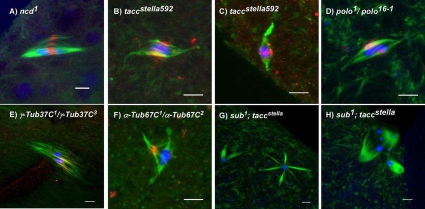
J. K. Jang et al.

linker region of all four proteins that are not present in other   were characterized by a disorganized array of microtubules
kinesins (Figure 1). PAV and MKLP1 have the highest level          (Figure 2A). SUB staining at this stage colocalized with
of amino acid identity or similarity and are probably ortho-       microtubules in the region that will become the MMCS.
logues. Similarly, there are several identical or similar amino    Thus, SUB localizes to the central spindle before bipolar
acids in the neck-linker regions of SUB and MKLP2 that are         spindle formation.
not found in other kinesins. This includes the serine residue         Stage 14 oocytes arrest at meiotic metaphase I; therefore,
in MKLP2 that is phosphorylated by Polo kinase (Neef et al.,       we used two methods to observe anaphase I spindles. First,
2003) and the corresponding acidic residue at ⫺2 that is           we looked at mei-218 mutants in which the metaphase arrest
often found at Polo kinase sites. Furthermore, although all        is bypassed (McKim et al., 1993). Second, we collected em-
four of these MKLP1 homologues have nonconserved N-                bryos after short periods of egg laying, which allows for the
terminal domains of ⬃100 amino acids, this domain is basic         isolation of oocytes undergoing the early meiotic divisions.
in PAV and MKLP1 but acidic in SUB and MKLP2. These                In both of these experiments, SUB remained in the spindle
sequence comparisons and the functional studies described          midzone as the chromosomes moved toward the poles (un-
here and in Neef et al. (2003) raise the possibility that SUB is   published data and Figure 2E, respectively).
the Drosophila ortholog of MKLP2.
                                                                   SUB Is a Mitotic Protein
SUB Localizes to the Central Spindle during Female                 Because sub mutants are viable, it is possible that sub has a
Meiosis I                                                          meiosis-specific function and is only required for the unique
Antibodies were raised against the poorly conserved C-             situation of assembling acentrosomal spindles during fe-
terminus of the SUB protein that follows the motor domain          male meiosis. There is, however, genetic evidence that sub is
(Figure 1). These antibodies recognize an ⬃75-kDa band on          expressed in mitotically dividing cells (Moore et al., 1994;
a Western blot that is absent in homozygotes of the null           Giunta et al., 2002). Consistent with these genetic observa-
alleles sub1 and sub131 (Figure 1C). To examine the localiza-      tions, we observed SUB staining in mitotically dividing cells
tion of SUB during meiosis, we stained mature Drosophila           of the embryo (Supplementary Figure 1). Similar to the
oocytes (stage 14), which in wild type, are arrested at meta-      metaphase oocytes, SUB protein was observed at the middle
phase I.                                                           of the spindle. SUB protein has also been observed at meta-
   Our identification of prometaphase and metaphase spin-          phase of larval neuroblast cells (B. Redding and K. McKim,
dles was based on previous studies of fixed (Theurkauf and         unpublished results). Thus, SUB protein may be a compo-
Hawley, 1992) and living oocytes (Matthies et al., 1996; Skold     nent of most or all metaphase spindles in Drosophila. Al-
et al., 2005). There is no congression to a metaphase plate in     though SUB has an important function in pronuclear fusion,
Drosophila oocytes. Instead, the chromosomes come together         some sub mutant embryos commence but never complete
and condense into a ball to form the karyosome much earlier        the early embryonic divisions (Giunta et al., 2002). Analysis
in oogenesis. After nuclear envelope breakdown (NEB), the          of these embryos shows evidence of spindle assembly de-
chromosomes in the karyosome initiate spindle formation.           fects and aneuploidy, indicating that sub also has a role in
The initial phases of spindle assembly are characterized by        mitotic spindle function (Supplementary Figure 1).
disorganized arrays of microtubules emanating from the
karyosome. Once a bipolar spindle forms, it remains stable         SUB Is Required for Central Spindle Formation
until the onset of anaphase when the oocyte is activated by        We previously reported that sub null mutant females de-
passage through the oviduct. As in previous studies (Theur-        velop monopolar and tripolar spindles in a majority of mei-
kauf and Hawley, 1992; McKim et al., 1993), staining for           osis I figures (see Table 4 in Giunta et al., 2002) and are sterile
tubulin in wild-type oocytes revealed that metaphase I spin-       because of a requirement during early embryogenesis. In
dles have a prominent band of microtubules that run pole-          addition, sub hypomorphic mutants exhibit a high frequency
to-pole and do not terminate at the chromosomes. The bright        of homologous chromosome nondisjunction at meiosis I
staining in the middle of this region probably represents the      (Giunta et al., 2002). Considered along with the SUB staining
antiparallel overlap of microtubules. We will refer to this        pattern, these results suggest that the MMCS may have an
region as the meiotic metaphase central spindle (MMCS) in          important role for in bipolar spindle formation and chromo-
order to distinguish it from the central spindle or midzone        some segregation. For this study, we reexamined the effects
present in anaphase of mitotic cells.                              on central spindle formation with a new sub mutant data set.
   Staining of wild-type oocytes with SUB antibodies re-           Similar to our previous report, 14/17 sub1/sub131 mutant
vealed that SUB protein was found exclusively in the MMCS          oocytes had abnormal spindle organization compared with
(Figure 2, A and B). Although SUB always colocalized with          only 4/36 in wild-type oocytes. Among the 14 abnormal
tubulin staining, it was also closely associated with the          sub1/sub131 spindles, 3 were monopolar, 9 were tripolar, and
chromosomes. In many spindles, 3D reconstruction revealed          2 had other problems such as fraying of the microtubules.
that SUB staining appeared more concentrated on one side           Thus, sub mutant spindles have a defect in maintaining or
or in some cases in two clusters on either side of the kary-       establishing bipolarity (Figure 2C), although in a minority of
osome. Whether this pattern reflects intrinsic features of the     cases, relatively normal bipolar spindles were observed (Fig-
spindle, such as asymmetry within the karyosome, or sto-           ure 2D). Notwithstanding these defects, the ability to taper
chastic properties of central spindle formation, remains to be     microtubules into poles is not usually affected in sub mu-
determined. SUB staining was not detected in the null alleles      tants.
sub1 and sub131 at prometaphase (see below) or metaphase              Given the localization of SUB, we examined the MMCS in
(Figure 2, C and D), confirming the specificity of the anti-       sub mutants. Strikingly, all sub mutant spindles, even those
body.                                                              that were bipolar, lacked metaphase central spindle tubulin
   Because genetic and cytogenetic studies (Giunta et al.,         staining (Figure 2, C and D, Supplementary Figure 2). Al-
2002) have suggested that SUB has an important role in             though the amount of central spindle microtubules was
spindle assembly, we investigated when SUB first appears           variable in wild type, the prominent bundles of central
on the meiotic spindle. SUB appeared from the earliest time        spindle microtubules often observed in wild-type oocytes
points after NEB, even on early prometaphase spindles that         were never observed in sub mutant oocytes. Instead, there

4686                                                                                                    Molecular Biology of the Cell
Acentrosomal Spindle Formation
were often small gaps of microtubules staining in the middle (Figure 4B). However, the level of Polo staining was variable
of the spindle or only limited evidence of antiparallel mi- and may be dependent on spindle structure, because in
crotubule overlap. Additional evidence that the central spin- some sub mutant oocytes, particularly those with the most
dle fails to form in sub mutants is that midzone proteins such disorganized spindles, Polo staining was weak (Figure 4C).
as Incenp and AurB did not localize to this region in sub
mutants (see below). These results suggest that the MMCS is Midzone Proteins Depend on Subito
normally organized or maintained by SUB. Our results suggest that the MMCS has an important func-
tion in organizing the meiotic spindle. This led us to exam-
SUB Localization Does Not Require Bipolar Spindle ine what other proteins are located in this region of the
Formation meiotic spindle and could contribute to microtubule assem-
To address whether SUB localization was dependent on bly. Several proteins have been localized to the spindle
bipolar spindle formation, SUB localization was examined in midzone at anaphase of mitotic cells including the passenger
mutants with disrupted spindle organization, including ncd, proteins AurB/Ial and Incenp. In mitotic cells, the localiza-
tacc, ␣ -tub67C, and ␥ -tub37C (Figure 3). Like sub, ncd en- tion pattern of the passenger proteins depends on the mi-
codes a kinesin required for bipolar spindle formation (Hat- totic stage. They localize to the centromeres during meta-
sumi and Endow, 1992; Matthies et al., 1996). Double mu- phase and then move to the spindle midzone at anaphase
tants with the hypomorph sub1794 have a meiotic phenotype (Adams et al., 2001; Giet and Glover, 2001). We examined the
similar to that of the single mutants (Giunta et al., 2002). tacc metaphase I localization pattern of AurB and Incenp by
has an important role in meiotic bipolar spindle formation with antibody staining to determine if the MMCS also contains
some similar phenotypes to sub mutants (Cullen and Ohkura, these proteins and whether they show an early (mitotic
2001). ␥-Tub37C is one of two Drosophila ␥-tubulin isoforms and metaphase) or late (mitotic anaphase) staining pattern. We
has previously been shown to have a role in female meiotic also examined RacGap50C, another mitotic midzone com-
spindle formation (Tavosanis et al., 1997). Mutants of the fe- ponent that forms a complex with the SUB paralog PAV
male-specific isoform ␣-Tub67C are sterile, although defects in (Somers and Saint, 2003).
spindle formation have not previously been shown (Matthews For Incenp (Figure 4D), AurB (Figure 4E), and RacGap50C
et al., 1993). Despite severe defects in bipolar spindle formation, (Figure 4G), we found staining similar or identical to SUB.
these mutants exhibited SUB staining in association with the For example, Incenp perfectly colocalized with SUB in the
central spindle. In addition, we have documented meiotic spin- MMCS (Figure 4D), including early prometaphase staining
dle defects in ␣ -tub67C mutant females for the first time. These before a bipolar spindle had formed. To determine the rela-
results suggest that the MMCS and SUB staining are not de- tionship of these proteins to the centromeres, we stained
pendent on bipolar spindle formation. Instead, SUB most likely with an antibody to MEI-S332 (Moore et al., 1998). SUB and
localizes and functions before the formation of a bipolar spin- MEI-S332 always occupied distinct regions around the kary-
dle. SUB may simply localize to any region of the spindle osome in both disorganized prometaphase and bipolar
containing antiparallel microtubules. metaphase spindles (Figure 4, I and J). By extension, because
the midzone proteins and SUB always colocalize, Incenp
Polo Localization during Female Meiosis and AurB do not localize to the centromere regions during
One candidate for regulating SUB localization is Polo kinase meiotic metaphase I. Indeed, the centromeres and MMCS
because, as described above, the SUB ortholog MKLP2 is appear to be properly organized and oriented before a bi-
phosphorylated by Polo in human cells. Observing the ef- polar spindle forms.
fects of polo null mutants is problematic because the ho- The localization of these proteins to the MMCS was de-
mozygotes are lethal. We were, however, able to examine pendent on sub activity. In sub mutants, AurB accumulated
females with a viable but female sterile allele heterozygous in a region surrounding the karyosome but not in the region
to a null allele and found that SUB staining was normal where the MMCS would have been (Figure 4F). Almost
(Figure 3). Indeed, these mutants did not have gross defects identical localization defects were observed with
in meiotic spindle formation, except for a possible reduction RacGap50C (Figure 4H) and Incenp (unpublished data).
in kinetochore microtubules. The absence of an effect on SUB This pattern appears to be more extensive than just centro-
staining in polo mutants is consistent with the results that in mere staining, at least when compared with the MEI-S332
HeLa-S3 cells, MKLP2 is a target for Polo phosphorylation staining and Polo staining described above. Instead, these
but this is not required for localization (Neef et al., 2003). proteins concentrated in the region where the microtubules
Because MKLP2 is required for the localization of Polo are in close proximity to the karyosome. This could occur if,
kinase to the midzone in HeLa-S3 cells (Neef et al., 2003), we in the absence of a central spindle in sub mutants, these
examined Polo localization during Drosophila meiosis. In proteins concentrate near the plus-ends of microtubules. A
addition, Drosophila Polo has been shown to have a localiza- similar effect of midzone disruption on AurB and MKLP1
tion pattern similar in mitotic cells to proteins such as AurB staining has been observed in HeLa cells (Kurasawa et al.,
and Incenp (Logarinho and Sunkel, 1998), which as de- 2004). It was suggested that localization toward the plus ends
scribed below, localize to the MMCS in Drosophila oocytes was an intermediate stage in development of the midzone. It is
and depend on SUB activity. Polo antibody staining in wild- possible these proteins have an affinity for the plus ends of
type metaphase Drosophila oocytes appeared weakly in the microtubules or other motors, such as the Drosophila MKLP1
MMCS and was not visible in all images. In contrast, it was ortholog Pavarotti, that actively transport them there (Matu-
stronger in foci that colocalized with the DNA (Figure 4A). liene and Kuriyama, 2002). Unfortunately, we have been un-
These Polo foci were probably the kinetochores, consistent able to test the role of the passenger proteins in acentrosomal
with previous studies of larval neuroblasts. In these mitoti- spindle formation because Incenp appears to be required dur-
cally dividing cells, Polo was localized to kinetochores dur- ing early stages of oogenesis (see Materials and Methods).
ing metaphase, and midzone staining was strong only dur-
ing anaphase (Logarinho and Sunkel, 1998). In sub mutant Two Pathways for Meiotic Spindle Formation
oocytes, the foci of Polo staining were still observed, which Although SUB has an important role in spindle formation,
is consistent with the absence of SUB protein at kinetochores most sub mutant spindles retain the ability for form poles.
Vol. 16, October 2005 4687J. K. Jang et al. Figure 2. SUB localizes to the meiotic metaphase central spindle in wild-type female meiosis I. SUB staining is in red, tubulin in green, and DNA in blue in addition to gray scale images of the separate SUB and tubulin channels. Before NEB, SUB is excluded from the nucleus 4688 Molecular Biology of the Cell
Acentrosomal Spindle Formation
Figure 3. SUB staining in spindle mutants. SUB is still found in the central spindle in a variety of mutants with disorganized meiotic
spindles, such as (A) ncd1, (B and C) taccstella592, (D) polo16 –1/polo1, (E) ␥-Tub37C1/␥-Tub37C3 and (F) ␣-Tub67C1/␣-Tub67C2. (G and H)
Synergistic effects on spindle formation in sub; tacc double mutant oocytes. SUB staining is in red, tubulin in green, and DNA in blue. Scale
bars, 5 m.
Therefore, SUB-independent activities must be functioning assembly, we constructed sub; tacc double mutants. The sub1;
to organize spindle poles. TACC and MSPS may have a role taccstella592 double mutant had severe spindle formation de-
in this function because these proteins localize to meiotic fects, more dramatic than either single mutant (compare
spindle poles. Previous genetic and cytological studies have Figure 2C and Figure 3B with Figure 3, G and H). Unlike the
shown that tacc and msps have an important role in meiotic single mutants, there were often multiple bundles of micro-
bipolar spindle formation (Cullen and Ohkura, 2001), and tubules, some associating with chromosomes. Although mi-
the mutants have been reported to have phenotypes similar crotubules were still associating with the chromosomes,
to sub mutants. To investigate the relationship between most bundles of microtubules were randomly organized.
TACC and SUB, we examined TACC localization in sub Similar defects were also observed in sub1794/sub1; taccstella592
mutants and examined the phenotype of double mutant but not sub1794/sub1794; taccstella592 females, demonstrating
combinations. As reported previously, TACC localized to this phenotype was dependent on severe loss of sub func-
the acentrosomal spindle poles in wild-type oocytes (Figure tion. The double mutants appear to retain the ability to
4K). In sub mutants, however, the spindle pole staining was assemble kinetochore microtubules. This phenotype could
weaker and in some cases accumulated near the chromo- be explained by a combination of tacc and sub mutant de-
somes (Figure 4L). Thus, it is possible that the phenotype of fects: a failure to stabilize the spindle poles (tacc) and a
SUB mutants may be related to defects in TACC localization. failure to organize the microtubules around the chromo-
To test if SUB and TACC function in distinct spindle somes (sub). These results suggest that sub and tacc contrib-
forming activities or have similar functions during spindle ute to different pathways that function to organize the mi-
crotubules of acentrosomal spindles. This is consistent with
the observation that SUB and TACC associate with distinct
structures or populations of microtubules.
Figure 2 (cont). (unpublished data). (A) Prometaphase oocyte: SUB
localizes to the presumptive central spindle, even though a mature
bipolar spindle has not yet formed. (B) Metaphase I oocyte: A DISCUSSION
bipolar spindle has formed and SUB staining is associated with the
central spindle, which is typically visible as bright bundles of pole-
Evidence that Subito Belongs to the MKLP1 Family of
to-pole microtubules. The chromosomes are under tension because Kinesinlike Proteins
they are being pulled toward to the poles but homologues are still Meiotic spindle microtubules in most oocytes must be orga-
connected by chiasmata (visible as thinner DNA staining between nized without centrosomes to organize the poles. Although
the two main masses). (C and D) SUB staining is absent in sub null the chromosomes play a critical role in spindle formation by
mutant metaphase I oocyte spindles. Although most sub mutants capturing free microtubules (replacing the nucleation step of
spindles have polarity defects (C), some have relatively normal centrosomes), it is not clear what organizes the bundling and
structure (D) (see text). In some cases, nonspecific signals are ob-
served. In either case, however, the bright microtubule staining in
elongation of microtubules into a bipolar spindle. Our re-
the central spindle is greatly reduced in sub mutants. (E) SUB sults suggest that the kinesinlike protein Subito has an im-
localization during meiotic anaphase I. SUB staining remains in the portant role in Drosophila acentrosomal spindle formation,
region between the chromosomes as they move toward the poles. possibly by organizing the prominent central spindle that
Scale bars, 5 m. assembles at meiotic prometaphase. Interestingly, SUB has
Vol. 16, October 2005 4689J. K. Jang et al. Figure 4. The meiotic metaphase central spindle contains proteins typically found on mitotic midzones. In these wild-type (A, D, E, G, I, J, and K) or sub protein null mutant (B, C, F, E,and L) oocytes, microtubules are shown in green and the DNA is i nblue, except in D, I, and J, where blue is another protein. In some cases, images of the less frequentbipolar sub mutant spindles were selected to more clearly show the abnormal 4690 Molecular Biology of the Cell
Acentrosomal Spindle Formation
several characteristics similar to MKLP2: SUB localizes to a suggests that PAV cannot compensate in a significant way
region of antiparallel microtubules, in this case the meiotic for the absence of SUB.
metaphase central spindle; it is required for central spindle
formation; it is required for the localization of other central Passenger Proteins Appear at the Midzone during Female
spindle proteins, and it has nonmotor domain sequence Meiotic Metaphase I
similarity including amino acids that could be phosphory- Our studies suggest a new role for the central spindle in
lated by Polo kinase. Similarly, a phylogenetic tree made bipolar spindle formation and chromosome segregation. The
from the alignment of kinesin motor domain sequences has localization pattern of central spindle components, such as
SUB in a cluster close to the MKLP1 group (Dagenbach and members of the passenger protein complex AurB and In-
Endow, 2004). As described below, we suggest these fea- cenp, is consistent with the idea that a central spindle is
tures allow SUB to contribute to the organizing of Drosophila forming precociously in oocytes. Although it is typical in
acentrosomal spindles by establishing or maintaining the Drosophila and human mitotic cells for AurB and Incenp to
central spindle at prometaphase and metaphase. initially associate with centromeres and then move to the
Mammalian MKLP1 (Matuliene and Kuriyama, 2002), midzone at anaphase (Adams et al., 2001; Giet and Glover,
Drosophila PAV (Adams et al., 1998), and the C. elegans or- 2001; Gruneberg et al., 2004), in Drosophila oocytes, these
tholog ZEN-4 (Raich et al., 1998; Mishima et al., 2002) have proteins appear on the central spindle much earlier in pro-
been found in the spindle midzone during anaphase and metaphase. Furthermore, the meiotic division of Drosophila
have an important function in cytokinesis. The midzone has oocytes appears to skip the stage in mitotic cells (metaphase)
been implicated in establishing the placement of the cyto- where passenger proteins associate with centromeres. We
plasmic furrow, although there are exceptions (D’Avino et have not observed SUB, AurB, or Incenp at the centromeres
al., 2005). Furthermore, MKLP1 was found to bundle micro- during female meiosis; they appear to be associated only
tubules and to promote anti-parallel sliding in vitro (Nislow with the nonkinetochore microtubules. This appears to be
et al., 1992). This is consistent with its localization in the specific only to a subset of midzone proteins. Polo exhibited
spindle midzone, where microtubules overlap in antiparallel kinetochore staining typical of mitotic metaphase at meiotic
orientation. In addition, from the direction of the antiparallel metaphase I. In addition, KLP3A, a kinesinlike protein that
sliding of microtubules it was concluded that MKLP1 is a associates with the anaphase midzone in mitotic cells, has
plus-end-directed motor. Although less is known about been reported to stain along the length of female meiotic
MKLP2, it is also required for the spindle midzone and spindles and only moves to the midzone at anaphase (Wil-
cytokinesis, and like other MKLP1 family members, the liams et al., 1997).
protein accumulates at the midzone (Hill et al., 2000; Fontijn AurB and Incenp localization to the oocyte MMCS de-
et al., 2001; Neef et al., 2003). Our characterization of SUB pends on SUB. Similarly, Incenp and AurB midzone local-
suggests that organisms with two MKLP1-like proteins are ization depends on MKLP2 in mammalian mitotic cells, and
not restricted to vertebrates. MKLP2 may even have a direct interaction with AurB
Two observations suggest that the MMCS is mostly or (Gruneberg et al., 2004). An important aspect of SUB func-
entirely absent in sub mutants. First, sub mutant spindles tion could be to recruit proteins like AurB in order to stim-
lack the prominent band of antiparallel microtubules arising ulate chromosome–microtubule interactions (Gassmann et
from the overlap of pole to pole spindle fibers. Second, al., 2004; Sampath et al., 2004). Consistent with this model,
proteins that normally associate with this region, such as phosphorylation of the microtubule-destabilizing kinesin
Incenp, AurB, and RacGap50C, are absent in sub mutant MCAK by AurB stimulates chromatin induced spindle as-
oocytes. Nonetheless, it is difficult to rule out if other pro- sembly in Xenopus extracts (Ohi et al., 2004). We have not,
teins are able to promote formation of a thin and fragile however, been able to determine the role, if any, of the
central spindle in sub mutants. A candidate with this func- passenger proteins in meiotic spindle formation.
tion could be PAV but, because of its lethal phenotype and
because pav mutant germlines do not make oocytes (Min- Antiparallel Microtubules Organize the Meiotic
estrini et al., 2002), we could not determine if PAV contrib- Acentrosomal Spindle
utes to the meiotic spindle assembly. However, the severe Previous models for acentrosomal spindle formation sug-
defect in meiotic central spindle formation in sub mutants gested that the process was initiated by the capture of free
microtubules by the chromosomes followed by bundling
and sorting of microtubules by minus-end-directed motors
to form the poles (Matthies et al., 1996; Skold et al., 2005).
Figure 4 (cont). localization of a midzone protein and that this was However, these models lack a mechanism to ensure that the
due to the absence of SUB rather than spindle structure. (A) POLO kinetochore microtubules are oriented toward only one of
(red) localizes to spots on the chromosomes, which are most likely two poles. For example, how are the two half spindles
the kinetochores. (B) Polo staining can still be observed in some sub
mutant oocytes. (C) In other sub mutant oocytes with more disor-
oriented relative to each other and what limits the spindle to
ganized spindles, however, Polo staining was reduced. The insets in have only two poles? On the basis of the localization pattern
B and C have the microtubule staining removed in order to see Polo of SUB and the phenotype of sub mutants, we present a
staining clearly. (D) Incenp (red) colocalizes with SUB (blue) in the model for acentrosomal spindle formation in Drosophila oo-
central spindle. The inset images show the separate channels for cytes that addresses these questions (Figure 5). We propose
SUB and Incenp. (E) AurB (red) localizes to the meiotic central that a structure composed of antiparallel microtubules is
spindle. (G) Similar results were obtained using antibodies against organized during prometaphase. The axis of the spindle is
RacGap50C. (F and H) In the absence of SUB, midzone proteins defined by this structure, the MMCS, which provides the
(AurB and RacGap50C are shown) accumulate around the karyo- scaffold on which to build a bipolar spindle during promet-
some instead of the MMCS. (I and J) MEI-S332 (blue) is a marker for
the meiotic centromeres and this staining does not overlap with SUB
aphase and metaphase. Proteins that localize to the spindle
(red). The inset shows SUB and MEI-S332 with DNA in green. (K) poles have a separate function in spindle pole formation and
TACC (red) normally localizes to the spindle poles. (L) In sub the functions of the central spindle or spindle-poles are
mutant oocytes, there is less staining at the poles and in some cases partially redundant for maintaining spindle integrity and
TACC it is observed near the center of the spindle. Scale bars, 5 m. establishing poles. As the sub; tacc double mutant phenotype
Vol. 16, October 2005 4691J. K. Jang et al.
must be inactive during metaphase. Repeated attempts at
new spindle pole formation could generate extra poles in sub
mutants. Indeed, we observed what appears to be newly
formed short half spindles and the ectopic appearance of
TACC in the middle of the spindle of sub mutants, suggest-
ing that de novo pole formation can occur at metaphase. The
presence of monopolar spindles could occur if the MMCS
has a role in maintaining half spindles, resulting in the
collapse of half spindles at metaphase in sub mutants. This
dynamic portrayal of the meiotic spindle in sub mutants is
consistent with real time observations in ncd mutants (Mat-
thies et al., 1996). Although wild-type spindles appear to be
stable structures over long periods of time, ncd mutant spin-
dles are dynamic structures, where bipolar spindles will
form only to lose their organization to become apolar, mo-
nopolar, or even completely disassemble and then reform
again.
The role for the MMCS described above in coordinating
spindle pole formation can explain the sub genetic and cy-
tological mutant phenotypes. However, we have not ruled
out other roles for SUB in chromosomes segregation. An
alternative is that SUB contributes to a balance of forces
between pushing apart or pulling together the spindle poles
(Sharp et al., 1999). In sub mutants, this could lead to a defect
in spindle pole positioning. Two observations argue against
this hypothesis. First, the sub mutant phenotype is not alle-
viated by defects in ncd (Giunta et al., 2002), in contrast to
Klp61F (Wilson et al., 2004), which has this role in mitotic
Figure 5. Model for acentrosomal spindle formation in Drosophila cells. Second, this function does not easily explain why sub
oocytes. (A) The chromosomes enter prometaphase clustered to- mutants often have multiple poles, whereas the length of the
gether in a ball, the karyosome, and capture microtubules that are half spindles are not dramatically shorter than wild-type.
not organized into a bipolar array. At this time, SUB protein accu- We also cannot rule out a role for SUB in facilitating inter-
mulates on microtubules adjacent to the karyosome. (B) Motor actions between the chromosomes and the microtubules.
proteins, possibly involving minus-end-directed motor proteins
This could have a role in aligning the homolog pairs at
such as NCD, bundle parallel microtubules, and taper them into
defined poles. In parallel with this process, the spindle is stabilized metaphase I, similar to what has been proposed for the
by proteins that accumulate at the poles. Proteins that localize to the chromokinesin NOD (Zhang et al., 1990; Theurkauf and
female meiotic spindle poles include Asp (Riparbelli et al., 2002), Hawley, 1992). Although in nod mutants, the nondisjunction
MSPS and TACC (Cullen and Ohkura, 2001). (C) The direction of phenotype is not associated with defects in spindle organi-
elongation/bundling/sliding is dictated by the metaphase central zation.
spindle. Critical to the model is that the orientation of the kineto- We thus favor a model in which SUB directly contributes
chore microtubules is established through interactions (via cross-
linking) with the central spindle. Examples of these interactions are to bipolar spindle formation by organizing and/or stabiliz-
shown with by the arrows. The “backbone” structure provided by ing the MMCS. An important implication of this model is
the central spindle defines the long axis of the spindle, ensuring that that, to compensate for the absence of centrosomes, the
the two poles form on opposite sides of the chromosomes and oocyte has modified the regulation of the central spindle so
prevents additional poles from forming. In fixed images, the SUB- that it appears earlier in order to direct spindle formation.
staining region appears localized to one side of the karyosome, but This is a novel function for the central spindle and contrasts
in other images there are two or more clusters of SUB staining.
Elongation of the spindle could involve the capture of additional with the suggestion for mitotic cells that the midzone accu-
microtubules to lengthen the spindle. Thus, spindle elongation mulation of Incenp and AurB needs to be inhibited until
could occur via microtubule capture and bundling with or without anaphase (Pereira and Schiebel, 2003; Mishima et al., 2004).
motor activity, microtubule sliding or growth at the plus ends. An important question that we are currently investigating is
what controls SUB localization. One possibility is that the
concentration of a factor that promotes microtubule assem-
demonstrates, in the absence of these structures, the spindle
bly, such as ran-GTP (Kahana and Cleveland, 1999), is great-
loses all organization.
est in one region of the karyosome. Given the SUB/MKLP2
SUB and the MMCS could be required at several points in
spindles assembly. The MMCS may have a role in the tran- conservation of sequence and function, it will be interesting
sition from prometaphase, with its disorganized microtu- to determine if the central spindle has an important role in
bules around the karyosome, to metaphase with a bipolar organizing the acentrosomal spindles of oocytes in mam-
spindle. The interaction of kinetochore microtubules with mals and other animals or in plants. Furthermore, as de-
pole-to-pole microtubules of the MMCS via parallel micro- scribed here for embryos and will be described elsewhere for
tubule bundling could determine the formation and relative other mitotic cells (B. Redding and K. McKim, unpublished
orientation of only two poles (Figure 5). In addition, SUB results), SUB also has a role in spindle assembly of mitotic
probably has a role in maintaining spindle bipolarity. By cells. This is consistent with the hypothesis that acentroso-
maintaining the MMCS, SUB could attenuate the activity mal spindle assembly occurs through the modification of
that is active to establish poles during prometaphase but functions already present in mitotic cells.
4692 Molecular Biology of the CellAcentrosomal Spindle Formation
ACKNOWLEDGMENTS Matthews, K. A., Rees, D., and Kaufman, T. C. (1993). A functionally special-
ized a-tubulin is required for oocyte meiosis and cleavage mitoses in Drosoph-
We are grateful to Li Nguyen for technical assistance and R. Scott Hawley, ila. Development 117, 977–991.
who initially suggested that the central spindle might be important for
Matthies, H. J., McDonald, H. B., Goldstein, L. S., and Theurkauf, W. E. (1996).
organizing the meiotic spindle. We also thank Mar Carmena, Terry Orr-
Anastral meiotic spindle morphogenesis: role of the non-claret disjunctional
Weaver, Jordan Raff, Robert Saint, and Claudio Sunkel for providing anti-
kinesin-like protein. J. Cell Biol. 134, 455– 464.
bodies and R. Scott Hawley and Cordelia Rauskolb for comments on the
manuscript. Some stocks used in this study were obtained from the Bloom- Matuliene, J., and Kuriyama, R. (2002). Kinesin-like protein CHO1 is required
ington Stock Center. This work was supported by a grant from the National for the formation of midbody matrix and the completion of cytokinesis in
Institutes of Health (GM 067142) to K.M. mammalian cells. Mol. Biol. Cell 13, 1832–1845.
McKim, K. S., and Hawley, R. S. (1995). Chromosomal control of meiotic cell
division. Science 270, 1595–1601.
REFERENCES
McKim, K. S., Jang, J. K., Theurkauf, W. E., and Hawley, R. S. (1993). Me-
Adams, R. R., Maiato, H., Earnshaw, W. C., and Carmena, M. (2001). Essential chanical basis of meiotic metaphase arrest. Nature 362, 364 –366.
roles of Drosophila inner centromere protein (INCENP) and aurora B in
Minestrini, G., Mathe, E., and Glover, D. M. (2002). Domains of the Pavarotti
histone H3 phosphorylation, metaphase chromosome alignment, kinetochore
kinesin-like protein that direct its subcellular distribution: effects of mislocal-
disjunction, and chromosome segregation. J. Cell Biol. 153, 865– 880.
isation on the tubulin and actin cytoskeleton during Drosophila oogenesis.
Adams, R. R., Tavares, A. A., Salzberg, A., Bellen, H. J., and Glover, D. M. J. Cell Sci. 115, 725–736.
(1998). pavarotti encodes a kinesinlike protein required to organize the central
Mishima, M., Kaitna, S., and Glotzer, M. (2002). Central spindle assembly and
spindle and contractile ring for cytokinesis. Genes Dev. 12, 1483–1494.
cytokinesis require a kinesin-like protein/RhoGAP complex with microtubule
Compton, D. A. (2000). Spindle assembly in animal cells. Annu. Rev. Biochem. bundling activity. Dev. Cell 2, 41–54.
69, 95–114. Mishima, M., Pavicic, V., Gruneberg, U., Nigg, E. A., and Glotzer, M. (2004).
Cullen, C. F., and Ohkura, H. (2001). Msps protein is localized to acentrosomal Cell cycle regulation of central spindle assembly. Nature 430, 908 –913.
poles to ensure bipolarity of Drosophila meiotic spindles. Nat. Cell Biol. 3, Moore, D. P., Miyazaki, W. Y., Tomkiel, J., and Orr-Weaver, T. L. (1994).
637– 642. Double or nothing: a Drosophila mutation affecting meiotic chromosome seg-
Dagenbach, E. M., and Endow, S. A. (2004). A new kinesin tree. J. Cell Sci. 117, regation in both females and males. Genetics 136, 953–964.
3–7. Moore, D. P., Page, A. W., Tang, T. T., Kerrebrock, A. W., and Orr-Weaver,
D’Avino, P. P., Savoian, M. S., and Glover, D. M. (2005). Cleavage furrow T. L. (1998). The cohesion protein MEI-S332 localizes to condensed meiotic
formation and ingression during animal cytokinesis: a microtubule legacy. and mitotic centromeres until sister chromatids separate. J. Cell Biol. 140,
J. Cell Sci. 118, 1549 –1558. 1003–1012.
Endow, S. A., and Komma, D. J. (1997). Spindle dynamics during meiosis in Neef, R., Preisinger, C., Sutcliffe, J., Kopajtich, R., Nigg, E. A., Mayer, T. U.,
Drosophila oocytes. J. Cell Biol. 137, 1321–1336. and Barr, F. A. (2003). Phosphorylation of mitotic kinesin-like protein 2 by
polo-like kinase 1 is required for cytokinesis. J. Cell Biol. 162, 863– 875.
Fontijn, R. D., Goud, B., Echard, A., Jollivet, F., van Marle, J., Pannekoek, H.,
and Horrevoets, A. J. (2001). The human kinesin-like protein RB6K is under Nislow, C., Lombillo, V. A., Kuriyama, R., and McIntosh, J. R. (1992). A
tight cell cycle control and is essential for cytokinesis. Mol. Cell. Biol. 21, plus-end-directed motor enzyme that moves antiparallel microtubules in
2944 –2955. vitro localizes to the interzone of mitotic spindles. Nature 359, 543–547.
Ohi, R., Sapra, T., Howard, J., and Mitchison, T. J. (2004). Differentiation of
Gassmann, R., Carvalho, A., Henzing, A. J., Ruchaud, S., Hudson, D. F.,
cytoplasmic and meiotic spindle assembly MCAK functions by Aurora B-
Honda, R., Nigg, E. A., Gerloff, D. L., and Earnshaw, W. C. (2004). Borealin:
dependent phosphorylation. Mol. Biol. Cell 15, 2895–2906.
a novel chromosomal passenger required for stability of the bipolar mitotic
spindle. J. Cell Biol. 166, 179 –191. Pereira, G., and Schiebel, E. (2003). Separase regulates INCENP-Aurora B
anaphase spindle function through Cdc14. Science 302, 2120 –2124.
Giet, R., and Glover, D. M. (2001). Drosophila aurora B kinase is required for
histone H3 phosphorylation and condensin recruitment during chromosome Raich, W. B., Moran, A. N., Rothman, J. H., and Hardin, J. (1998). Cytokinesis
condensation and to organize the central spindle during cytokinesis. J. Cell and midzone microtubule organization in Caenorhabditis elegans require the
Biol. 152, 669 – 682. kinesin-like protein ZEN-4. Mol. Biol. Cell 9, 2037–2049.
Giunta, K. L., Jang, J. K., Manheim, E. M., Subramanian, G., and McKim, K. S. Riparbelli, M. G., Callaini, G., and Glover, D. M. (2000). Failure of pronuclear
(2002). subito encodes a kinesin-like protein required for meiotic spindle pole migration and repeated divisions of polar body nuclei associated with MTOC
formation in Drosophila melanogaster. Genetics 160, 1489 –1501. defects in polo eggs of Drosophila. J. Cell Sci. 113, 3341–3350.
Gruneberg, U., Neef, R., Honda, R., Nigg, E. A., and Barr, F. A. (2004). Riparbelli, M. G., Callaini, G., Glover, D. M., and Avides Md Mdo, C. (2002).
Relocation of Aurora B from centromeres to the central spindle at the meta- A requirement for the Abnormal Spindle protein to organise microtubules of
phase to anaphase transition requires MKlp2. J. Cell Biol. 166, 167–172. the central spindle for cytokinesis in Drosophila. J. Cell Sci. 115, 913–922.
Hatsumi, M., and Endow, S. A. (1992). Mutants of the microtubule motor Rothwell, W. F., and Sullivan, W. (2000). Fluorescent analysis of Drosophila
protein, nonclaret disjunctional, affect spindle structure and chromosome embryos. In: Drosophila Protocols, ed. W. Sullivan, M. Ashburner, and R. S.
movement in meiosis and mitosis. J. Cell Sci. 101, 547–559. Hawley, Cold Spring Harbor, NY: Cold Spring Harbor Laboratory Press,
141–157.
Hill, E., Clarke, M., and Barr, F. A. (2000). The Rab6-binding kinesin, Rab6-
KIFL, is required for cytokinesis. EMBO J. 19, 5711–5719. Sampath, S. C., Ohi, R., Leismann, O., Salic, A., Pozniakovski, A., and Fun-
abiki, H. (2004). The chromosomal passenger complex is required for chro-
Kahana, J. A., and Cleveland, D. W. (1999). Beyond nuclear transport. Ran- matin-induced microtubule stabilization and spindle assembly. Cell 118, 187–
GTP as a determinant of spindle assembly. J. Cell Biol. 146, 1205–1210. 202.
Karsenti, E., and Vernos, I. (2001). The mitotic spindle: a self-made machine. Schupbach, T., and Wieschaus, E. (1989). Female sterile mutations on the
Science 294, 543–547. second chromosome of Drosophila melanogaster. I. Maternal effect mutations.
Kramer, J., and Hawley, R. S. (2003). The spindle-associated transmembrane Genetics 121, 101–117.
protein Axs identifies a membranous structure ensheathing the meiotic spin- Sharp, D. J., Yu, K. R., Sisson, J. C., Sullivan, W., and Scholey, J. M. (1999).
dle. Nat. Cell Biol. 5, 261–263. Antagonistic microtubule-sliding motors position mitotic centrosomes in Dro-
sophila early embryos. Nat. Cell Biol. 1, 51–54.
Kurasawa, Y., Earnshaw, W. C., Mochizuki, Y., Dohmae, N., and Todokoro, K.
(2004). Essential roles of KIF4 and its binding partner PRC1 in organized Skold, H. N., Komma, D. J., and Endow, S. A. (2005). Assembly pathway of
central spindle midzone formation. EMBO J. 23, 3237–3248. the anastral Drosophila oocyte meiosis I spindle. J. Cell Sci. 118, 1745–1755.
Lee, M. J., Gergely, F., Jeffers, K., Peak-Chew, S. Y., and Raff, J. W. (2001). Somers, W. G., and Saint, R. (2003). A RhoGEF and Rho family GTPase-
Msps/XMAP215 interacts with the centrosomal protein D-TACC to regulate activating protein complex links the contractile ring to cortical microtubules
microtubule behavior. Nat. Cell. Biol. 3, 643– 649. at the onset of cytokinesis. Dev. Cell 4, 29 –39.
Logarinho, E., and Sunkel, C. E. (1998). The Drosophila POLO kinase localises Stapleton, M. et al. (2002). The Drosophila gene collection: identification of
to multiple compartments of the mitotic apparatus and is required for the putative full-length cDNAs for 70% of D. melanogaster genes. Genome Res. 12,
phosphorylation of MPM2 reactive epitopes. J. Cell Sci. 111(Pt 19), 2897–2909. 1294 –1300.
Vol. 16, October 2005 4693J. K. Jang et al.
Tavosanis, G., Llamazares, S., Goulielmos, G., and Gonzalez, C. (1997). Es- behavior of male and female pronuclei at fertilization. Development 124,
sential role for gamma-tubulin in the acentriolar female meiotic spindle of 2365–2376.
Drosophila. EMBO J. 16, 1809 –1819.
Wilson, P. G., Simmons, R., and Shigali, S. (2004). Novel nuclear defects in
Theurkauf, W. E., and Hawley, R. S. (1992). Meiotic spindle assembly in KLP61F-deficient mutants in Drosophila are partially suppressed by loss of
Drosophila females: behavior of nonexchange chromosomes and the effects of Ncd function. J. Cell Sci. 117, 4921– 4933.
mutations in the nod kinesin-like protein. J. Cell Biol. 116, 1167–1180.
Yamamoto, A. H., Komma, D. J., Shaffer, C. D., Pirrotta, V., and Endow, S. A.
Walczak, C. E., Vernos, I., Mitchison, T. J., Karsenti, E., and Heald, R. (1998). (1989). The claret locus in Drosophila encodes products required for eye color
A model for the proposed roles of different microtubule-based motor proteins and for meiotic chromosome segregation. EMBO J. 8, 3543–3552.
in establishing spindle bipolarity. Curr. Biol. 8, 903–913.
Zhang, P., Knowles, B. A., Goldstein, L.S.B., and Hawley, R. S. (1990). A
Williams, B. C., Dernburg, A. F., Puro, J., Nokkala, S., and Goldberg, M. L. kinesin-like protein required for distributive chromosome segregation in
(1997). The Drosophila kinesin-like protein KLP3A is required for proper Drosophila. Cell 63, 1053–1062.
4694 Molecular Biology of the CellYou can also read