Maheshvara regulates JAK/STAT signaling by interacting and stabilizing hopscotch transcripts which leads to apoptosis in Drosophila melanogaster ...
←
→
Page content transcription
If your browser does not render page correctly, please read the page content below
Maurya et al. Cell Death and Disease (2021)12:363
                                    https://doi.org/10.1038/s41419-021-03649-0                                                                                                             Cell Death & Disease

                                     ARTICLE                                                                                                                                                    Open Access

                                    Maheshvara regulates JAK/STAT signaling by
                                    interacting and stabilizing hopscotch transcripts
                                    which leads to apoptosis in Drosophila
                                    melanogaster
                                    Bhawana Maurya1, Satya Surabhi1,2, Rituparna Das1, Pranjali Pandey1, Ashim Mukherjee1 and Mousumi Mutsuddi                                                                                       1

                                      Abstract
                                      Maheshvara (mahe), an RNA helicase that is widely conserved across taxa, regulates Notch signaling and neuronal
                                      development in Drosophila. In order to identify novel components regulated by mahe, transcriptome profiling of
                                      ectopic mahe was carried out and this revealed striking upregulation of JAK/STAT pathway components like upd1,
                                      upd2, upd3, and socs36E. Further, significant downregulation of the pathway components in mahe loss-of-function
                                      mutant as well as upon lowering the level of mahe by RNAi, supported and strengthened our transcriptome data.
                                      Parallelly, we observed that mahe, induced caspase-dependent apoptosis in photoreceptor neurons, and this
                                      phenotype was significantly modulated by JAK/STAT pathway components. RNA immunoprecipitation unveiled the
                                      presence of JAK/STAT tyrosine kinase hopscotch (hop) transcripts in the complex immunoprecipitated with Mahe,
                                      which ultimately resulted in stabilization and elevation of hop transcripts. Additionally, we also observed the surge in
                  1234567890():,;
1234567890():,;
                  1234567890():,;
1234567890():,;

                                      activity of downstream transcription factor Stat92E, which is indicative of activation of the JAK/STAT signaling, and this
                                      in turn led to apoptosis via upregulation of hid. Taken together, our data provide a novel regulation of JAK/STAT
                                      pathway by RNA helicase Maheshvara, which ultimately promotes apoptosis.

                                    Introduction                                                                                        approaches have identified mutations in DEAD box heli-
                                      With the development of a deeper understanding of the                                             case genes as major cause of neurodevelopmental dis-
                                    importance of RNA in maintaining the fidelity of biological                                          abilities, these include DDX59, DDX6, DHX37, DHX30,
                                    processes, the pivotal roles played by RNA binding proteins                                         DHX16, DHX34, and DDX546–9.
                                    (RBPs)1,2 such as the DEAD box RNA helicases (DBRHs)                                                  We have previously characterized maheshvara (mahe), a
                                    are being unraveled. The DBRHs are a set of multitasking                                            Drosophila homolog of human DDX59, as a novel mod-
                                    proteins that are involved in multitudes of RNA processes                                           ulator of Notch signaling10. Misregulated mahe leads to
                                    depending largely on the partners they interact with in                                             neuronal defects and reduced lifespan in Drosophila,
                                    various biological contexts3,4. The involvement of DBRHs in                                         which parallels similarities with patients having frameshift
                                    RNA processing events in neurons implicates their role in                                           deletion in DDX59 presenting orofaciodigital syndrome
                                    numerous neurological disorders4,5. High-throughput                                                 associated with broad neurological defects11.
                                                                                                                                          mahe and its human orthologues have been implicated
                                                                                                                                        in disease and development of nervous system. To delve
                                    Correspondence: Mousumi Mutsuddi (mousumi@bhu.ac.in)                                                deeper into the mechanism of the neuronal defects caused
                                    1
                                     Department of Molecular and Human Genetics, Institute of Science, Banaras                          due to ectopic mahe, we carried out whole transcriptome
                                    Hindu University, Varanasi 221005 Uttar Pradesh, India. 2Present address:
                                                                                                                                        analysis. Interestingly, we observed significant upregula-
                                    Department of Molecular and Developmental Biology, Albert Einstein College
                                    of Medicine, New York, NY 10461, USA                                                                tion of JAK/STAT pathway components. In addition, alike
                                    Edited by K. McCall

                                    © The Author(s) 2021
                                                        Open Access This article is licensed under a Creative Commons Attribution 4.0 International License, which permits use, sharing, adaptation, distribution and reproduction
                                                        in any medium or format, as long as you give appropriate credit to the original author(s) and the source, provide a link to the Creative Commons license, and indicate if
                                    changes were made. The images or other third party material in this article are included in the article’s Creative Commons license, unless indicated otherwise in a credit line to the material. If
                                    material is not included in the article’s Creative Commons license and your intended use is not permitted by statutory regulation or exceeds the permitted use, you will need to obtain
                                    permission directly from the copyright holder. To view a copy of this license, visit http://creativecommons.org/licenses/by/4.0/.

                                    Official journal of the Cell Death Differentiation Association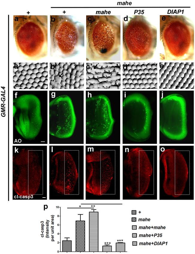
Maurya et al. Cell Death and Disease (2021)12:363                                                                Page 2 of 14

to the case of many DEAD box helicase associated neu-             Interestingly, DAVID analysis revealed positive regula-
rological diseases12,13, we also noted that overexpression      tion of JAK/STAT cascade (Fig. 1a). To reconfirm our
of mahe leads to increased apoptosis in the developing          RNA-Seq data, real-time PCR was done in mahe loss-of-
photoreceptor neurons. Drosophila JAK/STAT signaling,           function mutant, remarkably each of the three Drosophila
though relatively simpler when compared to that of ver-         JAK/STAT ligands upd1, upd2, upd3, and socs36E, the
tebrates, regulates patterning, organizer establishment,        downstream target of the transcription factor Stat92E
proliferation, apoptosis and maintains the size of the adult    were significantly downregulated (Fig. 1f). As there were
Drosophila eye14. Mechanistically, secreted ligand family       very few first instar escaper larvae of mahe loss-of-
unpaired (upd) and upd like proteins (upd2 and upd3)            function mutant and none developed up to third instar
binding dimerizes the receptor (domeless/dome) and              larval stage, further analysis was done using mahe-RNAi.
activates the receptor-associated JAK tyrosine kinases          In addition, in line with that of mahe mutants, JAK/STAT
(hopscotch/hop). In the canonical pathway, these tyrosine       pathway components were downregulated upon lowering
kinases phosphorylate themselves and their associated           the levels of mahe by RNAi (Fig. 1f). Further, in agreement
receptors and this creates docking sites for signal-            with the above results ectopically expressed mahe tissue
transducer and activator of transcription protein at 92E        revealed upregulation of the JAK/STAT signaling com-
(Stat92E). Stat92E transcription factor dimerizes upon          ponents, and this corresponded with the RNA-Seq data
phosphorylation and enters into the nucleus to activate or      (Fig. 1e, f). Together, these data indicated mahe to be a
repress transcription of target genes such as socs36E15,16.     positive regulator of JAK/STAT pathway.
During development, the decision of a cell to die or sur-
vive requires the integration of several regulatory path-       mahe induces Caspase-dependent cell death in a dosage
ways among which JAK/STAT signaling also plays a                sensitive manner
significant role. In our study, genetic analysis of mahe           Parallelly, a gain-of-function assay was carried out and
with members of JAK/STAT pathway displayed modula-              the effect of ectopic expression of mahe was monitored in
tion of apoptotic phenotype in the developing photo-            the developing photoreceptors. An HA tagged full length
receptor neurons. Our study has uncovered the novel role        Mahe protein was ectopically expressed posterior to the
of an RNA helicase Mahe that binds to hop tyrosine              morphogenetic furrow using GMR-GAL4 driver. Over-
kinase transcripts, and leads to a surge in its levels          expression of mahe resulted in rough and reduced eye
resulting in activation of JAK/STAT signaling. This             phenotype when compared to the GMR-GAL4 control
ectopic JAK/STAT activation culminates into hid medi-           adult eye phenotype (Fig. 2b, b′ and c, c′ to a, a′). The
ated cell death. To the best of our knowledge, this is the      rough eye phenotype was found to be largely due to
first report in either vertebrates or invertebrates, where       apoptosis, as seen by acridine orange staining as well as
the pivotal role played by an RNA helicase in activating        immunostaining of cleaved caspase and the immuno-
JAK/STAT cascade has been unraveled. This activation            fluorescence signal was dosage sensitive to the levels of
ultimately ensues in an upregulation of hid mediated            mahe expression (Fig. 2g, l and h, m to f, k). Cell survival
apoptosis.                                                      is facilitated by factors which directly inhibit caspases.
                                                                DIAP1 and P35 are such known inhibitors of caspases.
Results                                                         Our observation was further consolidated by the sig-
Transcriptome analysis of mahe reveals upregulation of          nificant rescue of mahe associated eye roughness and
JAK/STAT pathway components                                     apoptosis by DIAP1 and P35 (Fig. 2d, d′, i, n and e, e′, j, o
  For dissecting out the molecular factors regulated by         to b, b′, g, l). Quantitative analysis of cleaved caspase
mahe during development, transcriptome analysis using           staining in terms of intensity per unit area was indicated
RNA extracted from GMR-GAL4 driven mahe over-                   by prominent reduction in apoptotic cells when P35 and
expressed eye tissues were compared to that of the tran-        DIAP1 are coexpressed with mahe (2 p). These results
script levels in GMR-GAL4 controls. RNA-sequencing              indicate that mahe induces cell death in canonical
analysis revealed a total of 12,225 differentially regulated    caspase-dependent manner and its level must be tightly
genes (with fold change > 0.5). Out of these, 1080 genes        regulated for proper development and differentiation of
were found to be upregulated and 519 genes were                 Drosophila eye.
downregulated significantly (with fold change >1.5). For
functional annotation of differentially expressed genes         mahe induced apoptosis is mediated via surge in JAK/STAT
mined from our RNA-Seq data, NIH DAVID 6.8 (The                 pathway components
Database for Annotation, Visualization, and Integrated            Since, major components of the JAK/STAT pathway
Discovery) was used. Gene Ontology (GO) analysis,               were upregulated as depicted in the RNA-Sequencing
identified the biological activities and molecular pathways      data, we next sought to identify whether the components
affected in response to mahe overexpression (Fig. 1a–d).        of JAK/STAT pathway themselves alter the mahe induced

Official journal of the Cell Death Differentiation Association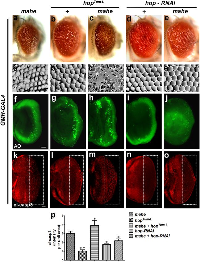
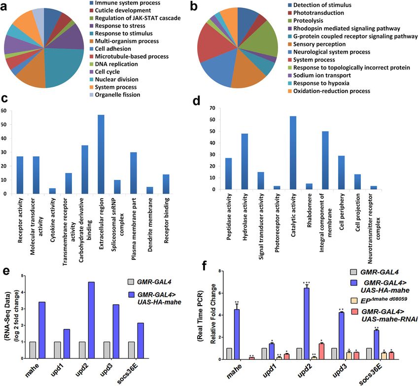
Maurya et al. Cell Death and Disease (2021)12:363                                                                                     Page 3 of 14

  Fig. 1 mahe promotes upregulation of members of JAK/STAT pathway as revealed by transcriptome analysis. Transcriptome analysis was
  done of eye tissue overexpressing mahe driven with GMR-GAL4 and was compared to GMR-GAL4 controls. a, b Functional categories enriched in
  upregulated (1080) and downregulated (519) genes based on their molecular functions using DAVID, which includes genes associated with
  phototransduction, JAK/STAT pathway activation, response to stress, nuclear division, and many more. c, d Enrichment of pathway specific
  upregulated (1080) and downregulated (519) genes based on their biological and cellular functions using DAVID. e Graph represents selected genes
  upd1, upd2, upd3 and socs36E were differentially expressed in the RNA-Seq data. f Real time PCR validated the candidate genes obtained from our
  transcriptome analysis data. The components of JAK/STAT signaling pathway were upregulated upon ectopic expression of mahe and were
  significantly downregulated in mahe loss-of-function mutant as well as in mahe-RNAi. One-way ANOVA. Genotypes (a–f) w/+; +; GMR-GAL4/+ and
  w/+; +; GMR-GAL4, UAS-HA-mahe/UAS-HA-mahe (f) EPΔmahe d08059;+;+ (f) +; UAS-mahe-RNAi/+; GMR-GAL4/+. One-way ANOVA *P < 0.05, **P < 0.01,
  ***P < 0.001.

apoptotic phenotype. Coexpression of both mahe and                          combination with mahe overexpression completely res-
upd2 exhibited severe disorganization of ommatidia with                     cued the apoptotic phenotype (Fig. 3e, e′, j, o to a, a′, f, k).
black necrotic patches that were observed in a few of the                   Quantitative analysis of cleaved caspase staining showed
escapers, while most of the pupae failed to eclose and                      enhancement of apoptotic cells, when both upd2 and
were dead. (Fig. 3c–c′). This was in contrast to the rough                  mahe were expressed together, whereas in contrary mahe
and enlarged eye phenotype exhibited by overexpression                      induced phenotype was suppressed in heterozygous
of mahe and upd2 alone (Fig. 3a, a′ and b, b′). Both,                       combination with upd2 loss of function allele (Fig. 3p).
acridine orange and cleaved caspase immunostaining                            We also observed that upd1 ligand that activates JAK/
showed dramatic enhancement in apoptosis when upd2                          STAT signaling17 also enhanced mahe induced apoptosis
and mahe were coexpressed in the developing eye-                            (Fig. S1). Our observation was supported by an indepen-
antennal discs (Fig. 3h, m to f, k and g, l). On similar                    dent transgenic RNA interference (RNAi) screen per-
lines, heterozygous upd2 mutant (Fig. 3d, d′, i, n) in                      formed for mahe modifiers (unpublished Pandey and

Official journal of the Cell Death Differentiation Association
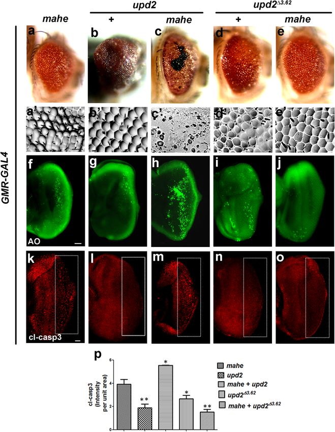
Maurya et al. Cell Death and Disease (2021)12:363                                                                                               Page 4 of 14

  Fig. 2 Ectopic expression of mahe promotes caspase-dependent dosage sensitive cell death in eye tissue of Drosophila. a–e Images of
  Drosophila adult eyes. a′–e′ Eye imprints of adult eye. GMR-GAL4 was used as a control (a), or to drive expression of mahe (b–e), P35 (d), DIAP1 (e). a, a′
  Control adult eye showed normal ommatidium arrangement. b, b′ GMR-GAL4 > mahe induced eye roughness with fused ommatidia (marked with
  arrow). c, c′ Ommatidial disarray and fusion were severely enhanced on increasing the dosage of mahe. d, d′ mahe induced eye phenotype was
  suppressed by expression of P35, an inhibitor of effector caspase. e, e′ Similar to P35, inhibitor of initiator caspase DIAP1 suppressed the eye
  roughening and ommatidium disarrangement. f–j Acridine orange staining of eye-antennal imaginal discs. f Acridine orange marks few dying cells in
  control eye-antennal disc. g, h Number of acridine orange positive cells were enhanced upon mahe overexpression in dosage sensitive manner. i, j
  P35 and DIAP1 suppressed cell death, shown by reduction in acridine positive nuclei. k–o Caspase immunostaining of eye-antennal discs from third
  instar larvae (Compare area within rectangle). l, m Ectopic mahe leads to increase in number of caspase positive cells in comparison to wild type (k).
  n, o Reduction in caspase positive cells were observed upon coexpression of P35 and DIAP1 along with mahe. (p) Graph represents intensity of cl-
  casp3 per unit area showing mahe triggers caspase-dependent cell death, which was suppressed by P35 and DIAP1. One-way ANOVA. Scale bar in
  50μm (a′–e′, f–j, k–o). Genotypes (a) w/+; +; GMR-GAL4/+ (b) w/+; +; GMR-GAL4, UAS-HA-mahe/+ (c) w/+; +; GMR-GAL4,UAS-HA-mahe/UAS-HA-
  mahe (d) w/+; +; GMR-GAL4,UAS-HA-mahe/UAS-P35 (e) w/+; +; GMR-GAL4,UAS-HA-mahe/UAS-DIAP1. One-way ANOVA * P < 0.05, ** P < 0.01, *** P <
  0.001.

Mutsuddi). Downregulation of Chaperonin containing                               role of mahe induced JAK/STAT signaling in promoting
TCP1 subunit 7 (CCT7) gene, which is known to be a                               cell death. Our genetic interaction studies are in agree-
negative regulator of upd118 led to enhancement of                               ment with our transcriptome data. Our results clearly
apoptosis induced by mahe (Fig. S2) suggesting, a positive                       display that ectopic expression of upd2 dramatically

Official journal of the Cell Death Differentiation Association
Maurya et al. Cell Death and Disease (2021)12:363                                                                                         Page 5 of 14

  Fig. 3 Ectopic expression of upd2 enhances, while loss of upd2 suppresses mahe induced apoptosis in developing eye. a–e Images of
  Drosophila adult eye. a′–e′ Eye imprints of adult eye. GMR-GAL4 was used to drive the expression of UAS-mahe (a, c, e) or UAS-upd2 (b, c). a, a′ GMR-
  GAL4 > mahe promotes eye roughening. b, b′ GMR-GAL4 driven upd2 resulted in severely overgrown eyes with regular patterned ommatidia. c, c′
  Coexpression of upd2 and mahe leads to pupal lethality whereas few escapers showed drastic eye roughness, with loss of pigmentation, black
  necrotic patches and loss of ommatidia in comparison to ectopic expression of mahe alone. d, d′ upd2Δ3-62 a loss-of-function allele shows slightly
  disorganized and fused ommatidia along the dorsal side of adult eye. e, e′ upd2Δ3-62 in combination with GMR-mahe suppressed the disorganized
  adult eye phenotype. f–j and k–o Acridine orange and caspase staining (compare area marked within rectangle) in eye-antennal disc of third instar
  larvae. f, k Acridine orange and caspase staining marked dying cells on ectopic expression of mahe. g, l Fewer acridine orange and caspase positive
  cells were observed upon upd2 overexpression. h, m Coexpression of upd2 and mahe enhanced cell death associated with mahe, which is reflected
  by increase in acridine orange and caspase positive cells when compared to mahe alone. i, n upd2Δ3-62 showed fewer acridine and caspase positive
  cells. j, o upd2Δ3-62 rescued the mahe induced cell death, which is depicted by reduced number of acridine orange and caspase positive cells. p Graph
  represents cl-caspase intensity per unit area which shows upd2 overexpression promotes mahe induced cell death, and was significantly reduced in
  combination with upd2 mutant. One-way ANOVA. Scale bar in 50 µm (a′–e′, f–j, k–o). Genotypes (a) w/+; +; GMR-GAL4,UAS-HA-mahe/+ (b) w/+;
  UAS-upd2:GFP/+;GMR-GAL4/+ (c) w/+;UAS-upd2:GFP/+;GMR-GAL4,UAS-HA-mahe/+ (d) upd2Δ3.62/+; +;+ (e) upd2Δ3.62/+; +; GMR-GAL4, UAS-HA-mahe.
  One-way ANOVA * P < 0.05, ** P < 0.01, *** P < 0.001.

enhances the apoptotic phenotype of mahe, while mod-                          led to massive suppression of the mahe induced pheno-
erate enhancement of apoptosis was seen in combination                        type. This suggested a vital role of JAK/STAT signaling in
of upd1 and mahe. Reduced levels of both upd1 and upd2                        mahe induced cell death.

Official journal of the Cell Death Differentiation Association
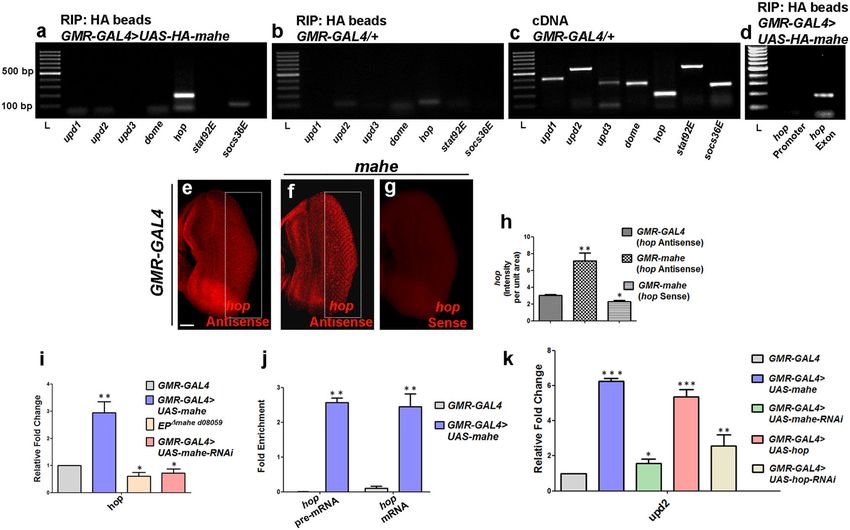
Maurya et al. Cell Death and Disease (2021)12:363                                                                                         Page 6 of 14

hopscotch transcripts coimmunoprecipitates with Mahe                          using anti-HA beads to pull down HA-tagged Mahe, while
and this interaction leads to its stabilization                               RNA-protein extracted from GMR-GAL4 alone served as
   In Drosophila, upd acts through the Dome/Hop/                              negative control. This was followed by RT-PCR using
STAT92 signaling pathway for proper photoreceptor                             gene-specific primers for the members of JAK/STAT
development14,19. We were further interested to check                         pathway, upd1, upd2, upd3, dome, hop, stat92E, and
how mahe may be involved in altering JAK/STAT sig-                            socs36E. Interestingly, anti-HA pulled down HA-Mahe
naling. To determine the mechanism that connects mahe                         was positive only for the presence of hop transcripts that
with JAK/STAT pathway-mediated apoptosis, we hypo-                            encode the tyrosine kinase involved in JAK/STAT sig-
thesized that Mahe which encodes an RNA binding pro-                          naling (Fig. 4a–c). Further, a complete absence of ampli-
tein, might physically interact with the transcripts of any/                  fication with hop promoter-specific primers and positive
some of the major components of JAK/STAT pathway via                          amplification with hop exon-intron boundary-specific
its RNA binding domain thereby ectopically activating                         primers ensured the specificity of hop RNA and Mahe
and enhancing JAK-STAT signaling output.                                      interaction. (Fig. 4d). This ruled out the interaction of
   To test this hypothesis, immunoprecipitation of RNA-                       Mahe RNA helicase with that of the promoter region or
protein complex was carried out from fly head extracts                         the DNA of hop.

  Fig. 4 Mahe directly binds to hop transcripts and increases/stabilizes its level leading to upregulation of upd2 ligand. a, b, d RNA
  immunoprecipitation was done using Anti-HA beads to immunoprecipitate HA-tagged-Mahe along with associated RNA-protein complex. a Protein
  lysates from GMR-GAL4 > UAS-HA-mahe was used for immunoprecipitation followed by RT-PCR with primers specific for components of JAK/STAT
  pathway. Out of the different components upd1, upd2, upd3, dome, hop,stat92E, and socs36E of JAK/STAT signaling, only hop was amplified indicating
  that Mahe binds with hop transcripts. b No amplification was observed in negative control in which GMR-GAL4/+ lysate was used for
  immunoprecipitation. c cDNA samples without RNA immunoprecipitation were used for positive control. d No amplification was observed with
  primer specific for hop promoter, while hop exon specific primers showed positive amplification. e–g RNA-FISH was done to check the level of hop
  transcripts in eye antennal discs. e FISH with hop-specific antisense riboprobe in wild-type eye antennal disc showed a very little signal. f Ectopic
  mahe expression revealed enhanced levels of hop transcript posterior to the morphogenetic furrow when compared to that of the control (area in
  rectangle). g hop sense riboprobe was used as negative control and no signal was seen in mahe overexpressed eye-antennal discs. h Graph
  represents hop intensity per unit area that clearly shows mahe overexpression leads to increase in hop transcript levels when compared to that of
  wild-type control. One-way ANOVA. i Quantitative real-time PCR shows increase in hop transcripts level upon mahe overexpression when compared
  to transcripts from GMR-GAL4 control tissue. One-way ANOVA. j Enrichment of both hop pre-mRNA and mRNA was observed by real-time PCR in RIP
  precipitate. One-way ANOVA. k Overexpression of hop alone leads to increase in upd2 transcript levels, similar to that of ectopic mahe. One-way
  ANOVA. Scale bar in 50 μm (e–g). Genotypes (b, c, e, h, i, j, k) w/+; +; GMR-GAL4/+ (a, d, f, g, h, i, j, k) w/+; +; GMR-GAL4,UAS-HA-mahe/UAS-HA-mahe
  (i, k) w/+; UAS-mahe-RNAi/+; GMR-GAL4/+ (k) w/+; UAS-hop/+; GMR-GAL4/+ (k) w/+; UAS-hop-RNAi/+; GMR-GAL4/+. One-way ANOVA * P < 0.05, **
  P < 0.01, *** P < 0.001.

Official journal of the Cell Death Differentiation Association
Maurya et al. Cell Death and Disease (2021)12:363 Page 7 of 14
Additionally, fluorescent RNA in situ hybridization (FISH) JAK/STAT signaling. To test this, we utilized 10X-
with hop-specific antisense RNA probe was done in mahe Stat92E-GFP line containing five tandem repeats of
overexpressed eye-antennal discs to visualize, whether the socs36E binding sites, a transcriptional reporter for JAK/
interaction of hop transcripts with Mahe led to any change in STAT signaling20. As predicted, we observed a surge in
the turnover rate of hop transcripts. Increase in hop tran- Stat92E-GFP reporter activity upon ectopic expression of
script levels was seen in mahe overexpressed discs in com- mahe, when compared to that of the basal Stat92E
parison to the wild-type control discs, whereas no signal was reporter gene expression (Fig. 6a–d). This supported our
observed with the hop sense probe (Fig. 4e–h). For transcriptome data where we saw elevated socs36E tran-
unchanged control hsrω probe was used, and no change in scripts, which is a downstream target of Stat92E (Fig. 1e,
transcript levels was observed in eye antennal discs over- f). The above results further strengthened our findings
expressing mahe when compared to GMR-GAL4 control that JAK/STAT pathway is activated and upregulated by
(Fig. S3). This observation clearly signifies that recruitment of mahe.
hop transcripts in the Mahe protein complex leads to stabi- We argued that an elevated expression of Stat92E pro-
lization and increase in hop RNA levels. RT-PCR analysis was tein can be correlated with increase in activation of JAK/
also in agreement with our FISH data (Fig. 4i). The next STAT pathway. We assessed the expression of Stat92E
question was whether mahe enhanced the levels of processed protein in gain-of-function mahe clones to check whether
hop transcripts or pre-mRNA. RT-PCR using hop pre- the levels of STAT92E protein were altered in the somatic
mRNA and mature RNA-specific primers identified the clones. The stock yw hsp70-flp; Act FRT y+ FRT-GAL4
presence of both types of RNAs in the ribonucleoprotein UAS-GFP/+; +, was used to generate clonal cells in the
complex (Fig. 4j). It is likely that the interaction of hop salivary gland, in which mahe levels were enhanced with
transcripts with Mahe RNA helicase reduces the turnover UAS-HA-mahe (Fig. 6e–l). The somatic clones with
rate of transcripts and stabilizes it, thus resulting in elevated enhanced Mahe expression were marked with GFP (Fig.
levels of hop RNA. We further hypothesized that ectopically 6i–l). A substantial increase in Stat92E expression in the
elevated hop transcripts might stimulate upregulation of nucleus and cytoplasm was observed in UAS-HA-mahe
upd2 ligand which in turn promotes a positive feedback clones, whereas in the non-clonal cells, the fluorescent
signal for ensuring prolonged JAK/STAT activation. Our signal was comparatively lower (Fig. 6e–h, m). We also
proof of principle was strengthened by the elevated levels of made an effort to generate mahe loss-of-function mutant
upd2 RNA in tissues where hop was overexpressed (Fig. 4k). clones using FLP-FRT system; however, mutant clones of
To further establish the link between active hop and mahe, mahe did not survive and hence we were unable to carry
we carried out genetic interaction with hyperactive JAK out mahe loss of function analysis. However, the accu-
Kinase UAS-hop(Tum-l) which carries a dominant mutation in mulation of Stat92E protein in mahe overexpression
hop that activates Stat92E and with UAS-hop-RNAi. Coex- clones is a strong proof that mahe leads to activation of
pression of GMR-GAL4 driven mahe and UAS-hop(Tum-l) JAK/STAT pathway.
resulted in an enhanced rough eye phenotype as compared It has been previously reported that upd via JAK/STAT
to that of expressing both the components by themselves signaling promotes apoptosis through hid in super-
(Fig. 5c, c′ to a, a′ and b, b′). Further, extensive cell death was numerary polar cells during Drosophila oogenesis21.
detected posterior to the morphogenetic furrow that was Suppression of mahe rough eye phenotype by DIAP1
depicted by acridine orange and anti-caspase antibody (Fig. 2), led us to examine the effect of proapoptotic genes
immunostaining in eye-antennal discs with overexpressed in mahe mediated apoptosis. H99 mutant in which all the
hop and mahe (Fig. 5h, m to f, k and g, l, p). In line with three genes hid, rpr and grim are deleted, in a hetero-
above genetic interaction, conversely downregulation of hop zygous combination led to suppression of ectopic mahe
led to suppression of mahe induced apoptotic phenotype induced phenotype. We then carried out genetic inter-
(Fig. 5e, e′, j, o to a, a′, f, k and d, d′, i, n). Thus, our action analysis with mutants of proapoptotic genes. It was
molecular and genetic interaction studies suggest that observed that mutations in hid, but not in rpr and grim
interaction of Mahe with hop RNA stabilizes the hop tran- suppressed mahe induced rough eye phenotype (Fig. 6n–r
scripts, leading to the activation of JAK/STAT pathway, and n’- r’). In addition, a 3 fold increase in hid transcript
which in turn induces apoptosis. levels were observed with mahe overexpression, sup-
porting the involvement of hid in mahe induced cell death
Increase in both cytoplasmic and nuclear Stat92E levels (Fig. 6s).
indicates activation of JAK/STAT pathway that ultimately Next, we quantified hid transcript levels after blocking
leads to hid mediated apoptosis JAK/STAT activity. Lowering levels of Stat92E via RNAi
To investigate the role of mahe in activating JAK/STAT in mahe overexpression background, led to a 2.5 fold
signaling we checked the status of the most downstream decrease in hid transcripts in comparison to transcripts
component Stat92E, a transcription factor required for from mahe overexpressed tissue (Fig. 6s). This clearly
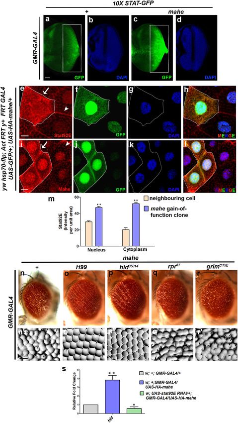
Official journal of the Cell Death Differentiation AssociationMaurya et al. Cell Death and Disease (2021)12:363 Page 8 of 14 Fig. 5 hopscotch enhances mahe induced cell death. (a–e) Images of Drosophila adult eye. a′–e′ Adult eye imprints. a, c, e GMR-GAL4 was used to drive UAS-mahe or (b, c) UAS-hop(Tum-l) (d, e)UAS-hop-RNAi. a, a′ Ectopic mahe expression promotes eye roughening. b, b′ Overexpression of constitutive active form of hop results in slight ommatidial disorganization. c, c′ Coexpression of hop and mahe enhanced the rough eye phenotype compared to mahe alone. d, d′ Reduction of hop levels by RNAi mediated knockdown leads to no observable change in eye roughening phenotype. e, e′ RNAi mediated depletion of hop rescued the mahe induced eye roughness. f–j, k–o Acridine orange and caspase staining in eye antennal disc. f, k Acridine and caspase positive cells show cell death induced by mahe. g, l hopTum-l overexpression showed few dying cells. h, m Coexpession of hopTum-l and mahe enhanced cell death in comparison to mahe alone. i, n No dying cells were observed on downregulating the level of hop. j, o Downregulation of hop rescued the mahe induced cell death as depicted by acridine orange and cl-caspase staining. p Graph represents cl- caspase intensity per unit area which shows activated hopTum-l promotes mahe induced cell death, which was significantly reduced in combination with hop-RNAi. One-way ANOVA. Scale bar in 50 µm (f–j, k–o). Genotypes (a) w; +; GMR-GAL4,UAS-HA-mahe/+ (b) w; UAS-hopTum-l/+;GMR-GAL4/+ (c) w; UAS-hopTum-l/+;GMR-GAL4,UAS-HA-mahe/+ (d) w; UAS-hop-RNAi/+;GMR-GAL4/+ (e) w; UAS-hop - RNAi/+; GMR-GAL4,UAS-HA-mahe/+. One-way ANOVA * P < 0.05, ** P < 0.01, *** P < 0.001. indicated that mahe positively regulates JAK/STAT Based on our findings we put forward a hypothetical pathway, which ultimately modulates the levels of hid model (Fig. 7), where we propose that Mahe regulates transcripts and lowering of the downstream effector JAK/STAT signaling by directly interacting with hop Stat92E alone was sufficient to rescue the hid mediated tyrosine kinase transcripts leading to its stabilization and apoptosis. this cascade the activation of downstream effector Official journal of the Cell Death Differentiation Association
Maurya et al. Cell Death and Disease (2021)12:363               Page 9 of 14

  Fig. 6 (See legend on next page.)

Official journal of the Cell Death Differentiation Association
Maurya et al. Cell Death and Disease (2021)12:363 Page 10 of 14
(see figure on previous page)
Fig. 6 JAK/STAT pathway activation by ectopic mahe induces hid dependent apoptosis. a–d Activation of JAK/STAT pathway was detected by
10X STAT92E-GFP reporter in eye-antennal discs. a, c Significant increase in GFP level was observed in GMR-GAL4 driven mahe eye antennal disc when
compared to the reporter line alone, indicating activation of JAK/STAT signaling (compare area in rectangle). b, d DAPI was used to mark the nucleus.
e–l Gain of function clone of mahe displays enhanced Stat92E activity. Gain of function clones of mahe using UAS-HA-mahe flies were generated with
FLP/FRT system in salivary gland. f, j GFP positive and non-GFP cells mark mahe gain-of-function clones (marked with arrow) and wild-type cells
(marked with arrowhead), respectively. e Level of Stat92E the transcription factor for JAK/STAT signaling was enhanced in mahe gain-of function
somatic clones when compared to wild-type neighboring cells. i Clonal area marked with GFP positive cells, shows elevated level of Mahe in the
gain-of-function clones. g, k DAPI was used to mark the nucleus. h, l Fourth column represents merged image with DAPI staining. m Graph
represents Stat92E intensity per unit area which shows marked enhancement in the level of Stat92E in nucleus and cytoplasm, in mahe
overexpressing clones when compared to neighboring cells. One-way ANOVA. n, n′ Ectopic expression of mahe results in rough eye phenotype.
o, o′ and p, p′ mahe induced rough eye phenotype was rescued by H99 and hid mutant, but not with rpr and grim mutants (q, q′ and r, r′). s Real
time PCR showed a threefold increase in hid transcript levels upon ectopic expression of mahe, while downregulation of JAK/STAT pathway by
stat92E-RNAi rescued the hid levels. One-way ANOVA. Scale bar in 50 µm (a–d, e–l). Genotypes (a, b) w/+; 10X STAT-GFP/+, GMR-GAL4/+ (c, d) w/+;
10X STAT-GFP/+; GMR-GAL4, UAS-HA-mahe/+ (n) w/+; +; GMR-GAL4,UAS-HA-mahe/+ (o) w/+; +; GMR-GAL4,UAS-HA-mahe/H99 (p) w/+; +; GMR-
GAL4,UAS-HA-mahe/hid05014 (q) w/+; +; GMR-GAL4,UAS-HA-mahe/rpr87 (r) w/+; +; GMR-GAL4,UAS-HA-mahe/grimC15E. One-way ANOVA * P < 0.05,
** P < 0.01, *** P < 0.001.
have recently reported that mutation in DDX59, the
human homolog of maheshvara (mahe), is associated with
neurological abnormalities and is crucial for development
of the nervous system11.
In this report we have elucidated a novel link between
mahe and JAK/STAT signaling, suggesting the sig-
nificance of mahe in regulation of JAK/STAT signaling in
the photoreceptor neurons of Drosophila. Our tran-
scriptome analysis has identified differentially regulated
genes that are predominantly related to neuronal func-
tion, phototransduction, stress response and regulation of
cell signaling. Interestingly, by quantitative-PCR analysis,
we found that all the ligands and downstream effector of
JAK/STAT pathway were significantly lowered in mahe
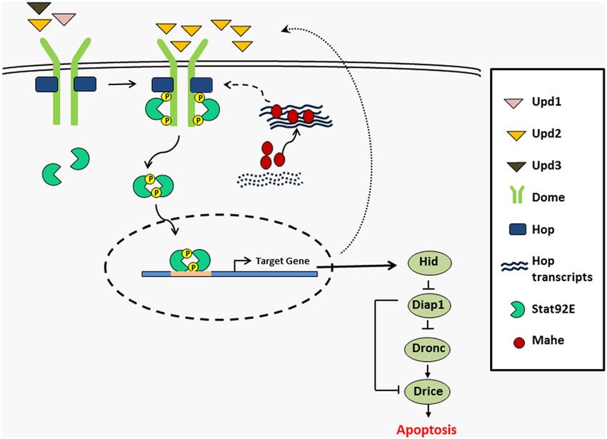
Fig. 7 A hypothetical model depicting recruitment and loss-of-function mutant and in mahe-RNAi. Similarly,
stabilization of hop kinase transcripts by RNA helicase Mahe upregulation of the pathway components suggested
which leads to activation of JAK/STAT signaling and primes hid ectopic activation of the JAK/STAT signaling by over-
mediated apoptosis. Our model depicts that Mahe encoding an RNA expressed mahe, which was in agreement with the gene
helicase binds and regulates hop transcript stability, and this results in
ontology analysis of transcriptome data.
the increase in levels of Hop kinase. This upregulation of Hop tyrosine
kinase leads to enhanced phosphorylation and activation of JAK/STAT signaling is an important pathway required
transcription factor Stat92E which enters into the nucleus, resulting in for Drosophila eye development and for proper axon
downstream target gene activation. An auto feedback loop is targeting in a cell autonomous manner during photo-
reflected by increase in the levels of Upd2, and thus the JAK/STAT receptor formation32. Further, genetic studies have pre-
signaling remains in an active state. This activated JAK/STAT signaling
viously shown that overexpression of upd and
then induces hid mediated apoptosis.
hopTumorous-lethal which encodes constitutively active form
of hop results in dramatically overgrown adult eyes. In
molecules in this pathway. This in turn leads to activation addition, downregulation of JAK/STAT pathway by
of proapoptotic gene hid, along with a surge in apoptosis. hypomorphic upd mutant and dominant negative form of
domeΔcyt leads to small or ablated eyes19,22,33–35. However,
Discussion in contrast in lobe mutants, ectopic upd expression is
A number of reports have shown that Decapentaplegic known to activate JAK/STAT pathway and induce apop-
(Dpp), Hedgehog (Hh), Notch, JAK/STAT and Wingless tosis, which leads to small eye phenotype36. Taken toge-
(Wg) signaling pathways are major regulators of devel- ther these findings suggest that JAK/STAT pathway can
opment and differentiation of photoreceptor neurons in induce both context-dependent proliferation as well as
Drosophila22–28. Dysregulation of JAK/STAT pathway has apoptosis and is needed for proper eye development in
been implicated in a wide variety of neurological disorders Drosophila. Although both upd2 and upd3 are ligands for
like the glial tauopathy and Parkinson’s Disease29–31. We JAK/STAT pathway but their role in Drosophila
Official journal of the Cell Death Differentiation AssociationMaurya et al. Cell Death and Disease (2021)12:363 Page 11 of 14 photoreceptor development, specifically cell death has not Mahe and hop interaction upregulates the JAK/STAT been investigated. Here, we observed that coexpression of pathway via an auto feedback loop. both mahe and upd2 along with other components of In summary, we report that mahe induces JAK/STAT JAK/STAT pathway results in massive apoptosis. Our signaling mediated apoptosis during Drosophila photo- findings reveal that mahe activated JAK/STAT signaling receptor development. We put forth a novel mechanism can induce apoptosis in Drosophila photoreceptors thus in which hop is post-transcriptionally regulated by a showing context-dependent role of JAK/STAT pathway DEAD box RNA helicase Maheshvara, which ultimately during development. Conversely, loss of JAK/STAT sig- results in activation of JAK/STAT signaling mediated naling components leads to rescue in the mahe-induced apoptosis. As deregulated JAK/STAT signaling and apoptosis. apoptosis have been linked to many human diseases, the RBPs are known to modulate post-transcriptional reg- identification of a novel regulator of this pathway will lead ulation, turnover, localization, and translational control of to a better understanding of development and disease. mRNA. Reports have shown that RBPs can act in both Identification of Maheshvara as a novel modulator of positive or negative manner to stabilize mRNA37,38. TTP JAK/STAT pathway has opened up a novel platform for and KSRP destabilizes several mRNA like c-fos, TNFα and understanding the role of RNA helicase associated cell COX-2, however HuR acts positively to maintain their death which may also hold good for human disorders stability39–42. A recent report revealed that RNA binding as well. protein Musashi 2 (MSI2) regulates IL-6 signal transducer (IL6ST) and promotes its degradation by binding to 3′ Material and methods UTR of mRNA. IL6ST cytokine receptor in turn after Drosophila genetics forming complex with kinases affects the phosphorylation All the fly stocks were maintained on standard corn- of STAT3 and other mitogen-activated protein kinase meal/yeast/molasses/agar medium at 25 °C. w1118 was ERK43. Similarly, in order to understand the mechanism used as the wild-type control. UAS-HA-mahe10 and of ectopic JAK/STAT activation, we argued whether EPΔmahe d0805911 were generated in our lab. UAS-upd2- Mahe a DEAD box RNA helicase might interact directly GFP and upd2Δ3-62were kind gift from Prof. James with the transcripts of the components of JAK/STAT Castelli-Gair Hombría. UAS-mahe-RNAi (v109465), UAS- transduction cascade to regulate the pathway. Interest- hop-RNAi, UAS-stat92E-RNAi, UAS-upd1-RNAi, UAS- ingly, hop transcripts were found to be associated with CCT7-RNAi were obtained from VDRC.GMR-GAL4, Mahe-containing RNA–protein complex. We propose a UAS-DIAP, UAS-P35, UAS-upd1, UAS-hop, 10X STAT- model in which this interaction may lead to decrease in GFP, H99, hid05014, rpr87, grimC15E, stocks were obtained the turnover rate of transcripts, and thus leads to stabi- from Bloomington Drosophila Stock Center. All the lization and elevation in hop transcripts. The elevated JAK crosses were performed at 25 °C, unless mentioned is activated possibly via interaction with the receptor and otherwise. To generate mahe gain-of-function clones ligand Upd2 in response to ectopic mahe. Mechanism females of hsp70-flp; Act FRT y+ FRT-GAL4 UAS-GFP/+; that could account for activation of the pathway is +/+ were crossed to UAS-HA-mahe males. Heat shock probably via augmented phosphorylation and activation/ was given at 37 °C for 10 min at 24 h AEL and the third stabilization of downstream transcription factor Stat92E, instar larvae were analyzed for GFP marked clones. as seen by its enhanced cytoplasmic to nuclear localiza- tion. Next, we hypothesized that this interaction can Eye imprints result in enhanced upd2 RNA levels, which is further Eye imprints using nail polish were prepared for analysis supported by a significant increase in upd2 transcript of ommatidial defects and were examined under differ- levels upon expression of hop alone. Our prediction that ential interference contrast (DIC) optics in a Nikon mahe activates downstream targets of JAK/STAT signal- Eclipse 80i microscope. ing was supported by our reporter gene analysis using 10X Stat92E GFP line. Further, a massive increase in both Acridine orange staining cytoplasmic and nuclear Stat92E transcription factor was To observe the extent of apoptosis we used the vital dye seen in mahe gain-of-function somatic clone. Activation acridine orange (AO). Eye-antennal discs from larvae of of the JAK/STAT pathway was further reflected by the desired genotypes were dissected in phosphate buffer increase in transcription of downstream target of socs36E. saline (PBS) (130 mM NaCl, 7 mM Na2HPO4, 3 mM The above finding was supported by a report where oral KH2PO4, pH 7.4) and stained with 1 μg/μl of Acridine infection initiates immune response in the gut of Droso- orange (AO) in PBS for 3 min. Followed by two washes phila followed by upregulation of JAK/STAT signaling, with PBS and were finally mounted in PBS. These were and the pathway activity was assessed by transcript levels than immediately viewed under Nikon Eclipse 80i of socs36E44. Based on these findings we propose that microscope. Official journal of the Cell Death Differentiation Association
Maurya et al. Cell Death and Disease (2021)12:363                                                             Page 12 of 14

Immunostaining                                                   hop reverse primer: 5′-GCATTCACGCACAATATAG
  Immunostaining was performed in various tissues               C-3′
dissected from third instar larvae. Larvae were dissected        hop promoter forward primer: 5′-CAAGAATATAGAC
in PBS (pH 7.4) and immunostaining was done as                  GCCATAGAGC-3′
described previously10. To mark the nuclei, staining             hop promoter reverse primer: 5′-GTCATCGATTGTCC
with 4′, 6- diamidino-2-phenylindole dihydrochloride            AATA ACCTG-3′
(DAPI) (1 µg/ml) was done. Tissues were mounted in               socs36E forward primer: 5′- GCTGCCAGTCAGCAAT
DABCO. All slides were observed under LSM 780 laser             ATGT-3′
scanning confocal microscope Zeiss (Carl Zeiss),                 socs36E reverse primer: 5′- GACTGCGGCAGCAACTG
Thornwood, NY. The images were further processed                T-3′
with Adobe Photoshop 7. The following primary anti-              hid forward primer: 5′-AGC GTCTGCAGGAGTTCA
bodies were used in this study: rabbit anti-Mahe (1:300)        AT-3′
generated in our laboratory, rabbit anti-cleaved Caspase         hid reverse primer: 5′-CTTCGCCTTTTGTCGTTCT
3 (1:100) (Cell Signalling Technology), rabbit anti-            C-3′
Stat92E (1:200) a kind gift from Erika Bach. Secondary           rps17 forward primer: 5′-AAGCGCATCTGCGAGGA
antibody used was Alexa Fluor 555 conjugated goat anti-         G-3′
rabbit IgG (1:200). Intensity profile graphs were made by         rps17 reverse primer: 5′-CCTCCTCCTGCAACTTGA
using Image J and Graph Pad Prism 5 software.                   TG-3′.

RNA extraction and Real-time PCR                                Transcriptome analysis
  Total RNA was extracted using Trizol reagent (Invi-             Total RNA was isolated from 200 adult fly heads using
trogen), from adult fly heads of desired genotypes. To           standard Trizol method (Sigma) from adult fly heads of
remove genomic DNA contamination the extracted RNA              GMR-GAL4 driven UAS-mahe and GMR-GAL4 alone
was treated with RNase free DNaseI for 30 min at 37 °C.         which served as control for comparison. Libraries were
Reverse transcription was performed with a cDNA                 made using standard protocol of TrueSeq RNA sample
synthesis kit (Applied Biosystems, Foster City, CA), using      Prep kit v2. Libraries were then sequenced by the paired-
aliquots of total RNA extracted. Real-time quantitative         end reads using Illumina HiSeq2500 platform and the
PCR was performed to check the expression of desired            resulting sequencing reads were aligned to the reference
genes of interest. Real-time PCR reactions were per-            Drosophila melanogaster genome downloaded from
formed using the ABI 7500 sequence detection system             Ensemble database (ftp://ftp.ensembl.org/pub/release-
(Applied Biosystems) with SYBR Green PCR Master Mix             81/fasta/drosophila_melanogaster/dna/Drosophila_mela-
(Thermo Scientific). The relative quantity of amplified           nogaster.BDGP6.dna.toplevel.fa.gz). The alignment was
cDNA/DNA corresponding to the gene was determined               performed by STAR program (version = STAR_2.4.1d).
using the ΔΔCt method and normalized for expression of          Further, the aligned reads were used for estimating
rps17 in each sample45. The graph was prepared using            expression of transcripts using cufflinks program (ver-
Graph Pad Prism 5 software.                                     sion: cufflinks-2.2.1). The expression values are reported
  Primers used for the study are as follows:                    in FPKM (Fragment per kilo per million) units for each of
  mahe forward primer: 5′-TTCGTGCGTTGGCCCTTG                    the genes and transcripts. Differential expression analysis
TTATTG-3′                                                       was performed using cuffdiffv2.2.1. Gene ontology ana-
  mahe reverse primer: 5′-GCTGGGCATCGAACGAGC                    lysis was done using DAVID 6.8 (The Database for
AAG-3′                                                          Annotation, Visualization and Integrated Discovery).
  upd1 forward primer: 5′- ATTGCCCTAAAGCGCTGG
TACCG-3′                                                        Immunoprecipitation of RNA-protein complex
  upd1 reverse primer: 5′- GTAGTAGTGGTGCTTCAC                     Protein lysates were prepared by homogenizing 200 adult
AAAGC-3′                                                        heads from 1 day old flies of GMR-GAL4 driven UAS-mahe
  upd2 forward primer: 5′- GTGAAGCTAAAGACTTG-3′                 and GMR-GAL4 alone which served as control for com-
  upd2 reverse primer: 5′- TCAAGACTCATTGGATC                    parison in lysis Buffer (100 mM KCl, 5 mM MgCl2, 10 mM
CGCCAT-3′                                                       HEPES, pH 7.0, 0.5% Nonidet P-40, 1 mM DTT, 100 U
  upd3 forward primer: 5′-TGCCCCGTCTGAATCTCA                    ml−1 RNase inhibitor (NEB), 2 mM vanadyl ribonucleoside
CT-3′                                                           complexes solution (Sigma- Aldrich, 25 μl ml−1 protease
  upd3 reverse primer: 5′-GTGAAGGCGCCCACGTAA-3′                 inhibitor cocktail (Roche). Supernatant was collected and
  hop forward primer: 5′- GGGTATCTACATCAGATTG                   centrifuged at 12,000g for 20 min at 4 °C. Equilibration of
TC-3′                                                           anti-HA agarose beads was done in lysis buffer. Protein

Official journal of the Cell Death Differentiation Association
Maurya et al. Cell Death and Disease (2021)12:363                                                                                                 Page 13 of 14

lysates were precleared by incubating with 20 μl of anti-HA                    We thank Prof. S.C. Lakhotia for providing hsrω construct for RNA-FISH control
agarose beads for 2 h at 4 °C. For immunoprecipitation,                        experiments. We acknowledge the real-time facility at ISLS (Interdisciplinary
                                                                               Centre for Life Sciences) and Department of Molecular and Human Genetics.
50 μl of anti-HA beads were used for every 250 μl of protein                   We also acknowledge confocal facility at ISLS and Department of Zoology,
lysate and were incubated for 5 h at 4 °C to pull down the                     Banaras Hindu University. B.M., R.D., and P.P. were supported by a fellowship
desired protein of interest. This was followed by brief                        from the Council of Scientific and Industrial Research, Government of India.
                                                                               SERB-POWER Fellowship to MM from Science and Engineering Research Board,
centrifugation to collect the beads. These beads were                          Department of Science and Technology, Government of India is
washed with 0.5 ml of polysome lysis buffer thrice by cen-                     acknowledged.
trifugation at 2000g for 5 min at 4 °C. Washed beads were
resuspended in 100 μl of lysis buffer with 30 μg of protei-                    Author contributions
nase k and 0.1% SDS followed by heating at 50 °C for 30 min                    Conceptualization, B.M., and M.M.; Methodology, B.M. and P.P.; Investigation, B.
in order to degrade the protein. Total RNA was extracted                       M., A.M., S.S., and M.M.; Software and Formal Analysis, B.M., R.D., and M.M.;
                                                                               Writing Original Draft, B.M. and M.M.; Supervision, M.M.
using Trizol reagent. Upper aqueous phase having RNA was
recovered by centrifugation and to it 10 μl of yeast tRNA                      Conflict of interest
(1 mg ml−1), 12 μl of 3 M sodium acetate and 250 μl of                         The authors declare no competing interests.
ethanol were added per 100 μl of aqueous phase and kept at
−20 °C overnight for precipitation. This was followed by
                                                                               Publisher’s note
centrifugation at 12,000g for 20 min at 4 °C, ethanol was                      Springer Nature remains neutral with regard to jurisdictional claims in
removed and the pellet rinsed with 70% ethanol followed by                     published maps and institutional affiliations.
air drying until all the liquid evaporated. The pellet was
                                                                               Supplementary information The online version contains supplementary
resuspended in nuclease free MQ water. cDNA was syn-                           material available at https://doi.org/10.1038/s41419-021-03649-0.
thesized from total RNA using M-MuLV Reverse Tran-
scriptase (New England Biolabs) as per the manufacturer’s                      Received: 14 December 2020 Revised: 17 March 2021 Accepted: 18 March
instructions. RT-PCR was performed using primers for                           2021
upd1, upd2, upd3, dome, hop, stat92E, and socs36E
listed above.

RNA: RNA in situ hybridization                                                 References
  RNA-RNA in situ hybridization was carried out in tis-                         1. Keene, J. D. RNA regulons: coordination of post-transcriptional events. Nat. Rev.
                                                                                    Genet. 8, 533–543 (2007).
sue from third instar larvae. Larvae were dissected and                         2. Moore, M. J. From birth to death: the complex lives of eukaryotic mRNAs.
RNA:RNA in situ hybridization was carried out as                                    Science 309, 1514–1518 (2005).
described earlier37. Hybridization was done with 100 ng                         3. Abdelhaleem, M. Do human RNA helicases have a role in cancer? Biochim.
                                                                                    Biophys. Acta 1704, 37–46 (2004).
hop antisense riboprobe in hybridization buffer at 50 °C                        4. Fuller-Pace, F. V. DEAD box RNA helicase functions in cancer. RNA Biol. 10,
for 12–16 h. hop sense riboprobe was used as negative                               121–132 (2013).
control and was hybridized at 50 °C for 12–16 h. hsrω                           5. Anthony, K. & Gallo, J.-M. Aberrant RNA processing events in neurological
                                                                                    disorders. Brain Res. 1338, 67–77 (2010).
antisense riboprobe served as positive control and was                          6. Shamseldin, H. E. et al. Mutations in DDX59 implicate RNA helicase in the
hybridized for 12–16 h at 50 °C. For fluorescent detection                           pathogenesis of orofaciodigital syndrome. Am. J. Hum. Genet. 93, 555–560
of the riboprobe, anti-DIG-Rhodamine conjugated anti-                               (2013).
                                                                                7. Paine, I. et al. Paralog studies augment gene discovery: DDX and DHX Genes.
body (1:200, Roche) was used. DAPI was used to mark the                             Am. J. Hum. Genet. 105, 302–316 (2019).
nucleus. Samples were mounted in DABCO and observed                             8. Balak, C. et al. Rare de novo missense variants in RNA helicase DDX6 cause
under LSM 780 laser scanning confocal microscope Zeiss                              intellectual disability and dysmorphic features and lead to P-Body defects and
                                                                                    RNA dysregulation. Am. J. Hum. Genet. 105, 509–525 (2019).
(Carl Zeiss). All the images were further processed using                       9. Lessel, D. et al. De novo missense mutations in DHX30 impair global trans-
Adobe Photoshop 7.0.                                                                lation and cause a neurodevelopmental disorder. Am. J. Hum. Genet. 101,
                                                                                    716–724 (2017).
                                                                               10. Surabhi, S. et al. Regulation of notch signaling by an evolutionary conserved
Statistical analysis                                                               DEAD box RNA helicase, maheshvara in Drosophila melanogaster. Genetics
  In our study experiments were conducted in triplicate,                           201, 1071–1085 (2015).
analysis was done using PRISM 5 as guided (GraphPad,                           11. Salpietro, V. et al. A loss‐of‐function homozygous mutation in DDX59 impli-
                                                                                   cates a conserved DEAD‐box RNA helicase in nervous system development
San Diego, CA) and results were given as the mean ±                                and function. Hum. Mutat. 39, 187–192 (2018).
standard deviation (S.D.). The statistical analysis was                        12. Honig, L. S. & Rosenberg, R. N. Apoptosis and neurologic disease. Am. J. Med.
performed using one-way ANOVA. The significant level                                108, 317–330 (2000).
                                                                               13. Cavallaro, S. Cracking the code of neuronal apoptosis and survival. Cell Death
was set as p-values below 0.05.                                                    Dis. 6, e1963 (2015).
                                                                               14. Wang, Y. H. & Huang, M. L. Organogenesis and tumorigenesis: insight from
Acknowledgements                                                                   the JAK/STAT pathway in the Drosophila eye. Dev. Dyn. 239, 2522–2533
The authors acknowledge the Bloomington Drosophila Stock Centre and                (2010).
Vienna Drosophila Resource Centre. We thank Prof. James Castelli-Gair          15. Zeidler, M. P. & Bausek, N. The Drosophila Jak-Stat pathway. JAKSTAT 2, e25352
Hombría and Prof. Erika Bach for kindly providing us fly stocks and antibody.       (2013).

Official journal of the Cell Death Differentiation Association
Maurya et al. Cell Death and Disease (2021)12:363 Page 14 of 14
16. Kisseleva, T., Bhattacharya, S., Braunstein, J. & Schindler, C. Signaling through 32. Hoi, C. S., Xiong, W. & Rebay, I. Retinal axon guidance requires integration of
the JAK/STAT pathway, recent advances and future challenges. Gene 285, Eya and the Jak/Stat pathway into phosphotyrosine-based signaling circuitries
1–24 (2002). in Drosophila. Genetics 203, 1283–1295 (2016).
17. Harrison, D. A., McCoon, P. E., Binari, R., Gilman, M. & Perrimon, N. Drosophila 33. Bach, E. A., Vincent, S., Zeidler, M. P. & Perrimon, N. A sensitized genetic screen
unpaired encodes a secreted protein that activates the JAK signaling pathway. to identify novel regulators and components of the Drosophila janus kinase/
Genes Dev. 12, 3252–3263 (1998). signal transducer and activator of transcription pathway. Genetics 165,
18. Mukherjee, T., Schäfer, U. & Zeidler, M. P. Identification of Drosophila genes 1149–1166 (2003).
modulating Janus kinase/signal transducer and activator of transcription signal 34. Harrison, D. A., Binari, R., Nahreini, T. S., Gilman, M. & Perrimon, N. Activation of a
transduction. Genetics 172, 1683–1697 (2006). Drosophila Janus kinase (JAK) causes hematopoietic neoplasia and develop-
19. Tsai, Y. C. & Sun, Y. H. Long‐range effect of upd, a ligand for Jak/STAT pathway, mental defects. EMBO J. 14, 2857–2865 (1995).
on cell cycle in Drosophila eye development. Genesis 39, 141–153 (2004). 35. Brown, S., Hu, N. & Hombrı ́a, J. C.-G. Identification of the first invertebrate
20. Bach, E. A. et al. GFP reporters detect the activation of the Drosophila JAK/ interleukin JAK/STAT receptor, the Drosophila gene domeless. Curr. Biol. 11,
STAT pathway in vivo. Gene Expr. Patterns 7, 323–331 (2007). 1700–1705 (2001).
21. Borensztejn, A., Boissoneau, E., Fernandez, G., Agnès, F. & Pret, A.-M. JAK/STAT 36. Wang, Y.-H. & Huang, M.-L. Reduction of Lobe leads to TORC1 hypoactivation
autocontrol of ligand-producing cell number through apoptosis. Development that induces ectopic Jak/STAT signaling to impair Drosophila eye develop-
140, 195–204 (2013). ment. Mech. Dev. 126, 781–790 (2009).
22. Hanratty, W. P. & Dearolf, C. R. The Drosophila Tumorous lethal hematopoietic 37. Tripathi, B. K., Surabhi, S., Bhaskar, P. K., Mukherjee, A. & Mutsuddi, M. The RNA
oncogene is a dominant mutation in the hopscotch locus. Mol. Gen. Genet. binding KH domain of Spoonbill depletes pathogenic non-coding spinocer-
238, 33–37 (1993). ebellar ataxia 8 transcripts and suppresses neurodegeneration in Drosophila.
23. Ma, C., Zhou, Y., Beachy, P. A. & Moses, K. The segment polarity gene Biochim. Biophys. Acta 1862, 1732–1741 (2016).
hedgehog is required for progression of the morphogenetic furrow in the 38. Maurya, B., Surabhi, S., Pandey, P., Mukherjee, A. & Mutsuddi, M. in Insights Into
developing Drosophila eye. Cell 75, 927–938 (1993). Human Neurodegeneration: Lessons Learnt From Drosophila 373–403 (Springer,
24. Treisman, J. E. & Rubin, G. M. wingless inhibits morphogenetic furrow 2019).
movement in the Drosophila eye disc. Development 121, 3519–3527 (1995). 39. Dean, J. L. et al. The 3′ untranslated region of tumor necrosis factor alpha
25. Heslip, T. R., Theisen, H., Walker, H. & Marsh, J. L. Shaggy and dishevelled exert mRNA is a target of the mRNA-stabilizing factor HuR. Mol. Cell Biol. 21,
opposite effects on Wingless and Decapentaplegic expression and on posi- 721–730 (2001).
tional identity in imaginal discs. Development 124, 1069–1078 (1997). 40. Sawaoka, H., Dixon, D. A., Oates, J. A. & Boutaud, O. Tristetraprolin binds to the
26. Greenwood, S. & Struhl, G. Progression of the morphogenetic furrow in the 3′-untranslated region of cyclooxygenase-2 mRNA a polyadenylation variant
Drosophila eye: the roles of Hedgehog, Decapentaplegic and the Raf path- in a cancer cell line lacks the binding site. J. Biol. Chem. 278, 13928–13935
way. Development 126, 5795–5808 (1999). (2003).
27. Baonza, A. & Freeman, M. Notch signalling and the initiation of neural 41. Katsanou, V. et al. HuR as a negative posttranscriptional modulator in
development in the Drosophila eye. Development 128, 3889–3898 (2001). inflammation. Mol. Cell 19, 777–789 (2005).
28. Fu, W. & Baker, N. E. Deciphering synergistic and redundant roles of Hedge- 42. Winzen, R. et al. Functional analysis of KSRP interaction with the AU-rich
hog, Decapentaplegic and Delta that drive the wave of differentiation in element of interleukin-8 and identification of inflammatory mRNA targets.
Drosophila eye development. Development 130, 5229–5239 (2003). Mol. Cell Biol. 27, 8388–8400 (2007).
29. Nicolas, C. S. et al. The role of JAK-STAT signaling within the CNS. JAKSTAT 2, 43. Duggimpudi, S. et al. Transcriptome-wide analysis uncovers the targets of the
e22925 (2013). RNA-binding protein MSI2 and effects of MSI2’s RNA-binding activity on IL-6
30. Colodner, K. J. & Feany, M. B. Glial fibrillary tangles and JAK/STAT-mediated signaling. J. Biol. Chem. 293, 15359–15369 (2018).
glial and neuronal cell death in a Drosophila model of glial tauopathy. J. 44. Buchon, N., Broderick, N. A., Poidevin, M., Pradervand, S. & Lemaitre, B. Dro-
Neurosci. 30, 16102–16113 (2010). sophila intestinal response to bacterial infection: activation of host defense
31. Qin, H. et al. Inhibition of the JAK/STAT pathway protects against α-synuclein- and stem cell proliferation. Cell Host Microbe 5, 200–211 (2009).
induced neuroinflammation and dopaminergic neurodegeneration. J. Neu- 45. Schmittgen, T. D. & Livak, K. J. Analyzing real-time PCR data by the com-
rosci. 36, 5144–5159 (2016). parative CT method. Nat. Protoc. 3, 1101–1108 (2008).
Official journal of the Cell Death Differentiation AssociationYou can also read