Box Jellyfish (Cnidaria, Cubozoa) Extract Increases Neuron's Connection: A Possible Neuroprotector Effect
←
→
Page content transcription
If your browser does not render page correctly, please read the page content below
Hindawi
BioMed Research International
Volume 2021, Article ID 8855248, 12 pages
https://doi.org/10.1155/2021/8855248

Research Article
Box Jellyfish (Cnidaria, Cubozoa) Extract Increases Neuron’s
Connection: A Possible Neuroprotector Effect

 Gian Lucas M. Arruda,1 Hugo Vigerelli ,2 Michelle C. Bufalo ,3 Giovanna B. Longato ,4
 Rodinei V. Veloso,1 Vanessa O. Zambelli ,3 Gisele Picolo ,3 Yara Cury ,3
 André C. Morandini ,5,6 Antonio Carlos Marques ,5 and Juliana Mozer Sciani 1
 1
 Laboratório Multidisciplinar de Pesquisa, Universidade São Francisco, Bragança Paulista 12916-900, Brazil
 2
 Laboratório de Genética, Instituto Butantan, São Paulo 05503-900, Brazil
 3
 Laboratório de Dor e Sinalização, Instituto Butantan, São Paulo 05503-900, Brazil
 4
 Laboratório de Pesquisa em Farmacologia Molecular e Compostos Bioativos, Universidade São Francisco,
 Bragança Paulista 12916-900, Brazil
 5
 Departamento de Zoologia, Instituto de Biociências, Universidade de São Paulo, São Paulo 05508-090, Brazil
 6
 Centro de Biologia Marinha, Universidade de São Paulo, São Sebastião 11612-109, Brazil

 Correspondence should be addressed to Juliana Mozer Sciani; juliana.sciani@usf.edu.br

 Received 28 September 2020; Revised 14 February 2021; Accepted 23 February 2021; Published 5 March 2021

 Academic Editor: Mauricio Budini

 Copyright © 2021 Gian Lucas M. Arruda et al. This is an open access article distributed under the Creative Commons Attribution
 License, which permits unrestricted use, distribution, and reproduction in any medium, provided the original work is
 properly cited.

 Neurodegenerative diseases are one of the major causes of death worldwide, characterized by neurite atrophy, neuron apoptosis,
 and synapse loss. No effective treatment has been indicated for such diseases so far, and the search for new drugs is being
 increased in the last years. Animal venoms’ secretion/venom can be an alternative for the discovery of new molecules, which
 could be the prototype for a new treatment. Here, we present the biochemical characterization and activity of the extract from
 the box jellyfish Chiropsalmus quadrumanus (Cq) on neurites. The Cq methanolic extract was obtained and incubated to human
 SH-SY5Y neurons, and neurite parameters were evaluated. The extract was tested in other cell types to check its cytotoxicity and
 was submitted to biochemical analysis by mass spectrometry in order to check its composition. We could verify that the Cq
 extract increased neurite outgrowth length and branching junctions, amplifying the contact between SH-SY5Y neurons, without
 affecting cell body and viability. The extract action was selective for neurons, as it did not cause any effects on other cell types,
 such as tumor line, nontumor line, and red blood cells. Moreover, mass spectrometry analysis revealed that there are no
 proteins but several low molecular mass compounds and peptides. Three peptides, characterized as cryptides, and 14 low
 molecular mass compounds were found to be related to cytoskeleton reorganization, cell membrane expansion, and
 antioxidant/neuroprotective activity, which act together to increase neuritogenesis. After this evaluation, we conclude that the
 Cq extract is a promising tool for neuronal connection recovery, an essential condition for the treatment of neurodegenerative
 diseases.

1. Introduction 7th cause of death. The expectancy is that, in 2050, this num-
 ber could reach 131 mi for Alzheimer solely [1–3].
Neurodegenerative diseases are one of the major causes of The most common neurodegenerative diseases included
death worldwide. The number of patients suffering from such Alzheimer’s disease (AD), Parkinson’s disease (PD), Hun-
diseases has been rising every year due to an increase in the tington’s disease (HD), and amyotrophic lateral sclerosis
life expectancy of the population. In 2015, 46.8 million peo- (ALS). All these pathologies are characterized by protein or
ple with dementia were reported worldwide, making it the peptide accumulation in certain regions of the brain, inside
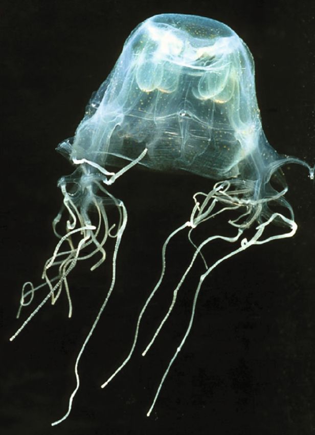
2 BioMed Research International

or outside neurons, after aggregation [2]. Key proteins Glycosaminoglycans, isolated from sea squirts, octo-
involved in these processes are β-amyloid and tau for AD, puses, and sea urchins, display strong neuroregenerative
α-synuclein for PD, huntingtin for HD, and ubiquitinated activity, by promoting neurite outgrowth [19].
proteins for ALS [2]. Jellyfishes (phylum Cnidaria) are abundant animals in
 Although the molecular and cellular mechanisms for many coastal areas around the world [20, 21]. They are
neurodegeneration development have not been completely known to cause unpleasant reactions on humans, due to
understood, it is known that precursor proteins are involved its venom present in specialized intracellular organelles
in the deposition of aggregates, which are not eliminated [4, called nematocysts [22]. The venom of jellyfishes is known
5]. Thus, it is suggested that the protein accumulation causes to cause several stings, mainly characterized by inflamma-
several damages to the brain, including an increase of reactive tion and pain [23]. The venom is constituted mainly by
oxygen species, inflammation, and neuron apoptosis [6]. proteins: phospholipases A2, metallopeptidases, serinepepti-
 Neurite atrophy and synapse loss are frequent in such dases, CRISPs, lectins, pore-forming toxins, and protease
diseases, followed by an axonal degeneration, which probably inhibitors [24–27]. Peptides have also been described; how-
initiates the neuronal death. The cell toxicity may cause ever, little is known about low molecular mass compounds
impairment of cognition, memory, and muscle control, from Cnidaria, regarding both structure and biological
depending on the affected area of the brain [7, 8]. activity.
 Molecules derived from natural products have been used Moreover, there are some studies showing the abundance
as prototypes in the development of new drugs, including the of neurotransmitters in cnidarians, such as acetylcholine,
treatment of neurological diseases [9]. Around 33% of the norepinephrine, serotonin, histamine, glutamate, and γ-ami-
world’s most selling drugs are derived from natural sources, nobutyric acid (GABA), involved in the animal’s physiology,
both animals and plants, indicated for treating cancer, viral including neurotransmission and neuromodulation [28].
infections, pain, inflammation, and diseases of the central Our goal was to verify the composition of the methanolic
nervous system [10, 11]. extract of the tentacle of the box jellyfish (Chiropsalmus
 Species from a marine habitat have provided several quadrumanus), as well as its activity on human neurons, ana-
molecules of therapeutic interest; some of them already lyzing its potential for neurite and branch formation, which
turned into commercial drugs and applied in the clinics would be useful in diseases characterized by neuronal loss.
[10, 12]. Examples are arabinose nucleosides from sea
sponges for treating several types of cancer [13]. Bioactive 2. Materials and Methods
molecules from marine animals are different from terrestrial
ones in terms of structures—in the last years, 13,000 new 2.1. Preparation of Extract. The box jellyfish Chiropsal-
molecules were identified from marine animals, with 3000 mus quadrumanus (Cnidaria, Cubozoa, Chridropida,
belonging to the phylum Cnidaria. Among them, there is Figure 1—photo kindly provided by Dr. Alvaro E. Migotto
no description of molecules from the class Cubozoa [14]. and Cifonauta-CEBIMar) was collected in a marina at Ilha-
Nevertheless, these numbers reflect the ocean as an impor- bela country, São Sebastião Island, São Paulo, Brazil (23°
tant source of new molecules, including terpenes, peptides, 46 ′ 23″ S, 45° 21 ′ 25″ W), under IBAMA license #16802-2.
alkaloids, piperazines, lactones, nucleosides, and glycolipids, After collection, animals had their tentacles removed and
the most frequent ones [9, 13]. placed in methanol containing 0.1% acetic acid for 48 hours
 Several natural compounds, specially from plants, have (Cq extract). After that, the solution was centrifuged at
proved to induce neurite length increase and regeneration. 5,000 x g for 10 minutes and the supernatant was lyophilized.
One example is platycodigenin, a terpenoid isolated from The content was resuspended in sterile phosphate buffer
Platycodon grandifloras (Chinese bellflower), with known solution (PBS 50 mM, pH 7.4) for the cell experiments and
neuroprotective activity. Yang et al. [15] showed its ability in water methanol (1 : 1 vol : vol) for mass spec and SDS-
in preventing Aβ25-35-induced neuronal death, besides PAGE analysis. Species identification and extra biological
promoting neurite regeneration. Another example is the information may be found in Jarms et al. [29].
alpha-toxin from the bacteria Clostridium perfringens that
induces phosphorylation of TrkA through the phospho- 2.2. Cell Culture and Neuronal Differentiation Protocol.
lipid metabolism, and this pathway was related to the SH-SY5Y cells (ECACC, Sigma Aldrich, St. Louis, MO,
induction of neurite outgrowth in PC12 cells [16]. USA) were cultured in a 1 : 1 mixture of Ham’s F12 and
 Compounds from marine animals have been poorly Dulbeco’s modified Eagle’s medium (DMEM) (Gibco Life
studied concerning neuron regeneration. A peptide frag- Technologies, Grand Island, NY, USA) supplemented with
ment of thymosin β4, found in sea cucumbers and sea 10% heat-inactivated fetal bovine serum (FBS) (HyClone
urchins, act as a regulator of neurogenesis, by facilitating Labs., Logan, UT) and 100 U/mL of penicillin/streptomy-
the hippocampal neurogenesis and increasing spatial mem- cin (Gibco Life Technologies, Grand Island, NY, USA) in
ory [17]. Moreover, a beta-thymosin from marine mol- a humidified atmosphere of 5% CO2 at 37°C. The medium
lusks, and its peptide fragments, through the neuronal was changed twice a week, and cells were split at about
proliferation and the increase of neurite outgrowth [17, 80% confluence. For neuronal differentiation, 5 × 10⁴
18], supports the anchoring of neurons, besides increasing cells/well was seeded in collagen-coated plates (100 μg/mL,
neurite regeneration (sprouting and total neurite out- Corning, USA). After 24 hours (day 1), the medium was
growth) in culture [18]. replaced by medium in which FBS concentration was
BioMed Research International 3

 (ii) Mean process length: total outgrowth (in μm)
 divided by number of processes of the cell
 (iii) Process: number of outgrowths connected to the cell
 body
 (iv) Branches: number of branching junctions of all the
 processes connected to the cell
 (v) Total cell body area: total μm2 of the cell bodies in
 the image (excluding outgrowths)
 (vi) Straightness: ratio varying between 0 (not straight) and
 1 (perfectly straight) defined as end-to-end Euclidean
 distance between segment junctions divided by corre-
 sponding actual neurite curve length (the sum of
 end-to-end lengths divided by the sum of curve
 lengths), averaged over all of the cells in the image

 2.4. Cell Viability. The Cq extract, in a range of 1.6 to
 100 μg/mL, was tested in a cell panel of human cultured
 cells of neuroblastoma (SH-SY5Y), glioblastoma (U-251),
 Figure 1: Box jellyfish C. quadrumanus collected for the study.
 breast (MCF7) and ovary (OVCAR-3) adenocarcinoma,
 multidrug-resistant ovary (NCI-ADR/RES), leukemia (K-
 562) and nontumoral keratinocytes (HaCaT), obtained
reduced to 2% (differentiation medium) and supplemented and cultured according to the instructions of American Type
with 10 μM all-trans retinoic acid (RA, Sigma Aldrich, Culture Collection (ATCC, Manassas, VA). Cells were main-
Saint Louis, MO). Cells were incubated for 5 days, with tained in a humidified 5% CO2 incubator at 37°C. After 48
the medium replaced every day, except on the second hours of treatment, the cell viability was determined by 3-
day. At day 5 of differentiation, the medium was removed, (4,5-dimethylthiazol-2-yl)-2,5-diphenyltetrazolium bromide
and cultures were stimulated with serum-free medium (MTT) assay, where the medium was discarded, and the
supplemented with human brain derived neurotrophic fac- reagent was incubated for 4 hours in a concentration of
tor (BDNF 50 ng/mL—R&D Systems, MN, USA). At day 7 0.5 mg/mL. The blue formazan product was dissolved in
of differentiation, neurons were used in the experiments. dimethyl sulfoxide (DMSO), and the absorbance was mea-
 sured in a microplate reader (EPOCH, BioTech Instrument
2.3. Neurite Outgrowth Assay Using High-Content Screening Inc., USA) at 540 nm.
(HCS) Platforms. Neurons were treated with the Cq extract The results were plotted in a graph of % viable cells accord-
for 24 hours with 2, 10, and 100 μg/mL. After treatments, ing to the extract concentration, and IC50 was calculated.
neurons were fixed for 30 min at room temperature in 3.7% Alternatively, the Cq extract was incubated in human
paraformaldehyde (Sigma-Aldrich, Saint Louis, MO) in red blood cells (RBC; approved by the Research Ethics
phosphate-buffered saline (PBS, Sigma-Aldrich, Saint Louis, Committee from USF—CAAE 25441719.0.0000.5514). The
MO) at pH 7.4. After washing in PBS, cells were perme- total blood was collected in EDTA tubes from 5 volunteers
abilized for 5 minutes in 0.5% Triton X-100 (Sigma-Aldrich, and pooled for being centrifuged at 1000 x g for 10 minutes
Saint Louis, MO) in PBS and washed 3 times for 10 minutes, under room temperature. RBC were separated and washed
also in PBS. Samples were blocked for 1 hour at room tem- with PBS (50 mM, pH 7.3). Then, RBC 4% suspension was
perature with 3% bovine serum albumin (BSA) (Amresco, obtained and 40 μL were mixed with 100 μL PBS and
MA, USA) in PBS. Cultures were incubated overnight at 100 μg/mL Cq extract. The reaction was incubated by 60
4°C with chicken anti neuron specific β-III tubulin primary minutes at 37°C and then centrifuged at 4000 x g for 5
antibody diluted in PBS and 3% BSA (1 : 500; Merck Milli- minutes under room temperature. The supernatant was
pore MA, USA). After this period, cells were incubated with placed in a 96-well plate, and the absorbance was measured
secondary PE-conjugated goat anti-chicken antibodies in a spectrophotometer at λ = 414 nm.
1 : 500 (Cell Signaling Technology, MA, USA) for 1 hour at As a negative control, PBS was used instead the Cq
room temperature. Nuclei were stained using nuclear dye extract, and for positive control, 0.1% Triton-X 100 was
DAPI 1 : 200 (Gibco Life Technologies, Grand Island, NY, added.
USA), and neurite analysis was performed using HCS
according to the following parameters: 2.5. Biochemical Analysis
 (i) Total outgrowth: total amount of skeletonized out- 2.5.1. Mass Spectrometry. The Cq extract was submitted to a
 growth in μm (corrected for diagonal lengths) asso- chromatography coupled to a mass spectrometry for peptides
 ciated with the cell and low molecular compound analysis.
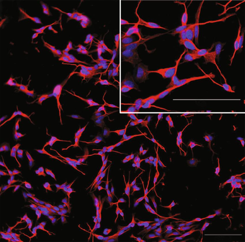
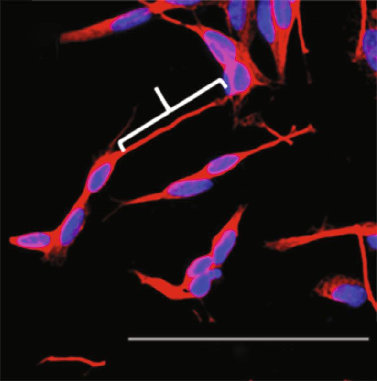
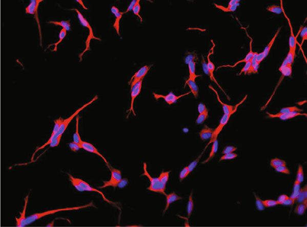
4 BioMed Research International

 100 m 100 m

 100 m 100 m

 (a) (b)

Figure 2: Neurons derived from the SH-SY5Y cell line. (a) Control, without treatment. (b) After 10 μg/mL C. quadrumanus extract
incubation. Inset: a zoom to show cell details.

 The extract was firstly analyzed in a reversed-phase ultra- weight) was applied in the gel, and a constant voltage of
performance liquid chromatography (RP-UPLC). Aliquots 120 V was applied. After the run, the gel was stained with
of the samples were loaded in a C18 column (ACE C18, silver, according to the method described by Laemmli [31].
5 m, 100 Å, 2:0 mm × 50 mm) in a two-solvent system: (A)
formic acid (FA)/H2O (1 : 1000) and (B) FA/acetonitri- 2.6. Statistical Analysis. All the cell experiments were per-
le/H2O (1 : 900 : 100). The content was eluted at a constant formed in triplicate, and the results are shown as mean ±
flow rate of 0.2 mL/min with a 0–100% gradient of solvent standard deviation. The treatment with compounds (3
B over 40 min, after a 5 min isocratic elution with 0% B. groups) was compared to the negative control (same condi-
The UPLC column eluates were monitored by a mass spec- tion without treatment) by one-way ANOVA, followed by
trometry (Q-ToF Xevo GS, Waters Co.), in a positive ioniza- Tukey’s posttest. Significance was considered if p < 0:05.
tion mode, in a range of 200 to 1800 m/z and FWHM 40000
resolution at 500 m/z. For the MS/MS analysis, argon colli- 3. Results
sion energy was applied. The instrument control and data
acquisition were conducted by MassLinx 4.2. 3.1. Neurons Analysis. Neurons derived from the SH-
 The results were automatically processed by PEAKS®7.0 SY5Y cell line were analyzed after treatment with the
software (Bioinformatics Solution Inc.) and then manually Cq extract, in order to investigate its impact on neurite
verified. De novo sequences were considered when average outgrowth. It was possible to verify neurites and branches
local confidence (ALC %) was >50. Peptides sequenced were without the treatment and after the neuron’s differentia-
submitted to protein BLAST (Basic Local Alignment Search tion (Figure 2(a)). However, 10 μg/mL Cq extract clearly
Tool), in order to find similar peptides already described. increased the neurites’ length, with apparent enhanced
For this analysis, the database nonredundant protein contact between them (Figure 2(b)).
sequences (nr) was used, with organism selection of Cnidaria The outgrowth (in μm) was quantified, and 10 and
(taxid: 6073) and blastp algorithm, with parameters of 10 100 μg/mL of extract significantly increased the total out-
expected threshold, matrix BLOSUM62, and gap existence growth length, as shown in the quantification of Figure 3(a).
11. In parallel, de novo sequences were searched against When we calculated the ratio between length and num-
peptide databank (PepBank) [30]. ber of outgrowths (mean process length), an increase after
 Alternatively, a fingerprinting of low molecular mass Cq extract treatment was observed, in both concentrations
compounds was performed by the spectra analysis on Pro- of 10 and 100 μg/mL, confirming the previous result, but
genesis QI Software (Waters Co.). Molecules were identified now analyzed by each cell (Figure 3(b)). The increase of neur-
by spectra similarity with the HMDB database, exact molec- ite length is evident after the extract treatment, as depicted in
ular mass, and m/z. Figure 3(d), in comparison to the control, without treatment,
 in Figure 3(c), analyzed in the same scale.
2.5.2. SDS-PAGE. SDS-PAGE (12%) was performed to ana- Besides the length, the number of outgrowths that con-
lyze proteins on the Cq extract under reducing and nonre- nects to the cell body (process) was significantly increased
ducing conditions. An aliquot (10 μg, determined by dry as well, with the same extract concentrations (10 and
BioMed Research International 5

 Total outgrowth Mean process length
 15 6 ⁎ ⁎
 ⁎ ⁎

 m/number of process
 10 4
 m

 5 2

 0 0
 Control 2 g/mL 10 g/mL 100 g/mL Control 2 g/mL 10 g/mL 100 g/mL
 (a) (b)

 m

 m

 100 m 100 m

 (c) (d)

Figure 3: Parameters of outgrowth measured in the neurons derived from the SH-SY5Y cell line after the incubation of C. quadrumanus
extract. (a) Quantification of total outgrowth, measured in μm. (b) Mean of the process length, obtained after quantification of total
outgrowth (μm) divided by the number of processes. (c) Representative image of an outgrowth of control neurons. (d) Representative
image of an outgrowth of neurons treated with 10 μg/mL Cq extract. ∗ Statistical difference p < 0:05.

100 μg/mL). Figure 4(a) shows the quantification of processes the Cq extract could induce neuron’s proliferation or even
and Figures 4(b) and 4(c) show the neuron with processes toxicity. As shown in Figure 6(a), the extract did not cause
indicated by white circles from the control and treated neu- any SH-SY5Y proliferation. Importantly, the extract did
rons, respectively. not cause cytotoxic effect, even in the highest concentra-
 Moreover, the number of branching junctions of all the tion evaluated (100 μg/mL), what confirms its activity only
processes (connected to the cell) was also increased signifi- on neurites and not in the cell body.
cantly with 10 and 100 μg/mL Cq extract, as shown in the The extract did not interfere with red blood cell viability
quantification graph (Figure 4(d)) and in the image in as well, by inducing only 1.3% hemolysis, compared to the
Figures 4(e) (control) and 4(f) (Cq treated). positive control, Triton-X 100, and negative control saline
 On the other hand, the cell body area was not affected solutions (Figure 6(b)). In order to confirm that the Cq
with the treatment, indicating the Cq extract effect only on extract would not induce alteration on viability of other lines,
outgrowths. Figure 5(a) shows the quantification, while neu- it was evaluated in a panel consisting of glioblastoma, breast
ron images are shown in Figures 5(b) (control) and 5(c) (Cq and ovary adenocarcinoma, multidrug-resistant ovary, leu-
extract). kemia, and keratinocytes. In all the tested concentrations
 The straightness was measured in order to verify if the (0.1 to 100 μg/mL), the extract did not cause any cytotoxic
path of a neurite’s growth would be deviated from a straight effect or cell proliferation (Figure 6(c)), reinforcing its activ-
line. Even 100 μg/mL Cq extract could not cause any effect on ity only on neurites.
this parameter (Figure 5(d)).
 3.3. Biochemical Analysis. In order to analyze the chemical
3.2. Cell Viability Analysis. Although there is an apparent composition of the Cq extract and identify important mol-
increase in number of cells when the images are analyzed ecules for neuron or neurite regeneration, the extract was
(e.g., Figure 2(b)), an MTT assay was performed to check if submitted to a reversed-phase chromatography coupled
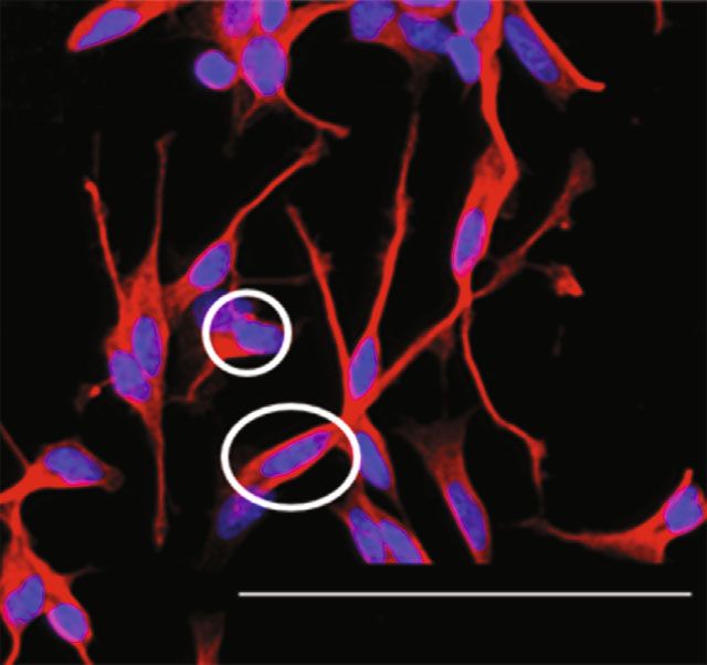
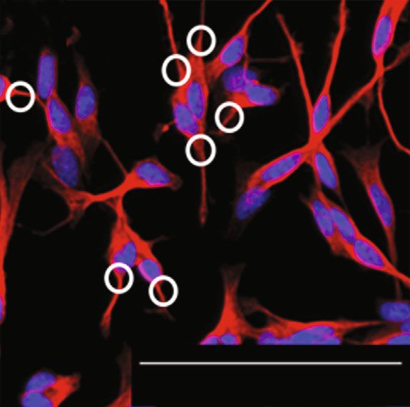
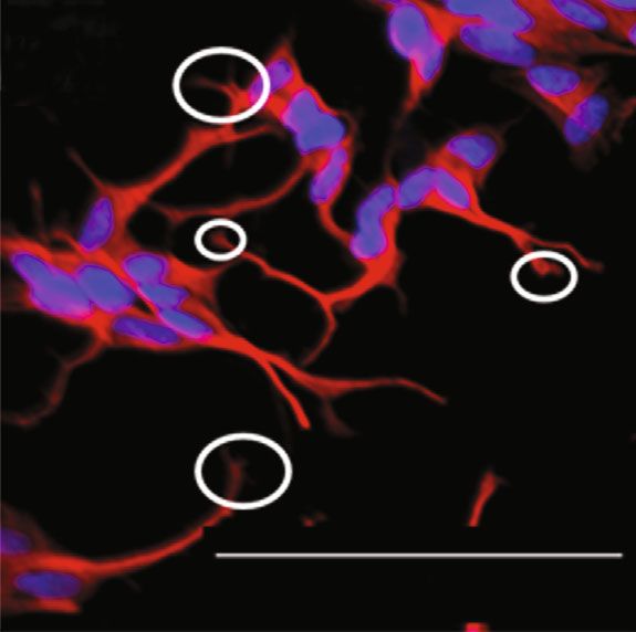
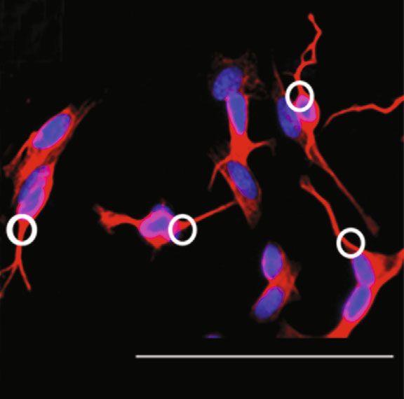
6 BioMed Research International

 Process
 2.0
 ⁎ ⁎
 Number of outgrowth

 1.5

 1.0

 0.5

 100 m 100 m
 0.0
 Control 2 mg/mL 10 mg/mL 100 mg/mL
 (a) (b) (c)

 Branches
 0.25
 Number of branching junctions

 ⁎ ⁎
 0.20

 0.15

 0.10

 0.05
 100 g 100 m
 0.00
 Control 2 mg/mL 10 mg/mL 100 mg/mL
 (d) (e) (f)

Figure 4: Processes and branches of neurons derived from the SH-SY5Y cell line after the incubation of the C. quadrumanus extract. (a)
Quantification of number of outgrowths connected to the cell body (process). (b) Representative image of processes from control neurons.
(c) Representative image of processes from neurons treated with 10 μg/mL C. quadrumanus extract. (d) Number of branching junctions.
(e) Representative image of branches from control neurons. (f) Representative image of branches from neurons treated with 10 μg/mL C.
quadrumanus extract. The white circles indicate the measured structures. ∗ Statistical difference p < 0:05.

to a mass spectrometry. This method was set to verify the 4. Discussion
presence of peptides and low molecular mass compounds.
In a general profile, it was possible to see qualitatively high Neurodegenerative diseases have no specific treatment, and
abundance of molecules, due to the presence of several the therapies currently available are applied for increasing
peaks along the chromatographic gradient (Figure 7(a)). the patient’s life expectation in some years, not representing
Through SDS-PAGE, we verified that the extract did not a cure [32]. Thus, the search for new drugs for such diseases
contain proteins: the extraction with methanol was able is essential, as well as the understanding of the mechanisms
to remove all the proteins (secreted or constitutive), and behind their effects. In this context, the research involving
only low molecular mass compounds were obtained for natural products is increasing and bioactive molecules have
the cell’s test (Figure 7(b)). been explored for drug discovery and development.
 Regarding peptides, we could identify and sequence Considering that cnidarians from Cubozoa is rarely
thirteen, shown in Table 1. These sequences were searched explored in terms of biotechnological application, we have
in the PepBank, and no description/activity was found for obtained a methanolic extract in order to verify its composi-
any of them. However, when searched in a protein data- tion and to study preliminary applications on neurite growth
base (BLAST), the peptides matched proteins with high for the application of neuron network regeneration.
score. The proteins are related somehow to neuron regen- In the present work, we show, for the first time, that the
eration and with high score and identity (Table 2). molecular composition of the methanolic extract of C. quad-
 The low molecular mass compounds were identified as rumanus is associated with a relevant biological effect, other
well, and the complete list of molecules is in the Supple- than envenomation. We demonstrated that peptides and
mentary Table 1. However, the molecules related to small molecules from the Cubozoa jellyfish extract act with
neuron regeneration could be identified and are shown synergy in different mechanisms to increase the neurite
in Table 3. length, processes, and branches.
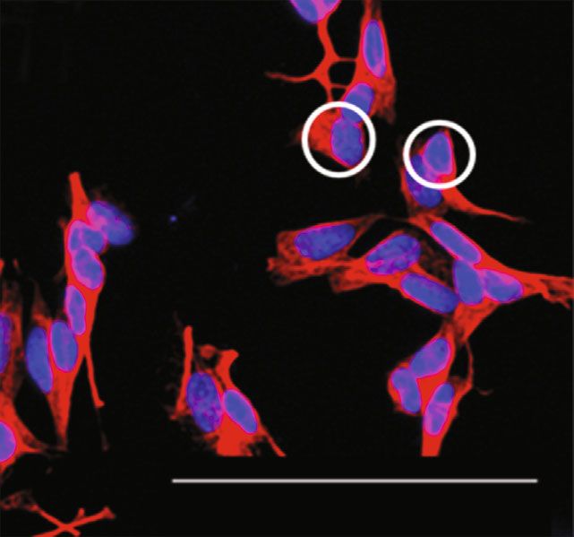
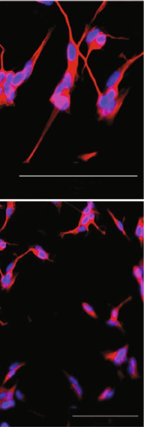
BioMed Research International 7

 Total cell body area
 80000

 60000
 m2

 40000

 20000

 100 m
 0
 Control 2 mg/mL 10 mg/mL 100 mg/mL
 (a) (b)

 Straightness
 1.0

 0.8

 0.6
 Ratio

 0.4

 0.2
 100 m
 0.0
 Control 2 mg/mL 10 mg/mL 100 mg/mL
 (c) (d)

Figure 5: Body cell and straightness of neurons derived from the SH-SY5Y cell line after the incubation of C. quadrumanus extract. (a)
Quantification of total body cell area (μm2). (b) Representative image of the total body cell area from control neurons. (c) Representative
image of the total body cell area from neurons treated with 10 μg/mL Cq extract. (d) Ratio between not straight and perfectly straight. The
white circles indicate the measure structures.

 The human SH-SY5Y neuroblastoma cell line is fre- peptidases from blood and tissues. Thus, the small molecules
quently used for different neuronal cell culture models, and peptides obtained from natural products can overpass
including neurodegenerative disorders, and has been chosen these issues, by presenting neuritogenic activity with good
for C. quadrumanus extract evaluation. Neurite evaluation pharmacokinetics properties [33].
has already been standardized for such cells, with similar It is important to mention that the proper neuronal func-
methods applied in this work, regarding the incubation of tion depends on the maintenance of axons and dendrites
natural products aiming neurite elongation, different mea- (collectively known as neurites) contributing to the precise
sures into the cells, and time of analysis [33]. Therefore, we neuronal network, which are essential for the establishment
analyzed the neurons 24 hours after Cq extract incubation, of synapses [37]. Thus, the neurite length is essential for the
time enough to promote neurite elongation, as observed here, entire cell recovery.
and also enough to activate intracellular pathways that would Thus, we have found molecules that act on neuron’s
cause changes in the cell body, not observed in any tested differentiation and growth, such as folinic acid and Pteroyl-
concentration. These data reinforce the selective action of D-glutamic acid, two derivative molecules from folic acid, a
the extract on neurites. known chemoprotectant that participates of the nerve injury
 SH-SY5Y can be differentiated into neuron-like pheno- repair, by acting on the proliferation and migration of
type cells, essential for functional analyses in neurosciences Schwann cell, and secretion of nerve growth factor [38].
[34]. The neuronal differentiation process was performed (6s)-5-methyltetrahydrofolic acid compounds were patented
using retinoid acid and BDNF, in a method already standard- to treat diseases associated with nervous system injuries, for
ized [35, 36]. Neurotrophic factors have been used for a neur- example, amyotrophic lateral sclerosis and Alzheimer’s
itogenic effect and neuronal regeneration; however, they have disease [39].
presented several disadvantages, such as the difficulties to Indeed, it is important to mention that the Cq extract did
cross the blood-brain barrier and inactivation/cleavage by not induce any cell death, in several cell lines tested, which
8 BioMed Research International

 Hemolysis
 MTT SH-SY5Y 2.5
 125

 2.0
 100

 Absorbance
 % viable cells

 1.5
 75

 50 1.0

 25 0.5
 ⁎⁎⁎
 0 0.0
 Control

 Estaurosporine

 2 g/mL

 5 g/mL

 10 g/mL

 20 g/mL

 30 g/mL

 40 g/mL

 50 g/mL

 100 g/mL
 Control + Control – Cq extract

 Cq extract []
 (a) (b)
 110
 100
 90
 80
 Cell growth (%)

 70
 60
 50
 40
 30
 20
 10
 0
 0.1 1 10 100
 g/mL

 U-251 OVCAR-3
 MCF7 K-562
 NCI-ADR/RES HaCaT
 (c)

Figure 6: Cell viability after treatment with C. quadrumanus extract. (a) Cell viability assay by MTT after Cq extract treatment on SH-SY5Y.
(b) Hemolysis rate of red blood cell after the treatment of Cq extract; control-: PBS incubation and control+: Triton-X 100 incubation. (c) Cell
viability assay by MTT after Cq extract treatment on cell line panel (U-251, MCF7, NCI-ADR/RES, OVCAR-3, K-562, and HaCaT).

shows that the Cq extract does not interfere with the neurons for neuritogenesis: we could identify molecules that act
body, regarding plasmatic membrane, cell signaling for on the organization of neuron’s cytoskeleton that contrib-
necrosis or apoptosis or even proliferation. It was clear that utes to the neurite expansion. Moreover, we have identi-
the extract alters only the neurite-related structures. fied molecules that act on the cell membrane formation,
 Here, the neurites were evaluated by the measurement of necessary to follow the neurite expansion. Antioxidants were
length of total skeletonized outgrowth associated with the found as well that contributes to the neuron regeneration,
cell, which has increased. Moreover, we observed increase with mechanism still unknown, besides neurotransmitter-
in the average, which means that the effects on neurite length like molecules that act on neuronal plasticity and neuropro-
was not an isolated phenomenon, but it was applied to all tective compounds. So, the action of several molecules in all
cells present in the culture, reinforcing the activity of the those mechanisms of action contributes to the significantly
extract. neuritogenic effect observed here.
 By the identification of molecules present in the Cq The neurite formation is the first step for the axon and
extract, it was possible to correlate them to mechanisms dendrite synthesis, essential for the development of a
BioMed Research International 9

 1: TOF MSES+ MM R NR
 5Moonc TIC
 100 5.06e6 225 K
 102 K
 76 K

 52 K

 38 K
 31 K

 24 K

 17 K

 12 K
 0 Time
 5.00 10.00 15.00 20.00 25.00 30.00 35.00 40.00 45.00
 (a) (b)

Figure 7: Profile of C. quadrumanus extract. (a) TIC chromatogram obtained after mass spectrometry analysis. (b) SDS-PAGE of reduced (R)
and nonreduced (NR) conditions of the extract.

Table 1: Peptides identified in the C. quadrumanus methanolic (PI4P), important for the plasma membrane identity and
extract. myelin development, as well as remyelination [43].
Peptide sequence m/z z Mass ALC (%) Besides peptides, low molecular mass compounds related
 to plasmatic membrane organization for neuritogenesis were
FCEHW 721.3490 1 720.2690 76
 identified in our Cq extract. Raloxifene is one of them, and its
YQGFAGKSS 472.7750 2 943.4399 59 analogue increases the number of regenerating sciatic nerve
CPKKDEVP 458.2280 2 914.4531 59 fibers in mice. Moreover, authors could observe that the mol-
PAYGARF 391.1820 2 780.3918 59 ecule increased the axonal transport [44].
PVGAAPAASVLF 550.3570 2 1098.6072 54 A neuroprotective action of 17β-estradiol was identified
DMYEFAQCA 539.1842 2 1076.3943 54 by Ishihara et al. [45] on injured neurons induced by several
GSKTFTL 377.2400 2 752.4069 52
 pathological conditions and by toxic compounds, such as
 organometallics. This protection has been related to the
GAVVLKGAALKT 564.3934 2 1126.7073 53
 action of 17β-estradiol promoting neurite extension ex vivo
KGPVTAASPLGHL 624.3920 2 1246.7034 51 and protecting neurons from oxidative stress in vitro. Here,
VAFFYVNA 465.7850 2 929.4647 51 we have found an estradiol-like molecule, the 4-hydroxy-
ELVHAASCK 479.2880 2 956.4749 51 17beta-estradiol-2-S-glutathione that can act in this mecha-
KNKNKLVPPPLHLGHR 616.7383 3 1847.1006 50 nism [46].
ALVTGGFNAR 503.3231 2 1004.5403 50 Regarding oxidative stress, antioxidant molecules were
 also found in the Cq extract—several compounds similar
 to flavonoidsa nd phenolic substances from plants with
 known antioxidant activity [47]. We have identified kuwa-
functional neuronal network, and takes part of the regenera- non J and 6-[(2-(Kumar and Khanum 2012)-7-hydroxy-4-
tion process after a nervous system injury. For neurite forma- oxo-3,4-dihydro-2H-1-benzopyran-5-yl)oxy]-3,4,5-trihy-
tion, the cytoskeleton has an important role, where actin, droxyoxane-2-carboxylic acid. These phytochemicals have
microtubule networks, and neurofilaments are essential [40]. been associated to neuroprotective activity [48].
 A cryptide from our extract derived from translin-like Two derivatives of serotonin, (E, E)-4,4″ -Bi (N-4-
protein is one molecule that contribute to neurite formation, hydroxycinnamoylserotonin) and 5-hydroxyindoleacetalde-
related to microtubules and motor proteins. When aug- hyde, were also observed. The hydroxycinnamoylserotonin
mented, they are associated with learning and memory, loco- belongs to the n-acylserotonin group. This molecule is fre-
motor activity, anxiety-related behavior, and sensorimotor quently found in the central nervous system, synthesized
gating [41]. Tyrosine-protein kinase ABL1-like is activated in the mammalian pineal gland and retina, and has dem-
after stimuli related to cytoskeletal reorganization [42]. onstrated antioxidant properties, with potential use for
 The hyccin, another cryptein found here, have impor- protection in neurological disorders, such as Alzheimer’s
tance in the neuron membrane, as it was demonstrated to disease, Parkinsonism, and age-related macular degenera-
regulate the synthesis of phosphatidylinositol 4-phosphate tion. Moreover, the acylserotonins seem to protect injured
10 BioMed Research International

 Table 2: Peptides found peptides in the C. quadrumanus methanolic extract and related proteins, with neuronal function.

Sequence Protein (organism) Score Identity (%) GAPs (%)
FCEHW Translin-like [(Pocillopora damicornis) 43 80 0
GAVVLKGAALKT Hyccin-like protein 46 100 0
 Cyclin-dependent kinase 18-like 59 67 0
KNKNKLVPPPLHLGHR
 Tyrosine-protein kinase ABL1-like 54 89 0

Table 3: LC-MS fingerprinting of low molecular mass compounds related to neuritogenesis, identified in the C. quadrumanus methanolic
extract.

Compound name HMDB ID Formula m/z Score
4-Hydroxy-17beta-estradiol-2-S-glutathione HMDB0060139 C28H39N3O9S 616.2241 31.4
5-Aminopentanoic acid HMDB0003355 C5H11NO2 118.0890 34.3
5-Hydroxyindoleacetaldehyde HMDB0004073 C10H9NO2 140.0572 30.8
5-Methyltetrahydrofolic acid HMDB0001396 C20H25N7O6 482.1733 34.2
6-[(2-{2-[2-(3,3-Dimethyloxiran-2-yl)ethyl]-7-hydroxy-2-methyl-
2H-chromen-6-yl}-7-hydroxy-4-oxo-3,4-dihydro-2H-1-benzopyran- HMDB0132837 C31H34O13 615.2183 30.2
5-yl)oxy]-3,4,5-trihydroxyoxane-2-carboxylic acid
(E,E)-4,4″ -Bi(N-4-hydroxycinnamoylserotonin) HMDB0041431 C38H34N4O6 643.2291 30.3
Folinic acid HMDB0001562 C20H23N7O7 496.1463 31.7
Indoleacetic acid HMDB0000197 C10H9NO2 140.0572 30.8
Kuwanon J HMDB0030112 C40H38O10 643.2291 35.5
N-Acetylgalactosaminyl lactose HMDB0041622 C20H35NO16 510.1827 37.5
N-Methyl-a-aminoisobutyric acid HMDB0002141 C5H11NO2 118.0890 34.3
Norvaline HMDB0013716 C5H11NO2 118.0890 34.3
Pteroyl-D-glutamic acid HMDB0002140 C20H23N7O7 496.1463 31.7
Raloxifene HMDB0014624 C28H27NO4S 496.1463 30.1

neurons against excitotoxic compounds by activating TrkB tic acid, also found in this study, was identified in the
receptors [49]. cerebrospinal fluid from epileptic patients, related to the
 Serotonin (5-HT) is an important neurotransmitter tryptamine metabolism [52].
that regulates neuronal connectivity during mammalian The use of N-acetylgalactosaminyl lactose has been sug-
development and has been associated with neuronal plas- gested by Vankar and Schmidt [53] for the application on
ticity by promoting secondary neurite outgrowth through Parkinson’s disease. This molecule is a carbohydrate present
5-HT1A and 5-HT7 receptors [50]. Here, we reported in the central nervous system, overall. Studies pointed out
the increase in the number of outgrowths attached to the that these molecules are neuroprotective and increase the
cell body and branches and junctions of all the processes regeneration of neurons.
connected to the cell, indicating primary and secondary
neurite formation and elongation. So, the presence of mol-
ecules related to 5-HT may contribute to this kind of 5. Conclusions
neuritogenesis.
 Regarding a neurotransmitter, N-methyl-a-aminoisobu- Considering that neuronal network regeneration is critical
tyric acid and 5-aminopentanoic acid are molecules that act for the neuronal regeneration and studies have shown that
as gamma-aminobutyric acid (GABA). This neurotransmit- it is fundamental for memory recovery on Alzheimer’s dis-
ter is involved in the neuronal protection and survival, ease and others [54], our study demonstrated that Cq extract
besides synapse recovery that contribute to the neuron regen- presented important molecules that, acting with synergy,
erative process, although the mechanism is not fully under- increased the neurites length and can be able to recover the
stood [33]. neuronal connection, which is useful for neurodegenerative
 Another important amino acid for the neurodegenera- diseases.
tion is arginine, and norvaline is a noncompetitive arginase
inhibitor that reduced the arginine loss in the brain. This
molecule is considered a candidate for Alzheimer’s disease Data Availability
treatment, as it has improved the memory and increased pro-
teins related to neuroplasticity [51]. Moreover, the indoleace- Supplementary files are available in this submission.
BioMed Research International 11 Disclosure [8] T. C. Dickson and J. C. Vickers, “The morphological pheno- type of β-amyloid plaques and associated neuritic changes in Gian Lucas M. Arruda present address is Biolab Sanus Farm- Alzheimer’s disease,” Neuroscience, vol. 105, no. 1, pp. 99– acêutica, Bragança Paulista 12929-600, Brazil. 107, 2001. [9] H. Malve, “Exploring the ocean for new drug developments: Conflicts of Interest marine pharmacology,” Journal of Pharmacy & Bioallied Sciences, vol. 8, no. 2, pp. 83–91, 2016. The authors declare that there is no conflict of interest [10] D. J. Newman and G. M. Cragg, “Natural products as regarding the publication of this paper. sources of new drugs over the last 25 years,” Journal of Natural Products, vol. 70, no. 3, pp. 461–477, 2007. [11] D. S. Rawat, M. C. Joshi, P. Joshi, and H. Atheaya, “Marine Acknowledgments peptides and related compounds in clinical trial+,” Current Medicinal Chemistry. Anti-Cancer Agents, vol. 6, no. 1, The authors are grateful to Dr. Carlos De Ocesano-Pereira, pp. 33–40, 2006. from Centre of Excellence in New Target Discovery (CENTD), Butantan Institute, for the high-content screening [12] N. L. Daly, J. Seymour, and D. Wilson, “Exploring the ther- apeutic potential of jellyfish venom,” Future Medicinal (HCS) parameters. The authors also thank Grant Chemistry, vol. 6, no. 15, pp. 1715–1724, 2014. 2015/50040-4 from São Paulo Research Foundation and GlaxoSmithKline. This work was funded by FAPESP (grant [13] S. Vignesh, A. Raja, and R. A. James, “Marine drugs: impli- cation and future studies,” International Journal of Pharma- # 2015/21007-9 to ACMo, grant # 2011/50242-5 to ACMa, cology, vol. 7, pp. 22–30, 2011. grant # 2016/06137-6 to GBL, and grant # 2019/19929-6 to [14] S.-K. Kim, Springer Handbook of Marine Biotechnology, JMS) and CNPq (grant # 309440/2019-0 to ACMo and Springer, 2015. 309995/2017-5 to ACMa). This work was funded in part by São Paulo Research Foundation (FAPESP) through the Cen- [15] Z. Yang, B. Liu, L.-E. Yang, and C. Zhang, “Platycodigenin as potential drug candidate for Alzheimer’s disease via mod- ter of Toxins, Immune Response and Cell Signaling ulating microglial polarization and neurite regeneration,” (CeTICS) (grant # 2013/07467-1). This is a contribution of Molecules, vol. 24, p. 3207, 2019. NP-BioMar, USP. [16] M. Oda, Y. Saito, Y. Morimune, M. Nagahama, and J. Sakurai, “Induction of neurite-outgrowth in PC12 cells by alpha-toxin Supplementary Materials from Clostridium perfringens,” Biochemical and Biophysical Research Communications, vol. 411, no. 2, pp. 241–246, 2011. Supplementary table: complete list of low molecular mass [17] D. H. Kim, E. Y. Moon, J. H. Yi et al., “Peptide fragment of thy- compounds identified in the C. quadrumanus methanolic mosin β4 increases hippocampal neurogenesis and facilitates extract. (Supplementary materials) spatial memory,” Neuroscience, vol. 310, pp. 51–62, 2015. [18] E. V. Romanova, M. J. Roth, S. S. Rubakhin et al., “Identifica- References tion and characterization of homologues of vertebrate β-thy- mosin in the marine mollusk Aplysia californica,” Journal of [1] I. Aprahamian, J. E. Martinelli, and M. S. Yassuda, “Doença de Mass Spectrometry, vol. 41, no. 8, pp. 1030–1040, 2006. Alzheimer: revisão da epidemiologia e diagnóstico,” Revista [19] J. Valcarcel, R. Novoa-Carballal, R. I. Pérez-Martín, R. L. Reis, Brasileira de Clínica Médica, vol. 7, pp. 27–35, 2009. and J. A. Vázquez, “Glycosaminoglycans from marine sources [2] J. T. Pedersen and N. H. H. Heegaard, “Analysis of protein as therapeutic agents,” Biotechnology Advances, vol. 35, no. 6, aggregation in neurodegenerative disease,” Analytical Chemis- pp. 711–725, 2017. try, vol. 85, no. 9, pp. 4215–4227, 2013. [20] D. Boltovskoy, South Atlantic Zooplankton, Backhuys Pub- [3] World Health O, Global action plan on the public health lishers, 1999. response to dementia 2017–2025, World Health Organization, 2017. [21] H. W. Mianzan and P. F. S. Cornelius, “Cubomedusae and scyphomedusae,” in South Atlantic Zooplankton, vol. 1, [4] C. A. Ross and M. A. Poirier, “Protein aggregation and neuro- pp. 513–559, Backhuys Publishers, 1999. degenerative disease,” Nature Medicine, vol. 10, no. S7, pp. S10–S17, 2004. [22] A. C. Morandini, M. R. Custódio, and A. C. Marques, “Phylum [5] M. Hashimoto, E. Rockenstein, L. Crews, and E. Masliah, porifera and cnidaria,” in Toxinology: Marine and Freshwater “Role of protein aggregation in mitochondrial dysfunction Toxins, pp. 287–316, Springer, 2016. and neurodegeneration in Alzheimer’s and Parkinson’s dis- [23] A. C. Marques, V. Haddad Jr., L. Rodrigo, E. Marques-da- eases,” NeuroMolecular Medicine, vol. 4, no. 1-2, pp. 21–36, Silva, and A. C. Morandini, “Jellyfish (Chrysaora lactea, 2003. Cnidaria, Semaeostomeae) aggregations in southern Brazil [6] C. Vidoni, C. Follo, M. Savino, M. A. B. Melone, and and consequences of stings in humans,” Latin American C. Isidoro, “The role of cathepsin D in the pathogenesis of Journal of Aquatic Research, vol. 42, no. 5, pp. 1194–1199, human neurodegenerative disorders,” Medicinal Research 2014. Reviews, vol. 36, no. 5, pp. 845–870, 2016. [24] L. B. Doonan, S. Lynham, C. Quinlan et al., “Venom composi- [7] W. M. Alobuia, W. Xia, and B. P. S. Vohra, “Axon degen- tion does not vary greatly between different nematocyst types eration is key component of neuronal death in amyloid-β isolated from the primary tentacles of Olindias sambaquiensis toxicity,” Neurochemistry International, vol. 63, no. 8, (Cnidaria: Hydrozoa),” The Biological Bulletin, vol. 237, no. 1, pp. 782–789, 2013. pp. 26–35, 2019.
12 BioMed Research International

[25] A. Jaimes-Becerra, R. Chung, A. C. Morandini et al., “Compar- [41] J. M. Stein, W. Bergman, Y. Fang et al., “Behavioral and
 ative proteomics reveals recruitment patterns of some protein neurochemical alterations in mice lacking the RNA-binding
 families in the venoms of Cnidaria,” Toxicon, vol. 137, pp. 19– protein translin,” Journal of Neuroscience, vol. 26, no. 8,
 26, 2017. pp. 2184–2196, 2006.
[26] A. Jaimes-Becerra, R. Gacesa, L. B. Doonan et al., ““Beyond [42] A. Khatri, J. Wang, and A. M. Pendergast, “Multifunctional
 primary sequence”—proteomic data reveal complex toxins in Abl kinases in health and disease,” Journal of Cell Science,
 cnidarian venoms,” Integrative and Comparative Biology, vol. 129, no. 1, pp. 9–16, 2016.
 vol. 59, no. 4, pp. 777–785, 2019. [43] J. M. Baskin, X. Wu, R. Christiano et al., “The leukodystrophy
[27] C. Wang, B. Wang, B. Wang et al., “Unique diversity of sting- protein FAM126A (hyccin) regulates PtdIns (4) P synthesis at
 related toxins based on transcriptomic and proteomic analysis the plasma membrane,” Nature Cell Biology, vol. 18, no. 1,
 of the Jellyfish Cyanea capillata and Nemopilema nomurai pp. 132–138, 2016.
 (Cnidaria: Scyphozoa),” Journal of Proteome Research, [44] R. McMurray, R. Islamov, and A. K. Murashov, “Raloxifene
 vol. 18, pp. 436–448, 2018. analog LY117018 enhances the regeneration of sciatic nerve
[28] G. Kass-Simon and P. Pierobon, “Cnidarian chemical neu- in ovariectomized female mice,” Brain Research, vol. 980,
 rotransmission, an updated overview,” Comparative Bio- no. 1, pp. 140–145, 2003.
 chemistry and Physiology Part A: Molecular & Integrative [45] Y. Ishihara, T. Takemoto, A. Ishida, and T. Yamazaki, “Pro-
 Physiology, vol. 146, no. 1, pp. 9–25, 2007. tective actions of 17β-estradiol and progesterone on oxidative
[29] G. Jarms, A. C. Morandini, A. Schmidt-Rhaesa, O. Giere, and neuronal injury induced by organometallic compounds,”
 I. Straehler-Pohl, World atlas of jellyfish, Dölling und Galitz Oxidative Medicine and Cellular Longevity, vol. 2015, Article
 Verlag, 2019. ID 343706, 16 pages, 2015.
[30] T. Shtatland, D. Guettler, M. Kossodo, M. Pivovarov, and [46] A. R. D’Amato, D. L. Puhl, S. A. T. Ellman, B. Balouch, R. J.
 R. Weissleder, “PepBank-a database of peptides based on Gilbert, and E. F. Palermo, “Vastly extended drug release from
 sequence text mining and public peptide data sources,” BMC poly (pro-17β-estradiol) materials facilitates in vitro neuro-
 Bioinformatics, vol. 8, no. 1, p. 280, 2007. trophism and neuroprotection,” Nature Communications,
[31] U. K. Laemmli, “Denaturing (SDS) discontinuous gel electro- vol. 10, pp. 1–12, 2019.
 phoresis,” Nature, vol. 277, pp. 680–685, 1970. [47] P.-G. Pietta, “Flavonoids as antioxidants,” Journal of Natural
[32] L. Gan, S. Ye, A. Chu et al., “Identification of cathepsin B as a Products, vol. 63, no. 7, pp. 1035–1042, 2000.
 mediator of neuronal death induced by Aβ-activated micro- [48] G. P. Kumar and F. Khanum, “Neuroprotective potential of
 glial cells using a functional genomics approach,” Journal of phytochemicals,” Pharmacognosy Reviews, vol. 6, no. 12,
 Biological Chemistry, vol. 279, no. 7, pp. 5565–5572, 2004. pp. 81–90, 2012.
[33] S. V. More, S. Koppula, I.-S. Kim, H. Kumar, B.-W. Kim, and [49] G. Tosini, K. Ye, and P. M. Iuvone, “N-acetylserotonin:
 D.-K. Choi, “The role of bioactive compounds on the promo- neuroprotection, neurogenesis, and the sleepy brain,” The
 tion of neurite outgrowth,” Molecules, vol. 17, no. 6, pp. 6728– Neuroscientist, vol. 18, no. 6, pp. 645–653, 2012.
 6753, 2012. [50] P. S. Rojas, D. Neira, M. Muñoz, S. Lavandero, and J. L. Fiedler,
[34] A. Kunzler, F. Zeidán-Chuliá, J. Gasparotto et al., “Changes in “Serotonin (5-HT) regulates neurite outgrowth through 5-
 cell cycle and up-regulation of neuronal markers during SH- HT1A and 5-HT7 receptors in cultured hippocampal neu-
 SY5Y neurodifferentiation by retinoic acid are mediated by rons,” Journal of Neuroscience Research, vol. 92, no. 8,
 reactive species production and oxidative stress,” Molecular pp. 1000–1009, 2014.
 Neurobiology, vol. 54, no. 9, pp. 6903–6916, 2017. [51] B. Polis, K. D. Srikanth, E. Elliott, H. Gil-Henn, and A. O.
[35] J. I. Forster, S. Köglsberger, C. Trefois et al., “Characterization Samson, “L-Norvaline reverses cognitive decline and synaptic
 of differentiated SH-SY5Y as neuronal screening model reveals loss in a murine model of Alzheimer’s disease,” Neurothera-
 increased oxidative vulnerability,” Journal of Biomolecular peutics, vol. 15, no. 4, pp. 1036–1054, 2018.
 Screening, vol. 21, no. 5, pp. 496–509, 2016. [52] S. N. Young, S. Gauthier, G. M. Anderson, and W. C. Purdy,
[36] J. Kovalevich and D. Langford, “Considerations for the use of “Tryptophan, 5-hydroxyindoleacetic acid and indoleacetic
 SH-SY5Y neuroblastoma cells in neurobiology,” in Neuronal acid in human cerebrospinal fluid: interrelationships and the
 Cell Culture, pp. 9–21, Springer, 2013. influence of age, sex, epilepsy and anticonvulsant drugs,” Jour-
[37] C. T. Chu, E. D. Plowey, R. K. Dagda, R. W. Hickey, S. J. nal of Neurology, Neurosurgery, and Psychiatry, vol. 43, no. 5,
 Cherra Iii, and R. S. B. Clark, “Autophagy in neurite injury pp. 438–445, 1980.
 and neurodegeneration: in vitro and in vivo models,” Methods [53] Y. D. Vankar and R. R. Schmidt, “Chemistry of glycosphingo-
 in Enzymology, vol. 453, pp. 217–249, 2009. lipids—carbohydrate molecules of biological significance,”
[38] W. B. Kang, Y. J. Chen, D. Y. Lu, and J. Z. Yan, “Folic acid Chemical Society Reviews, vol. 29, no. 3, pp. 201–216, 2000.
 contributes to peripheral nerve injury repair by promoting [54] Z. Yang, T. Kuboyama, and C. Tohda, “A systematic strategy
 Schwann cell proliferation, migration, and secretion of nerve for discovering a therapeutic drug for Alzheimer’s disease
 growth factor,” Neural Regeneration Research, vol. 14, no. 1, and its target molecule,” Frontiers in Pharmacology, vol. 8,
 pp. 132–139, 2019. p. 340, 2017.
[39] B. S. Iskandar and K. J. Hogan, (6s)-5-Methyltetrahydrofolic
 acid for therapy of tissue injury, Google Patents, 2009.
[40] R. Sainath and G. Gallo, “Cytoskeletal and signaling mech-
 anisms of neurite formation,” Cell and Tissue Research,
 vol. 359, no. 1, pp. 267–278, 2015.
You can also read