Lawrence Berkeley National Laboratory - Recent Work - eScholarship
←
→
Page content transcription
If your browser does not render page correctly, please read the page content below
Lawrence Berkeley National Laboratory
Recent Work

Title
Simultaneous ambient pressure x-ray photoelectron spectroscopy and grazing
incidence x-ray scattering in gas environments.

Permalink
https://escholarship.org/uc/item/8f19k1n5

Journal
The Review of scientific instruments, 92(4)

ISSN
0034-6748

Authors
Kersell, Heath
Chen, Pengyuan
Martins, Henrique
et al.

Publication Date
2021-04-01

DOI
10.1063/5.0044162

Peer reviewed

 eScholarship.org Powered by the California Digital Library
 University of California
Simultaneous ambient pressure X-ray photoelectron spectroscopy
 and grazing incidence X-ray scattering in gas environments

Heath Kersell1, Pengyuan Chen2, Henrique Martins,1,3 Qiyang Lu1,4, Felix Brausse,5 Bo-Hong Liu,1,5
Monika Blum,1,5 Sujoy Roy,1 Bruce Rude, 1 Arthur Kilcoyne,1 Hendrik Bluhm,1,5,6 Slavomír Nemšák1

1
 Advanced Light Source, Lawrence Berkeley National Laboratory, Berkeley, California, 94720, United
States
2
Department of Electrical Engineering and Computer Sciences, University of California, Berkeley,
California, 94720, United States
3
 Department of Physics, University of California, Davis, California, 95616, United States
4
Department of Materials Science and Engineering, Stanford University, Stanford, California, 94305,
United States
5
Chemical Science Division, Lawrence Berkeley National Laboratory, Berkeley, California, 94720,
United States
6
Department of Inorganic Chemistry, Fritz Haber Institute of the Max Planck Society, 14195, Berlin,
Germany

Keywords: Ambient Pressure, X-ray Photoelectron Spectroscopy, Grazing Incidence X-ray Scattering, Silver
Behenate

ABSTRACT: We have developed an experimental system to simultaneously observe surface structure,
morphology, composition, chemical state, and chemical activity for samples in gas phase
environments. This is accomplished by simultaneously measuring X-ray photoelectron spectroscopy
(XPS) and grazing incidence X-ray scattering (GIXS) in gas pressures as high as the multi-Torr regime,
while also recording mass spectrometry. Scattering patterns reflect near-surface sample structures from
the nano- to the meso-scale. The grazing incidence geometry provides tunable depth sensitivity while
scattered X-rays are detected across a broad range of angles using a newly designed pivoting-UHV-
manipulator for detector positioning. At the same time, XPS and mass spectrometry can be measured,
all from the same sample spot and in ambient conditions. To demonstrate the capabilities of this
system, we measured the chemical state, composition, and structure of Ag-behenate on a Si(001) wafer
in vacuum and in O2 atmosphere at various temperatures. These simultaneous structural, chemical, and
gas phase product probes enable detailed insights into the interplay between structure and chemical
state for samples in gas phase environments. The compact size of our pivoting-UHV-manipulator
makes it possible to retrofit this technique into existing spectroscopic instruments installed at
synchrotron beamlines. Because many synchrotron facilities are planning or undergoing upgrades to
diffraction limited storage rings with transversely coherent beams, a newly emerging set of coherent
X-ray scattering experiments can greatly benefit from the concepts we present here.
1. Introduction
 Nano- and atomic- scale understanding of chemical and structural processes is highly
sought-after in many technologically relevant areas; from photovoltaics [1] and information
storage [2] to batteries [3] and heterogeneous catalysis. [4] Additionally, practically all
important technological or natural processes take place in ambient gas environments. These
environments often induce complex relationships between structure, chemistry, and function.
For example, as dimensions reach the nanoscale, transition metals and transition metal oxides
exhibit size dependent behavior, contributing to heavy metal migration in geological systems.
[5] [6] [7] Reactant gas environments can lead to pressure-dependent adsorbate coverages [8]
[9] [10], and potentially initiate nanocluster formation at catalyst surfaces, subsequently
altering reaction pathways. [8] [11] [12] Active species in bimetallic alloy catalysts can
segregate under reaction conditions, altering their operando surface composition. [13] [14]
Polyamide membranes for water purification exhibit correlations between water permeability
and the presence of nanoscale Turing structures. [15] Detailed understanding of the correlation
between chemistry, structure, and function in these systems requires multimodal investigations.
Additionally, each of these processes occurs in heterogeneous environments, and measuring
them in ambient gas mixtures requires in-situ or operando techniques.
 In the past two decades, a number of operando spectroscopic and structural probes were
developed to study heterogeneous interfaces. [16] [17] For example, environmental
transmission electron microscopy (ETEM) enables cross-sectional elemental mapping in
ambient pressure environments. [18] Polarization-modulation infrared reflection-absorption
spectroscopy (PM-IRRAS) provides an operando probe of interfacial adsorbates. [16] Dynamic
surface restructuring at pressures from ultrahigh vacuum (UHV) to the bar regime can be
measured with atomic resolution using high pressure scanning tunneling microscopy (HPSTM)
and atomic force microscopy (HPAFM). [17] [19] [20] Operando studies of surface chemical
composition and chemical states, as well as adsorbate species can be pursued with ambient
pressure X-ray photoelectron spectroscopy (APXPS). [21]
 In spite of the growth of operando methods over the last two decades, understanding
the detailed relationship between dynamic surface restructuring and chemical transformations
remains a difficult task. One challenge lies in correlating chemical transformations with
structural evolution, each measured with separate techniques. Currently, this is often done

 2
using separate samples, separate measurement sets, and frequently in entirely separate
experimental systems. Differences in pressure measurements, sample temperatures, time
resolutions, and sample quality can lead to difficulties comparing structural or chemical
changes in separate measurements. This opens a gap between the two experiments,
complicating the interpretation of separately collected data, and hindering the accuracy of such
separate experiments. Therefore, multi-modal approaches, like the one presented in this work,
have been undertaken at various synchrotron facilities, with a few successful results. For
example, a combined APXPS and IRRAS study utilized the sensitivity introduced by IRRAS
to identify species that were not detected in APXPS measurements. [22] Some experimental
systems are now also equipped to measure X-ray based spectroscopies and X-ray diffraction
(XRD). [23] For example, a setup at Argonne National Laboratory allows for simultaneous
analysis of chemistry and structure using hard X-ray photoelectron spectroscopy and X-ray
diffraction in a specifically designed pulsed laser deposition chamber. [24] To address the need
for simultaneous structural and chemical probes under ambient conditions, we have combined
APXPS with grazing incidence X-ray scattering (GIXS). This combination enables the
simultaneous correlation of sample chemical states and adsorbate species, with surface
structures that emerge in heterogeneous environments, where ambient pressures reach into the
Torr regime.

2. Combining ambient pressure photoemission and X-ray scattering
 In XPS, X-ray photons incident on a material cause photoelectron emission if the
photon energy exceeds the binding energy of electrons in the material. The resulting
photoelectron intensity at various kinetic energies provides information about surface
chemistry, composition, valence states, and adsorbate coverage. [25] Because electrons are
strongly scattered by matter, these photoelectrons are predominantly from the surface and near-
surface regions, with typical probing depths in the few nm range. Electrons are also strongly
scattered by gas molecules, and thus most XPS experiments are performed in high or ultra-
high vacuum conditions. To measure in ambient pressures, APXPS apparatuses are equipped
with a small (sub-mm) aperture in front of the sample surface, which photoelectrons pass
through. [21] [26] This aperture leads to a differentially pumped electrostatic lens system, and

 3
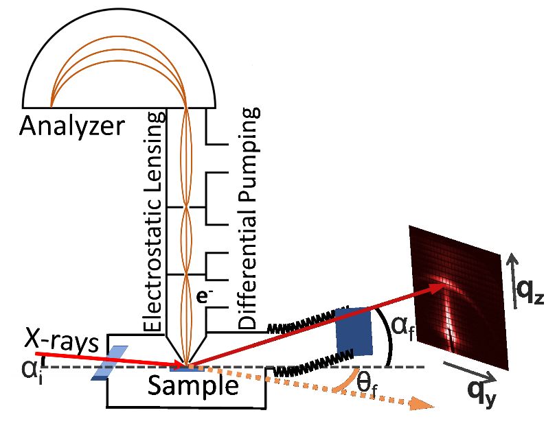
Fig. 1. Schematic of a combined ambient pressure XPS/GIXS system. Incident X-rays pass
through a 100 nm thick silicon nitride window (blue, lower left side) separating the beamline from
ambient gas environments in the sample chamber. Photoelectrons are collected by the
differentially-pumped electrostatic lens system and analyzer, which are separated from the sample
environment by a small aperture next to the sample. In the APGIXS measurements, photons
scattered from the surface are measured by a detector (right side). A flexible edge-welded bellows
(zig-zag lines) mounted on a rotating 2D joint allows the detector to be scanned across scattering
angles of interest. The X-ray detector is separated from the gas environment by an X-ray
transparent silicon nitride window with a 150 nm thickness (blue, lower right side). The two
silicon nitride windows (blue) and the cone aperture separate the beamline, X-ray detector, and
the XPS analyzer from the gas phase environment present in the sample chamber. X-ray scattering
angles correspond uniquely to specific values of qy and qz, where the longitudinal and transverse
scattering angles are denoted αf and θf, respectively. The detector range is 0°≤ αf ≤ 20°, and -20°≤
θf ≤ 20°.

then to a hemispherical electron energy analyzer, as shown in Fig. 1. [27] The differential
pumping strongly reduces scattering of photoelectrons in the lens system by gas molecules,
allowing them to reach the hemispherical analyzer where differential pumping maintains high
vacuum.
 While some photons incident on the sample are absorbed to produce photoelectrons,
some are also elastically scattered. The resulting scattering pattern reflects the arrangement of
scattering centers in the sample (i.e. the sample structure). Using X-rays incident at grazing
angles enables X-ray scattering measurements in the reflection geometry (as in Fig. 1), [28]
(opposite to the transmission geometry) allowing simultaneous measurements of ambient
pressure GIXS (APGIXS) and APXPS.
 4
Scattered X-rays from samples form an interference pattern with maxima (constructive
interference) at angles inversely related to the location of the scattering centers. When the
scattering patterns are generated by X-rays at grazing incidence, the corresponding X-ray
scattering techniques are broadly grouped into grazing incidence wide angle X-ray scattering
(GIWAXS) and grazing incidence small angle X-ray scattering (GISAXS).
 Depending on the wavelength of the incident X-rays, GIWAXS provides access to
atomic scale information at large scattering angles, making it ideal for studies of crystal
structures and their transformations. [30] [31] Meanwhile, at smaller scattering angles,
GISAXS has been applied to the study of more extended structures in thin film growth,
nanoparticle stability, block copolymer thin film evolution, and quantum dots, for example.
[32] [33] [34] [35] There is no unambiguous delineation between the small and wide-angle
regimes in GIXS, and combined GISAXS/GIWAXS investigations provide a detailed picture
of crystal structures and the growth or evolution of structures from the nanoscale to the
mesoscale. [36]

3. Description of the experimental apparatus
 Measurement of GIXS across the small- and wide-angle regimes in ambient gas
environments poses a number of challenges. For example, simultaneous APGIXS and APXPS
measurements require (i) that X-rays be incident on samples at positions that optimize the
photoelectron intensity reaching the analyzer, while also scattering into angles accessible by
the X-ray detector. Detecting structures from the atomic- to the meso-scale also requires a
detector capable of both (ii) measurement over a wide range of scattering angles, and (iii) high
angular resolution (especially for coherent scattering and observing speckles). Additionally,
(iv) the photon detector itself often needs to be in vacuum, isolated from gas phase sample
environments.
 To address these issues, we developed the experimental configuration illustrated in Fig.
1. In this layout, a custom UHV sample chamber is connected to the X-ray source at beamline
11.0.2 of the Advanced Light Source in Berkeley [37] with a practical photon energy range of
180 – 1800 eV, and to a differentially pumped commercial APXPS analyzer (SPECS Phoibos
150 NAP), as well as a moveable differentially pumped X-ray detector (Andor iKon-L SO
CCD). X-ray transparent silicon nitride windows separate the sample environment from both
the X-ray source (beamline) and the X-ray detector. The APXPS detector is separated from the
 5
sample environment by the differential pumping stages as discussed in the previous section.
Although not shown in Fig. 1, the chamber is also connected to a sample preparation and load-
lock chamber, and a four-axis sample manipulator. How this layout addresses the challenges
(i-iv) enumerated in the previous paragraph is discussed next.
 Addressing challenge (i): A commercially available four-axis manipulator
(Thermionics Inc.) enables three dimensional cartesian positioning of the sample, and polar
rotation with respect to both the X-ray beam source and the APXPS analyzer. This positioning
system enables sample alignment such that grazing incidence X-rays generate photoelectrons
which enter the APXPS aperture, while some photons simultaneously scatter from the sample
toward the detector. In our system, a ~100 μm wide X-ray beam is incident on the sample
surface approximately 0.5 mm from the 0.3 mm wide APXPS aperture. When the X-ray beam
is at grazing incidence with respect to the sample surface, this geometry elongates the X-ray
beam along the beam direction, generating scattered X-rays from an elongated sample region.
 The grazing incidence geometry also enables control over the information depth for
these techniques: the penetration depth of X-rays drastically decreases with decreasing
incidence around the critical angle, which can be as high as several degrees at 1000 eV.
Meanwhile the emission angle in XPS affects the depth contribution of photoelectrons reaching
the analyzer. We note that in principle, a transmission scattering geometry could also be used,
although sample thickness becomes an important consideration in that case.
 Addressing challenge (ii): To measure GIXS over a wide range of scattering angles, we
developed a new pivoting-UHV-manipulator capable of scanning an X-ray detector in two
angular dimensions (Fig. 2a). As shown in Fig. 2, this device is attached to our vacuum
chamber by a 6” CF flange at one end (left side of Fig. 2a). At the opposite end is a 6” CF
flange where a vacuum compatible X-ray detector can be mounted (right side of Fig. 2a). UHV
is maintained between these two sides by a combination of flexible edge welded bellows, 4”
stainless steel tubes, and 6” CF flanges. At the chamber-side, the pivoting-UHV-manipulator
contains a pair of rotation axes that enable movement of the detector in two angular dimensions
(straight blue arrows in Fig. 2a). The vacuum bellows is flexible, bending and maintaining
vacuum when the detector is moved. Detector motion is controlled by a pair of precision linear
actuators, with a step resolution of 10 μm. The top of each actuator is attached to the pivoting-
UHV-manipulator by a rotatable ball joint, so that actuator extensions or retractions precisely
translate the manipulator across two angular directions (Fig 2a). Each actuator base is

 6
connected to a universal joint fixed to the system frame (not shown), allowing the actuators to
pivot while controlling the detector position. This layout facilitates movement of the detector
in two angular directions across a pseudo-spherical cap, illustrated by green curved arrows at
the right side of Fig. 2a. An important aspect of X-ray scattering measurements is the range of
available scattering angles where the camera has direct, unobstructed line-of-sight to the
sample. This range is primarily determined by the size and position of the main vacuum flange
between the chamber and pivoting-UHV-manipulator. This set of unobstructed angles

 Fig. 2. Pivoting-UHV-manipulator. The model (a) and picture (b), show an edge welded bellows
 (1) attached to two CF flanges. The flange indicated at the left (chamber side) is rigidly attached to
 a UHV chamber. The flange at the right (detector side) pivots around a set of bearings (at the
 chamber side), allowing it to independently rotate about the two bearing axes (designated by
 straight blue arrows). This facilitates movement of a detector (a CCD camera in our case) through
 a solid angle (green curved arrows) with approximately ±12° around either axis. The detector
 mounts onto the CF flange at the right (2), where it is separated from the gas environment by an X-
 ray transparent 150 nm thick silicon nitride window (3). The area between the window and detector
 can be pumped through one of the CF mini ports (4). Control over the rotation is facilitated by the
 use of a pair of approximately orthogonal precision linear actuators (5).

 7
determines the range over which scattered X-rays can be detected, and is illustrated in Fig. 1
by the red arrow scattered by the sample. We optimized this range, and the range of detector
motion, to coincide with X-ray scattering angles relevant to many ambient pressure
investigations. Scattered X-rays beyond the angular range represented by the red arrow in Fig.
1 will be obstructed. In our present layout, the angular range can be as large as approximately
±12° azimuthally and 0° to +24° in polar direction (with respect to the sample), which
accommodates measurement of features as small as approximately 2 nm for the photon energy
range available at beamline 11.0.2 at the ALS. This range can be increased by using a larger
diameter bellows and a larger port on the chamber, which provides more space for scattered X-
rays to pass unobstructed to the detector. This design facilitates GIXS measurements in both
the small-angle and wide-angle regimes. One major advantage of this design is its relatively
compact size. As shown in Fig. 2b, the entire pivoting-UHV-manipulator, which moves the X-
ray detector, is not much larger than our eight-inch-wide UHV sample chamber. This is
significantly smaller than conventional SAXS flight tubes, by approximately an order of
magnitude or more. As a result, this design can be implemented for GIXS measurements in
existing systems whose size is constrained by the amount of experiment floor available around
the instrument.
 Addressing challenge (iii): One critical experiment that requires high angular
resolution is coherent X-ray scattering. Coherent illumination of a distribution of scattering
centers gives rise to a so-called ‘speckle pattern’, which is characterized by interference of
photons scattered by that distribution. Changes in the speckle pattern can be measured to
understand changes in the distribution of scattering centers. To extract meaningful information
about the evolution of a distribution of scattering centers in such measurements, a detector
must have resolution at least as fine as the speckle size, , which depends on the photon
wavelength, , the distance between the sample and detector, D, and the width of the coherently
illuminated area, a, as [38]

 = . (1)

In the experimental setup described in this section, we use a CCD camera as our detector, with
2048 × 2048 pixels and 13.5μm × 13.5μm pixel size (Andor iKon-L SO). Given formula (2),
the minimum sample-detector distance for a 30 μm spot-size and 1.5 keV radiation at beamline

 8
11.0.2.1 of the Advanced Light Source is ca. 50 cm. To measure with high angular resolution
across the small- and wide-angle regimes, and hence probe features from approximately the
single nanometer scale to the mesoscale, the pixel size of the detector and its positioning have
to work with a resolution on the order of ~10 μm. Such conditions are also necessary to pursue
more sophisticated investigations of, for example, structural dynamics.
 Addressing challenge (iv): Using a CCD camera for measuring scattered X-rays
presents a unique challenge in ambient pressure environments. To lower electronic thermal
noise, CCD detectors are usually cooled using multiple-stage Peltier elements. As a result, any
humidity or other condensable gases can coat the camera surface, causing electrical shorts and
artifacts in the imaging, and inevitably reducing the lifetime of the camera. It is therefore
necessary to isolate the detector from the gas phase sample environments. For the experimental
setup we have described in this section, this is accomplished using a 150 nm thick X-ray
transparent silicon nitride membrane with an area of 27 mm × 27 mm (Silson Ltd). The window
is divided into a 3×3 array of 8.33 mm sections, divided by 1 mm thick frames to enhance the
membrane stability against high pressure differentials (Fig. 2a). Scattered X-rays pass through
the silicon nitride membrane to reach the CCD detector, while the membrane isolates the CCD
chip from the gas phase environment of the sample chamber. These membranes are stable up
to pressure differentials of several tens of Torr, which enables X-ray scattering measurements
in ambient pressure gas environments across a similar pressure regime to that accessible in our
APXPS system (
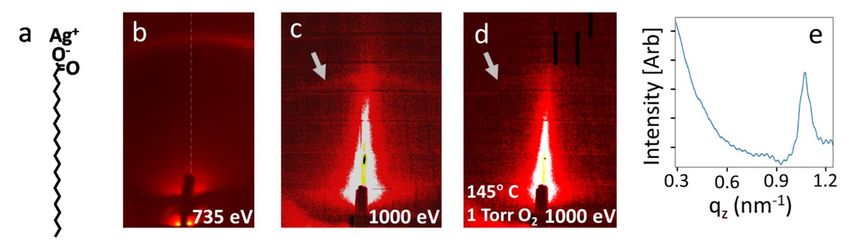
4. Proof-of-principle measurements of Ag-behenate
 To confirm the capabilities of this newly built system, we measured changes in Ag-
behenate (AgC22H43O2) layers deposited on SiOx as a function of temperature in the presence
of O2 gas. The Ag-behenate molecule (Fig. 3a) contains a long-chain silver carboxylate which,
when deposited onto a substrate, forms dimers that arrange into a layer-by-layer structure with
an interlayer spacing of approximately 5.84 nm. Because of its regular interlayer spacing, Ag-
behenate is a common reference standard in X-ray scattering experiments. [39] [40] Its layered
structure yields a scattering pattern characterized by rings of high intensity at Q = 0.108 Å-1
and at successively higher order scattering maxima. [41] In our measurements, Ag-behenate
was deposited onto a Si(001) surface (covered by a native oxide layer) by drop-casting a Ag-
behenate-in-ethanol solution onto the surface and allowing the solution to dry in air. Once
placed in vacuum, this system generated a clear scattering feature at 0.108 Å-1 in our GIXS
measurements, shown in Figs. 3b and c. In Fig. 3b, we note that the intensity of the scattering
feature decreases somewhat from the side of the image where the recording was started (right)
to the end (left), suggesting that over the course of the image, some beam damage may occur.

 Fig. 3. X-ray scattering from Ag-behenate. a) Ag-behenate molecular structure; b) scattering pattern
 containing the first order peak formed by X-rays scattered from Ag-behenate. c) Low exposure
 acquisition (117 seconds) of Ag-behenate scattering pattern. A grey arrow designates the scattering
 intensity maximum from the Ag-behenate layers. d) Low exposure acquisition of Ag-behenate
 scattering pattern after heating in oxygen gas. The grey arrow designates the location where the
 scattering feature from Ag-behenate layers existed before heating. e) Intensity line profile across
 the vertical centerline shown in the scattering pattern in b). Note that 735 eV photons were used in
 (b), and 1000 eV photons were used to produce (c) and (d). The different scattering angles for (b)
 as compared with (c) and (d) are generated by the different photon energy used to generate the
 scattering pattern.

 10
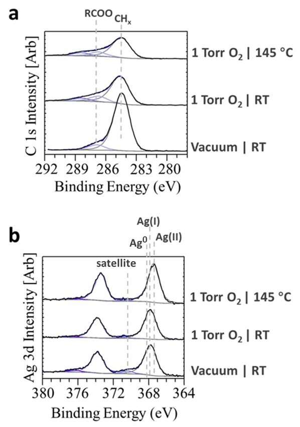
As a result, we recorded the images shown in Fig. 3c,d, and the spectra shown in Fig. 4, on a
different sample, and with less exposure to the X-ray beam to mitigate beam damage. Figs.
3c,d were recorded with exposure times of 117 seconds per image. Because the photon flux at
 photons
beamline 11.0.2 of the ALS is 3.3×1012 mm2 ∙s
 , that duration yields approximately 1.5×10¹⁴
photons incident on the sample per image, more than an order of magnitude lower than for the
image in Fig. 3a. The APXPS spectra discussed next were also collected in the same
measurement set, increasing the total exposure to 3.5×10¹⁴ photons per condition, still
significantly less than for the image shown in Fig. 3a.
 Fig. 4a shows C 1s XPS spectra for Ag-behenate/Si(001) under various temperatures
and oxygen pressures. Ag-behenate contains a CHx backbone with 21 carbon atoms and a
terminating carboxyl group (-COO) with two O atoms. As a result, the XPS intensity in the C
1s region is expected to have its largest contribution from C-C species, generally appearing
near 284 eV. [42] Adventitious carbon species accumulate on surfaces exposed to air, and also
contribute to the C 1s spectrum near 284 eV. [43] The bottom spectrum in Fig. 4a was taken in
vacuum after introducing the sample into the chamber, with overlapping C-C and adventitious
carbon peaks appearing in the C 1s region, along with a peak produced by the carboxyl group
in Ag-behenate at 286.9 eV. [44] [45] The ratio of these two peaks was approximately 10:1
(Fig. 4a). This contrasts with the composition of the molecule, which has 22 C atoms, only one
of which exists in a carboxyl group. Experimentally, the ratio of these two peaks reflects not
only the ratio of each type of carbon species in the molecule, but is also influenced by the
presence of contaminant species on the sample, by the orientation of the molecules on the
surface, and by the integrity of the molecular structure. Regarding the molecular integrity, Ag-
behenate is known to decompose upon prolonged exposure to air and light, which can
contribute to different spectral weights in the C 1s spectra, and create a diffuse photon
scattering background. Hence, the vacuum XPS in the C 1s region in Fig. 4a reflects that Ag-
behenate has undergone some decomposition, and the sample contains other carbon-containing
species, potentially altering the area ratios of the relevant peaks. Fig. 4b shows the Ag 3d
spectra corresponding to each condition from Fig. 4a. In vacuum, the Ag 3d spectrum contains
a peak at 367.8 eV, characteristic of Ag+. [46] The presence of a feature above 370 eV is
indicative of Ag satellite features which can contain contributions from Ag0 and/or Ag partially
oxidized by interaction Ag-behenate. [47]

 11
Fig. 4. (a) APXPS of the C 1s region on Ag-behenate under various conditions. Three features
appear in the spectrum at 283.8 eV, 285.9 eV, and 287 eV. The C-C peak near 284 eV contains
contributions from the hydrocarbon chain in Ag-behenate and from adventitious carbon. The
RCOO peak arises from the carboxyl group in Ag-behenate. The appearance of the higher BE peak
is characteristic of more highly oxidized carbon species in response to added oxygen. (b) Ag 3d
 +
region. Contributions appear from Ag in Ag-behenate, some metallic Ag prior to adding oxygen,
 2+
and Ag upon heating in O2(g). Each spectrum in (a) and (b) is fitted with a Shirley background
shape, represented by a grey line at the base of each spectrum. Photons with 1000 eV excitation
energy were used to collect the C 1s and Ag 3d spectra.

 After characterizing the surface in vacuum, we added 1 Torr of O2 gas into the chamber.
Oxygen exposure reduced the intensity of the C-C peak at 284.5 eV, suggesting a reaction with
some of the carbon species at the surface. At the same time, the intensity of the peak at 286.9
eV remained approximately the same as in vacuum. This indicates that the carboxyl groups in
Ag-behenate do not rapidly decompose upon O2 exposure in these conditions, even when parts

 12
of the molecule decompose slowly when exposed to light and ambient air conditions. The
decrease in the C-C peak is accompanied by the appearance of a feature at 288.5 eV,
characteristic of more highly oxidized carbon species resulting from exposure to O 2 gas. [48]
In the C 1s region, when the sample was heated to 145 °C in 1 Torr of O2, only a 10% decrease
in the intensity of the C-C peak was observed, indicating that the remaining carbon did not
react readily with oxygen at this temperature. Nevertheless, the scattering feature generated by
regularly spaced layers of Ag-behenate nearly vanished. Ag-behenate undergoes thermally
induced carbon chain folding above 120 °C, where it adopts a new molecular packing
arrangement that decreases the Ag-behenate interlayer spacing. [40] This is consistent with our
observation that the feature due to scattering from regularly spaced Ag-behenate layers nearly
vanishes under these conditions.
 The chemical transformation of Ag-behenate, as observed by APXPS, is consistent
with the results of the simultaneous scattering experiments. Nevertheless, the altered packing
arrangement of the Ag-behenate layers has a structure which should also produce a scattering
pattern detectable in the angular range of our GIXS system [40]. However, we note that the C-
C and carboxyl intensities decreased somewhat while heating in oxygen, and at the same time,
the BE of the Ag 3d peak shifted to 367.3 eV upon heating in O2. This latter BE shift is
characteristic of a transformation to Ag2+ [46], suggesting that the Ag atoms from the Ag-
behenate layered structure further oxidize into AgO. The oxidation of Ag under these
conditions could coincide with a change in the structure of the Ag-behenate layers, explaining
the disappearance of the diffraction ring. Additionally, the diminished intensity of both the
carboxyl and C-C peaks suggests that the molecules begin to decompose with some
components becoming volatile.

5. Conclusions
 These combined APXPS and APGIXS measurements on Ag behenate illustrate the
power of correlating chemical and structural transformations for samples in gas phase
environments. Changes in the Ag behenate layered structure were observed using APGIXS,
and were correlated with changes in the Ag oxidation state and carbon chemistry observed in
the Ag 3d and C 1s XPS regions. Combining APXPS and APGIXS provided a simultaneous
picture of the chemical state and structure of the sample, and provided a detailed picture of
their evolution in an O2 gas environment and in different thermal conditions. Our approach to
 13
understanding correlations between structure and behavior is applicable in a variety of fields,
where important phenomena often involve complex relationships between structure, chemistry,
and activity. [5-15]
 The geometry of APXPS measurements involves X-rays incident on surfaces in a way
that inherently enables scattering measurements in the reflection geometry (Fig. 1).
Additionally, our development of a compact pivoting-UHV-manipulator to control the detector
motion makes this configuration highly customizable. Using an X-ray transparent SiNx
membrane to isolate the X-ray detector enables GIXS measurements for samples in a wide
variety of gas phase environments. These developments make it possible to retrofit many
existing APXPS, XPS, and other experimental systems to include simultaneous (AP)-GIXS
measurements with relatively low cost and effort.
 Further related techniques can also be enabled using this approach. For example, X-
ray photon correlation spectroscopy (XPCS) measures correlations in scattering intensities
from a distribution of scattering centers illuminated by coherent photons. The evolution of the
correlation between speckle patterns over time provides a measurement of the evolution of
scattering center distributions in a sample. [49] This opens new possibilities to study dynamic
systems and transformations on their natural timescales. Several soon-to-be-emerging
diffraction limited synchrotron sources around the world will offer coherent flux intensities
increased by orders of magnitude, allowing measurements with time resolution into the
microsecond and nanosecond regimes.

AUTHOR’S CONTRIBUTIONS

H.B., S.N. initiated the project. H.K., S.R., A.K., S.N. contributed to the system design. H.K.,
S.N. performed the experiments with support from B.H.L. and M.B. Data analysis was
performed by H.K. with support from Q.L., H.M., P.C., and F.B. H.K. and S.N. wrote the
manuscript, S.N. supervised the project.

ACKNOWLEDGEMENTS

The instrumentation presented in this work was developed through the Laboratory Directed
Research and Development (LDRD) Program of Lawrence Berkeley National Laboratory
under a grant titled “Correlation of structural and chemical processes at interfaces under
operating conditions using multimodal ambient pressure X-ray photoelectron spectroscopy and
 14
surface X-ray scattering”. H.K. acknowledges the same LDRD grant for support for salary.
The experiments were performed at beamline 11.0.2 of the Advanced Light Source, a U.S.
DOE Office of Science User Facility, which is funded under contract no. DE-AC02-
05CH11231. Q.L. acknowledges the ALS Postdoctoral Fellowship Program for the fellowship.
H.P.M. has been supported for salary by the U.S. Department of Energy (DOE) under Contract
No. DE-SC0014697. F.B. acknowledges funding through the LDRD project “Ultrafast Science
Beyond Pump–Probe”. H.B. and M.B. also acknowledge support by the Director, Office of
Science, Office of Basic Energy Sciences, and by the Division of Chemical Sciences,
Geosciences and Biosciences of the U.S. Department of Energy at LBNL under Contract No.
DE-AC02-05CH11231. Authors would like to thank ALS technical and other staff for their
support, and acknowledge Caroline Bavasso for her contributions to the sample manipulation
automation.

AIP PUBLISHING DATA SHARING POLICY

The data that support the findings of this study are available from the corresponding author
upon reasonable request.

AUTHOR INFORMATION

Corresponding Author
* SNemsak@lbl.gov

 15
REFERENCES

[1] Y. Yuan and J. Huang, "Ion Migration in Organometal Trihalide Perovskite and Its
 Impact on Photovoltaic Efficience and Stability," Acc. Chem. Res., vol. 49, no. 2, pp.
 286-293, 2016.

[2] B. Kiraly, A. N. Rudenko, W. M. van Weerdenburg, D. Wegner, M. I. Katsnelson
 and A. A. Khajetoorians, "An orbitally derived single-atom magnetic memory," Nat.
 Commun., vol. 9, pp. 1-8, 2018.

[3] T. Shang, Y. Wen, D. Xiao, L. Gu, Y.-S. Hu and H. Li, "Atomic-Scale Monitoring
 of Electrode Materials in Lithium-Ion Batteries using In Situ Transmission Electron
 Microscopy," Adv. Energy Mater., vol. 7, p. 1700709, 2017.

[4] W. Gao, Z. D. Hood and M. Chi, "Interfaces in Heterogeneous Catalysts: Advancing
 Mechanistic Understanding through Atomic-Scale Measurements," Acc. Chem. Res.,
 vol. 50, pp. 787-795, 2017.

[5] M. F. Hochella Jr., S. K. Lower, P. A. Maurice, R. L. Penn, N. Sahai, D. L. Sparks
 and B. S. Twining, "Nanominerals, Mineral Nanoparticles, and Earth Systems,"
 Science, vol. 319, no. 5870, pp. 1631-1635, 2008.

[6] A. S. Madden and M. F. J. Hochella, "A test of geochemical reactivity as a function
 of mineral size: Manganese oxidation promoted by hematite nanoparticles,"
 Geochim. Cosmochim. Acta, vol. 69, no. 2, pp. 389-398, 2005.

[7] J. M. F. Hochella, T. Kasama, A. Putnis, C. V. Putnis and J. Moore, "Environmentally
 important, poorly crystalline Fe/Mn hydrous oxides: Ferrihydrite and possibly new
 vernadite-like mineral from the Clark Fork River Superfund Complex," Am. Mineral,
 vol. 90, no. 4, pp. 718-724, 2005.

[8] Tao, S. Dag, L.-W. Wang, Z. Liu, D. R. Butcher, H. Bluhm, M. Salmeron and G. A.
 Somorjai, "Break-Up of Stepped Platinum Catalyst Surfaces by High CO Coverage,"
 Science, vol. 327, pp. 850-853, 2010.

[9] S. R. Longwitz, J. Schnadt, E. K. Vestergaard, R. T. Vang, E. Lægsgaard, I.
 Stensgaard, H. Brune and F. Besenbacher, "High-Coverage Structures of Carbon
 Monoxide Adsorbed on Pt(111) Studied by High-Pressure Scanning Tunneling
 Microscopy," J. Phys. Chem. B, vol. 108, pp. 14497-14502, 2004.

[10] B. Eren and M. Salmeron, "Predicting Surface Clustering at Ambient Conditions
 from Thermodynamic Data," J. Phys. Chem. C, vol. 123, pp. 8171-8176, 2019.

 16
[11] B. Eren, D. Zherebetskyy, L. L. Patera, C. H. Wu, H. Bluhm, C. Frich, L.-W. Wang,
 G. A. Somorjai and M. Salmeron, "Activation of Cu(111) surface by decomposition
 into nanoclusters driven by CO adsorption," Science, vol. 351, no. 6272, pp. 475-
 478, 2016.

[12] B. Eren, D. Zherebetskyy, Y. Hao, L. L. Patera, L.-W. Wang, G. A. Somorjai and M.
 Salmeron, "One-dimensional nanoclustering of the Cu(100) surface under CO gas in
 the mbar pressure range," Surf. Sci., vol. 651, pp. 210-214, 2016.

[13] J. Kim, W. H. Park, W. H. Doh, S. W. Lee, M. C. Noh, J.-J. Gallet, F. Bournel, H.
 Kondoh, K. Mase, Y. Jung, B. S. Mun and J. Y. Park, "Adsorbate-driven reactive
 interfacial Pt-NiO1-x nanostructure formation on the Pt3Ni(111) alloy surface," Sci.
 Adv., vol. 4, no. 7, p. 3151, 2018.

[14] F. Gao, M. E. Grass, Y. Zhang, D. R. Butcher, J. R. Renzas, Z. Liu, J. Y. Chung, B.
 S. Mun, M. Salmeron and G. A. Somorjai, "Reaction-Driven Restructuring of Rh-Pd
 and Pt-Pd Core-Shell Nanoparticles," Science, vol. 322, no. 5903, pp. 932-934, 2008.

[15] Z. Tan, S. Chen, X. Peng, L. Zhang and C. Gao, "Polyamide membranes with
 nanoscale Turing structures for water purification," Science, vol. 360, no. 6388, pp.
 518-521, 2018.

[16] D. M. Meier, A. Urakawa and A. Baiker, "Polarization-modulation infrared
 reflection-absorption spectroscopy affording time-resolved simultaneous detection
 of surface and liquid phase species at catalytic solid-liquid interfaces," Analyst, vol.
 134, pp. 1779-1780, 2009.

[17] C. T. Herbschleb, P. C. van der Tuijn, S. B. Roobol, V. Navarro, J. W. Bakker, Q.
 Liu, D. Stolz, M. E. Cañas-Ventura, G. Verdoes, M. A. van Spronsen, M. Bergman,
 L. Crama, I. Taminiau, A. Ofitserov and G. J. C. van Baarle, "The ReactorSTM:
 Atomically resolved scanning tunneling microscopy under high-pressure, high-
 temperature catalytic reaction conditions," Rev. Sci. Instrum., vol. 85, p. 083703,
 2014.

[18] J. F. Creemer, S. Helveg, G. H. Hoveling, S. Ullmann, A. M. Molenbroek, P. M.
 Sarro and H. W. Zandbergen, "Atomic-scale electron microscopy at ambient
 pressure," Ultramicroscopy, vol. 108, no. 9, pp. 993-998, 2008.

[19] F. Tao, D. Tang, M. Salmeron and G. A. Somorjai, "A new scanning tunneling
 microscope reactor used for high-pressure and high-temperature catalysis studies,"
 Rev. Sci. Instrum., vol. 79, p. 0840101, 2008.

[20] S. B. Roobol, M. E. Cañas-Ventura, M. Bergman, M. A. van Spronsen, W. G.
 Onderwaater, P. C. van der Tuijn, R. Koehler, A. Ofitserov, G. J. C. van Baarle and
 J. W. M. Frenken, "The ReactorAFM: Non-contact atomic force microscope

 17
operating under high-pressure and high-temperature catalytic conditions," Rev. Sci.
 Instrum., vol. 86, p. 033706, 2015.

[21] D. F. Ogletree, H. Bluhm, E. D. Hebenstreit and M. Salmeron, "Photoelectron
 spectroscopy under ambient pressure and temperature conditions," Nucl. Instrum.
 Methods Phys. Res, vol. 601, pp. 151-160, 2009.

[22] A. R. Head, O. Karslioǧlu, T. Gerber, Y. Yu, L. Trotachaud, J. Raso, P. Kerger and
 H. Bluhm, "CO adsorption on Pd(100) studied by multimodal ambient pressure X-
 ray photoelectron and infrared reflection absorption spectroscopies," Surf. Sci., vol.
 665, pp. 51-55, 2017.

[23] M. A. Marcus, A. A. MacDowell, R. Celestre, A. Manceau, T. Miller, H. A. Padmore
 and R. E. Sublett, "Beamline 10.3.2 at ALS: a hard X-ray microprobe for
 environmental and materials sciences," J. Synchrotron Rad., vol. 11, pp. 239-247,
 2004.

[24] G. Eres, C. M. Rouleau, Q. Lu, Z. Zhang, E. Benda, H. N. Lee, J. Z. Tischler and D.
 D. Fong, "Experimental setup combining in situ hard X-ray photoelectron
 spectroscopy and real-time surface X-ray diffraction for characterizing atomic and
 electronic structure evolution during complex oxide heterostructure growth," Rev.
 Sci. Instrum., vol. 90, no. 093902, pp. 1-11, 2019.

[25] C. S. Fadley, "X-ray photoelectron spectroscopy: Progress and perspectives," J.
 Electron Spectros. Relat. Phenomena, vol. 178, pp. 2-32, 2010.

[26] D. E. Starr, Z. Liu, M. Hävecker, A. Knop-Gericke and H. Bluhm, "Investigation of
 solid/vapor interfaces using ambient pressure X-ray photoelectron spectroscopy,"
 Chem. Soc. Rev., vol. 42, p. 5833, 2013.

[27] D. F. Ogletree, H. Bluhm, G. Lebedev, C. S. Fadley, Z. Hussain and M. Salmeron,
 "A differentially pumped electrostatic lens system for photoemission studies in the
 millibar range," Rev. Sci. Instrum., vol. 73, no. 11, pp. 3872-3877, 2002.

[28] P. Müller-Buschbaum, "A Basic Introduction to Grazing Incidence Small-Angle X-
 Ray Scattering," Lect. Notes Phys., Vols. 61-89, p. 776, 2009.

[29] D. Altamura, T. Sibillano, D. Siliqi, L. De Caro and C. Giannini, "Assembled
 Nanostructure Architectures Studied by Grazing Incidence X-ray Scattering,"
 Nanomater. nanotechnol., vol. 2, no. 16, pp. 1-23, 2012.

[30] C. D. Liman, S. Choi, D. W. Breiby, J. E. Cochran, M. F. Toney, E. J. Kramer and
 M. L. Chabinyc, "Two-dimensional GIWAXS reveals a transient crystal phase in
 solution-processed thermally converted tetrabenzoporphyrin," J. Phys. Chem. B, vol.
 117, no. 46, pp. 14557-14567, 2013.

 18
[31] M. He, J. Li, M. L. Sorensen, F. Zhang, R. R. Hancock, H. H. Fong, V. A. Pozdin,
 D.-M. Smilgies and G. G. Malliaras, "Alkylsubstituted Thienothiophene
 Semiconducting Materials: Structure-Property Relationships," J. Am. Chem. Soc.,
 vol. 131, pp. 11930-11938, 2009.

[32] G. Kaune, M. A. Ruderer, E. Mewalli, W. Wang, S. Couet, K. Schlage, R.
 Röhlsberger, S. V. Roth and P. Müller-Buschbaum, "In Situ GISAXS Study of Gold
 Film Growth on Conducting Polymer Films," ACS Appl. Mater. Interfaces, vol. 1,
 no. 2, pp. 353-360, 2009.

[33] R. E. Winans, S. Vajda, B. Lee, S. J. Riley, S. Seifert, G. Y. Tikhonov and N. A.
 Tomczyk, "Thermal Stability of Supported Platinum Clusters Studied by in Situ
 GISAXS," J. Phys. Chem. B, vol. 108, no. 47, pp. 18105-18107, 2004.

[34] T. H. Metzger, I. Kegel, R. Paniago, A. Lorke, J. Peisl, J. Schulze, I. Eisele, P.
 Schittenhelm and G. Abstreiter, "Shape, size, strain and correlations in quantum dot
 systems studied by grazing incidence X-ray scattering methods," Thin Solid Films,
 vol. 336, pp. 1-8, 1998.

[35] T. H. Metzger, I. Kegel, R. Paniago and J. Peisl, "Grazing incidence x-ray scattering:
 an ideal tool to study the structure of quantum dots," J. Phys. D: Appl. Phys., vol. 32,
 pp. A202-A207, 1999.

[36] A. Hexemer and P. Müller-Buschbaum, "Advanced grazing-incidence techniques for
 modern soft-matter materials analysis," IUCrJ, vol. 2, no. 1, pp. 106-125, 2015.

[37] H. Bluhm, K. Andersson, T. Araki, K. Benzerara, G. E. Brown, J. J. Dynes, S. Ghosal,
 M. K. Gilles, H.-C. Hansen, J. C. Hemminger, A. P. Hitchcock, G. Ketteler, A. L.
 Kilcoyne, E. Kneedler, J. R. Lawrence, G. G. Leppard, J. Majzlam, B. S. Mun, S. C.
 Myneni, A. Nilsson, H. Ogasawara, D. F. Ogletree, K. Pecher, M. Salmeron, D. K.
 Shuh, B. Tonner, T. Tyliszczak, T. Warwick and T. H. Yoon, "Soft X-ray microscopy
 and spectroscopy at the molecular environmental science beamline at the Advanced
 Light Source," J. Electron Spectrosc., vol. 150, pp. 86-104, 2006.

[38] T. Hoshino, M. Kikuchi, D. Murakami, Y. Harada, K. Mitamura, K. Ito, Y. Tanaka,
 S. Sasaki, M. Takata, H. Jinnai and A. Takahara, "X-ray photon correlation
 spectroscopy using a fast pixel array detector with a grid mask resolution enhancer,"
 J. Synchrotron Rad., vol. 19, pp. 988-993, 2012.

[39] T. N. Blanton, T. C. Huang, H. Toraya, C. R. Hubbard, S. B. Robie, D. Louër, H. E.
 Göbel, G. Will, R. Gilles and T. Raftery, "JCPDS-International Centre for Diffraction
 Data round robin study of silver behenate. A possible low-angle X-ray diffraction
 calibration standard," Powder Diffraction, vol. 10, no. 2, pp. 91-95, 1995.

 19
[40] T. Blanton, S. Zdzieszynski, M. Nicholas and S. Misture, "In situ high-temperature
 X-ray diffraction study of phase transformations in silver behenate," Powder
 Diffraction, vol. 20, no. 2, pp. 94-96, 2005.

[41] M. Nyam-Osor, D. V. Soloviov, Y. S. Kovalev, A. Zhingunov, A. V. Rogachev, O.
 I. Ivankov, R. V. Erhan and A. I. Kuklin, "Silver behenate and silver stearate powders
 for calibration of SAS instruments," J. PHys. Conf. Ser., vol. 351, no. 012024, 2012.

[42] M. Morkel, V. V. Kaichev, G. Rupprechter, H. J. Freund, I. P. Prosvirin and V. I.
 Bukhtiyarov, "Methanol Dehydrogenation and Formation of Carbonaceous
 Overlayers on Pd(111) Studied by High-Pressure SFG and XPS Spectroscopy," J.
 Phys. Chem. B, vol. 108, pp. 12955-12961, 2004.

[43] G. Greczynski and L. Hultman, "C 1s Peak of Adventitious Carbon Aligns to the
 Vacuum Level: Dire Consequences for Material's Bonding Assignment by
 Photoelectron Spectroscopy," ChemPhysChem, vol. 18, pp. 1507-1512, 2017.

[44] J. Hasselström, O. Karis, M. Weinelt, N. Wassdahl, A. Nilsson, M. Nyberg, L. G. M.
 Pettersson, M. G. Samant and J. Stöhr, "The adsorption structure of glycine adsorbed
 on Cu(110); comparison with formate and acetate/Cu(110)," Surf. Sci., vol. 407, pp.
 221-236, 1998.

[45] H. Ju, X. Feng, Y. Ye, L. Zhang, H. Pan, C. T. Campbell and J. Zhu, "Ca Carboxylate
 Formation at the Calcium/Poly(methyl methacrylate) Interface," J. Phys. Chem. C,
 vol. 116, pp. 20465-20471, 2012.

[46] T. C. Kaspar, T. Droubay, S. A. Chambers and P. S. Bagus, "Spectroscopic Evidence
 for Ag(III) in Highly Oxidized Silver Films by X-ray Photoelectron Spectroscopy,"
 J. Phys. Chem. C, vol. 114, pp. 21562-21571, 2010.

[47] A. Dolatkhah, P. Jani and L. D. Wilson, "Redox-Responsive Polymer Template as an
 Advanced Multifunctional Catalyst Support for Silver Nanoparticles," Langmuir,
 vol. 34, pp. 10560-10568, 2018.

[48] T. E. Felter, W. H. Weinberg, G. Y. Lastushkina, A. I. Boronin, P. A. Zhdan, G. K.
 Boreskov and J. Hrbek, "Ag(111), An XPS and UPS Study of the Kinetics of Carbon
 Monoxide Oxidation over," Surf. Sci., vol. 118, pp. 369-386, 1982.

[49] O. G. Shpyrko, "X-ray photon correlation spectroscopy," J. Synchrotron Rad., vol.
 21, pp. 1057-1064, 2014.

 20
You can also read