Protective effect of Yi-Qi-Huo-Xue Decoction against ischemic heart disease by regulating cardiac lipid metabolism
←
→
Page content transcription
If your browser does not render page correctly, please read the page content below
Available online at www.sciencedirect.com

                                         Chinese Journal of Natural Medicines 2020, 18(10): 779-792
                                                    doi: 10.1016/S1875-5364(20)60018-8

•Research article•

Protective effect of Yi-Qi-Huo-Xue Decoction against ischemic heart
           disease by regulating cardiac lipid metabolism
                   LI Fang-He1, 2, HUANG Xiao-Lou3, WANG Hui2, GUO Shu-Wen4*, LI Ping1*
1
  The Third Affiliated Hospital, Beijing University of Chinese Medicine, Beijing 100029, China;
2
  School of Traditional Chinese Medicine, Beijing University of Chinese Medicine, Beijing 100029, China;
3
  College of Acupuncture and Orthopedics, Guizhou University of Traditional Chinese Medicine, Guiyang 550025, China;
4
  Fangshan Hospital, Beijing University of Chinese Medicine, Beijing 102400, China
                                                        Available online 20 Oct., 2020

    [ABSTRACT] Yi-Qi-Huo-Xue Decoction (YQHX) is the recombination of Dang-Gui-Bu-Xue Decoction (DBD), which is one of the
    well-known traditional Chinese Medicine (TCM) prescription, and has long been shown to have significant protective effects against
    myocardial ischemic injury. In previous studies, we found that YQHX could regulate lipid and glucose metabolism, promote angiogen-
    esis, attenuate inflammatory response, and ameliorate left ventricular function in myocardial ischemia rat models. However, the under-
    lying mechanism of how YQHX involves in lipid metabolism remains unclear so far. In this study, the underlying mechanism of
    YQHX in lipid metabolism disorders was elucidated in a myocardial ischemia rat model and a hypoxia-induced H9c2 cell injury mod-
    el. YQHX (8.2 g·kg−1) and positive-control drug trimetazidine (10 mg·kg−1) were administered daily on the second day after left anteri-
    or descending (LAD) operation. At 7 days and 28 days after surgery, changes of cardiac morphology, structure, and function were
    evaluated by H&E staining and echocardiography, respectively. The plasma lipid levels and mitochondrial ATP content were also eval-
    uated. Western blot and RT-PCR were used to determine the protein and mRNA expressions of AMPK, PGC-1α, CPT-1α, and
    PPARα. YQHX improved cardiac function and ameliorated lipid metabolism disorders. Furthermore, YQHX increased the expression
    of p-AMPK, PGC-1α, and CPT-1α without changing PPARα in ischemic rat myocardium. In vitro, YQHX activated the protein and
    mRNA expression of PGC-1α, CPT-1α, and PPARα in hypoxia-induced H9c2 cells injury, whereas AMPK inhibitor Compound c
    blocked the effects of YQHX. Taken together, the results suggest that YQHX reduces lipid metabolism disorders in myocardial
    ischemia via the AMPK-dependent signaling pathway.
    [KEY WORDS] Yi-Qi-Huo-Xue Decoction; Lipid metabolism; Ischemic heart disease; AMPK-dependent pathway; Fatty acid oxida-
    tion
    [CLC Number] R965           [Document code] A         [Article ID] 2095-6975(2020)10-0779-14

Introduction                                                              significant clinical problem, which urgently requires novel
                                                                          therapeutic strategies. The 2016 Heart Outcomes Prevention
     In recent years, according to the lifestyle changes, diet-
                                                                          Evaluation-3 (HOPE-3) trial supports that using rosuvastatin,
ary habits, and an aging population, the incidence of ischem-
                                                                          at a dose of 10 mg per day to lower cholesterol, decreases the
ic heart disease has gradually become a leading cause of
                                                                          risk of cardiovascular events among 12 705 participants in 21
death, which affected more than 17 million people world-
                                                                          countries without cardiovascular diseases [3]. Given that fatty
wide in 2008. Furthermore, the number is estimated to rise by             acids provide more energy production than glucose, recently
23.6 million in 2030 [1, 2]. Despite a series of significant thera-       people have gradually realized that lipid metabolic dysfunc-
peutic developments that share the aim to rapidly restore                 tion plays an important role in the pathogenesis of cardiovas-
coronary artery blood flood, poor prognosis of IHD remains a              cular diseases [4-7]. It suggests that reverting cardiac metabol-
                                                                          ism back to utilizing fatty acid metabolism may be a plaus-
[Received on] 09-Apr.-2020                                                ible therapeutic choice for treating myocardial ischemia [6, 8, 9].
[Research funding] This work was supported by the National Natur-              As a “metabolic master switch”, AMP-activated protein
al Science Foundation of China (No. 81473552), and the China              kinase (AMPK) maintains energy homeostasis and regulates
Postdoctoral Science Foundation (No. 2019TQ0043).
[*Corresponding author] Tel/Fax: 86-10-64286482, E-mails: guo11
                                                                          lipid and glucose metabolism for adaptation to stresses re-
63@163.com (GUO Shu-Wen); pearll2008@126.com (LI Ping)                    sponse, especially during and following ischemia [10-12].
These authors have no conflict of interest to declare.                    Ischemia causes a significant increase in the myocardial

                                                                   – 779 –
LI Fang-He, et al. / Chin J Nat Med, 2020, 18(10): 779-792
levels of AMPK phosphorylation and activity [10]. AMPK ac- SQ). The YQHX preparations were extracted by refluxing
tivates or inactivates a series of metabolic pathways, includ- with boiling distilled water (1∶10, g·mL−1) three times, re-
ing peroxisome proliferator activated receptor gamma co- spectively. After filtration, the aqueous extracts of YQHX
activator 1α (PGC-1α), carnitine palmitoyltransferase-1α were concentrated into a constant volume for use in animal
(CPT-1α), and peroxisome proliferator activated receptor α experiments, and were prepared in powder form by freeze-
(PPARα), which is involved in fatty acid and cholesterol syn- drying in vacuo, respectively. YQHX freeze-drying powders
thesis [6, 12-14]. However, the mechanism by which myocardial (25 mg) were dissolved in 50 mL of DMEM in vitro. Sub-
AMPK phosphorylation is activated in myocardial ischemia sequently, YQHX extracts were filtered through a 0.45 μm
remains unclear so far. millipore filter prior to use. Then, a part of each filtrate was 4-
Yi-Qi-Huo-Xue decoction (YQHX) is a compound fold diluted with distilled water and was further subjected to
Chinese medicine, which has been widely applied in the treat- HPLC and UHPLC-LTQ-Orbitrap-MS, respectively [17].
ment of ischemic heart disease with efficacy and safety [15]. Animal model and grouping
Results of high-performance-liquid-chromatography (HPLC) A total of 120 male Sprague-Dawley (SD) rats were pur-
and HPLC-linear ion trap-Orbitrap mass spectrometry ana- chased from Vital River Laboratory Animal Technology to be
lyses indicated that the active substances of YQHX include used for myocardial infarction surgery (Beijing, China, li-
Astragaloside IV, Calycosin, Ferulic acid, ginsenoside Rg1 cense number: SCXK2016-0006). Animals were housed at
and ginsenoside Rb1 [16, 17]. Our previous studies reported that Dongzhimen Hospital Science cage under consistent condi-
YQHX could exert cardioprotective effects in ischemic rat tions (12 h light or dark cycles). All experiments and proto-
myocardium by inhibiting oxidative stress and inflammation cols were performed in accordance with the Animal Care
response, promoting angiogenesis and protecting mitochon- Committee and Use Committee of Dongzhimen Hospital Af-
drial function [15-19]. Results of nuclear magnetic resonance
filiated to Beijing University of Chinese Medicine (2017-11).
metabolomics also showed YQHX could effectively regulate
The myocardial infarction rat model was established by a left
plasma and urine levels of lipid, amino acid and carbo-
anterior descending (LAD) coronary artery occlusion [22, 23].
hydrate metabolism in rat models of IHD [20]. Recently, we re-
Briefly, SD rats were anesthetized with 1% pentobarbital so-
ported that YQHX could further activate AMPK phos-
dium at the dosage of 40 mg·kg−1 by intraperitoneal injection.
phorylation in hypoxia-induced cardiomyocytes injury.
Then, the thorax was opened to expose the heart, and the
However, as of right now, the mechanisms of YQHX on en-
LAD was ligated with a 5−0 polypropylene suture except the
ergy metabolism especially the activation of AMPK phos-
phorylation have not been completely evaluated. To address Sham group. Finally, the chest was closed, and we used the
these unanswered questions, we use a rat myocardial infarc- sodium penicillin for 3 days after surgery for the purpose of
tion (MI) model, which is similar to the IHD patients af- anti-inflammation of the wound. From the second day after
fected by myocardial infarction to evaluate the change of lip- MI, rats in the YQHX group were treated with YQHX
id metabolism between the subacute (7 days) and chronic (28 aqueous extract at the daily dosage of 8.2 g·kg−1. Rats in the
days) stages after myocardial infarction [21]. Then we use the positive control drug Trimetazidine (Servier, Tianjin, China,
hypoxia-induced H9c2 cell injury model with YQHX or/and Series: H20055465) group were treated with Trimetazidine
AMPK inhibitor (Compound c) to assess the underlying lipid aqueous solution at the daily dosage of 10 mg·kg−1 [24]. Rats
mechanisms of YQHX. were acclimated for 1 week and randomly divided into four
groups during the subacute (7 days) and chronic (28 days)
Materials and Methods stages, including Sham group (Sham), myocardial infarction
Preparation of YQHX model group (MI), YQHX group (YQHX) and positive-con-
The herbs of YQHX were purchased from Dongzhimen trol drug Trimetazidine group (TMZ). All rats were anaes-
Hospital of Beijing University of Chinese Medicine. YQHX thetized by pentobarbital sodium at the end of the study, and
is composed of five herbs (Table 1), including Astragalus the cardiac samples were collected to analyze mRNA and
membranaceus (Huang Qi, HQ), Angelica sinensis (Dang protein expression levels.
Gui, DG), Panax ginseng (Ren Shen, RS), Ligusticum wal- Cell culture and grouping
lichii (Chuan Xiong, CX), and Panax notoginseng (San Qi, Rat H9c2 cells were seeded 5 × 10⁴ cells/mL in 6-well
Table 1 Prescription of YQHX
Chinese name English name Pharmaceutical name Family Medicinal parts
Huang Qi Astragali Radix Astragalus Membranaceus Astragalus membranaceus (Fisch.) Bunge Root
Dang Gui Angelica Sinensis Angelica Sinensis Angelica sinensis (Oliv.) Diels. Root
Ren Shen Ginseng Radix Ginseng Panax ginseng C. A. Mey. (Araliaceae) Root
Chuan Xiong Sichuan lovage rhizome Rhizoma Ligustici Chuanxiong Ligusticum chuanxiong Hort. Root and rhizoma
San Qi Sanchi Notoginseng radix et rhizoma Panax notoginseng (Burkill) F. H. Chen (Araliaceae) Root
– 780 –LI Fang-He, et al. / Chin J Nat Med, 2020, 18(10): 779-792
plates (Corning, New York, USA). H9c2 cells were main- genized in a cold homogenate buffer for 30 min. Then these
tained in high-glucose DMEM (Gibco, California, USA) sup- were centrifuged at 12 000 g for 5 min at 4 °C to collect the
plemented with 10% fetal bovine serum FBS (BI, Israel), 1% supernatant. The myocardial mitochondrial ATP content were
of penicillin/streptomycin in a cell incubator with 5% CO2 measured by a corresponding ATP Assay Kit (Beyotime,
and 95% air at 37 ºC. After 24−30 h culture, the culture medi- Shanghai, China).
um was completely replaced with serum-free DMEM. After Western blot analysis
synchronization culture for 6 h, H9c2 cells were exposed to a Samples of myocardial tissues from the infarcted
hypoxic atmosphere with 1% O2, 5% CO2 and 94% N2 for myocardial border, as well as H9c2 cells were prepared for
12 h. The treatment of H9c2 cells with YQHX at the dosage protein analysis. Firstly, the heart tissues were homogenized
of 200 μg·mL−1 (Dongzhimen Hospital, China) or/and AMPK and H9c2 cells were collected in an ice-cold RIPA lysis buf-
inhibitor Compound c (5 μmol·L−1) (Selleck, Shanghai, fer. The protein extracts (40 μg) were separated by 12% SDS-
China) were used before the initiation of hypoxia [25, 26]. H9c2 PAGE blots (Bio-Rad, California, USA) and then transferred
cells were randomly divided into four groups, including to the nitrocellulose membranes (Millipore, MA, USA). The
(1) Control group (C), (2) Ischemia/Hypoxia group (I/H), nitrocellulose membranes were blocked with 5% fat-free dry
(3) YQHX at the concentrations of 200 μg·mL−1 during hyp- milk in Tris-buffered saline for 2 h, washed, and then incub-
oxia group (Y2), and (4) 200 μg·mL−1 YQHX supplemented
ated with the following primary antibodies, including p-
with 5 μmol·L−1 Compound c during hypoxia group (Y2cc).
AMPK and AMPK (1: 1000, Cell Signaling Technology, Bo-
Transthoracic echocardiography
ston, USA), PGC-1α (1∶1000, Abcam, MA, USA), CPT-1α
Transthoracic echocardiography was used to assess car-
(1∶500, Proteintech, Rosemont, USA), and PPARα
diac function 7 and 28 days after MI using a Vevo770 (Visu-
(1∶1000, Proteintech, Rosemont, USA). The primary anti-
al Sonics Inc., Toronto, Canada). The parasternal left vent-
bodies were incubated at 4 °C overnight, washed, and then in-
ricle was measured in the short-axis view. The parameters of
cubated with the secondary antibodies for 2 h. After three
cardiac function were as follows: the left ventricular internal
washed, the blots were incubated with ECL and then detec-
dimension-systole/diastole (LVIDs/d), left ventricular ejec-
ted by using the Gene Genius Bio Imaging System.
tion fraction (LVEF), and left ventricular fractional shorten-
Real-time quantitative PCR analysis
ing (LVFS).
The mRNA expressions of AMPK, PGC-1α, CPT-1α,
Histopathology examination
Hematoxylin and eosin (HE) staining was used to visual- and PPARα were determined by reverse transcriptase poly-
ize cardiomyocyte architectures. Heart tissue samples were merase chain reaction (RT-PCR). Total RNA was extracted
extracted from sacrificed rats, and immediately fixed in 4% from cardiac tissues and H9c2 cells using Trizol reagent (In-
paraformaldehyde solution. Then the cardiac samples were vitrogen, California, USA) and subjected to reverse transcrip-
embedded in paraffin and were cut into 5 μm slices, which tion. Thereafter, the first-strand cDNA was synthesized using
were stained with HE and observed under a light microscope a Revert Aid TM First Strand cDNA Synthesis kit (Invitro-
(Olympus, Tokyo, Japan). gen, California, USA). Next, real-time quantitative PCR was
Plasma lipid analysis performed in triplicate using SYBR® Premix Ex Taq kit on an
At 7 and 28 days after MI, blood samples were collected ABI PRISM 7500 PCR instrument (Applied Biosystems,
from sacrificed rats, and centrifuged at 3000 r·min−1 at 4 ºC FosterCity, USA). The cDNA was denatured by 35 PCR
for 20 min to obtain serum. Plasma levels of free fatty acids cycles (2 min at 94 °C, 30 s at 94 °C, 30 s at 61 °C, 30 s at
(FFA), total cholesterol (TC), triglyceride (TG), high-density 72 °C). GAPDH was used for the invariant control, and the
lipoprotein (HDL), and low-density lipoprotein (LDL) were relative level of mRNA was calculated using the 2−ΔΔCt meth-
measured using a TBA-120 automatic biochemical analyzer od. The forward and reverse sequences for the primers are lis-
(Toshiba TBA-120, Tokyo, Japan). ted in Table 2.
Transmission electron microscope Statistical analysis
Fresh samples of the left ventricle in the margin area All data are expressed as mean ± SD, and the statistic-
were cut into thick slices, and rapidly fixed them in 2% para- ally significant differences between Sham group, MI group,
formaldehyde and 2.5% glutaraldehyde at 4 °C for 2 h. After YQHX group and other differences groups were calculated
washing with (phosphate buffered saline) PBS three times, via one-way ANOVA and Duncan’s multiple range test. P
the samples were fixed with 1% osmium tetroxide for 2 h, values less than 0.05 were considered as statistically signific-
and then dehydrated using acetone solutions. Next, the speci- ant. All statistical analyses were conducted using SPSS 17.0
mens were embedded with epoxy resin and heated polymeriz- software and performed by GraphPad 6.0 Prism software.
ation at 60 °C. Finally, the embedding blocks were sliced in-
Results
to ultrathin sections. Images of different stained areas were
recorded using a Hitachi-H7650 transmission electron micro- Effects of YQHX on cardiac function in MI rats
scopy (Tokyo, Japan). In order to assess whether YQHX improves cardiac func-
ATP content tion after myocardial infarction, we initially examined the
The fresh myocardial samples were dissected and homo- cardiac function in different groups after MI or Sham opera-
– 781 –LI Fang-He, et al. / Chin J Nat Med, 2020, 18(10): 779-792
Table 2 Primers used in this study
Gene Species Forward (5′−3′) Reverse (5′−3′)
AMPK Rat GTTAAATCCCACTACCACAA GAGGACTCGGCATCAATA
PGC-1α Rat GACCGTCCAAAGCATTCA GGTTCTTGTCCACGCCTC
CPT-1α Rat GGCTCTGGGTGGCAGTCAT CCGTGTTCTGCAAACATCCA
PPARα Rat TCCACAAGTGCCTGTCCGT CTTTCCTGCGAGTATGACCC
GAPDH Rat GGCAAGTTCAACGGCACAG CGCCAGTAGACTCCACGAC
Table 3 Indicators of cardiac function in different groups 7 days after myocardial infarction (mean ± SD, n = 6)
Group EF% FS% LVIDs (mm) LVIDd (mm)
Sham 85.68 ± 3.67 55.78 ± 4.24 2.43 ± 0.33 5.48 ± 0.39
MI 34.99 ± 2.76*** 17.45 ± 1.54*** 6.62 ± 0.49** 8.02 ± 0.57***
YQHX 50.62 ± 10.66### 27.03 ± 6.55## 5.09 ± 0.96# 7.39 ± 1.10
TMZ 59.82 ± 7.33### 33.24 ± 5.01### 5.33 ± 0.91# 7.94 ± 0.95
** *** # ## ###
P < 0.01, P < 0.001 vs Sham group; P < 0.05, P < 0.01, P < 0.001 vs MI group
Table 4 Indicators of cardiac function in different groups 28 days after myocardial infarction (mean ± SD, n = 8)
Group EF% FS% LVIDs (mm) LVIDd (mm)
Sham 80.24 ± 7.64 50.81 ± 7.90 2.98 ± 1.05 6.40 ± 1.06
MI 31.11 ± 3.82*** 16.32 ± 2.90*** 7.86 ± 1.03*** 9.49 ± 1.31***
YQHX 48.03 ± 5.42### 25.36 ± 3.27### 6.02 ± 0.83### 8.27 ± 0.73#
TMZ 42.84 ± 5.59## 22.08 ± 3.28## 6.38 ± 0.83## 8.29 ± 0.97#
***
P < 0.001 vs Sham group; #P < 0.05, ##P < 0.01, ###P < 0.001 vs MI group
tion between the subacute (7 days) and chronic stages (28 histopathological examination showed that the myocardial
days) (Tables 3−4). 7 days after MI, echocardiography ana- fibers were orderly arranged with no sign of inflammation in
lysis showed that LVEF and LVFS values in the MI group the Sham group. Compared with Sham group, there were
were significantly reduced by 34.99% and 17.45%, accom- myocardium fracture, edema and even necrosis, along with
panied by an increase in LVIDs/d compared with the Sham massive infiltration of inflammatory cells in the MI group.
group (Fig. 1A). After treatment with YQHX, the LVEF and YQHX and TMZ treatment significantly decreased those
LVFS values were significantly recovered by 50.62% and events, including reduced myocardial edema and infiltration
27.03% . In TMZ treatment group, the LVEF and LVFS val- of inflammatory cells. 28 days after MI (Fig. 2B), the MI
ues were upregulated by 59.82% and 33.24%, respectively. group myocardial fibers were arranged irregularly and dis-
The LVIDs values were downregulated in the YQHX group orderly as an interstitial substance with edema and cardiac
and TMZ group, whereas LVIDd values had no significant muscle membrane damage. A considerable number of cardi-
change compared with MI group. 28 days after MI, com- omyocytes were replaced by increased fibroblasts and colla-
pared with Sham group, LVEF and LVFS values were re- gen. YQHX and TMZ treatment significantly improved
duced by 31.11% and 16.32%, accompanied by an increase in myocardial edema and necrosis, and reduced inflammatory
LVIDs/d (Fig. 1B). After treatment with YQHX, LVEF and cell infiltration.
LVFS values were recovered by 48.03% and 25.36% (P < Effects of YQHX on plasma lipid levels in MI rats
0.001), accompanied by a decrease in LVIDs/d. In the TMZ To determine the effects of YQHX on lipid metabolism
group, the LVEF and LVFS values were respectively re- in MI rats, we next detected plasma lipid levels in different
covered by 42.84% and 22.08%, indicating that YQHX and groups after MI or sham operation. 7 days after MI, the levels
TMZ improved cardiac function in MI rats. of TC in the MI group were significantly increased compared
Effects of YQHX on cardiac histopathology changes in MI with the Sham group (P < 0.05). FFA, TG, and LDL levels in
rats the MI group also were increased, but there were no statistic-
Next, we investigated the cardiac morphological changes al differences (P > 0.05). Compared with the MI group,
after myocardial infarction (Fig. 2). 7 days after MI (Fig. 2A), YQHX group significantly decreased serum FFA and TG
– 782 –LI Fang-He, et al. / Chin J Nat Med, 2020, 18(10): 779-792
A
Sham MI YQHX TMZ
100 80
80
### 60
LVEF (%)
LVFS (%)
##
60
###
40 ##
40 ***
***
20
20
0 0
Sham MI YQHX TMZ Sham MI YQHX TMZ
8 10
** ***
#
# 8
6
LVIDd (mm)
LVIDs (mm)
6
4
4
2
2
0 0
Sham MI YQHX TMZ Sham MI YQHX TMZ
B Sham MI YQHX TMZ
100 60
80
40
LVEF (%)
LVFS (%)
60 ###
## ###
40 *** ***
##
20
20
0 0
Sham MI YQHX TMZ Sham MI YQHX TMZ
10 15
***
8
LVIDd (mm)
LVIDs (mm)
## ***
### 10
6 # #
4
5
2
0 0
Sham MI YQHX TMZ Sham MI YQHX TMZ
Fig. 1 YQHX and TMZ improved cardiac function including LVEF, LVFS, and LVIDs/d in MI rats. (A) Effect of YQHX (8.2
g·kg−1·d−1) and TMZ (10 mg·kg−1·d−1) on cardiac function 7 days after myocardial infarction (B) Effect of YQHX and TMZ on
cardiac function 28 days after myocardial infarction. Data are expressed as mean ± SD, n = 6−8. **P < 0.01, ***P < 0.001 vs Sham
group; #P < 0.05, ##P < 0.01, ###P < 0.001 vs MI group
– 783 –LI Fang-He, et al. / Chin J Nat Med, 2020, 18(10): 779-792
Sham MI YQHX TMZ
A
7d
100 μm 100 μm 100 μm 100 μm
B
28 d
100 μm 100 μm 100 μm 100 μm
Fig. 2 YQHX improved cardiomyocyte architecture after myocardial infarction (cardiac tissue morphology under the light mi-
croscope at magnification 200 ×). HE staining of the myocardial tissues in the infarcted marginal area from the Sham group, MI
group, YQHX group, and TMZ group at 7 days (A) and 28 days (B) after surgery. Scale bar, 100 μm
levels (P < 0.05). However, TMZ group had no significant sponse after myocardial infarction.
change, suggesting that metabolic pathways may be different Effects of YQHX on p-AMPK and AMPK expression in MI
between YQHX group and TMZ group (Table 5). 28 days rats
after MI (Table 6), the levels of TG were significantly higher As a central regulator of cardiomyocyte energy homeo-
in the MI group than in the Sham group (P < 0.001). stasis, AMPK controls fatty acid oxidation, glucose transport
However, YQHX treatment significantly decreased TC and and intracellular signaling pathways in response to low fuel
TG levels compared with the MI group (P < 0.01). The ef- supplies [27, 28]. Hence, Western Blot analysis confirmed
fects of TMZ treatment on TC and TG levels were similar whether YQHX in regulated AMPK phosphorylation at
with those of YQHX (P < 0.05). But no significant differ- threonine 172 protein expression 7 days and 28 days after
ences were found between YQHX group and TMZ group. acute myocardial infarction. As shown in Fig. 3A, the phos-
The above results showed that TG levels were lesser than TC phorylation level of AMPK was significantly activated in the
levels in the YQHX group 7 days or 28 days after MI, sug- MI group (P < 0.05) compared with the Sham group 7 days
gesting YQHX treatment has better efficacy on TG than TC, after MI. After treatment with YQHX and TMZ, AMPK
especially at the subacute stage. Furthermore, we found that phosphorylation was further up-regulated (P < 0.05). Similar
MI-induced HDL levels all had an increasing trend at two increase was observed by Western Blot at 28 days after MI
points time, indicating that it may be a compensatory re- (Fig. 3B). Compared with the Sham group, AMPK phos-
Table 5 Effects of YQHX and TMZ on plasma lipid levels in MI rats at 7 d (mean ± SD, n = 5−9)
Group FFA/(mmol·L−1) TC/(mmol·L−1) TG/(mmol·L−1) HDL/(mmol·L−1)) LDL/(mmol·L−1)
Sham 0.90 ± 0.24 1.26 ± 0.15 1.07 ± 0.36 0.48 ± 0.07 0.56 ± 0.05
*
MI 0.94 ± 0.47 1.48 ± 0.22 1.09 ± 0.41 0.59 ± 0.10 0.75 ± 0.18
# ###
YQHX 0.53 ± 0.26 1.43 ± 0.10 0.45 ± 0.15 0.64 ± 0.10 0.74 ± 0.05
&
TMZ 1.02 ± 0.26 1.36 ± 0.21 1.37 ± 0.28 0.54 ± 0.11 0.55 ± 0.12
* # ### &
P < 0.05 vs Sham group; P < 0.05, P < 0.001 vs MI group; P < 0.05 vs YQHX group
Table 6 Effects of YQHX and TMZ on plasma lipid levels in MI rats at 28 d (mean ± SD, n = 5−8)
Group FFA/(mmol·L−1) TC/(mmol·L−1) TG/(mmol·L−1) HDL/(mmol·L−1) LDL/(mmol·L−1)
Sham 1.08 ± 0.32 1.64 ± 0.17 0.94 ± 0.21 0.56 ± 0.03 0.98 ± 0.18
***
MI 1.14 ± 0.59 1.96 ± 0.19 1.70 ± 0.55 0.68 ± 0.08 0.80 ± 0.14
## ###
YQHX 0.97 ± 0.35 1.27 ± 0.42 0.64 ± 0.22 0.51 ± 0.10 0.62 ± 0.24
# ##
TMZ 0.88 ± 0.44 1.43 ± 0.43 0.96 ± 0.31 0.47 ± 0.10 0.77 ± 0.36
*** # ## ###
P < 0.001 vs Sham group; P < 0.05, P < 0.01, P < 0.001 vs MI group
– 784 –LI Fang-He, et al. / Chin J Nat Med, 2020, 18(10): 779-792
A Sham MI YQHX TMZ B Sham MI YQHX TMZ
p-AMPKα p-AMPKα
AMPK AMPK
β-actin β-actin
C
p-AMPKα protein expression
5 3 6
AMPK mRNA expression
AMPK protein expression
7d 7d 7d ##
28 d 28 d 5 28 d
(Relative to sham)
(Relative to sham)
4 #
###
2 4
3
(fold)
##
#
3 &
2 *
1 2
*
&
1 1
0 0 0
Sham MI YQHX TMZ Sham MI YQHX TMZ
am
Q I
TMX
Z
am
Q I
TMX
Z
Y M
Y M
H
H
Sh
Sh
Fig. 3 YQHX increased p-AMPK and AMPK expression in rats MI model. p-AMPK and AMPK protein expression in myocar-
dium were assayed by Western Blot using β-actin as a loading control in the upper panel, intensities of p-AMPK and AMPK rel-
ative to β-actin in the lower panel. (A) Effects of YQHX and TMZ on phosphorylated AMPK (Thr172) and AMPK protein ex-
pression in rats MI model at 7 d. (B) Effects of YQHX and TMZ on phosphorylated AMPK (Thr172) and AMPK protein expres-
sion in rats MI model at 28 d. (C) Effects of YQHX and TMZ treatment on AMPK mRNA expression in vivo. Data are expressed
as mean ± SD, n = 3−4. *P < 0.05 vs Sham group; #P < 0.05, ##P < 0.01, ###P < 0.001 vs MI group; &P < 0.05 vs YQHX group; P >
0.05 was considered as no statistical significance
phorylation at threonine 172 protein expression was signific- at mRNA and protein level (P < 0.05). Then we observed a
antly increased in the MI group (P < 0.05). YQHX treatment similar decrease in the MI group at 28 days (P < 0.05).
further markedly increased AMPK phosphorylation at threon- YQHX was shown to markedly increase PGC-1α mRNA and
ine 172 protein expression compared with MI group (P < protein expression (P < 0.05). However, TMZ significantly
0.001). TMZ alone treatment did slightly increase after MI 28 decreased PGC-1α protein expression at 28 days after MI
days, but there was no statistical difference (P > 0.05). These (P < 0.01, Fig. 4B). TMZ alone treatment did slightly de-
data indicate that YQHX and TMZ-induced the balance of crease in PGC-1α mRNA level at 28 days after MI (Fig. 4C),
glucose or lipid metabolism may be attributable to further ac- but there was no statistical difference (P > 0.05). Compared
tivating the phosphorylation level of AMPK. We next quanti- with YQHX group, PGC-1α mRNA and protein levels were
fied AMPK mRNA expression using quantitative real-time significantly decreased in the TMZ group (P < 0.05), indicat-
PCR. Compared with the Sham group, the AMPK mRNA ing that TMZ could affect energy metabolism by inhibiting
levels had an increase trend in the MI group (Fig. 3C), while fatty acid oxidation.
those in the YQHX group were higher than the MI group, es- As well as regulating the activity of PGC-1α, AMPK
pecially at 7 days after MI (P < 0.01). also regulates lipid metabolism through direct phosphoryla-
Effects of YQHX on lipid metabolism-related gene and pro- tion of acetylCoA carboxylase (ACC), promoting fatty acid
tein expression in MI rats oxidation via indirect relieving the suppression of CPT1. CPT-
To determine the molecular mechanisms responsible for 1, a key enzyme of fatty acid oxidation, catalyzes the rate-
regulating lipid metabolism by YQHX treatment, we next ex- limiting step for mitochondrial fatty acid uptake. The CPT-1
amined the lipid metabolism-related gene and protein expres- activity is strongly inhibited by the cytosolic malonyl-CoA,
sion by RT-PCR and Western Blot. As AMPK can directly which is controlled by the balance of two enzymes, ACC and
stimulate PGC-1α activity, which subsequently increases malonyl-CoA decarboxylase (MCD) [6, 7]. CPT-1α is the one
mitochondrial biogenesis and regulates of fatty acid oxida- of subtypes of CPT-1. Studies showed that TMZ as a weak
tion [13, 29]. We investigated changes of PGC-1α gene and pro- CPT-1 inhibitor blocks fatty acid oxidation. We then invest-
tein expression at 7 days and 28 days after MI. The results re- igated changes of CPT-1α expression at 7 days and 28 days
vealed that the PGC-1α mRNA and protein in the MI group at after MI. RT-PCR and Western Blot revealed that CPT-1α
7 days after MI were significantly down-regulated compared expressions in the MI group after MI 7 days were down-regu-
with the Sham group (P < 0.05, Figs. 4A and 4C). YQHX lated compared with the Sham group, respectively (P < 0.05,
treatment caused a significant increase in PGC-1α expression Figs. 4A and 4D). After treatment with YQHX, CPT-1α
– 785 –LI Fang-He, et al. / Chin J Nat Med, 2020, 18(10): 779-792
A Sham MI YQHX TMZ B Sham MI YQHX TMZ
PGC-1α PGC-1α
CPT-1α CPT-1α
PPARα PPARα
β-actin β-actin
1.5 1.5 1.5
PGC-1α protein expression
CPT-1α protein expression
PPARα protein expression
7d 7d 7d
28 d 28 d 28 d
(Relative to sham)
(Relative to sham)
(Relative to sham)
#
1.0 #
1.0 1.0
**
* * *
## #
&&&
0.5 0.5 **
0.5
0 0 0
Sham MI YQHX TMZ Sham MI YQHX TMZ Sham MI YQHX TMZ
C D E
4 8 6
PGC-1α protein expression
CPT-1α protein expression
PPARα protein expression
#
7d 7d 7d
28 d 28 d 28 d
3 6 #
4
(fold)
(fold)
2 # 4 #
# (fold)
#
2
1 *
& 2 &
* *
* &
*
0 0 0
am
am
am
am
am
am
Q I
TMX
Z
Q I
TMX
Z
Q I
TMX
Z
Q I
TMX
Z
Q I
TMX
Z
Q I
TMX
Z
Y M
Y M
Y M
Y M
Y M
Y M
H
H
H
H
H
H
Sh
Sh
Sh
Sh
Sh
Sh
Fig. 4 YQHX increased PGC-1α, CPT-1α, and PPARα proteins and mRNA expression in the rat MI model. (A) Effects of
YQHX and TMZ on PGC-1α, CPT-1α, and PPARα protein expression in rats MI model at 7 d. (B) Effects of YQHX and TMZ on
PGC-1α, CPT-1α, and PPARα protein expression in rats MI model at 28 d. (C) PGC-1α mRNA expression, (D) CPT-1α mRNA
expression, (E) PPARα mRNA expression. Values from Sham were set to 1. Data are expressed as mean ± SD, n = 3−4. *P <
0.05,**P < 0.01 vs Sham group; #P < 0.05, ##P < 0.01 vs MI group; &P < 0.05, &&&P < 0.001 vs YQHX group
mRNA and protein levels were up-regulated (P < 0.05). action with the PPARγ coactivator-1α PGC-1α [7, 30].
However, TMZ treatment was shown to further inhibit the ac- However, the effect of PPARα on ischemia is still controver-
tivation of CPT-1α expression compared with the MI group, sial. Some studies showed that PPARα was chronically activ-
but there was no statistical difference. Then we observed a ation after ischemia that is harmful to cardiac recovery [31]. As
similar significant decrease in the MI group at 28 days (P < shown in Figs. 4A and 4E, we observed that the expression of
0.05), whereas YQHX alone treatment did slightly increase at PPARα was significantly decreased in the MI group (P <
28 days after MI (P > 0.05, Fig. 4B). Interestingly, compared 0.05), whereas YQHX treatment did slightly increase and
with MI group, TMZ alone treatment did slightly decrease TMZ slightly decrease 7 days after MI. However, different
after MI 28 days, but there was no statistical difference groups showed no effect on PPARα (P > 0.05), except for
between MI group and TMZ group (P > 0.05). This agreed TMZ significantly decreased PPARα protein expression 28
with the TMZ results of 7 days, indicating that YQHX and days after MI (P < 0.05) (Fig. 4A). The above data indicated
TMZ could exert differences in their metabolic patterns, and that the metabolic pattern of YQHX on lipid metabolism was
the lipid metabolism effects of YQHX had more advantages different with those of TMZ.
in 7 days after MI than 28 days after MI. PPARα, one of three Effects of YQHX on mitochondrial structures and functions in
isoforms of PPARs, plays a key role in fatty acid β-oxida- MI rats
tion [7]. PPARα activity is controlled at multiple levels, in- The mitochondria are recognized as the powerhouse of
cluding its downstream lipid oxidation gene CPT-1 and inter- cardiomyocytes, which generate about 90% of the ATP to
– 786 –LI Fang-He, et al. / Chin J Nat Med, 2020, 18(10): 779-792
maintain cardiac function (systolic and diastolic) and energy chondrial proliferation, accumulation and content, suggesting
metabolism (lipid and glucose) [32]. We investigated the ef- that mitochondrial aggregation the phenomenon of mitochon-
fects of YQHX on mitochondrial ultrastructure and function drial aggregation may be an intrinsic characteristic of YQHX
in rat MI models. As shown in Fig. 5A, the changes in mito- in protecting cardiac function. In addition, we also measured
chondrial ultrastructure were observed by transmission elec- myocardial mitochondrial ATP levels in a rat MI model
tron microscopy. 7 days after MI, there were irregular and between 7 days and 28 days (Figs. 5B and 5C). Compared
disorderly ultrastructure of the myocardium, along with with the Sham group, MI group significantly decreased ATP
massive mitochondria destruction such as swelling, rupture levels (P < 0.001). However, YQHX group showed a signi-
and fracture of cristae in the MI group compared with the ficant increase in ATP content (P < 0.001). The data indic-
Sham group. After YQHX treatment, the destruction of ated that YQHX improved mitochondrial energy metabolism.
myocardium and mitochondria were significantly improved. Effects of YQHX on PGC-1α, CPT-1α, and PPARα expres-
Specifically, YQHX caused massive accumulation of mito- sion hypoxia-induced myocardial injury in H9c2 cells
chondria in the left ventricle marginal areas. 28 days after MI, To confirm the mechanism of YQHX regulated lipid
the MI group showed that myocardial and mitochondrial ul- metabolism, the lipid metabolism-related mRNA and protein
trastructure further were damaged, including irregular and expression were further examined in vitro (Fig. 6). Com-
disorderly myocardial ultrastructure, mitochondria swelling, pared with the Sham group, the levels of PGC-1α, CPT-1α,
loss of cristae, rupture and even vacuoles. However, YQHX and PPARα mRNA and protein were down-regulated at 12 h
treatment improved those events, especially increased mito- of hypoxia (P < 0.05). After treatment with YQHX, levels of
A Sham MI YQHX
7d
28 d
2 μm
B 1.5 C 1.5
ATP level (fold)
ATP level (fold)
1.0 1.0
###
###
0.5 *** 0.5
***
0 0
Sham MI YQHX Sham MI YQHX
Fig. 5 YQHX improves cardiac mitochondrial ultrastructure and function in rat MI model. The mitochondrial ultrastructure
and ATP content were measured by transmission electron microscope and ATP Assay Kits. (A) YQHX improves cardiac mito-
chondrial structures in rat MI model for 7 days and 28 days (cardiac mitochondrial structures under transmission electron mi-
croscope). (B) Effect of YQHX on the concentration of ATP in rat MI model for 7 days. (C) Effect of YQHX on ATP content in
rat MI model for 28 days. Data are expressed as mean ± SD, n = 5. ***P < 0.001 vs Sham group; ###P < 0.001 vs MI group
– 787 –LI Fang-He, et al. / Chin J Nat Med, 2020, 18(10): 779-792
A B C
Control Hypoxia Y2 Y2cc Control Hypoxia Y2 Y2cc Control Hypoxia Y2 Y2cc
PGC-1α CPT-1α PPARα
β-actin β-actin β-actin
2.0 2.0 2.0
PGC-1α protein expression
CPT-1α protein expression
PPARα protein expression
#
(Relative to control)
(Relative to control)
(Relative to control)
1.5 ### 1.5 1.5
1.0 1.0 # 1.0 &
**
*
0.5 0.5 0.5
#&&&
* &
0 0 0
C 1/H Y2 Y2CC C 1/H Y2 Y2CC C 1/H Y2 Y2CC
D E F
PGC-1α mRNA expression
4 10 8
CPT-1α mRNA expression
PPARα mRNA expression
#
8 #
3 6
6 ##
(fold)
(fold)
(fold)
2 4
4
1 2
* 2 &
* **
0 0 0
C 1/H Y2 Y2CC C 1/H Y2 Y2CC C 1/H Y2 Y2CC
Fig. 6 YQHX increased PGC-1α, CPT-1α, and PPARα protein and mRNA expression in H9c2 cells subject to normoxia or hyp-
oxia. (A) PGC-1α protein expression. (B) CPT-1α protein expression. (C) PPARα protein expression. (D) PGC-1α mRNA expres-
sion. (E) CPT-1α mRNA expression. (F) PPARα mRNA expression. Data are expressed as mean ± SD, n = 3−6. *P < 0.05, **P <
0.01 vs control group; #P < 0.05, ##P < 0.01, ###P < 0.001 vs I/H group; &P < 0.05, &&&P < 0.001 vs YQHX group
PGC-1α, CPT-1α, and PPARα mRNA and protein were sig- naling pathway.
nificantly up-regulated, compared with those in the Lipid metabolism disorder is not only one of the major
ischemia/hypoxia group (P < 0.05). However, treatment with mechanisms of ischemic heart disease induced by coronary
compound c, an AMPK inhibitor, completely blocked the atherosclerosis according to the lipid infiltration theory of
YQHX-induced up-regulated effects (P < 0.05), suggesting atherosclerosis, but also considered as a part of cardiac en-
that YQHX regulated PGC-1α, CPT-1α and PPARα protein ergy substrate selection according to energy depletion hypo-
via AMPK pathway. thesis [5, 7, 33]. Under the normal condition, 60%−90% of the
energy ATP supply to cardiac function comes from β-oxida-
Discussion
tion of fatty acids, and 10%−40% comes from pyruvate oxid-
Traditional Chinese medicine (TCM) has been widely ation [34]. Under the fasting state, the myocardium also prefer-
applied in the treatment of ischemic heart disease for thou- entially selects fatty acid oxidation for energy supply. Accu-
sands of years. YQHX is re-developed based on the theory of mulating evidence has supported that pure myocardial isch-
supplementing “Qi ” and nourishing “blood ” in the well- emia with no lipid intervention can indirectly or directly cau-
known TCM formulas Danggui Buxue Decoction (DBD). se the lipid metabolism disorders [8, 9, 20, 22]. Wang Y et al. [8]
There have been accumulating studies showing the signific- found that pure ischemic myocardial infarction could cause
ant protective effects of YQHX on myocardial ischemic in- plasma lipid disorder, with manifestation of up-regulation of
jury [15]. And accumulating evidence further supports that the triglyceride (TG), low density lipoprotein (LDL), Apolipo-
multiple activities of YQHX may involve multiple molecular protein B (Apo-B), and 3-hydroxy-3-methyl glutaryl coen-
targets and pathways [16,17]. However, the underlying metabol- zyme A reductase (HMGCR). The FATP-CPT1 lipid trans-
ic mechanisms and particular pathways have remained un- port pathway was down-regulated in ischemic model group.
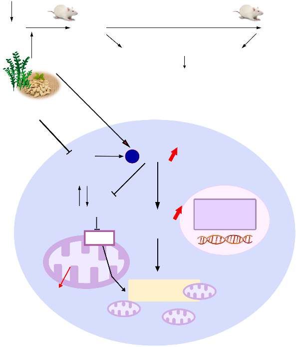
clear so far. The main findings (Fig. 7) of this study are that Danqi Pill could improve lipid metabolism disorder induced
YQHX can regulate lipid metabolism by activating p-AMPK, by myocardial infarction through up-regulating FATP-CPT1
PGC-1α, CPT-1α, and PPARα expression, whereas these ef- lipid transport pathway. Wang Q et al. [9] further reported that
fects were blocked with AMPK inhibitor Compound c. These Lipoprotein lipase (LPL) and key membrane proteins in-
results suggest that YQHX reduces lipid metabolism dis- volved in lipid transport and uptake, such as CD36, FABP4,
orders in myocardial ischemia via the AMPK-dependent sig- and CPT-1α was down-regulated in ischemic heart tissues
– 788 –LI Fang-He, et al. / Chin J Nat Med, 2020, 18(10): 779-792
Myocardial infarction rat model
Day 1 Day 7 Day 28
At two time points
YQHX
Evaluation of cardioprotective effects
(cardiac morphology, structure and function)
Compound c
Hypoxia AMPK p -AMPK
Myocardium
Acetyl-CoA
Nucleus
MCD ACC
Malonyl-CoA Transcriptional
PGC-1α factor
PPARα
CPT1
TCA cycle
Fatty acid
ATP oxidation
Fig. 7 The underlying mechanism of YQHX in lipid metabolism disorders was elucidated in a myocardial infarction rat model
and a hypoxia-induced H9c2 cell injury model. YQHX could regulate multiple lipid metabolism-related molecules, including
PGC-1α, CPT-1, and PPARα, whereas the regulation of PGC-1α, CPT-1α, and PPARα expression were blocked with AMPK in-
hibitor Compound c
compared with the Sham group. Furthermore, PPARα, (Table 6), the plasma TG levels were significantly increased
PPARγ, and PGC-1α relevant transcriptional factors, encod- in the MI group, but the changes of FFA, TC, HDL, and LDL
ing enzymes involved in fatty acid and glucose utilization, in serum were not obvious. Treatment with YQHX and TMZ
were all down-regulated in ischemic model group. Treatment could markedly decrease the TG and TC levels. These data
with Danqi Pill regulated lipid metabolism disorder through indicate that the metabolic mechanism of YQHX and TMZ
PPARs-PGC1α pathway. However, up to now, there are few exists differently at the early stage of myocardial infarction.
reports on whether YQHX treatment can regulate lipid meta- To our knowledge, energy substrate preference of myocardi-
bolism in pure myocardial ischemia at two time points. We al ischemia has a complex pathophysiology. The role of alter-
further aim to explore the metabolic mechanism by which ations in myocardial energetics has been controversial for
YQHX regulates lipid metabolism in myocardial ischemic decades. Here, we also observed the activity of glucose
injury. TMZ, as a well-known metabolic anti-ischemic agent, (GLU) and lactic acid (LA) in serum (Supplementary Table 1).
inhibits fatty acid metabolism and indirectly increases As shown in Supplementary Table 1, there was no signific-
the pyruvate oxidation, hence it is used as a positive control ant change in GLU and LA content in 7 days after MI, but
drug [24, 35]. It is reported that plasma FFA, cholesterol, trigly- GLU and LA content were significantly increased in 28 days
cerides, HDL, and LDL are primary components of plasma after MI. These data indicate that the cardiac metabolic pro-
lipids, which are high risk factors for Coronary Heart Dis- file reverts back to the “fetal phase” at the early stage of car-
ease (CHD) [36]. At the subacute stage (Table 5), our study ob- diac remodeling, which may be a compensatory response.
served that the levels of plasma TC significantly were in- However, glycolysis and lactic acid oxidation exist metabol-
creased, and TG, LDL, and FFA levels had an increased trend ic decompensation at the chronic stage, leading to lactic acid
in pure myocardial ischemia. Treatment with YQHX could accumulation. TMZ treatment merely reduced glucose 28
markedly decrease the FFA and TG levels. However, TMZ days after MI, while failed to reduce LA levels in serum.
group did not show significant change. At the chronic stage However, YQHX treatment significantly reduced the levels
– 789 –LI Fang-He, et al. / Chin J Nat Med, 2020, 18(10): 779-792
of glucose and LA in serum, especially 28 days after MI. reduce free fatty acid esterification and allow fatty acid entry
These results further indicate that YQHX treatment can regu- into mitochondria for fatty acid oxidation [43]. TMZ is con-
late glucose and lipid metabolism at 7 days and 28 days after sidered as a weak CPT-1 inhibitor that prevents fatty acid ox-
MI. However, the metabolisms of YQHX on the cardiac en- idation. Our study showed that YQHX treatment signific-
ergy substrate selection especially lipid metabolism in the antly up-regulated the expression of p-AMPK, PGC-1α, CPT-
ischemic heart remain to be answered. 1α in model rats with myocardial ischemia at the subacute
AMPK is a critical regulator of energy metabolism, stages, whereas at the chronic stages YQHX merely signific-
which plays a critical role in cardiovascular diseases [37]. Un- antly increased p-AMPK and PGC-1α expression, and attenu-
der normal conditions, AMPK catalyzes the substrates of ated the plasma glucose levels such as glucose and lactic
downstream metabolic enzymes such as CPT-1α and PPARα, acid, indicating that YQHX might have a better effect on lip-
and increases catabolism whilst it decreases anabolism, id metabolism at the subacute stages. However, after treat-
thereby relatively increasing myocardial ATP level [37]. Dur- ment with positive drug TMZ, our study showed that TMZ
ing myocardial ischemia, cellular depletion of ATP and the increased the expression of p-AMPK, whereas attenuated the
increased the AMP/ATP ratio induced AMPK phosphoryla- expression of CPT-1α and PPARα, indicating TMZ could
tion at threonine 172. Activated AMPK is involved in mul- protect myocardial metabolism by inhibiting fatty acid entry
tiple cellular processes, including autophagy, the change of into mitochondria for oxidation. Therefore, these data also in-
cardiac fatty acid and glucose metabolism [11, 38], especially dicate that the metabolic way of YQHX and TMZ may exist
importing myocardial cytosolic fatty acid into the mitochon- differently. Furthermore, we also observed that YQHX signi-
dria. Furthermore, activated AMPK accelerates production of ficantly restored mitochondrial ultrastructure and mitochon-
lactate, which forms a vicious cycle, further threatening drial ATP production after the MI different time points.
ischemic myocardium [38]. In the present study, we investig- These above data further indicate that YQHX may have a bet-
ated the effect of YQHX on the expression of AMPK phos- ter effect on lipid metabolism at the subacute stages, whereas
phorylation in model rats with myocardial ischemia (Fig. 3). at the chronic stages YQHX seemed to be more effective in
The results of Western Blot and RT-PCR showed that YQHX
glucose oxidation.
and TMZ further activated AMPK phosphorylation at two
To confirm the mechanism that YQHX regulated lipid
time points in the ischemic myocardium. Although there was
metabolism, Western blotting was analyzed by hypoxia-in-
no statistical difference between YQHX and TMZ group,
duced myocardial injury in H9c2 cells after treatment of
TMZ was a higher AMPK phosphorylation than YQHX at 7
YQHX or/and AMPK inhibitor Compound c. In this study,
days after MI. Interestingly, at 28 days after MI, we found
we found that PGC-1α, CPT-1α, and PPARα mRNA and pro-
that AMPK phosphorylation in myocardium of YQHX was
tein expression were significantly reduced in H9c2 cells dur-
higher than that seen in TMZ. These findings suggest that
ing hypoxia (Fig. 6). Moreover, treatment with YQHX signi-
YQHX and TMZ may have a different way to optimize en-
ficantly increased the expressions of PGC-1α, CPT-1α and
ergy substrate metabolism such as lipid and glucose metabol-
PPARα protein, whereas AMPK inhibitor compound c
ism further improving cardiac function at different stages,
blocked the expressions of these molecules. The data indic-
which were via the activation of AMPK-dependent signaling
ate that YQHX can activate PGC-1α, CPT-1α, and PPARα
pathway. To compare the differences between YQHX and
expression to maintain the balance of lipid metabolism that
TMZ, we study the mechanisms of YQHX on lipid metabol-
may be related to AMPK-dependent signaling pathway.
ism, and further explore the different mechanisms between
YQHX and TMZ. Conclusions
AMPK is involved in multiple signaling pathways. Stud-
ies have demonstrated that AMPK can directly stimulate PGC- The present findings provided evidence that YQHX
1α activity, which further regulates mitochondrial biogenesis could regulate lipid metabolism by activating p-AMPK, PGC-
and electron transport chain via co-activating other transcrip- 1α, CPT-1α, and PPARα expression, whereas the regulation
tion factors, including nuclear respiratory factor 1/2 (NRF- of PGC-1α, CPT-1α, and PPARα expression were blocked
1/2), the synthesis of transcription factor A mitochondrial with AMPK inhibitor compound c. YQHX may be a potent
(Tfam) and estrogen-related receptors (ERRs) [39]. PGC-1α drug for prevention of ischemic heart disease.
also regulates fatty acid oxidation together with PPARα [15, 35]. Supplementary Materials
PPARα, as one of the predominant subtypes of PPARs, is in-
volved in the regulation of fatty acid oxidation (FAO) en- All the supporting information of this paper can be re-
zyme genes such as CPT-1α to reduce lipid accumulation and quested by sending E-mails to the corresponding authors.
maintain the balance of lipid metabolism [30, 40-42]. However, References
the effect of PPARα on ischemia remains controversial. Some
studies showed that PPARα was chronic activated after [1] He H, Li N, Zhao Z, et al. Ischemic postconditioning improves
ischemia, which is harmful to cardiac recovery [31]. Mean- the expression of cellular membrane connexin 43 and attenu-
while, as a key enzyme of fatty acid β-oxidation, CPT-1α can ates the reperfusion injury in rat acute myocardial infarction [J].
– 790 –LI Fang-He, et al. / Chin J Nat Med, 2020, 18(10): 779-792
Biomed Rep, 2015, 3(5): 668-674. [17] Li F, Guo S, Wang C, et al. Yiqihuoxue decoction protects
[2] Pei H, Yang Y, Zhao H, et al. The role of mitochondrial func- against post-myocardial infarction injury via activation of car-
tional proteins in ROS production in ischemic heart diomyocytes PGC-1alpha expression [J]. BMC Complement Al-
diseases [J]. Oxid Med Cell Longev, 2016, 2016: 5470457. tern Med, 2018, 18(1): 253.
[3] Yusuf S, Bosch J, Dagenais G, et al. Cholesterol lowering in [18] Zheng CL, Yang PC, Zhang L, et al. Angiogenesis and expres-
intermediate-risk persons without cardiovascular disease [J]. sion of vascular endothelial growth factor and endostatin pro-
New Engl J Med, 2016, 374(21): 2021-2031. tein in myocardial infarction rat model [J]. Bangladesh J Phar-
[4] Li N, Yuan Y, Li S, et al. PDE5 inhibitors protect against post- macol, 2016, 11: S1-7.
infarction heart failure [J]. Front Biosci (Landmark Ed), 2016, [19] Wu JG, Chen X, Guo SW, et al. Effect of Yiqihuoxue prescrip-
21: 1194-1210. tion on myocardial energy metabolism after myocardial infarc-
[5] Tian Z, Miyata K, Kadomatsu T, et al. ANGPTL2 activity in tion via cross talk of liver kinase B1-dependent Notchl and ad-
cardiac pathologies accelerates heart failure by perturbing car- enosine 5′-monophosphateactivated protein kinase [J]. J Tradit
diac function and energy metabolism [J]. Nat Commun, 2016, Chin Med, 2017, 37: 378-386.
7: 13016. [20] Zhang L, Zheng CL, Jiang M, et al. Effect of Yiqi Huoxue De-
[6] Arumugam S, Sreedhar R, Thandavarayan RA, et al. Targeting coction on the metabolomics of acute myocardial infarction
fatty acid metabolism in heart failure: is it a suitable therapeut- rats [J]. J Tradit Chin Med Sci, 2017, 4(2): 195-206.
ic approach? [J]. Drug Discov Today, 2016, 21(6): 1003-1008. [21] Kanamori H, Takemura G, Goto K, et al. The role of auto-
[7] Fukushima A, Milner K, Gupta A, et al. Myocardial energy phagy emerging in postinfarction cardiac remodeling [J]. Car-
substrate metabolism in heart failure: from pathways to thera- diovasc Res, 2011, 91(2): 330-339.
peutic targets [J]. Curr Pharm Des, 2015, 21(25): 3654-3664. [22] Wang Y, Li C, Ouyang Y, et al. QSYQ attenuates oxidative
[8] Wang Y, Li C, Wang Q, et al. Danqi Pill regulates lipid meta- stress and apoptosis induced heart remodeling rats through dif-
bolism disorder induced by myocardial ischemia through FATP- ferent subtypes of NADPH-oxidase [J]. Evid Based Comple-
CPTI pathway [J]. BMC Complement Altern Med, 2015, 15: 28. ment Alternat Med, 2013, 2013: 824960.
[9] Chang H, Wang Q, Shi T, et al. Effect of DanQi Pill on PPAR- [23] Pang LZ, Ju AC, Zheng XJ, et al. YiQiFuMai Powder Injec-
alpha, lipid disorders and arachidonic acid pathway in rat mod- tion attenuates coronary artery ligation-induced myocardial re-
el of coronary heart disease [J]. BMC Complement Altern Med, modeling and heart failure through modulating MAPKs signal-
2016, 16: 103. ing pathway [J]. J Ethnopharmacol, 2017, 202: 67-77.
[10] Sambandam N, Lopaschuk GD. AMP-activated protein kinase [24] Li X, Liu J, Lin L, et al. Traditional chinese medicine shuang
(AMPK) control of fatty acid and glucose metabolism in the shen ning xin attenuates myocardial ischemia/reperfusion in-
ischemic heart [J]. Prog Lipid Res, 2003, 42(3): 238-256. jury by preserving of mitochondrial function [J]. Evid Based
[11] Schindler M, Pendzialek M, Grybel KJ, et al. Adiponectin Complement Alternat Med, 2014, 2014: 180965.
stimulates lipid metabolism via AMPK in rabbit blastocysts [J]. [25] Li FH, Guo SW, Hu JB, et al. Comparison on pharmacodynam-
Hum Reprod, 2017, 32(7): 1-11. ics effects between different extracts of Yiqihuoxue on H9c2
[12] Liao CC, Ou TT, Huang HP, et al. The inhibition of oleic acid myocardial cells injury induced by hypoxia [J]. J Tradit Chin
induced hepatic lipogenesis and the promotion of lipolysis by Med Pharm, 2018, 24(3): 29-32.
caffeic acid via up-regulation of AMP-activated kinase [J]. J [26] Pei X, Li X, Chen H, et al. Thymoquinone inhibits angiotensin
Sci Food Agric, 2014, 94(6): 1154-1162. II-induced proliferation and migration of vascular smooth
[13] Scarpulla RC. Metabolic control of mitochondrial biogenesis muscle cells through the AMPK/PPARgamma/PGC-1alpha
through the PGC-1 family regulatory network [J]. Biochim Bio- pathway [J]. DNA Cell Biol, 2016, 35(8): 426-433.
phys Acta, 2011, 1813(7): 1269-1278. [27] Zaha VG, Young LH. AMP-activated protein kinase regulation
[14] Kim B, Woo MJ, Park CS, et al. Hovenia dulcis extract re- and biological actions in the heart [J]. Circ Res, 2012, 111(6):
duces lipid accumulation in oleic acid-induced steatosis of Hep 800-814.
G2 cells via activation of AMPK and PPARalpha/CPT-1 path- [28] Bairwa SC, Parajuli N, Dyck JR. The role of AMPK in cardi-
way and in acute hyperlipidemia mouse model [J]. Phytother omyocyte health and survival [J]. Biochim Biophys Acta, 2016,
Res, 2017, 31(1): 132-139. 1862(12): 2199-2210.
[15] Guo SW, Yang PC, Zheng CL, et al. Clinical research on the [29] Rowe GC, Jiang A, Arany Z. PGC-1 coactivators in cardiac de-
treatment of CHD myocardial ischemia-induced syndrome of velopment and disease [J]. Circ Res, 2010, 107(7): 825-838.
Qi-deficiency and blood stasis with TCM Yiqihuoxue herbs [30] Tailleux A, Wouters K, Staels B. Roles of PPARs in NAFLD:
combined with conventional western medicine [J]. Chin J In- potential therapeutic targets [J]. Biochim Biophys Acta, 2012,
form Tradit Chin Med, 2012, 19(9): 15-17. 1821(5): 809-818.
[16] Li F, Guo S, Wang H, et al. Yiqi Huoxue Decoction attenuates [31] Sambandam N, Morabito D, Wagg C, et al. Chronic activation
ischemia/hypoxia-induced oxidative stress injury in H9c2 car- of PPAR alpha is detrimental to cardiac recovery after
diomyocytes [J]. J Tradit Chin Med Sci, 2018, 5(3): 271-282. ischemia [J]. Am J Physiol-Heart C, 2006, 290(1): H87-H95.
– 791 –LI Fang-He, et al. / Chin J Nat Med, 2020, 18(10): 779-792
[32] Murphy E, Ardehali H, Balaban RS, et al. Mitochondrial func- kinase regulation of fatty acid oxidation in the ischaemic
tion, biology, and role in disease: a scientific statement from heart [J]. Biochem Soc Trans, 2003, 31(1): 207-212.
the American heart association [J]. Circ Res, 2016, 118(12): [39] Jager S, Handschin C, St-Pierre J, et al. AMP-activated protein
1960-1991. kinase (AMPK) action in skeletal muscle via direct phos-
[33] Gao K, Zhang J, Gao P, et al. Qishen granules exerts cardiopro- phorylation of PGC-1alpha [J]. Proc Natl Acad Sci USA, 2007,
tective effects on rats with heart failure via regulating fatty acid 104(29): 12017-12022.
and glucose metabolism [J]. Chin Med, 2020, 15: 21. [40] Schmidt K, Hughes C, Chudek JA, et al. Cholesterol metabol-
[34] Stanley WC, Recchia FA, Lopaschuk GD. Myocardial sub-
ism: the main pathway acting downstream of cytochrome P450
strate metabolism in the normal and failing heart [J]. Physiol
oxidoreductase in skeletal development of the limb [J]. Mol
Rev, 2005, 85(3): 1093-1129.
Cell Biol, 2009, 29(10): 2716-2729.
[35] Lee SO, Simons AL, Murphy PA, et al. Soyasaponins lowered
[41] Zhou G, Myers R, Li Y, et al. Role of AMP-activated protein
plasma cholesterol and increased fecal bile acids in female
kinase in mechanism of metformin action [J]. J Clin Invest,
golden Syrian hamsters [J]. Exp Biol Med (Maywood), 2005,
2001, 108(8): 1167-1174.
230(7): 472-478.
[42] Lee MS, Kim D, Jo K, et al. Nordihydroguaiaretic acid pro-
[36] Rana JS, Visser ME, Arsenault BJ, et al. Metabolic dyslip-
idemia and risk of future coronary heart disease in apparently tects against high-fat diet-induced fatty liver by activating AMP-
healthy men and women: the EPIC-Norfolk prospective popu- activated protein kinase in obese mice [J]. Biochem Biophys
lation study [J]. Int J Cardiol, 2010, 143(3): 399-404. Res Commun, 2010, 401(1): 92-97.
[37] Qi D, Young LH. AMPK: energy sensor and survival mechan- [43] Yang YL, Li J, Liu K, et al. Ginsenoside Rg5 increases cardi-
ism in the ischemic heart [J]. Trends Endocrinol Metab, 2015, omyocyte resistance to ischemic injury through regulation of
26(8): 422-429. mitochondrial hexokinase-II and dynamin-related protein 1 [J].
[38] Hopkins TA, Dyck JR, Lopaschuk GD. AMP-activated protein Cell Death Dis, 2017, 8(2): e2625.
Cite this article as: LI Fang-He, HUANG Xiao-Lou, WANG Hui, GUO Shu-Wen, LI Ping. Protective effect of Yi-Qi-Huo-
Xue Decoction against ischemic heart disease by regulating cardiac lipid metabolism [J]. Chin J Nat Med, 2020, 18(10): 779-
792.
– 792 –You can also read