Neonatal Extracellular Superoxide Dismutase Knockout Mice Increase Total Superoxide Dismutase Activity and VEGF Expression after Chronic Hyperoxia
←
→
Page content transcription
If your browser does not render page correctly, please read the page content below
antioxidants

Article
Neonatal Extracellular Superoxide Dismutase Knockout Mice
Increase Total Superoxide Dismutase Activity and VEGF
Expression after Chronic Hyperoxia
Maxwell Mathias 1,2, * , Joann Taylor 1 , Elizabeth Mendralla 1 and Marta Perez 1,2

                                          1   Division of Neonatology, Department of Pediatrics, Northwestern University Feinberg School of Medicine,
                                              Chicago, IL 60611, USA; j-hinz@northwestern.edu (J.T.); emendral@student.touro.edu (E.M.);
                                              mtperez@luriechildrens.org (M.P.)
                                          2   Ann & Robert H. Lurie Children’s Hospital of Chicago, Chicago, IL 60611, USA
                                          *   Correspondence: maxwell-mathias@ouhsc.edu

                                          Abstract: Bronchopulmonary dysplasia (BPD) is a common lung disease affecting premature infants
                                          that develops after exposure to supplemental oxygen and reactive oxygen intermediates. Extracellular
                                          superoxide dismutase (SOD3) is an enzyme that processes superoxide radicals and has been shown
                                          to facilitate vascular endothelial growth factor (VEGF) and nitric oxide (NO) signaling in vascular
                                          endothelium. We utilized a mouse model of neonatal hyperoxic lung injury and SOD3 knockout
                                          (KO) mice to evaluate its function during chronic hyperoxia exposure. Wild-type age-matched
                                          neonatal C57Bl/6 (WT) and SOD3−/− (KO) mice were placed in normoxia (21% FiO2 , RA) or chronic
                                hyperoxia (75% FiO2 , O2 ) within 24 h of birth for 14 days continuously and then euthanized. Lungs

                                          were harvested for histologic evaluation, as well as comparison of antioxidant enzyme expression,
Citation: Mathias, M.; Taylor, J.;
                                          SOD activity, VEGF expression, and portions of the NO signaling pathway. Surprisingly, KO-O2 mice
Mendralla, E.; Perez, M. Neonatal
                                          survived without additional alveolar simplification, microvascular remodeling, or nuclear oxidation
Extracellular Superoxide Dismutase
                                          when compared to WT-O2 mice. KO-O2 mice had increased total SOD activity and increased VEGF
Knockout Mice Increase Total
                                          expression when compared to WT-O2 mice. No genotype differences were noted in intracellular
Superoxide Dismutase Activity and
VEGF Expression after Chronic
                                          antioxidant enzyme expression or the NO signaling pathway. These results demonstrate that SOD3
Hyperoxia. Antioxidants 2021, 10,         KO mice can survive prolonged hyperoxia without exacerbation of alveolar or vascular phenotype.
1236. https://doi.org/10.3390/
antiox10081236                            Keywords: extracellular superoxide dismutase; bronchopulmonary dysplasia; vascular endothelial
                                          growth factor; nitric oxide signaling
Academic Editor: Antonella Casola

Received: 2 July 2021
Accepted: 29 July 2021                    1. Introduction
Published: 1 August 2021
                                                Bronchopulmonary dysplasia (BPD) is a common lung disease of prematurity, affecting
                                          more than 10,000 infants in the United States annually and up to 60% of infants born under
Publisher’s Note: MDPI stays neutral
                                          28 weeks [1,2]. BPD is a consequence of lung immaturity combined with the side-effects
with regard to jurisdictional claims in
                                          of life-sustaining therapeutic interventions, including supplemental oxygen (hyperoxia),
published maps and institutional affil-
                                          which result in injury to the developing lung [3]. Hyperoxia is hypothesized to induce
iations.
                                          injury through the production of excess reactive oxygen intermediates (ROIs) [4]. Preterm
                                          infants are particularly susceptible to injury from ROI because of the immaturity of enzy-
                                          matic antioxidant systems and relative deficiency of nonenzymatic antioxidants relative to
                                          term infants [5–9]. Because of the hypothesized causal pathway of hyperoxia–ROI–lung in-
Copyright: © 2021 by the authors.
                                          jury, there have been multiple clinical trials of administration of either antioxidant enzymes
Licensee MDPI, Basel, Switzerland.
                                          or nonenzymatic antioxidants; however, to date, none have demonstrated superiority over
This article is an open access article
                                          standard care or have received approval from the FDA [10–14]. Since these trials were
distributed under the terms and
                                          conducted, our knowledge of cell-specific and compartment-specific redox signaling has
conditions of the Creative Commons
                                          vastly expanded, and it is clear that therapeutic intervention for neonatal hyperoxic lung
Attribution (CC BY) license (https://
creativecommons.org/licenses/by/
                                          injury (HLI) will require a more granular understanding of antioxidant system function
4.0/).
                                          during hyperoxia exposure [15].

Antioxidants 2021, 10, 1236. https://doi.org/10.3390/antiox10081236                                    https://www.mdpi.com/journal/antioxidants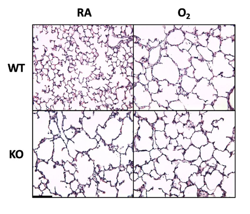
Antioxidants 2021, 10, 1236                                                                                         2 of 13

                                    Extracellular superoxide dismutase (SOD3) is an antioxidant enzyme found in high
                              concentration in the lungs, and it is 95% tissue-bound at the interface between pulmonary
                              vascular smooth muscle and pulmonary vascular endothelium [16,17]. SOD3 converts
                              superoxide radical to hydrogen peroxide (H2 O2 ), which can cross lipid bilayers and be
                              converted to water by numerous intracellular antioxidant enzymes or can modify cysteine
                              thiols on proteins for redox-dependent signaling [18]. SOD3 plays an important role in the
                              development of the newborn lung. Its expression increases fivefold between weeks 1 and 3
                              of life and sevenfold between weeks 1 and 8 [5]. SOD3 knockout (KO) mice have simplified
                              alveoli, pulmonary vascular remodeling, and increased susceptibility to bleomycin-induced
                              lung injury at 3 and 4 weeks of life in comparison to controls [19–21]. In contrast, SOD3
                              overexpression protects against HLI in newborn animals, but does not appear to have any
                              effect on alveolar or pulmonary vascular development during normoxia (21% FiO2 ) [22–24].
                              The specific function of SOD3 in neonatal lung development and how this function is altered
                              by hyperoxia exposure remains unknown.
                                    One hypothesized function of SOD3 is that it protects nitric oxide (NO) signaling,
                              whose function is critical to lung and pulmonary vascular development [24–28]. In the
                              pulmonary vascular endothelium, NO is produced by the endothelial nitric oxide synthase
                              (eNOS), diffuses into vascular smooth muscle, and binds soluble guanylate cyclase (sGC).
                              Activated sGC catalyzes the conversion of GTP to cGMP, an important second messenger
                              in vascular smooth muscle function and lung growth. Each step of this pathway has been
                              shown to play an important role in early lung development, and sGC depletion has been
                              shown to increase susceptibility to HLI [29–31]. SOD3 may protect NO bioavailability
                              by reducing the extracellular concentration of superoxide, which can attack NO to form
                              peroxynitrite (ONOO− ), itself a highly reactive and toxic intermediate [25,32,33]. In addi-
                              tion to direct effects on NO bioavailability, SOD3 can enhance NO signaling by increasing
                              NO production in the vascular endothelium through its effect on vascular endothelial
                              growth factor (VEGF) signaling. SOD3-derived H2 O2 has been shown to potentiate the
                              effect of VEGF on VEGF receptor 2 (VEGFR-2) phosphorylation [34]. VEGFR-2 phospho-
                              rylation induces the expression and activation of the endothelial nitric oxide synthase
                              (eNOS), providing a link between the effects of SOD3 on VEGF and the NO signaling
                              pathway [35,36].
                                    In the present study, we evaluated the combined effects of SOD3 KO and chronic
                              hyperoxia (O2 ) on lung and pulmonary vascular development, and we tested the hypothesis
                              that SOD3 protects lung development through VEGF-mediated effects on NO signaling.
                              To our knowledge, this is the first study to investigate the effects of SOD3 KO on neonatal
                              adaptations to chronic hyperoxia.

                              2. Materials and Methods
                              2.1. Animal Model
                                    This study was approved by the Northwestern University Institutional Animal Care
                              and Use Committee. SOD3−/− mice (The Jackson Laboratory, Bar Harbor, ME, USA) were
                              bred on a background of C57Bl/6N mice (Charles River, Wilmington, MA, USA) and
                              back-crossed through multiple generations. Heterozygotes were then crossed to produce
                              SOD3−/− (KO) and SOD3+/+ (WT) offspring. Cages were checked daily for litters. New
                              litters were placed in either room air (RA, 21% FiO2 ) or a plexiglass chamber (Biospherix,
                              Lacona, NY, USA) at 75% FiO2 (Hyperoxia, or O2 ) from birth to P14 continuously. Dams
                              were rotated daily to prevent oxygen toxicity. Mice were euthanized at 14 days.

                              2.2. Tissue Collection and Processing
                                   For histology, morphometry, and immunofluorescence, the chest was exposed, and
                              lungs were removed en bloc and inflated to 25 cm H2 O with 10% formalin solution, fixed
                              for 24 h, and paraffin-embedded. Blocks were cut into 5 µm thick sections and stained
                              with hematoxylin and eosin (H&E) for basic morphometry by the Northwestern University
                              Mouse Histology and Phenotyping Laboratory and the Stanley Manne Children’s Research
Antioxidants 2021, 10, 1236                                                                                            3 of 13

                              Institute Histology Laboratory. For Western blotting, enzyme activity assays, and immuno-
                              precipitation, the RV was punctured, and the pulmonary artery was flushed with 3 mL of
                              PBS to remove blood; then, the lung tissue was removed and snap-frozen in liquid nitrogen.
                              Samples were stored at −80 ◦ C until they were analyzed.

                              2.3. Lung Histology
                                   H&E slides were blinded to the researcher. For alveolar area, 4–8 nonoverlapping
                              images of the lungs were taken at 20× magnification using an Olympus BX40 microscope
                              (Olympus America, Melville, NY, USA) with a Moticam Pro 252A microscope camera
                              (Motic, Kowloon, Hong Kong). Alveolar area was calculated using ImagePro 9.2 (Media
                              Cybernetics, Rockland, MD, USA). Obvious airways and blood vessels were excluded,
                              as were alveoli without complete borders. For pulmonary artery smooth muscle wall
                              thickness index (MWT), 4–8 distinct resistance pulmonary arteries adjacent to airways
                              measuring less than 100 µm diameter were photographed per slide at 40× magnification.
                              Inner and outer borders were traced in triplicate using ImageJ Software (ImageJ, NIH,
                              Bethesda, MD, USA) to calculate circumference, and the ratio of smooth muscle to total
                              area was compared between groups as described previously [37,38].

                              2.4. Immunofluorescence
                                    Unstained 5 µm thick sections were obtained from paraffin-embedded lung samples.
                              For von Willebrand Factor (vWF) immunofluorescence, slides were heated to 60 ◦ C for
                              10 min, and then cleaned and deparrafinized by serial dilutions of ethanol. Samples were
                              then incubated in proteinase K solution (Sigma-Aldrich, St. Louis, MO, USA) for antigen
                              retrieval at 37 ◦ C for 15 min. Samples were blocked with 5% bovine serum albumin
                              (BSA; Sigma-Aldrich) in Tris-buffered saline containing 0.1% Tween-20 (1× TBST) at room
                              temperature for 30 min, and then probed with von Willebrand Factor antibody (Agilent,
                              Santa Clara, CA, USA; 1:50 in 5% BSA) overnight in a dark chamber. Slides were then
                              washed in PBS twice and probed with Rhodamine Red conjugated goat anti-rabbit antibody
                              (ThermoFisher, Waltham, MA, USA; 1:100 in 5% BSA) for 1 h in a dark chamber. Slides
                              were again washed twice in PBS, blotted dry, and mounted prior to storage at 4 ◦ C. Slides
                              were imaged at 10× magnification on a NIKON Eclipse TE300 fluorescent microscope and
                              captured using a Photometrics SenSys KAF0400 G-2 camera. Eight nonoverlapping images
                              per slide were obtained for vessel counts. Incompletely stained vessels or vessels with
                              diameter >100 µm were excluded.
                                    8-Hydroxydeoxyguanosine (8-OHdG) is a product of DNA oxidation. To measure
                              intracellular oxidative stress, 8-OHdG was quantified using immunofluorescence. Samples
                              were deparaffinized on an automated platform (Leica Autostainer XL) and incubated in
                              Sodium Citrate solution (pH 6) at 110 ◦ C for 10 min in a pressure cooker for antigen retrieval.
                              Slides were then incubated in 8-OHdG antibody (Sigma-Aldrich, 1:100) overnight at 4 ◦ C,
                              washed, incubated in species-specific secondary antibody (Jackson ImmunoResearch,
                              West Grove, PA, USA; 1:500), and counterstained with DAPI. Slides were imaged at 40×
                              magnification on a KEYENCE BZ-X fluorescent microscope (KEYENCE America, Itasca,
                              IL, USA). A total of 4–8 nonoverlapping regions were selected, and duplicate images
                              were obtained on blue (DAPI) and green (8-OHdG) channels. Images were converted to
                              grayscale and analyzed using ImageJ. Nuclear borders were delineated by applying the
                              same grayscale intensity threshold to all the DAPI images. These borders were overlaid on
                              the 8-OHdG images, and the average 8-OHdG intensity of nuclear regions was calculated
                              to filter out non-nuclear background fluorescence.

                              2.5. Western Blotting
                                   Frozen lung tissue was crushed, suspended in protein lysis buffer (EMD Millipore Mg2+ )
                              with phosphatase inhibitor and protease inhibitor (EMD Millipore and Sigma-Aldrich), and
                              sonicated. Protein concentration was calculated using the Bradford method [39]. A total of
                              40 µg of protein lysate per sample was run on 4–20% gel (ThermoFischer) and transferred to
Antioxidants 2021, 10, 1236                                                                                            4 of 13

                              a nitrocellulose membrane (GE Life Sciences Amersham). Membranes were blocked with 5%
                              BSA in TBST and incubated overnight with primary antibodies at 4 ◦ C on a rocker. Membranes
                              were washed and incubated with horseradish peroxidase (HRP)-conjugated, species-specific
                              secondary antibodies, and again washed three times prior to imaging. SuperSignal West
                              Femto Maximum Sensitivity Substrate (ThermoFischer Scientific, Rockford, IL, USA) was
                              added, and chemiluminescence was detected using a molecular imager (BioRad, Hercules,
                              CA, USA). Bands were analyzed using Image Lab (BioRad, Hercules, CA, USA), normalized
                              to β-actin, and shown as fold difference relative to controls.
                                    Primary antibodies were diluted in 5% BSA unless otherwise specified. Primary
                              antibodies and concentrations used were as follows: SOD3 goat polyclonal antibody (pAb)
                              at 1:1000 (R + D), cytosolic superoxide dismutase (SOD1) rabbit pAb at 1:1000 (Enzo
                              P07632), mitochondrial superoxide dismutase (SOD2) rabbit pAb at 1:750 (Enzo P04179),
                              glutathione peroxidase 1 (GPX-1) rabbit pAb at 1:1000 (Abnova 5038), catalase rabbit pAb at
                              1:2000 (Abcam ab52477), VEGF-A mouse monoclonal antibody (mAb) at 1:500 (Santa Cruz),
                              sGC-α rabbit pAb at 1:2000 (Abcam), sGC-β rabbit pAb at 1:1000 (Cayman Chemical),
                              endothelial nitric oxide synthase (eNOS) mouse mAb at 1:500 (Cayman Chemical), and
                              β-actin rabbit mAb at 1:5000 (Cell Signaling). HRP-conjugated secondary antibodies and
                              concentrations were as follows: goat anti-rabbit at 1:2000 (Cell Signaling), horse anti-mouse
                              at 1:2000 (Cell Signaling), and donkey anti-goat at 1:2000 (Santa Cruz).

                              2.6. SOD Activity Assay
                                   Protein lysates were obtained as described above. SOD enzyme activity was measured
                              as described previously using a commercially available SOD Activity Kit (Enzo) [40]. Briefly,
                              whole lung tissue lysates were passed through 0.45 µm filter, and protein concentration was
                              measured. A total of 150 µg of lung protein was diluted to 75 µL (2 µg/µL) and incubated
                              in SOD buffer, xanthine oxidase, and Water-Soluble Tetrazolium Salt 1 (WST-1) as per
                              the manufacturer’s instructions. Xanthine was added to initiate superoxide formation,
                              which was measured as the rate of reduction of WST-1 to WST-1 formazan, which absorbs
                              light at 450 nm. Absorbance readings were taken at 1 min intervals until WST-1 formazan
                              formation ceased. SOD activity was calculated as rate of inhibition of WST-1 formazan
                              formation. These were plotted against standard SOD enzyme concentrations (0.1 U/25 µL
                              to 10 U/25 µL) on a logarithmic scale to estimate total SOD activity.

                              2.7. sGC Activity Assay
                                   Protein lysates were obtained as described above. The cGMP ELISA kit was pur-
                              chased from Cayman Chemical. A total of 30 µg of protein was diluted to 100 µL in
                              incubation buffer consisting of 50 mM Tris-HCl (pH 7.5, Fisher Scientific), 4 mM MgCl2
                              (Fisher Scientific), 0.5 mM 3-isobutyl-1-methylxanthine (Enzo), 7.5 mM creatine phosphate
                              (Sigma-Aldrich), 0.2 mg/mL creatine phosphokinase (Sigma-Aldrich), 10 mM sodium
                              nitroprusside (Sigma-Aldrich), and 1 mM GTP (Sigma-Aldrich) for 10 min at 37 ◦ C. The
                              reaction was terminated with 900 µL of ice-cold 0.1 N HCl (Sigma-Aldrich) and then placed
                              in a 90 ◦ C water bath for 3 min to denature the protein. Samples were centrifuged for
                              15 min at 2000× g to precipitate protein. The supernatant was collected, dried in a speed
                              vacuum, and resuspended in cGMP EIA buffer (Cayman Chemical, Ann Arbor, MI, USA).
                              Samples were diluted 1:2 in EIA buffer for optimal calculation. Absorbance was detected
                              at 405 nm using an iMark automated plate reader (BioRad, Hercules, CA, USA).

                              2.8. Statistics
                                   Outliers were excluded using Grubb’s test prior to analysis. Data are presented as
                              means ± SEM, and experimental groups were compared using Prism 8 software (GraphPad,
                              San Diego, CA, USA) by two-way ANOVA with multiple comparisons, unless otherwise
                              specified, to assess genotype and hyperoxia exposure effects and their interaction. Statistical
                              significance was set at p < 0.05.
Outliers were excluded using Grubb’s test prior to analysis. Data are presented as
means ± SEM, and experimental groups were compared using Prism 8 software
(GraphPad, San Diego, CA, USA) by two-way ANOVA with multiple comparisons, unless
otherwise specified, to assess genotype and hyperoxia exposure effects and their interac-
Antioxidants 2021, 10, 1236 tion. Statistical significance was set at p < 0.05. 5 of 13
3. Results
3.3.1. Combined SOD3 KO and Hyperoxia Exposure Does Not Cause More Alveolar
Results
Simplification
3.1. ThanKO
Combined SOD3 SOD3 KO or Hyperoxia
and Hyperoxia Alone
Exposure Does Not Cause More Alveolar Simplification
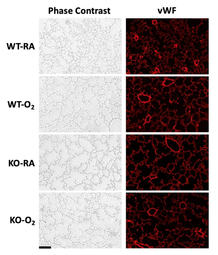
Than SOD3 KO or Hyperoxia Alone
Mean alveolar area for the control group (WT-RA) was 1055 ± 41 μm2. SOD3 KO and
Mean alveolar area for the controlinduced
group (WT-RA) 1055 ± 41inµm
was increase . SOD3 2KOwhich
hyperoxia exposure independently a significant alveolar area,
and hyperoxia exposure independently induced a significant increase in alveolar
was 1863 ± 242 μm for the KO-RA group and 2932 ± 270 μm for the WT-O2 group (Figure
2 2 area,
which was 1863 ± 242 µmof2 for the KO-RA group and 2932 ± 270 µm2 for the WT-O group
1A). The combination SOD3 KO and hyperoxia exposure (KO-O2 group) did 2 not induce
(Figure 1A). The combination of SOD3 KO and hyperoxia exposure (KO-O
further alveolar simplification (2364 ± 134 μm2). Representative images 2 group) did notin Fig-
are shown
induce further alveolar simplification (2364 ± 134 µm2 ). Representative images are shown
ure 1B.
in Figure 1B.
A B
Figure 1. Hyperoxia exposure and SOD3 KO alone induced alveolar simplification, but there was no additive effect of
genotype and exposure. (A) WT and KO mice were exposed to 14 days of either 75% O2 or RA prior to analysis. Lungs
Figure
were 1. Hyperoxia
sectioned, exposure
H&E-stained, andand SOD3 at
analyzed KO20alone induced alveolar
× magnification. simplification,
Alveolar but therehigher
area was significantly was no in Oadditive effect of
2 -exposed
genotype
mice in theand
WTexposure.
genotype (A)
but WT andKO
not the KOgenotype.
mice wereData
exposed to 14 days
are presented as of
± either
means75%
SEM;O2**orp RA prior
< 0.01, ****topanalysis.
< 0.0001;Lungs
nwere sectioned,
= 8–15 H&E-stained,
per group. and analyzed
(B) Representative at 20×
(20×) images of magnification.
H&E-stained P14Alveolar area Scale
mouse lung. was significantly
bar is 100 µm.higher in O2-exposed
mice in the WT genotype but not the KO genotype. Data are presented as means േ SEM; ** p < 0.01, **** p < 0.0001; n = 8–
15 per group. (B) Representative (20×) images of H&E-stained P14 mouse lung. Scale bar is 100 μm.
3.2. Combined SOD3 KO and Hyperoxia Exposure Does Not Cause More Microvascular
Remodeling Than SOD3 KO or Hyperoxia Alone
3.2.Pulmonary
Combined SOD3 KO and Hyperoxia Exposure Does Not Cause More Microvascular
vascular MWT for WT-RA was 0.31 ± 0.006. SOD3 KO and hyperoxia
Remodeling
exposure Than SOD3induced
independently KO or Hyperoxia Alone
a significant increase in MWT, which was 0.43 ± 0.03 for
Pulmonary
the KO-RA vascular
group and 0.42 ±MWT for the
0.03 for WT-RA
WT-Owas 0.31 (Figure
2 group ± 0.006.2A).
SOD3TheKO and hyperoxia
combination of ex-
genotype and hyperoxia exposure
posure independently induced did not induceincrease
a significant a furtherinincrease
MWT,in MWTwas
which ± 0.03).
(0.390.43 ± 0.03 for
Representative
the KO-RA groupimages
andare0.42
shown infor
± 0.03 Figure 2B. Mean
the WT-O vessel
2 group density
(Figure as The
2A). quantified by
combination of
vWF-stained vessels per 10 × field was 6.5 ± 0.6 in WT-RA. KO-RA and WT-O2 both
genotype and hyperoxia exposure did not induce a further increase in MWT (0.39 ± 0.03). had
decreased vessel density
Representative images are ± 0.6 and
(4.1shown 3.5 ± 2B.
in Figure 0.4, Mean
respectively; Figure 2C)
vessel density comparedby
as quantified to vWF-
WT-RA. KO-O 2 mice showed no additional reduction in vascular density
stained vessels per 10× field was 6.5 ± 0.6 in WT-RA. KO-RA and WT-O2 both had de- (Figure 2C).
Representative phase-contrast and fluorescent images are shown in Figure 2D.
creased vessel density (4.1 ± 0.6 and 3.5 ± 0.4, respectively; Figure 2C) compared to WT-
3.3. DNA Oxidation Was Not Affected by Genotype
There was an overall increase in 8-OHdG immunofluorescent intensity in hyperoxia-
exposed mice; however, post hoc Tukey’s test for multiple comparisons did not reach
statistical significance (Figure 3A). There was no effect of genotype on DNA oxidation.
Representative phase-contrast, fluorescent, and overlaid images are shown in Figure 3B.Antioxidants 2021, 10, x FOR PEER REVIEW 6 of 14
Antioxidants 2021, 10, 1236 6 of 13
RA. KO-O2 mice showed no additional reduction in vascular density (Figure 2C). Repre-
sentative phase-contrast and fluorescent images are shown in Figure 2D.
A B
C D
Figure 2. Hyperoxia exposure and SOD3 KO alone induced microvascular remodeling, but there was no additive effect of
Figure 2. Hyperoxia exposure and SOD3 KO alone induced microvascular remodeling, but there was no additive effect of
genotype and exposure. (A) Lung sections were analyzed at 40× magnification. Cross-sections of peri-bronchiolar arterioles
genotype and exposure. (A) Lung sections were analyzed at 40× magnification. Cross-sections of peri-bronchiolar arteri-
were identified for analysis. Medial wall thickness was significantly higher in O2 -exposed mice in the WT genotype but not
oles were identified for analysis. Medial wall thickness was significantly higher in O2-exposed mice in the WT genotype
the KO genotype; n = 5–7. (B) Representative high-power (40×) images of H&E-stained cross-sections of peribronchiolar
but not the KO genotype; n = 5–7. (B) Representative high-power (40×) images of H&E-stained cross-sections of peribron-
pulmonary arterioles.arterioles.
chiolar pulmonary Arrowheads indicate theindicate
Arrowheads medial the
layer. Scale layer.
medial bar is 20 (C)isLung
µm.bar
Scale sections
20 μm. were stained
(C) Lung sectionswith
werevWFstained
antibody to highlight the vascular endothelium. Vascular density was analyzed by counting resistance
with vWF antibody to highlight the vascular endothelium. Vascular density was analyzed by counting resistance pulmo- pulmonary blood
vessels (3.3. DNA Oxidation Was Not Affected by Genotype
There was an overall increase in 8-OHdG immunofluorescent intensity in hyperoxia-
exposed mice; however, post hoc Tukey’s test for multiple comparisons did not reach sta-
Antioxidants 2021, 10, 1236 tistical significance (Figure 3A). There was no effect of genotype on DNA oxidation.7 of 13
Rep-
resentative phase-contrast, fluorescent, and overlaid images are shown in Figure 3B.
A B
Figure 3. SOD3 KO did not affect DNA oxidation. (A) Lung sections were stained for 8-OHdG and counterstained with
DAPI. Nuclei were identified on the basis of DAPI fluorescent intensity, and these regions were analyzed for 8-OHdG
Figure 3. SOD3
fluorescent KOtodid
intensity not analyzing
avoid affect DNA oxidation.autofluorescence.
background (A) Lung sectionsThere
werewas
stained for 8-OHdG
an overall increaseand counterstained
in 8-OHdG with
fluorescent
DAPI. Nuclei
intensity were identified mice
in hyperoxia-exposed on the
bybasis of DAPI
two-way ANOVAfluorescent intensity,
(p < 0.05); and
however, these
post hocregions were
individual analyzed
group for 8-OHdG
comparisons did
fluorescent
not intensity
reach statistical to avoid analyzing
significance. backgroundimages
(B) Representative autofluorescence. There was
of phase-contrast, an overall
8-OHdG increase in 8-OHdG
immunofluorescent fluores-
(green), and
cent intensity in hyperoxia-exposed mice by two-way ANOVA (p < 0.05); however, post hoc individual group comparisons
DAPI-stained (blue) lungs are presented. An overlay of the 8-OHdG and DAPI images is shown in the far-right column.
did not reach statistical significance. (B) Representative images of phase-contrast, 8-OHdG immunofluorescent (green),
and DAPI-stained (blue) lungs are presented. An overlay of the 8-OHdG and DAPI images is shown in the far-right col-
umn. 3.4. SOD3 Protein Was Absent in KO Mice, but Genotype Did Not Affect Other Antioxidant
Protein Expression
There Protein
3.4. SOD3 was noWas SOD3 protein
Absent in KOdetected inGenotype
Mice, but the KO Did
mice,
Notalong
Affectwith noAntioxidant
Other effect of O2
exposure on SOD3 expression. In addition to SOD3, SOD1, SOD2, GPX-1, and catalase
Protein Expression
expression was assayed by Western blot (Figure 4A). Hyperoxia exposure increased SOD2
There was no SOD3 protein detected in the KO mice, along with no effect of O2 ex-
expression in both WT and KO mice (1.0 ± 0.1-fold vs. 2.6 ± 0.2-fold in WT; 0.9 ± 0.1-fold
posure on SOD3 expression. In addition to SOD3, SOD1, SOD2, GPX-1, and catalase ex-
vs. 1.9 ± 0.3-fold in KO), as described previously [40]. Hyperoxia exposure also decreased
pression was
Antioxidants 2021, 10, x catalase
FOR PEER
assayed by Western blot (Figure 4A). Hyperoxia exposure increased SOD2
REVIEW in WT mice (1.0 ± 0.03-fold vs. 0.72 ± 0.06-fold). However, there
expression
expression in both WT and KO mice (1.0 ± 0.1-fold vs. 2.6 ± 0.2-fold in WT; 0.9 ± 0.1-fold
was no effect of genotype on measured antioxidant enzyme expression between groups.
vs. 1.9 ± 0.3-fold in KO), as described previously [40]. Hyperoxia exposure also decreased
Representative blots are shown in Figure 4B.
catalase expression in WT mice (1.0 ± 0.03-fold vs. 0.72 ± 0.06-fold). However, there was
no effect of genotype on measured antioxidant enzyme expression between groups. Rep-
A resentative blots are shown in Figure 4B.
Figure 4. Cont.
BAntioxidants 2021, 10, 1236 8 of 13
B
Figure 4. SOD3 protein was absent in KO mice, but other antioxidant enzyme expression was not affected by genotype.
(A) Western blotFigure 4. was
analysis SOD3 protein
used was absent
to evaluate lung in KO mice,
protein but other
expression antioxidant
of SOD3, SOD1,enzyme expression
SOD2, GPX-1, and was not in
catalase affected
WT by genotype
(A) Western blot analysis was used to evaluate lung protein expression of SOD3, SOD1, SOD2,
and KO mice exposed to either RA or O2 for 14 days. Protein expression was normalized to β-actin. No SOD3 protein GPX-1, and catalase in W
and KO mice exposed to either RA or O2 for 14 days. Protein expression was normalized to β-actin. No SOD3 protein wa
was detected in KO mice, and O2 exposure did not affect SOD3 expression in WT mice. O2 exposure was associated with
detected in KO mice, and O2 exposure did not affect SOD3 expression in WT mice. O2 exposure was associated with in
increased SOD2 expression in both genotypes (as previously described [1]) and decreased catalase expression in WT mice.
creased SOD2 expression in both genotypes (as previously described [1]) and decreased catalase expression in WT mice
Data are presented
Dataasare
means ± SEM;
presented pAntioxidantsFigure 5. 1236
2021, 10, Total SOD activity was increased in KO-O2 mice. SOD activity was measured as the rate of elimination of super-9 of 13
oxide compared to standard concentrations of SOD enzyme. Whole-lung SOD enzyme activity was increased only in KO-
O2 mice. Data are presented as means േ SEM; * p < 0.05 vs. WT O2; n = 6–9 per group.
3.6.3.6. KO-O2
KO-O2 Mice
Mice Increased
Increased VEGF
VEGF andand DecreasedeNOS
Decreased eNOSininResponse
ResponsetotoHyperoxia
Hyperoxia
KO-OKO-O 2 mice had increased VEGF expression when compared to WT-O2 mice (2.7 ±
2 mice had increased VEGF expression when compared to WT-O2 mice
(2.7 ± 0.4-fold1.5
0.4-fold vs. vs.±1.5
0.1-fold; FigureFigure
± 0.1-fold; 6A). In6A).
order
Into evaluate
order downstream
to evaluate effects on
downstream the NO-
effects on
the NO-signaling pathway, we assessed eNOS and sGC protein subunit expressionfunc-
signaling pathway, we assessed eNOS and sGC protein subunit expression and sGC and
sGCtion (Figure(Figure
function 6B–D). eNOS
6B–D).expression was decreased
eNOS expression in O2-exposed
was decreased in OKO mice but not WT
2 -exposed KO mice
mice. sGC-α and -β expression were both significantly reduced
but not WT mice. sGC-α and -β expression were both significantly reduced in the O 2 groups (sGC-α
in the O2
not shown),
groups (sGC-αbut notthere was but
shown), no genotype
there was effect. Representative
no genotype effect.blots for VEGF, eNOS,
Representative blotsand
for
sGC-β are shown. Despite protein expression differences, there were no
VEGF, eNOS, and sGC-β are shown. Despite protein expression differences, there were significant group
differences in
no significant measured
group sGC activity
differences (Figure sGC
in measured 6D). activity (Figure 6D).
A B
Antioxidants 2021, 10, x FOR PEER REVIEW 10 of 14
C D
Figure 6. VEGF was increased and eNOS was decreased in combined SOD3 KO and hyperoxia. Western blot analysis was
Figure 6. VEGF was increased and eNOS was decreased in combined SOD3 KO and hyperoxia. Western blot analysis was
used to evaluate lung protein expression of VEGF-A, eNOS, and sGC-β1 in WT and KO mice exposed to either RA or O2 for
used to evaluate lung protein expression of VEGF-A, eNOS, and sGC-β1 in WT and KO mice exposed to either RA or O2
14 days. Protein expression was normalized to β-actin. Representative blots are presented. (A) VEGF-A expression was
for 14 days. Protein expression was normalized to β-actin. Representative blots are presented. (A) VEGF-A expression was
significantly increased
significantly by theby
increased combination of O2of
the combination andO2 SOD3 KO. KO.
and SOD3 (B) eNOS expression
(B) eNOS waswas
expression significantly decreased
significantly byby
decreased thethe
combination of O
combination and SOD3 KO. (C) sGC-β1 expression was decreased in both genotypes following O
2 of O2 and SOD3 KO. (C) sGC-β1 expression was decreased in both genotypes following2 O2 exposure, but exposure, but
there
there was nowas no genotype
genotype effect. effect.
sGC-αsGC-α protein
protein was measured
was also also measured
and and
had had an identical
an identical expression
expression pattern
pattern (data
(data notnot shown).
shown).
(D) sGC activity was measured in whole-lung lysates and was not affected by O exposure or genotype.
(D) sGC activity was measured in whole-lung lysates and was not affected by O2 exposure or genotype. Data are presented
2 Data are presented
as means ± SEM;േ* SEM;
as means * pAntioxidants 2021, 10, 1236 10 of 13
function is affected by hyperoxia remains understudied in the neonatal lung. In the present
study, neonatal SOD3 KO mice survive prolonged hyperoxia without exacerbation of
alveolar simplification or vascular remodeling that occurs in hyperoxia-exposed WT mice or
KO mice in RA ( Figure 1; Figure 2). These results are surprising, as both hyperoxia exposure
and SOD3 KO alone induce significant damage to alveoli and pulmonary vasculature.
One possibility for these findings is that SOD3 KO induces changes to alveoli and
pulmonary vasculature through local effects on redox state in the extracellular space,
while hyperoxia exposure induces changes through mitochondrial reactive oxygen species
production [41]. We found overall increased nuclear DNA oxidation in hyperoxia-exposed
mice but did not find any genotype differences (Figure 3). The lack of increased DNA
oxidation in KO mice could mean that local changes in redox state in SOD3 KO are
alone sufficient for the phenotypic changes seen in more global “oxidative stress”. This
hypothesis is supported by the fact that a delocalizing mutation that converts arginine to
glycine (R213G) allows SOD3 to retain enzymatic function, but induces similar morphologic
changes in the pulmonary vasculature [42].
If global hyperoxia exposure and SOD3 KO induce similar phenotypic changes
through different mechanisms (local extracellular redox state vs. mitochondrial ROI),
then why did the dual hit KO-O2 mice not exhibit a worse phenotype? We hypothesized
that these mice adapt through upregulation of alternative antioxidants. While we found
no differences in measured SOD protein expression (Figure 4), our results demonstrate
increased total SOD activity in KO-O2 mice when compared to WT-O2 mice (Figure 5). One
possibility for this discrepancy between protein expression and activity is post-translational
modification. Numerous post-translational modifications to SODs, including by reactive
oxygen and nitrogen species (such as ONOO− ), have been shown to inhibit SOD1 and
SOD2, although limited study has been done on SOD3 [43]. Another hypothesis for the
observed lack of phenotypic differences between WT and SOD3 KO mice exposed to hyper-
oxia is that SOD3 KO and oxygen exposure cause phenotypic changes through a common
pathway that is maximally affected by SOD3 KO or O2 exposure. The phenotype of KO-RA
mice suggests some degree of baseline injury that may activate the transcription factor
nuclear factor erythroid 2-related factor 2 (NRF2). NRF2 is a redox-sensitive regulator of
myriad antioxidant genes, including SOD3 [44,45]. It is not known, however, whether
SOD3 KO activates NRF2, which would confer resistance to further oxidative stress in the
SOD3 KO mice. Further study is needed to investigate these possibilities, as well as the
importance of post-translational modifications of SOD enzymes on hyperoxic responses in
neonatal lungs.
We initially hypothesized that SOD3 protects lung development through VEGF-
mediated effects on NO signaling. In a study using 7 days of postnatal exposure at 95%
FiO2 , Perveen et al. found decreased pulmonary VEGF-A protein and vascular endothelial
progenitor cell population density in neonatal O2 -exposed mice [24]. In the same model,
SOD3 overexpression protected against these hyperoxic changes. Interestingly, we did not
find decreased VEGF-A protein expression in WT mice after O2 exposure in our model but
did see significantly increased VEGF-A protein in KO mice after O2 exposure (Figure 6A).
In addition, our KO mice demonstrated decreased eNOS expression after O2 exposure that
was not seen in WT mice (Figure 6B).
Indeed, eNOS expression has been shown to be decreased in a lamb model of persis-
tent pulmonary hypertension of the newborn (PPHN) only after O2 -exposure but not in RA,
and eNOS expression was restored in these PPHN-O2 lambs with exogenous SOD admin-
istration [28]. Lastly, decreased eNOS expression in the KO-O2 mice compared to KO-RA
animals was not associated with altered sGC activity despite reduced sGC expression
(Figure 6B–D). Contrary to our initial hypothesis, this would suggest that NO-independent
pathways are responsible for the phenotypic differences between WT and KO mice and for
the adaptation of the KO-O2 mice to the dual hit. NO-independent pathways stimulated
by VEGF might contribute to relative attenuation of hyperoxic lung injury in our KO mice,
and this deserves further study [46,47].Antioxidants 2021, 10, 1236 11 of 13
There are several limitations to the present study. Firstly, as it relates to animal models
of bronchopulmonary dysplasia, the study was designed to be narrow in scope and assess
only the effect of hyperoxia exposure and a single antioxidant enzyme, SOD3. BPD is a
chronic illness that results from prematurity combined with prenatal exposures, inflam-
mation, oxygen exposure, mechanical lung injury, genetic predisposition, and nutritional
status, among other factors. In addition, neonatal hyperoxic lung injury involves far
broader and more complex mechanisms than can be elucidated in a single gene knockout.
Secondly, we used a global SOD3 knockout rather than a tissue- or cell-specific knock-
out. SOD3 is expressed in vascular smooth muscle throughout the body and has been used
to model disease processes in other organ systems, including the kidneys [48], eyes [49],
and ischemia/reperfusion [50]. However, SOD3 is found in highest content per gram tissue
in the vasculature of the lungs, and, despite global knockout, these mice otherwise have a
normal lifespan [20]. In addition, we demonstrated decreased eNOS expression in KO-O2
mice, but did not assess eNOS activity, which might be affected by factors unrelated to pro-
tein expression [51]. Lastly, enzyme activity assays can be limited in their generalizability to
in vivo function. Features such as substrate and cofactor concentrations, post-translational
modifications, and compartmentalization can all affect enzymatic function in ways that we
did not assess.
5. Conclusions
We demonstrated that SOD3 KO mice survive prolonged hyperoxia without exacerba-
tion of HLI phenotype. This survival is associated with increased overall SOD activity and
increased VEGF expression. Further study is needed to assess whether these mice survive
the dual hit due to a common pathway affected by SOD3 KO and hyperoxia exposure or by
adaptation through upregulation of SOD activity and/or NO-independent VEGF signaling.
Author Contributions: Conceptualization, M.M. and M.P.; methodology, M.M., J.T., E.M., and M.P.;
formal analysis, M.M., J.T., and M.P.; investigation, M.M., E.M., J.T., and M.P.; resources, M.P.;
writing—original draft preparation, M.M.; writing—review and editing, M.M., J.T., E.M., and M.P.;
visualization, M.M.; supervision, M.P.; funding acquisition, M.M. and M.P. All authors read and
agreed to the published version of the manuscript.
Funding: This research was funded by the National Heart, Lung, and Blood Institute (grant K08HL124295
to M.P.) and the Little Giraffe Foundation Neonatal Research Grant (to M.M.).
Institutional Review Board Statement: This study was approved by the Northwestern University
Animal Care and Use Committee (Protocol IS4076).
Informed Consent Statement: Not applicable.
Data Availability Statement: A supplemental figure is available on FigShare at http://doi.org/
10.6084/m9.figshare.15109872. All other data are contained within the article.
Acknowledgments: Histology services were provided by the Northwestern University Mouse
Histology and Phenotyping Laboratory, which is supported by NCI CCSG P30 CA060553, awarded
to the Robert H. Lurie Comprehensive Cancer Center. Gregory Waypa provided guidance on
8-OHdG immunofluorescence analysis.
Conflicts of Interest: The authors declare no conflict of interest.
References
1. Stroustrup, A.; Trasande, L. Epidemiological Characteristics and Resource Use in Neonates With Bronchopulmonary Dysplasia:
1993–2006. Pediatrics 2010, 126, 291–297. [CrossRef]
2. Poindexter, B.B.; Feng, R.; Schmidt, B.; Aschner, J.L.; Ballard, R.A.; Hamvas, A.; Reynolds, A.M.; Shaw, P.A.; Jobe, A.H. Prematurity
and Respiratory Outcomes Program Comparisons and Limitations of Current Definitions of Bronchopulmonary Dysplasia for
the Prematurity and Respiratory Outcomes Program. Ann. Am. Thorac. Soc. 2015, 12, 1822–1830. [CrossRef]
3. Baraldi, E.; Filippone, M. Chronic Lung Disease after Premature Birth. N. Engl. J. Med. 2007, 357, 1946–1955. [CrossRef]
4. Saugstad, O.D. Bronchopulmonary Dysplasia-Oxidative Stress and Antioxidants. Semin. Neonatol. 2003, 8, 39–49. [CrossRef]Antioxidants 2021, 10, 1236 12 of 13
5. Berkelhamer, S.K.; Kim, G.A.; Radder, J.E.; Wedgwood, S.; Czech, L.; Steinhorn, R.H.; Schumacker, P.T. Developmental Differences
in Hyperoxia-Induced Oxidative Stress and Cellular Responses in the Murine Lung. Free Radic. Biol. Med. 2013, 61, 51–60.
[CrossRef]
6. Heinonen, K.; Mononen, I.; Mononen, T.; Parviainen, M.; Penttilä, I.; Launiala, K. Plasma Vitamin C Levels Are Low in Premature
Infants Fed Human Milk. Am. J. Clin. Nutr. 1986, 43, 923–924. [CrossRef]
7. Ochoa, J.J.; Ramirez-Tortosa, M.C.; Palomino, N.; Robles, R.; Mataix, J.; Huertas, J.R.; Quiles, J.L. Oxidative Stress in Erythrocytes
from Premature and Full-Term Infants During Their First 72 h of Life. Free Radic. Res. 2003, 37, 317–322. [CrossRef]
8. Autor, A.P.; Frank, L.; Roberts, R.J. Developmental Characteristics of Pulmonary Superoxide Dismutase: Relationship to Idiopathic
Respiratory Distress Syndrome. Pediatric Res. 1976, 10, 154–158. [CrossRef] [PubMed]
9. Ofman, G.; Tipple, T.E. Antioxidants & Bronchopulmonary Dysplasia: Beating the System or Beating a Dead Horse? Free Radic.
Biol. Med. 2019, 142, 138–145. [CrossRef]
10. Rosenfeld, W.; Evans, H.; Concepcion, L.; Jhaveri, R.; Schaeffer, H.; Friedman, A. Prevention of Bronchopulmonary Dysplasia by
Administration of Bovine Superoxide Dismutase in Preterm Infants with Respiratory Distress Syndrome. J. Pediatrics 1984, 105,
781–785. [CrossRef]
11. Davis, J.M.; Parad, R.B.; Michele, T.; Allred, E.; Price, A.; Rosenfeld, W. Pulmonary Outcome at 1 Year Corrected Age in Premature
Infants Treated at Birth With Recombinant Human CuZn Superoxide Dismutase. Pediatrics 2003, 111, 469–476. [CrossRef]
12. Suresh, G.; Davis, J.M.; Soll, R. Superoxide Dismutase for Preventing Chronic Lung Disease in Mechanically Ventilated Preterm
Infants. Cochrane Database Syst. Rev. 2001. [CrossRef]
13. Buonocore, G.; Groenendaal, F. Anti-Oxidant Strategies. Semin. Fetal Neonatal Med. 2007, 12, 287–295. [CrossRef]
14. Jr, C.A.S.; McEvoy, C.T.; Aschner, J.L.; Kirk, A.; Rosas-Salazar, C.; Cook-Mills, J.M.; Moore, P.E.; Walsh, W.F.; Hartert, T.V. Update
on Vitamin E and Its Potential Role in Preventing or Treating Bronchopulmonary Dysplasia. NEO 2018, 113, 366–378. [CrossRef]
15. Finkel, T. Signal Transduction by Reactive Oxygen Species. J. Cell Biol. 2011, 194, 7–15. [CrossRef]
16. Marklund, S.L. Human Copper-Containing Superoxide Dismutase of High Molecular Weight. Proc. Natl. Acad. Sci. USA 1982, 79,
7634–7638. [CrossRef]
17. Oury, T.D.; Day, B.J.; Crapo, J.D. Extracellular Superoxide Dismutase in Vessels and Airways of Humans and Baboons. Free Radic.
Biol. Med. 1996, 20, 957–965. [CrossRef]
18. Woo, H.A.; Yim, S.H.; Shin, D.H.; Kang, D.; Yu, D.-Y.; Rhee, S.G. Inactivation of Peroxiredoxin I by Phosphorylation Allows
Localized H2O2 Accumulation for Cell Signaling. Cell 2010, 140, 517–528. [CrossRef]
19. Delaney, C.; Wright, R.H.; Tang, J.-R.; Woods, C.; Villegas, L.; Sherlock, L.; Savani, R.C.; Abman, S.H.; Nozik-Grayck, E. Lack of
EC-SOD Worsens Alveolar and Vascular Development in a Neonatal Mouse Model of Bleomycin-Induced Bronchopulmonary
Dysplasia and Pulmonary Hypertension. Pediatr. Res. 2015, 78, 634–640. [CrossRef]
20. Carlsson, L.M.; Jonsson, J.; Edlund, T.; Marklund, S.L. Mice Lacking Extracellular Superoxide Dismutase Are More Sensitive to
Hyperoxia. Proc. Natl. Acad. Sci. USA 1995, 92, 6264–6268. [CrossRef] [PubMed]
21. Xu, D.; Guo, H.; Xu, X.; Lu, Z.; Fassett, J.; Hu, X.; Xu, Y.; Tang, Q.; Hu, D.; Somani, A.; et al. Exacerbated Pulmonary Arterial
Hypertension and Right Ventricular Hypertrophy in Animals with Loss of Function of Extracellular Superoxide Dismutase.
Hypertension 2011, 58, 303–309. [CrossRef]
22. Folz, R.J.; Abushamaa, A.M.; Suliman, H.B. Extracellular Superoxide Dismutase in the Airways of Transgenic Mice Reduces
Inflammation and Attenuates Lung Toxicity Following Hyperoxia. J. Clin. Investig. 1999, 103, 1055–1066. [CrossRef]
23. Ahmed, M.N.; Suliman, H.B.; Folz, R.J.; Nozik-Grayck, E.; Golson, M.L.; Mason, S.N.; Auten, R.L. Extracellular Superoxide
Dismutase Protects Lung Development in Hyperoxia-Exposed Newborn Mice. Am. J. Respir. Crit. Care Med. 2003, 167, 400–405.
[CrossRef]
24. Perveen, S.; Patel, H.; Arif, A.; Younis, S.; Codipilly, C.N.; Ahmed, M. Role of EC-SOD Overexpression in Preserving Pulmonary
Angiogenesis Inhibited by Oxidative Stress. PLoS ONE 2012, 7, e51945. [CrossRef]
25. Oury, T.D.; Day, B.J.; Crapo, J.D. Extracellular Superoxide Dismutase: A Regulator of Nitric Oxide Bioavailability. Lab. Invest.
1996, 75, 617–636.
26. Brady, T.C.; Chang, L.Y.; Day, B.J.; Crapo, J.D. Extracellular Superoxide Dismutase Is Upregulated with Inducible Nitric Oxide
Synthase after NF-Kappa B Activation. Am. J. Physiol. 1997, 273, L1002–L1006. [CrossRef]
27. Friebe, A.; Schultz, G.; Koesling, D. Stimulation of Soluble Guanylate Cyclase by Superoxide Dismutase Is Mediated by NO.
Biochem. J. 1998, 335 Pt 3, 527–531. [CrossRef]
28. Farrow, K.N.; Lakshminrusimha, S.; Reda, W.J.; Wedgwood, S.; Czech, L.; Gugino, S.F.; Davis, J.M.; Russell, J.A.; Steinhorn, R.H.
Superoxide Dismutase Restores ENOS Expression and Function in Resistance Pulmonary Arteries from Neonatal Lambs with
Persistent Pulmonary Hypertension. Am. J. Physiol. Lung Cell. Mol. Physiol. 2008, 295, L979–L987. [CrossRef]
29. Young, S.L.; Evans, K.; Eu, J.P. Nitric Oxide Modulates Branching Morphogenesis in Fetal Rat Lung Explants. Am. J. Physiol. Lung
Cell. Mol. Physiol. 2002, 282, L379–L385. [CrossRef]
30. Han, R.N.N.; Stewart, D.J. Defective Lung Vascular Development in Endothelial Nitric Oxide Synthase-Deficient Mice. Trends
Cardiovasc. Med. 2006, 16, 29–34. [CrossRef]
31. Bachiller, P.R.; Cornog, K.H.; Kato, R.; Buys, E.S.; Roberts, J.D. Soluble Guanylate Cyclase Modulates Alveolarization in the
Newborn Lung. Am. J. Physiol. Lung Cell Mol. Physiol. 2013, 305, L569–L581. [CrossRef]Antioxidants 2021, 10, 1236 13 of 13
32. Parton, R.G.; del Pozo, M.A. Caveolae as Plasma Membrane Sensors, Protectors and Organizers. Nat. Rev. Mol. Cell Biol. 2013, 14,
98–112. [CrossRef]
33. Ahmed, M.N.; Codipilly, C.; Hogg, N.; Auten, R.L. The Protective Effect of Overexpression of Extracellular Superoxide Dismutase on
Nitric Oxide Bioavailability in the Lung after Exposure to Hyperoxia Stress. Exp. Lung Res. 2011, 37, 10–17. [CrossRef] [PubMed]
34. Oshikawa, J.; Urao, N.; Kim, H.W.; Kaplan, N.; Razvi, M.; McKinney, R.; Poole, L.B.; Fukai, T.; Ushio-Fukai, M. Extracellular
SOD-Derived H2O2 Promotes VEGF Signaling in Caveolae/Lipid Rafts and Post-Ischemic Angiogenesis in Mice. PLoS ONE
2010, 5, e10189. [CrossRef]
35. Shen, B.-Q.; Lee, D.Y.; Zioncheck, T.F. Vascular Endothelial Growth Factor Governs Endothelial Nitric-Oxide Synthase Expression
via a KDR/Flk-1 Receptor and a Protein Kinase C Signaling Pathway*. J. Biol. Chem. 1999, 274, 33057–33063. [CrossRef]
36. Simons, M.; Gordon, E.; Claesson-Welsh, L. Mechanisms and Regulation of Endothelial VEGF Receptor Signalling. Nat. Rev. Mol.
Cell Biol. 2016, 17, 611–625. [CrossRef]
37. Archer, S.L.; Johnson, G.J.; Gebhard, R.L.; Castleman, W.L.; Levine, A.S.; Westcott, J.Y.; Voelkel, N.F.; Nelson, D.P.; Weir, E.K. Effect
of Dietary Fish Oil on Lung Lipid Profile and Hypoxic Pulmonary Hypertension. J. Appl. Physiol. 1989, 66, 1662–1673. [CrossRef]
38. Perez, M.; Lee, K.J.; Cardona, H.J.; Taylor, J.M.; Robbins, M.E.; Waypa, G.B.; Berkelhamer, S.K.; Farrow, K.N. Aberrant CGMP
Signaling Persists during Recovery in Mice with Oxygen-Induced Pulmonary Hypertension. PLoS ONE 2017, 12, e0180957. [CrossRef]
39. Bradford, M.M. A Rapid and Sensitive Method for the Quantitation of Microgram Quantities of Protein Utilizing the Principle of
Protein-Dye Binding. Anal. Biochem. 1976, 72, 248–254. [CrossRef]
40. Gupta, A.; Perez, M.; Lee, K.J.; Taylor, J.M.; Farrow, K.N. SOD2 Activity Is Not Impacted by Hyperoxia in Murine Neonatal
Pulmonary Artery Smooth Muscle Cells and Mice. Int. J. Mol. Sci. 2015, 16, 6373–6390. [CrossRef]
41. Datta, A.; Kim, G.A.; Taylor, J.M.; Gugino, S.F.; Farrow, K.N.; Schumacker, P.T.; Berkelhamer, S.K. Mouse Lung Development and
NOX1 Induction during Hyperoxia Are Developmentally Regulated and Mitochondrial ROS Dependent. Am. J. Physiol. Lung Cell
Mol. Physiol. 2015, 309, L369–L377. [CrossRef] [PubMed]
42. Sherlock, L.G.; Trumpie, A.; Hernandez-Lagunas, L.; McKenna, S.; Fisher, S.; Bowler, R.; Wright, C.J.; Delaney, C.; Nozik-Grayck,
E. Redistribution of Extracellular Superoxide Dismutase Causes Neonatal Pulmonary Vascular Remodeling and PH but Protects
Against Experimental Bronchopulmonary Dysplasia. Antioxidants 2018, 7, 42. [CrossRef]
43. Yamakura, F.; Kawasaki, H. Post-Translational Modifications of Superoxide Dismutase. Biochim. Biophys. Acta (BBA)–Proteins
Proteom. 2010, 1804, 318–325. [CrossRef] [PubMed]
44. Ma, Q. Role of Nrf2 in Oxidative Stress and Toxicity. Annu. Rev. Pharmacol. Toxicol. 2013, 53, 401–426. [CrossRef]
45. Ma, D.; Gao, W.; Liu, J.; Kong, D.; Zhang, Y.; Qian, M. Mechanism of Oxidative Stress and Keap-1/Nrf2 Signaling Pathway in
Bronchopulmonary Dysplasia. Medicine (Baltimore) 2020, 99. [CrossRef]
46. Gerber, H.-P.; McMurtrey, A.; Kowalski, J.; Yan, M.; Keyt, B.A.; Dixit, V.; Ferrara, N. Vascular Endothelial Growth Factor Regulates
Endothelial Cell Survival through the Phosphatidylinositol 30 -Kinase/Akt Signal Transduction Pathway: REQUIREMENT FOR
Flk-1/KDR ACTIVATION*. J. Biol. Chem. 1998, 273, 30336–30343. [CrossRef]
47. Brown, K.R.S.; England, K.M.; Goss, K.L.; Snyder, J.M.; Acarregui, M.J. VEGF Induces Airway Epithelial Cell Proliferation in
Human Fetal Lung in Vitro. Am. J. Physiol. Lung Cell. Mol. Physiol. 2001, 281, L1001–L1010. [CrossRef]
48. Hong, Y.A.; Lim, J.H.; Kim, M.Y.; Kim, Y.; Park, H.S.; Kim, H.W.; Choi, B.S.; Chang, Y.S.; Kim, H.W.; Kim, T.-Y.; et al. Extracellular
Superoxide Dismutase Attenuates Renal Oxidative Stress Through the Activation of Adenosine Monophosphate-Activated
Protein Kinase in Diabetic Nephropathy. Antioxid. Redox Signal. 2017, 28, 1543–1561. [CrossRef]
49. Wert, K.J.; Velez, G.; Cross, M.R.; Wagner, B.A.; Teoh-Fitzgerald, M.L.; Buettner, G.R.; McAnany, J.J.; Olivier, A.; Tsang, S.H.;
Harper, M.M.; et al. Extracellular Superoxide Dismutase (SOD3) Regulates Oxidative Stress at the Vitreoretinal Interface. Free
Radic Biol. Med. 2018, 124, 408–419. [CrossRef]
50. Kim, H.W.; Lin, A.; Guldberg, R.E.; Ushio-Fukai, M.; Fukai, T. Essential Role of Extracellular SOD in Reparative Neovasculariza-
tion Induced by Hindlimb Ischemia. Circ. Res. 2007, 101, 409–419. [CrossRef]
51. Förstermann, U.; Sessa, W.C. Nitric Oxide Synthases: Regulation and Function. Eur Heart J. 2012, 33, 829–837. [CrossRef]You can also read