Germline masculinization by Phf7 in D. melanogaster requires its evolutionarily novel C terminus and the HP1 family protein HP1D3csd - Nature
←
→
Page content transcription
If your browser does not render page correctly, please read the page content below
www.nature.com/scientificreports
OPEN Germline masculinization by Phf7
in D. melanogaster requires its
evolutionarily novel C‑terminus
and the HP1‑family protein
HP1D3csd
Shu Yuan Yang
Germ cells in Drosophila melanogaster need intrinsic factors along with somatic signals to activate
proper sexual programs. A key factor for male germline sex determination is PHD finger protein 7
(Phf7), a histone reader expressed in the male germline that can trigger sex reversal in female germ
cells and is also important for efficient spermatogenesis. Here we find that the evolutionarily novel
C-terminus in Phf7 is necessary to turn on the complete male program in the early germline of D.
melanogaster, suggesting that this domain may have been uniquely acquired to regulate sexual
differentiation. We further looked for genes regulated by Phf7 related to sex determination in the
embryonic germline by transcriptome profiling of FACS-purified embryonic gonads. One of the genes
positively-regulated by Phf7 in the embryonic germline was an HP1family member, Heterochromatin
Protein 1D3 chromoshadow domain (HP1D3csd). We find that this gene is needed for Phf7 to induce
male-like development in the female germline, indicating that HP1D3csd is an important factor acting
downstream of Phf7 to regulate germline masculinization.
Establishing a correct sexual identity and implementing a sex-specific developmental program is a fundamental
cell fate decision to be made for cells that are sexually dimorphic. In the germline, the choice to be female or
male is typically determined early during embryogenesis, and subsequent development is closely intertwined
with this early decision.
In the germline of D. melanogaster, sexual information comes from both the surrounding somatic gonad as
well as from intrinsic knowledge of a cell’s sex chromosome c omposition1–4. The sex of the soma and the germline
needs to be the same for germ cells to develop normally, and discordance between the two leads to germline
atrophy. The signal from the male soma to germline is transmitted via the JAK-STAT pathway while the identity
of the female signal is not yet identified5.
A few germline factors have been shown to cause germline sex reversal in D. melanogaster. In the female ger-
mline, Sex lethal (Sxl) is a critical female-determining factor and can induce male germ cells to undergo oogenesis
if Sxl is ectopically e xpressed1–3,6. In the male germline, the histone code reader Phf7 (PHD finger protein 7)
acts in reverse to how Sxl works: overexpression of Phf7 in the female germline can lead to masculinization7.
Phf7 contains three PHD domains in its N-terminus and can bind methylated H 3K47,8. Phf7 expression is highly
enriched in the male germline; expression of this gene in the male germline starts during mid-embryogenesis and
continues to be present in the undifferentiated fraction of germ cells in the developing testis including germline
stem cells and spermatogonia9.
Interestingly, only amniotes and some insects of Diptera have a Phf7 gene10, and Drosophila Phf7 genes are
unique in that they contain extended C-termini that do not match any known protein domains. Moreover, this
portion of Drosophila Phf7 appears to be rapidly evolving as homology between more distant Drosophila species is
already lost. These observations lead one to wonder what the role of the C-terminus of Drosophila Phf7 would be.
HP1 proteins is a well-known family of factors that promote heterochromatin propagation and these pro-
teins contain histone-associating chromodomains and chromoshadow domains (CSDs) that can serve as an
1
Department and Graduate Institute of Biomedical Sciences, College of Medicine, Chang Gung University,
Taoyuan 333, Taiwan. 2Division of Reproductive Endocrinology and Infertility, Department of Obstetrics and
Gynecology, Linkou Chang Gung Memorial Hospital, Taoyuan 333, Taiwan. email: yangsy@mail.cgu.edu.tw
Scientific Reports | (2021) 11:6308 | https://doi.org/10.1038/s41598-021-85560-4 1
Vol.:(0123456789)
www.nature.com/scientificreports/
interconnecting platform with other chromatin-associated factors11,12. In Drosophila genomes, “half ” HP1 pro-
teins exist that have just CSDs, and intriguingly most of them appear to be expressed in a germline-biased
fashion13. These half-HP1 factors have been suggested to regulate retrotransposon silencing in the germline, but
they can potentially regulate additional aspects of germline biology.
Here we examine how different domains of Phf7 help regulate the establishment of the male germline sex,
especially regarding the unique C-terminus. Our findings indicate that this novel portion of D. melanogaster Phf7
is required for the male-determining function of Phf7 and for proper control of the male germline program. We
further looked for factors that are regulated by Phf7 in the embryonic germline and reveal a downstream gene,
HP1D3csd (Heterochromatin Protein 1D3 chromoshadow domain) which belongs to the family of HP1 proteins,
that is positively-regulated by Phf7 in the process of germline masculinization.
Materials and methods
Fly stocks and crosses. The flies used in this study are cultured on standard media. The strains include:
w1118, Phf7ΔN2 7, Phf7ΔN18 7, vas-GFP14, EY03023, UAS-Phf7-FL7, UAS-Phf7-N10, UAS-Phf7-C, nos-Gal415, tra1,
Df(3L)st-j7, HP1D3csdf07323, UAS-HP1D3csd.ORF.3xHA.GW, Df(1)ED7441, Dp(1:Y)Bs , FM7a,Dfd-YFP. Stocks
were obtained from the Bloomington Stock Center unless otherwise noted.
Sexing was performed using Dp(1:Y)Bs as a Y-chromosome marker in adults whereas the FM7a,Dfd-YFP
chromosome was used for sexing embryos by crossing male carriers to unmarked females and performing immu-
nofluorescence to detect YFP expression with an α-GFP antibody. The genetic combination used for generating
tra mutant flies was tra1/Df(3L)st-j7.
Plasmid construction and S2 cell transfections. UAS-PHF7.C was made by cloning the C terminal
domain of fruit fly PHF7 protein, amplified by primers (forward: 5′-GAATGCGGCCGCATGGCAGTGCCC
GTTGCCG-3′, reverse: 5′-CATAACTAGTCTAATCCTTGCGGCTGGCC-3′), into the pUASpB vector7 via
NotI and SpeI sites. The construct was used for transgenesis by integration into the attP2 landing site via ΦC31-
based recombination (WellGenetics).
Construction of a fusion protein of Gal4 DNA binding domain (DBD) and Phf7 C-terminus controlled by the
ubiquitin promoter was achieved by first cloning the ubiquitin promoter-Gal4 segment into pBKS via SacII and
ApaI sites. Gal4 was then excised with MluI and NcoI, and a segment containing the Gal4 DBD (forward primer:
5′-TCTG CCC GCA GAA TAA TCC -3′, reverse primer: 5′-GATT
CGG
CAA CGG GCA CTG CCG ATA
CAG TCA AC
TGTCTTTG-3′) and Phf7 C-terminus (forward primer: 5′-CAAAGACAGT TGACTGTATCGGCAGTGCCC
GTTGCCGAATC-3′, reverse primer: 5′-CCAAACGCGTTTACTAATCCTTGCGGCTGGCC-3′) assembled by
overlap-extension PCR was inserted into those sites.
For transfections, 5 μg each of two plasmids, either pBKS-Ubi-Gal4 and pUASt-6Xmyc-EGFP (a gift from Lab
of Haiwei Pi), or pBKS-Ubi-Gal4DBD/PHF7.C and pUASt-6Xmyc-EGFP were electroporated into 1 07 S2 cells
(250 kV, 950 μFD, infinite resistance). GFP expression was analyzed 48 h after transfection on a FACSAria (BD).
Immunofluoresence staining, in situ hybridization, and hybridization chain reaction
(HCR). To carry out immunofluorescence staining, gonads dissected in PBS were fixed in 4% paraformalde-
hyde for 15 min at room temperature and incubated with the appropriate primary antibodies overnight at 4 °C
followed by staining with secondary antibodies for at least 2 h at room temperature.
Primary antibodies used were: rabbit-α-Vasa (1:250, Santa Cruz Biotechnology), rat-α-N-cadherin (1:20,
DSHB), mouse-α-α-spectrin (1:5, DSHB), mouse-α-Sxl (M18, 1:50, DSHB), mouse-α-Bam (1:25, DSHB), rabbit-
α-Phf7 (1:25009). Alexa Flour 488, 694, and 647 secondary antibodies (Thermo Scientific) made in goat or donkey
were used at 1:500. Images were taken on APOTOME.2 (Zeiss).
For embryonic in situ hybridization, the probe for HP1D3csd was synthesized in vitro using T7 RNA poly-
merase and a template fragment generated by PCR (Primers: 5′-TGCT AAA
CGA
TGG CGG ACA
-3′; 5′-GAAT TA
ATAC GAC TCA CTA
TAG GGG GGT GCA CAT
GTT TGA TCT CC-3′). Embryo preparation and hybridization was
performed as previously described9.
Hybridization chain reaction (HCR v3.0, Molecular Instruments) staining of HP1D3csd transcripts was per-
formed as recommended by the vendor on embryos. Briefly, embryos were dechorionated for 2 min with 100%
bleach, fixed in 4.5% formaldehyde, cleared with xylene substitute, hybridized with 2 pmol of probes overnight
at 37 °C, and incubated with 6 pmol of fluorescently-labeled hairpins overnight at room temperature for signal
amplification. The embryos were subsequently stained for Vas expression to determine the location of germ cells
before confocal imaging (Zeiss).
RNA sequencing of embryonic gonads. 18-22 h embryos that contain the vas-GFP transgene either
normal or mutant for Phf7 (Phf7ΔN18) were homogenized by 7 strokes using the loose pestle in a Dounce homog-
enizer, filtered twice through 70 μm mesh, centrifuged at 850 g for 2 min to collect gonads16. GFP-positive
gonads were isolated by FACS-sorting on a FACSAria (BD). RNA was then purified from those gonads using
RNA reagent (Bioman). Two biological replicates were done for each genotype, and the RNA samples were used
for 3′-end cDNA library construction (QuantSeq 3′ mRNA-seq Library Prep Kit, Lexogen) and high-throughput
sequencing (NextSeq 500, Illumina) in which 15–20 M 50 bp, single-end reads were collected for each sample.
Sequence analysis was performed on the Galaxy platform17. The reads were mapped to the Drosophila genome
(dm6) using HISAT218, expression counts for each gene were tallied by htseq-count19, gene expression values
were calculated by C ufflinks20, and differential gene expression was determined using DESeq2 with the FDR
21
cutoff set at 0.05 .
Scientific Reports | (2021) 11:6308 | https://doi.org/10.1038/s41598-021-85560-4 2
Vol:.(1234567890)
www.nature.com/scientificreports/
Figure 1. The C-terminus of D. melanogaster Phf7 is necessary to cause female germline loss. (a), Schematic
diagrams of different constructs used in this experiment. “FL” is full-length Phf7 protein, “N” denotes the
fruit fly protein without the C-terminus, “C” indicates just the C-terminus alone. Dm is D. melanogaster and
Hs is short for H. sapiens. (b–f), Ovaries with germline expression of various forms of Phf7. Vasa is in green,
N-cadherin is in red. Genotypes for the panels are: (b), nos-Gal4/ + , (c), nos-Gal4/UAS-Phf7.FL, (d), nos-Gal4/
UAS-Phf7.N, (e), nos-Gal4/UAS-Phf7.C, (f), nos-Gal4/UAS-hPhf7.
Results
The unique C‑terminus of Phf7 is necessary to drive masculinization of female germline. Phf7
is comprised of three zinc fingers classified as PHD domains at the N-terminus and this region makes up about
60% of the length of the protein. The remaining 40% in the C-terminal portion of Phf7 is mysterious in structure
and function. This C-terminus of Phf7 shares no significant homology with any known domains, in and outside
of the Drosophila genus, and prediction programs did not suggest specific structures or domains that may be
formed in this region. Previous results have indicated that the Phf7 C-terminus is not always required for all
functions of the protein. Male flies lacking Phf7 exhibit reduced fecundity and this defect can be rescued by a
Phf7 gene without its C-terminus10. Such results indicated that Phf7 can regulate spermatogenesis without this
part of the protein, and prompted us to ponder if this part of the protein would have any function in the other
role Phf7 is known to play: establishment of the male germline sex.
To test if the Phf7 C-terminus is involved in male germline sex determination, different Phf7 fragments were
overexpressed in female germ cells using the germline-specific nanos-Gal4 (nos-Gal4) to express UAS-driven Phf7
transgenes. Overexpression of full-length D. melanogaster Phf7 gene results in a clear loss of germ cell phenotype
(Fig. 1a–c), and this is presumed to be due to masculinization of the germ cells, thereby making them sexually
incompatible with the surrounding female somatic g onad7. This assay was extended to test whether Phf7 without
the C-terminus or the C-terminus alone could also trigger the same effects in the female germline (Fig. 1d,e).
Intriguingly, neither caused any female germline phenotypes, suggesting that the entirety of the Phf7 gene is
necessary for this effect. Human PHF7 also lacks this portion of protein and does not cause female germline loss
when ectopically expressed (Fig. 1f).
Next we wanted to ascertain if the female germline loss caused by overexpression of Phf7 is indeed due to
masculinization of such XX germ cells. This was done by letting these XX germ cells develop in a male somatic
environment by manipulating the somatic sex determination genes. Briefly, we utilize XX flies mutant for trans-
former (tra, tra1/Df(3L)st-j7) whose soma would be masculinized whereas the germ cells would be XX and
overexpressing various fragments of Phf7 by a germline driver (nos-Gal4, UAS-Phf7). It is well documented that
XX germ cells developing in a male soma cannot survive and differentiate properly2,22,23, and there are two typical
phenotypes of these XX germ cells. They are either almost all lost in the adult pseudotestes, or substantial germ
cells remain albeit in a relatively undifferentiated state in which the germ cells are small and lack the character-
istics of later-stage development (Fig. 2a,b). In this paper we name these two types as “sparse” and “abnormal”,
respectively, and they each make up about half of the pseudotestes we have examined in the absence of other
genetic manipulations (Fig. 2m).
Overexpression of Phf7 changes the distribution of these phenotypes of the XX germ cells. When the N-ter-
minus alone construct is expressed, most of the gonads contain significant numbers of small germ cells that fail
to progress (“abnormal”, Fig. 2c,d,m). When full-length Phf7 is expressed, most pseudotestes contain minimal
numbers of germ cells (“sparse”, Fig. 2e), but about 10% contain germ cells that mirror normal spermatogenesis
at least up to the late spermatocyte stage (“partial rescue”, Fig. 2g–m). This partial rescue requires higher levels of
Scientific Reports | (2021) 11:6308 | https://doi.org/10.1038/s41598-021-85560-4 3
Vol.:(0123456789)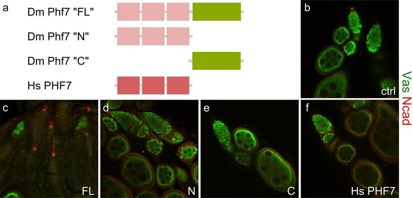
www.nature.com/scientificreports/

                                            Figure 2.  Stimulation of male-like development in female germ cells growing in XX pseudotestes by Phf7.
                                            (a,b), Δtra/tra1, nos-Gal4 pseudotestes. (a), sparse type, (b), abnormal type. (c,d), UAS-Phf7.N, Δtra/tra1, nos-
                                            Gal4 pseudotestes. (c), sparse type, (d), abnormal type. E–G, UAS-Phf7.FL, Δtra/tra1, nos-Gal4 pseudotestes. (e),
                                            sparse type, (f), abnormal type, (g), partial rescue type. (h), tra1, nos-Gal4/ + testis as a normal control. (a–h),
                                            Vasa in green, N-cadherin in red. (i–l), comparison between “partial rescue” pseudotestes and normal testes.
                                            (i,j) show Vasa staining, (k–l) show the DAPI signals. (m), distribution of different types of pseudotestes under
                                            various conditions. Blue, orange, and purple bars indicate the sparse, abnormal, and partial rescue categories
                                            respectively. Sample sizes are indicate for each genotype on the graph. The temperatures at which each set of
                                            data was performed under as well as the Phf7 construct expressed are indicated below the graph. N is UAS-
                                            Phf7.N and FL is UAS-Phf7.FL.

          Scientific Reports |   (2021) 11:6308 |                https://doi.org/10.1038/s41598-021-85560-4                                                    4

Vol:.(1234567890)
www.nature.com/scientificreports/
Figure 3. Expression of sex-specific markers in the pseudotestes. (a–d), Sxl staining in wild-type ovaries (a),
wild-type testis (b), and abnormal (c) and partial rescue (d) type pseudotestes of UAS-Phf7.FL, Δtra/tra1, nos-
Gal4. Vasa is in red, Sxl in green, and N-cadherin in blue. (a′–d′) display just the Sxl signals alone. The yellow
arrow in A′ indicate the germline stem cells in an ovariole that stain Sxl clearly. (e–h), Staining of Bam in wild-
type ovary (e), wild-type testis (f), and abnormal (g) and partial rescue (h) type pseudotestes of UAS-Phf7.FL,
Δtra/tra1, nos-Gal4. Vasa is in red, Bam in green, and N-cadherin in blue. (i–k), Phf7 expression in wild-type
testes (i), and abnormal (j) and partial rescue (k) type pseudotestes of UAS-Phf7.FL, Δtra/tra1, nos-Gal4. Vasa is
in red, Phf7 in green, and Armadillo in blue. (i′–k′) display the Phf7 channel alone.
Phf7 expression which is done with flies being grown at 29 °C to enhance the activity of GAL4 to drive transgene
expression; growing them at 25 °C is insufficient to initiate this level of male germline rescue (Fig. 2m). Interest-
ingly, there was also a higher ratio of pseudotestes that contained small, undifferentiated germline (“abnormal”,
Fig. 2f,m) at 29 °C compared to when flies were grown at 25 °C.
The temperature-dependent germline masculinization by Phf7 allowed us to further address the develop-
mental time point at which this process occurs. This was tested by raising the flies for 5 days at 25 °C and the
remaining 5 days at 29 °C, or the reverse. We find that pseudotestes containing germline that underwent sub-
stantial differentiation (“partial rescue”) were only observed when flies were grown first at 29 °C and then 25 °C
(Fig. 2m). The pseudotestes phenotype of flies grown initially at 25 °C and then 29 °C were very similar to those
continuously maintained at 25 °C. All the results from these Phf7-expressing experiments indicate three things.
First, the period most critical for choosing the male germline fate is early in development, in line with obser-
vations from other studies that showed the earliest signs of sexual dimorphism in the germline begins during
embryogenesis5,6. Second, expressing Phf7 highly later in development is not sufficient to overcome the defects
in determining the proper sexual fate earlier. Lastly, as the female germline loss assay correctly suggested, the
masculinizing effect of Phf7 in the germline requires its C-terminus.
The unique C‑terminus of Phf7 is necessary for sex‑specific gene expression. The partial pen-
etrance of the spermatogenesis rescue phenotype induced by full length Phf7 in female germ cells developing
in male somatic environments prompted us to investigate possible differences in the pseudotestes that exhibit
different grades of germline development. Specifically, expression of several genes known to have sex-dependent
expression patterns in the germ cells were examined.
Sex lethal (Sxl) is involved in the sexual development of both the female germline and s oma6,24, and its expres-
sion is the highest in germline stem cells and cystoblasts in the ovary while undetectable in normal testes25,26
(Fig. 3a,b). Intriguingly, in the “abnormal” category of pseudotestis, most germ cells clearly express Sxl cytoplas-
mically regardless of whether they over-express Phf7, or which form thereof (Fig. 3c, Supplementary Fig. S1a,b).
The “sparse” type pseudotestes were difficult to analyze due to the very small numbers of remaining germ cells.
In contrast, germ cells in the “partial rescue” category do not express Sxl (Fig. 3d). These observations indicate
that germ cells in the “abnormal” category are not able to fully suppress the female germline program.
We next looked at expression of Bam, a pro-differentiation factor in both male and female germline that is
expressed in slightly different stages: in males it is mainly in the 4–8 cell spermatogonia and in females 2–4 cell
oogonia27,28 (Fig. 3e–f). The BAM expression patterns in all pseudotestes examined, regardless of phenotype
and genotype, were quite similar and we find the protein to be present largely in the 4–8 cell stage (Fig. 3e–h,
Supplementary Fig. 1e,f), in line with what is observed in normal testes.
Phf7 is yet another protein exhibiting sex-biased expression: it is substantially expressed in the nuclei of
male germline stem cells and spermatogonia up to the 8-cell stage but undetectable in the female germline by
Scientific Reports | (2021) 11:6308 | https://doi.org/10.1038/s41598-021-85560-4 5
Vol.:(0123456789)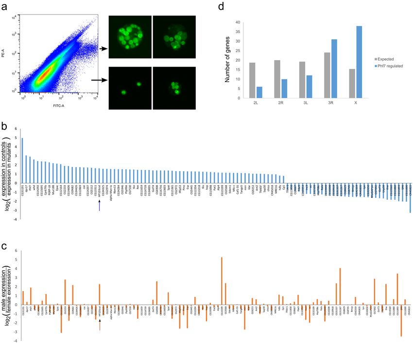
www.nature.com/scientificreports/
immunostaining9. The fact that the experiments here involve overexpression of Phf7 complicates the analysis
of Phf7 staining results. However, as the epitope of the Phf7 antibody is in the C-terminus, it is still meaningful
to examine Phf7 expression in pseudotestes without Phf7 overexpression as well as those expressing just the
N-terminus of Phf7 as we wondered whether Phf7 expression from the endogenous locus would be different
in XX germ cells exhibiting different degrees of male-like development. We found that Phf7 is expressed in all
germ cells of the “abnormal” type as well as those in “partial rescue” that resemble germline stem cells and sper-
matogonia (Fig. 3i–k, Supplementary Fig. 1c,d). Surprisingly, we did not see higher expression in germ cells that
overexpress the full-length Phf7 construct, even if they are of the “partial rescue” category.
These results, taken together with the Bam staining results, indicate that most XX germ cells developing in
the male gonad take on a partial male identity, possibly with help from signals from the male soma. Nonethe-
less, without the coordination of germline-intrinsic mechanisms, the male germline program cannot be fully
installed. Phf7, with its C-terminus, is able to provide that critical germline-intrinsic information to trigger the
proper male program.
Phf7 up‑regulates expression of most of its downstream genes in the embryonic germline. To
understand how Phf7 initiates the male germline program, we wanted to examine the genes regulated by Phf7 to
reveal the factors that are important in turning on the male germline program. We have previously performed
a related experiment of looking for Phf7 target genes in the adult testis of bam mutants which are enriched for
spermatogonia that express Phf7 highly9. However, our latest results highlight the importance of addressing this
question in a stage-specific manner as the role of Phf7 in male germline development changes over time, and the
genes that Phf7 regulates likely differ for these distinct roles.
To identify the genes Phf7 regulates in the embryonic germline, we FACS-sorted control or Phf7-deficient late
embryonic gonads that carry the germline-specific vas-GFP transgene. Gonads from 18 to 22 h embryos were
obtained by mechanical disruption to dislodge the gonads from the surrounding tissues, and the gonads mostly
stay as a whole though some germ cells are released from the gonads in this process (Fig. 4a). The homogenates
were subsequently passed through a FACS sorter so that we can separate out GFP + particles, whose distribution
on the FACS plot turned out to be rather broad (Fig. 4a). Examination of the morphology of GFP + particles from
different parts of the FACS plot under a fluorescence microscope confirmed that the sorted particles are enriched
for more intact gonads. We opted against further treatment of the gonads with enzymes to obtain single germ
cells as we wanted to reduce disturbance on the germ cell transcriptome. This means that the sample contains
both germ cells and somatic cells, but we reasoned that the contribution of germ cells to the total RNA purified
from gonads is substantial enough that we could detect expression changes resulting from the absence of Phf7.
The samples we collected included both male and female samples; male gonads are bigger than female gonads
in late embryogenesis, but we found out that this difference was not sufficient for effective separation by FACS.
The impact of this impurity is likely also limited as female germ cells are quiescent in this period and our samples
would contain more male germ cells than female germ cells. Previous experiments looking for gene expression
differences between male and female germline have also resulted mostly in those with higher expression levels
in the male embryonic g ermline4.
Two biological replicates for both control and Phf7-mutant gonads were processed for transcriptome analysis
by next generation sequencing. The correlation coefficients between the replicates were good: 0.91547 for the
control datasets and 0.94868 for the mutant datasets. Next we examined whether expression of genes known
to be present in the germline or gonad of early stage 17 embryos is indeed observed and at comparable levels
in all samples. We observed clear and consistent expression for germ cell genes such as nos, vasa, and piwi,
whereas genes present in earlier pole cells like pgc or are turned on after differentiation has started such as bam
are expressed minimally (Supplementary Fig. 2a). We also looked at genes reported to exhibit sex-enriched
expression patterns at this stage of gonads and found most of them to be expressed (Supplementary Fig. 2b,c)4,
indicating that our preparations contained a mix of embryonic testes and ovaries.
We found 97 genes to show significantly different expression levels between control and Phf7-deficient sam-
ples (Fig. 4b). Of those, expression levels of roughly two-thirds were lower in Phf7-mutants indicating that Phf7
would normally stimulate their expression. Intriguingly, this is opposite of what we found of genes regulated by
Phf7 in the adult germline9. We will note that this analyses does not identify whether these genes are directly tar-
geted by Phf7 or whether their expression changes are secondary to the immediate effects of Phf7. We examined
whether the genes regulated by Phf7 in the embryonic germline exhibit sex-biased expression and found that
about they are evenly split between being male- and female-biased in undifferentiated adult germline (Fig. 4c).
There are currently no comprehensive gene expression databases for the male versus female embryonic germline,
thus we do not know if the Phf7-regulated genes exhibit a stronger male-biased trend during embryogenesis.
Curiously, there is minimal overlap between the genes regulated in the embryonic and adult germline by Phf7
(Supplementary Fig. 2d), though we do note that all of the ones that overlap are male-biased. These differences
in downstream genes likely reflect the differing roles of Phf7 in the embryonic and adult germline. Interestingly,
the genes regulated by Phf7 in embryogenesis are enriched for being on the X chromosome (Fig. 4d). When we
re-examined the chromosomal distribution of those regulated by Phf7 in adults, we found a similar trend (Sup-
plementary Fig. 2e), suggesting that X chromosome genes could be preferentially targeted by Phf7.
As Phf7 appears to regulate gene expression differently in the embryonic and adult germline, and the Phf7
C-terminus appears to act mainly in germline masculinization, a process that occurs normally during embryo-
genesis, we wondered whether the C-terminal portion of Phf7 may function like a transcriptional activating
domain to enhance target gene expression. This was tested by replacing the activation domain of GAL4 with the
Phf7 C-terminus to create a fusion protein between the DNA binding domain of GAL4 with the C-terminus
of Phf7. A construct containing this fusion protein placed under the regulation of the ubiquitin promoter was
Scientific Reports | (2021) 11:6308 | https://doi.org/10.1038/s41598-021-85560-4 6
Vol:.(1234567890)www.nature.com/scientificreports/
Figure 4. Transcriptome profiling of embryonic gonads with or without Phf7. (a), FACS plot of homogenate
from vas-GFP late embryos. The GFP + particles are distributed in two patches, and representative images of
gonads from each population are shown to the right of the FACS plot. Gonads from the upper population are
more intact and were collected for RNA-seq. (b), Genes whose expression are significantly changed by the
Phf7 mutation are plotted. The y-axis is the log-2 value of the expression ratio of control to mutant. HP1D3csd
is indicated with an arrow. (c), Sex-expression ratio of Phf7-regulated genes in bam-mutant adult testis versus
ovary based on another study29. HP1D3csd is indicated with an arrow. (d), Distribution of Phf7-regulated genes
across different major fruit fly chromosomes. Gray bars are the expected number of genes on that chromosome
based on their relative numbers of genes; blue bars represent distribution of Phf7-regulated embryonic targets.
The observed distribution is significantly different from the expected one based on chi-squared analysis
(P < 0.001).
co-transfected with UAS-GFP into S2 cells to test the ability of the Phf7 C-terminus to activate expression of
GFP. However, we did not observe GFP expression in such S2 cells whereas the positive control treatment (Ubi-
GAL4, UAS-GFP) resulted in a clear population of GFP + cells (Supplementary Fig. 3a,b). Thus this part of the
Phf7 protein does not function as a canonical transcriptional activation domain in S2 cells.
HP1D3csd acts downstream of Phf7 to induce germline masculinization. One of the candidate
genes regulated by Phf7 in embryogenesis is the X-encoded HP1D3csd. This gene encodes a small, ~ 20 kD
protein predicted to resemble chromoshadow domains that are typically found in HP1 proteins and mediate
dimerization and interactions with other chromatin f actors12,30–32. As Phf7 is a chromatin-associated protein, we
became intrigued by the possibility that HP1D3csd may assist Phf7 in regulating expression of genes important
for setting up the male germline sex. HP1D3csd is expressed germline-specifically in the embryo (Fig. 5a–c). Its
transcripts are present in germline of both sexes but become male-biased later (Figs. 4c, 5a,b).
To test for possible interactions between Phf7 and HP1D3csd, we asked if HP1D3csd could modify the loss of
female germline caused by ectopic expression of Phf7. We first addressed what would happen with a lower dose
of HP1D3csd by using two mutants of HP1D3csd, a transposon-insertion in the 3′ end of the coding sequence of
Scientific Reports | (2021) 11:6308 | https://doi.org/10.1038/s41598-021-85560-4 7
Vol.:(0123456789)www.nature.com/scientificreports/
Figure 5. Genetic interaction between Phf7 and HP1D3csd on the female germline loss caused by Phf7
overexpression. (a,b), in situ hybridization of stage 17 embryos with an HP1D3csd probe. (a), Female embryo;
arrow indicates position of gonad on one side. (b), Male embryo. (c), HCR staining of HP1D3csd transcripts
in an unsexed stage 17 embryo. Colocalization with the Vas (green) signal indicate the HP1D3csd signals
are in germ cells. (c’), HCR signal alone. (d–k), Ovaries of various genotypes stained with Vasa (green) and
N-cadherin (red). (d), nos-Gal4/ + , (e), nos-Gal4/UAS-Phf7.FL, (f), Df(1)ED7441/ + ; nos-Gal4/UAS-Phf7.FL,
(g), HP1D3csdf07323/ + ; nos-Gal4/UAS-Phf7.FL, (h), Df(1)ED7441/ + , (i), HP1D3csdf07323/ + , (j), nos-Gal4/UAS-
HP1D3csd.HA, (k), nos-Gal4/UAS-HP1D3csd.HA, UAS-Phf7.FL. (l), Quantitation of the fraction of ovarioles
that contain no germline in different genotypes. Sample sizes and genotypes are indicated on the bottom of
the bars. Dark and light pink portions of the bars denote the fraction of ovaries with and without germline. *
indicates P < 0.05 with chi-square tests.
Scientific Reports | (2021) 11:6308 | https://doi.org/10.1038/s41598-021-85560-4 8
Vol:.(1234567890)www.nature.com/scientificreports/
Figure 6. HP1D3csd affects the ability of Phf7 to induce germline masculinization in a dose-dependent
manner. (a,b), Example of a pseudotestis with its XX germ cells overexpressing both Phf7 and HP1D3csd
(UAS-Phf7.FL, UAS-HP1D3csd.HA, Δtra/tra1, nos-Gal4) that exhibited “partial rescue” of spermatogenesis.
Vas is in green and N-cad is in red. (a), View of the entire pseudotestis. (b), View of the pseudotestis apical
tip. (c), Quantitation of the different types of phenotypes observed for pseudotestes with XX germ cells
overexpressing Phf7 and either HP1D3csd or a control construct (LacZ). The numbers in the graph indicate
sample sizes. * indicates P < 0.05 with chi-square tests. (d,e), A pseudotestis overexpressing Phf7 but lacking a
functional copy of HP1D3csd (HP1D3csdf07323/ + ; UAS-Phf7.FL, Δtra/tra1, nos-Gal4) exhibiting “partial rescue”
of spermatogenesis. Vas is in green and N-cad is in red. (d), Image of the entire pseudotestis. (e), Image of the
pseudotestis apical tip. (f), Quantitation of the different types of phenotypes observed for pseudotestes with their
XX germ cells overexpressing Phf7 and either wildtype or harboring a mutant copy of HP1D3csd. The numbers
in the graph indicate sample sizes. * indicates P < 0.05 with chi-square tests.
HP1D3csd which is possibly a hypomorphic allele (HP1D3csdf07323), and a deficiency covering HP1D3csd (Df(1)
ED7441). When one copy of HP1D3csd was mutated (HP1D3csdf07323/ + or Df(1)ED7441/ +), the extent of female
germline loss caused by Phf7 was exacerbated compared to when HP1D3csd is intact (Fig. 5d–g,l). Ovaries het-
erozygous for HP1D3csdf07323 or Df(1)ED7441 alone did not show any germline defects (Fig. 5h,i,l), indicating
that the phenotype enhancement is due to a genetic interaction between Phf7 and HP1D3csd.
The relationship between Phf7 and HP1D3csd was further investigated by overexpressing HP1D3csd along
with Phf7 in female germ cells. We used an HA-tagged transgene driven by UAS (UAS-HP1D3csd.HA) from the
FlyORF collection. Quite surprisingly, overexpressing the two genes together also causes more severe germline
loss than when just Phf7 is overexpressed (Fig. 5j–l). It should be noted that when HP1D3csd and Phf7 are co-
expressed using nos-Gal4, expression levels of each of the genes are expected to drop, which should alleviate the
germline loss effect induced by Phf7 overexpression. The fact that the opposite was observed indicates that over-
expressing HP1D3csd clearly leads to a stronger germline loss phenotype. These results suggest that HP1D3csd
is functionally associated with Phf7, but as both reduced and increased doses of HP1D3csd enhanced the female
germline loss phenotype mediated by Phf7, the exact relationship between Phf7 and HP1D3csd is unclear.
To clarify the role of HP1D3csd in germline sexual development and how it interacts with Phf7, we carried
out a second set of experiments to test for genetic interaction between these two factors: we investigated whether
HP1D3csd can modify the ability of Phf7 to induce partial spermatogenesis in female germ cells that develop
in a male somatic gonad. These experiments were carried out at 29 °C, the temperature at which we observed
partial spermatogenesis to occur in the female germline overexpressing Phf7 developing in a male soma. When
HP1D3csd was overexpressed along with Phf7, we found that the frequencies of pseudotestes that exhibited
partial spermatogenesis (“partial rescue” phenotype) was increased compared to samples that expressed an
unrelated protein (LacZ) together with Phf7 (Fig. 6c). The extent of spermatogenesis that occurred in these
samples is comparable to what is achieved with just Phf7 overexpression (Figs. 6a,b vs. 2g,i,k). The fraction of
partial rescue type in samples that overexpress both Phf7 and LacZ is lower than that of pseudotestes that only
overexpress Phf7 (Fig. 6f); this is most likely because expression of Phf7 is reduced in the former genotype with
both Phf7 and LacZ being driven by a single nos-Gal4 driver.
We also performed the reverse experiment by examining pseudotestes that overexpress Phf7 and are het-
erozygous for the f07323 allele of HP1D3csd. The rate of such pseudotestes that show partial spermatogenesis was
lower than what we observed for samples that overexpressed Phf7 but were wild-type for HP1D3csd (Fig. 6f). In
addition, the extent of spermatogenesis rescue in most of the pseudotestes that have a mutant copy of HP1D3csd
Scientific Reports | (2021) 11:6308 | https://doi.org/10.1038/s41598-021-85560-4 9
Vol.:(0123456789)www.nature.com/scientificreports/
was more limited than most of those in the same phenotypic category for other genotypes (Figs. 6c,d vs. a,b,
2g,i). These results show that a single HP1D3csd mutant copy diminished the ability of Phf7 to trigger male
germline development. Taken together, results of our experiments clearly demonstrate that Phf7 and HP1D3csd
interact genetically. Furthermore, they indicate that HP1D3csd acts downstream of Phf7 to facilitate germline
masculinization.
Discussion
In this study we investigated how Phf7 regulates sex determination in the embryonic germline, and one of our
interesting finding is that the unusual C-terminus of Phf7 is necessary for its effects in germline masculinization.
The N-terminus of Phf7 is a conserved module comprised of three zinc fingers, of which at least one is function-
ally essential33, and this part of the Phf7 protein evolved from G2E3 (G2/M E3 Ubiquitin Ligase), a protein also
made up of three zinc fi ngers10. In contrast, the C-terminus of Phf7 is evolutionarily novel and is not similar to
any known domains, suggesting that this domain is undergoing very rapid evolution, a feature not uncommon
for factors involved in sex determination34,35.
We previously conducted a phylogenetic analysis of Phf7 proteins across the species tree, and surprisingly
found that Phf7 in insects and amniotes do not share a common a ncestor10. Those findings with our latest results
indicate that Phf7 in these two animal branches are not orthologous to each other, and that the emergence of
the novel C-terminus is likely a unique event that occurred in Drosophila to regulate sexual differentiation in
the germline. Recently, mouse Phf7 was demonstrated to be expressed in spermatocytes and can ubiquitinate
histones to facilitate histone to protamine e xchange8,36. These show that the expression patterns and functions
of D. melanogaster and mouse Phf7 are different, albeit both acting on the male germline. These observations
further suggest that the C-terminus of D. melanogaster PHF7 evolved onto an existing module of three zinc
fingers, thereby creating new ways to regulating germline sexual development. This is a very interesting example
that adds to the collection of diverse mechanisms in sex determination.
What does this uncommon C-terminus of PHF7 do? The two most intuitive ideas are that it acts as a transac-
tivation domain like those found in transcription factors, or that it can recruit other effector molecules through
protein–protein interactions. The former idea did not hold up when tested in S2 cells. The possibility that the
Phf7 C-terminus acts as a bridge between its histone-associating N-terminus and other transcription factors or
chromatin factors to alter target gene expression is an appealing one but there is currently no direct data that
support this idea.
We further looked for downstream effectors of Phf7 in the embryonic germline, and revealed that HP1D3csd
is activated by Phf7 to regulate germline masculinization. We performed two different genetic tests, and while
both indicated that Phf7 and HP1D3csd genetically interact, there were some differences in the results. In the
Phf7-induced female germline loss assay, we found both reduction and gain of HP1D3csd expression exacerbated
the Phf7-induced phenotype. In comparison, in the spermatogenesis rescue assay, loss of one HP1D3csd copy
hampered rescue whereas HP1D3csd overexpression enhanced spermatogenesis in XX germ cells. The latter
experiment is a more direct assay of germline masculinization whereas germline loss can potentially be caused
by secondary effects unrelated to sexual development. Therefore, we think the results of the spermatogenesis
rescue experiments more accurately reflect the relationship between Phf7 and HP1D3csd. In addition to our
transcriptome results, HP1D3csd has been identified along with Phf7 to be a part of sex-biased mechanisms in
other contexts not limited to the g ermline37,38. These also support the model that Phf7 and HP1D3csd function
synergistically.
Phf7 regulates male germline development, and it can associate with the active histone mark methylated
H3K47, but it is unclear what Phf7 then does to regulate expression of target genetic loci. H3K9 methylation has
also been reported to be important for maintaining sexual differentiation programs in the Drosophila germline39.
The identification of HP1D3csd as an important downstream factor provides interesting new ideas regarding
how the male germline program is initiated and regulated. CSDs have been shown to interact with various
chromatin remodelers, thus one appealing model would be that Phf7 can activate or even recruit HP1D3csd to
loci important for germline masculinization. This would in turn bring chromatin remodelers to such genes for
expression activation and regulation and initiate male-development of the germline. Given our other finding
in this study that the C-terminus of Phf7 is an essential part of this process, it would be very interesting to now
study which of these factors interact and cooperate with one another.
Data availability
The datasets generated during the current study are available at the NCBI Gene Expression Omnibus (GEO;
https://www.ncbi.nlm.nih.gov/geo/) underaccession number GSE167380.
Received: 27 August 2020; Accepted: 25 February 2021
References
1. Steinmann-Zwicky, M., Schmid, H. & Nöthiger, R. Cell-autonomous and inductive signals can determine the sex of the germ line
of drosophila by regulating the gene Sxl. Cell 57, 157–166 (1989).
2. Nöthiger, R., Jonglez, M., Leuthold, M., Meier-Gerschwiler, P. & Weber, T. Sex determination in the germ line of Drosophila
depends on genetic signals and inductive somatic factors. Development 107, 505–518 (1989).
3. Schüpbach, T. Normal female germ cell differentiation requires the female X chromosome to autosome ratio and expression of
sex-lethal in Drosophila melanogaster. Genetics 109, 529–548 (1985).
4. Casper, A. L. & Van Doren, M. The establishment of sexual identity in the Drosophila germline. Development 136, 3821–3830
(2009).
Scientific Reports | (2021) 11:6308 | https://doi.org/10.1038/s41598-021-85560-4 10
Vol:.(1234567890)www.nature.com/scientificreports/
5. Wawersik, M. et al. Somatic control of germline sexual development is mediated by the JAK/STAT pathway. Nature 436, 563–567
(2005).
6. Hashiyama, K., Hayashi, Y. & Kobayashi, S. Drosophila Sex lethal gene initiates female development in germline progenitors.
Science 333, 885–888 (2011).
7. Yang, S. Y., Baxter, E. M. & Van Doren, M. Phf7 controls male sex determination in the Drosophila germline. Dev. Cell 22,
1041–1051 (2012).
8. Wang, X. et al. PHF7 is a novel histone H2A E3 ligase prior to histone-to-protamine exchange during spermiogenesis. Development
146, dev175547 (2019).
9. Yang, S. Y. et al. Control of a novel spermatocyte-promoting factor by the male germline sex determination factor PHF7 of dros-
ophila melanogaster. Genetics 206, 1939–1949 (2017).
10. Wang, X. R. et al. Evidence for parallel evolution of a gene involved in the regulation of spermatogenesis. Proc. R. Soc. B Biol. Sci.
284, 20170324 (2017).
11. Aasland, R. & Stewart, F. The chromo shadow domain, a second chromo domain in heterochromatin-binding protein 1, HP1.
Nucl. Acids Res. 23, 3168–3173 (1995).
12. Lechner, M. S., Schultz, D. C., Negorev, D., Maul, G. G. & Rauscher, F. J. The mammalian heterochromatin protein 1 binds diverse
nuclear proteins through a common motif that targets the chromoshadow domain. Biochem. Biophys. Res. Commun. 331, 929–937
(2005).
13. Levine, M. T. et al. Phylogenomic analysis reveals dynamic evolutionary history of the Drosophila heterochromatin protein 1
(HP1) gene family. PLoS Genet. 8, e1002729 (2012).
14. Shigenobu, S., Kitadate, Y., Noda, C. & Kobayashi, S. Molecular characterization of embryonic gonads by gene expression profiling
in Drosophila melanogaster. Proc. Natl. Acad. Sci. U. S. A. 103, 13728–13733 (2006).
15. Van Doren, M., Williamson, A. L. & Lehmann, R. Regulation of zygotic gene expression in Drosophila primordial germ cells. Curr.
Biol. 8, 243–246 (1998).
16. Shigenobu, S., Arita, K., Kitadate, Y., Noda, C. & Kobayashi, S. Isolation of germline cells from Drosophila embryos by flow
cytometry. Dev. Growth Differ. 48, 49–57 (2006).
17. Goecks, J., Nekrutenko, A. & Taylor, J. Galaxy: a comprehensive approach for supporting accessible, reproducible, and transparent
computational research in the life sciences. Genome Biol. 11, R86 (2010).
18. Pertea, M., Kim, D., Pertea, G. M., Leek, J. T. & Salzberg, S. L. Transcript-level expression analysis of RNA-seq experiments with
HISAT, StringTie and Ballgown. Nat. Protoc. 11, 1650–1667 (2016).
19. Anders, S., Pyl, P. T. & Huber, W. HTSeq–a Python framework to work with high-throughput sequencing data. Bioinformatics 31,
166–169 (2015).
20. Trapnell, C. et al. Transcript assembly and quantification by RNA-Seq reveals unannotated transcripts and isoform switching
during cell differentiation. Nat. Biotechnol. 28, 511–515 (2010).
21. Love, M. I., Huber, W. & Anders, S. Moderated estimation of fold change and dispersion for RNA-seq data with DESeq2. Genome
Biol. 15, 550 (2014).
22. Sturtevant, A. H. A gene in drosophila melanogaster that transforms females into males. Genetics 30, 297–299 (1945).
23. Marsh, J. L. & Wieschaus, E. Is sex determination in germ line and soma controlled by separate genetic mechanisms?. Nature 272,
249–251 (1978).
24. Salz, H. & Erickson, J. W. Sex determination in Drosophila: the view from the top. Fly (Austin). 4, 60–70 (2010).
25. Bopp, D., Horabin, J. I., Lersch, R. A., Cline, T. W. & Schedl, P. Expression of the Sex-lethal gene is controlled at multiple levels
during Drosophila oogenesis. Development 118, 797–812 (1993).
26. Vied, C., Halachmi, N., Salzberg, A. & Horabin, J. I. Antizyme is a target of sex-lethal in the drosophila germline and appears to
act downstream of hedgehog to regulate sex-lethal and cyclin B. Dev. Biol. 253, 214–229 (2003).
27. McKearin, D. M. & Spradling, A. C. bag-of-marbles: a Drosophila gene required to initiate both male and female gametogenesis.
Genes Dev. 4, 2242–2251 (1990).
28. Gönczy, P., Matunis, E. & DiNardo, S. bag-of-marbles and benign gonial cell neoplasm act in the germline to restrict proliferation
during Drosophila spermatogenesis. Development 124, 4361–4371 (1997).
29. Primus, S., Pozmanter, C., Baxter, K. & Van Doren, M. Tudor-domain containing protein 5-like promotes male sexual identity in
the Drosophila germline and is repressed in females by Sex lethal. PLoS Genet. 15, e1007617 (2019).
30. Ye, Q., Callebaut, I., Pezhman, A., Courvalin, J.-C. & Worman, H. J. Domain-specific interactions of human HP1-type chromo-
domain proteins and inner nuclear membrane protein LBR. J. Biol. Chem. 272, 14983–14989 (1997).
31. Lechner, M. S., Begg, G. E., Speicher, D. W. & Rauscher, F. J. Molecular determinants for targeting heterochromatin protein 1-medi-
ated gene silencing: direct chromoshadow domain-KAP-1 corepressor interaction is essential. Mol. Cell. Biol. 20, 6449–6465 (2000).
32. Ekblad, C. M. S. et al. Binding of EMSY to HP1β: implications for recruitment of HP1β and BS69. EMBO Rep. 6, 675–680 (2005).
33. Smolko, A. E., Shapiro-Kulnane, L. & Salz, H. K. An autoregulatory switch in sex-specific phf7 transcription causes loss of sexual
identity and tumors in the Drosophila female germline. Development 147, dev192856 (2020).
34. Bachtrog, D. et al. Sex determination: Why so many ways of doing it?. PLoS Biol. 12, e1001899 (2014).
35. Meccariello, A. et al. Maleness-on-the-Y (MoY) orchestrates male sex determination in major agricultural fruit fly pests. Science
365, 1457–1460 (2019).
36. Kim, C. R. et al. PHF7 modulates BRDT stability and histone-to-protamine exchange during spermiogenesis. Cell Rep. https://
doi.org/10.1016/j.celrep.2020.107950 (2020).
37. Shapiro-Kulnane, L., Smolko, A. E. & Salz, H. K. Maintenance of Drosophila germline stem cell sexual identity in oogenesis and
tumorigenesis. Dev. 142, 1073–1082 (2015).
38. Molnar, C. et al. The histone code reader PHD finger protein 7 controls sex-linked disparities in gene expression and malignancy
in Drosophila. Sci. Adv. 5, eaaw7965 (2019).
39. Smolko, A. E., Shapiro-Kulnane, L. & Salz, H. K. The H3K9 methyltransferase SETDB1 maintains female identity in Drosophila
germ cells. Nat. Commun. 9, 1 (2018).
Acknowledgements
This work was carried out with technical assistance from Hsing-Chun Chen, Chien-Cheng Wang, Li Bing Lin,
and Yi-Ru Li. Funding support for this study include a Chang Gung Memorial Hospital Grant (CMRPD1F0593)
and a MOST Grant from Taiwan (108-2628-B-182-007). The author declares no competing financial interests.
Strains and plasmids are available upon request. The author affirms that all data necessary for confirming the
conclusions of the article are present within the articles and figures. Data from this manuscript will be deposited
in a repository upon acceptance of publication.
Author contributions
S.Y.Y. conceived the study, carried out the experiments and analysis, and wrote the manuscript.
Scientific Reports | (2021) 11:6308 | https://doi.org/10.1038/s41598-021-85560-4 11
Vol.:(0123456789)www.nature.com/scientificreports/
Competing interests
The author declares no competing interests.
Additional information
Supplementary Information The online version contains supplementary material available at https://doi.org/
10.1038/s41598-021-85560-4.
Correspondence and requests for materials should be addressed to S.Y.Y.
Reprints and permissions information is available at www.nature.com/reprints.
Publisher’s note Springer Nature remains neutral with regard to jurisdictional claims in published maps and
institutional affiliations.
Open Access This article is licensed under a Creative Commons Attribution 4.0 International
License, which permits use, sharing, adaptation, distribution and reproduction in any medium or
format, as long as you give appropriate credit to the original author(s) and the source, provide a link to the
Creative Commons licence, and indicate if changes were made. The images or other third party material in this
article are included in the article’s Creative Commons licence, unless indicated otherwise in a credit line to the
material. If material is not included in the article’s Creative Commons licence and your intended use is not
permitted by statutory regulation or exceeds the permitted use, you will need to obtain permission directly from
the copyright holder. To view a copy of this licence, visit http://creativecommons.org/licenses/by/4.0/.
© The Author(s) 2021
Scientific Reports | (2021) 11:6308 | https://doi.org/10.1038/s41598-021-85560-4 12
Vol:.(1234567890)You can also read