Interferon Regulatory Factor 7 Mediates Activation of Tap-2 by Epstein-Barr Virus Latent Membrane Protein
←
→
Page content transcription
If your browser does not render page correctly, please read the page content below
JOURNAL OF VIROLOGY, Jan. 2001, p. 341–350                                                                                   Vol. 75, No. 1
0022-538X/01/$04.00⫹0 DOI: 10.1128/JVI.75.1.341–350.2001
Copyright © 2001, American Society for Microbiology. All Rights Reserved.

       Interferon Regulatory Factor 7 Mediates Activation of Tap-2 by
               Epstein-Barr Virus Latent Membrane Protein 1
                                       LUWEN ZHANG1,2*          AND    JOSEPH S. PAGANO1,2,3
           Lineberger Comprehensive Cancer Center, Department of Microbiology and Immunology,2 and Department of
                                                      1

                        Medicine,3 University of North Carolina, Chapel Hill, North Carolina 27599-7265
                                             Received 6 June 2000/Accepted 2 October 2000

            Transporter associated with antigen processing 2 (Tap-2) is responsible for ATP-dependent transport of
         peptides from the cytosol to the endoplasmic reticulum, where peptides bind to newly synthesized human

                                                                                                                                              Downloaded from http://jvi.asm.org/ on December 23, 2020 by guest
         leukocyte antigen (HLA) class I molecules, which are essential for cellular immune responses. Epstein-Barr
         virus (EBV) latent membrane protein 1 (LMP-1) has been shown to induce the expression of Tap-2. In this
         study, the induction of endogenous Tap-2 by LMP-1 is shown to be associated with and requires the expression
         of interferon regulatory factor 7 (IRF-7). In DG75 Burkitt’s lymphoma (BL) cells, in which LMP-1 induces the
         expression of IRF-7, LMP-1 induced endogenous Tap-2, and ectopic expression of IRF-7 could enhance the
         induction. In Akata BL cells, in which LMP-1 could not induce IRF-7, LMP-1 could not induce Tap-2. Addition
         of IRF-7, which complements the defect in Akata cells, could stimulate the expression of Tap-2. Furthermore,
         LMP-1 and IRF-7A but not other IRF-7 splicing variants could activate endogenous Tap-2. A Tap-2 promoter
         reporter construct could be activated by the overexpression of IRF-7A. The activation could be specifically
         enhanced by LMP-1 and was dependent on an intact interferon-stimulated response element (ISRE) present
         in the Tap-2 promoter. Also, IRF-7 can bind to the Tap-2 promoter under physiological conditions in vivo, as
         shown by formaldehyde cross-linking, as well as to the Tap-2 ISRE in vitro, as shown by gel mobility shift
         assays. Furthermore, LMP-1 facilitates the phosphorylation and nuclear translocation of IRF-7. These data
         point to the role of IRF-7 as a secondary mediator of LMP-1-activated signal transduction for Tap-2 as follows:
         LMP-1 stimulates the expression of IRF-7 and facilitates its phosphorylation and nuclear translocation, and
         then the activated IRF-7 mediates the activation of the cellular Tap-2 gene. The induction of Tap-2 by IRF-7
         and LMP-1 may have an important implication for the immune response to EBV and its persistence in vivo.

   Infection by Epstein-Barr virus (EBV) may contribute to the             peptides from the cytosol to the endoplasmic reticulum, where
development of malignant diseases such as Hodgkin’s disease,               peptides bind to newly synthesized HLA class I molecules,
Burkitt’s lymphoma (BL), nasopharyngeal carcinoma, and                     which are essential for cellular immune responses (for a re-
posttransplant lymphoproliferative diseases (reviewed in refer-            view, see references 3 and 57).
ences 20, 37, 41, and 44). In vitro, EBV efficiently infects and              Tap-2 appears to be involved in some human diseases.
immortalizes primary B lymphocytes (reviewed in references                 Mutation of Tap-2 may cause defective precessing of HLA I,
19 and 44), and latent membrane protein 1 (LMP-1) expres-                  leading to primary immunodeficiency (55). Tap-2 mutation
sion is required for this immortalization process (18).                    has been associated with familial bronchiectasis and with
   LMP-1 is an integral membrane protein with six membrane-                susceptibility to Sjögren’s syndrome (10, 21). Tap-2B may
spanning domains with a long C-terminal domain, which is                   increase the risk for nickel allergy (52). Tap-2 polymorphism
located in the cytoplasm (19, 23). LMP-1 acts as a constitu-               may also be involved in inflammatory bowl disease (15).
tively active receptor-like molecule, which does not need the              Understanding the regulation of Tap-2 is essential to elucidate
binding of a ligand (14). The six transmembrane domains me-                its role in the pathogenesis of these as well as EBV-associated
diate oligomerization of LMP-1 molecules in the plasma mem-                diseases.
brane, a prerequisite for LMP-1 function (11, 14). So far, two                The mechanism leading to upregulation of Tap-2 by LMP-1
domains in the C terminus of LMP-1 have been shown to                      is currently unknown. Previous results suggest that LMP-1
initiate signaling processes: C-terminal activator region 1                stimulates the expression of interferon-regulatory factor 7
(CTAR-1, amino acids 194 to 231) and region 2 (CTAR-2,                     (IRF-7) (66). IRF-7 was first cloned by its binding activity to
amino acids 332 to 386) (16, 29).                                          the EBV BamHI Q promoter (Qp), used in latent EBV infec-
   LMP-1 can induce a variety of cellular genes that enhance               tion for transcription of EBNA-1, and has subsequently proven
cell survival and adhesive and invasive potential (12, 27, 59, 60,         to be a negative regulator of Qp (65–67). IRF-7 belongs to the
64). Interestingly, LMP-1 can stimulate the expression of the
                                                                           IRF family, a group of transcription factors with multiple func-
transporter associated with antigen processing 2 (TAP-2) gene
                                                                           tions (reviewed in reference 33). The hallmark of this family is
(46). Tap-2 is involved in the ATP-dependent transport of
                                                                           the conserved N-terminal DNA-binding domain which has the
                                                                           potential to bind to interferon-stimulated response elements
                                                                           (ISREs) and regulate the activity of promoters containing
   * Corresponding author. Mailing address: Lineberger Comprehen-
sive Cancer Center, University of North Carolina, Campus Box 7295,
                                                                           ISREs. Because there is a putative ISRE in the Tap-2 pro-
Chapel Hill, NC 27599. Phone: (919) 966-1183. Fax: (919) 966-9673.         moter region, whether IRF-7 is involved in regulation of the
E-mail: luzhang@med.unc.edu.                                               Tap-2 gene was investigated. In this paper, we report that

                                                                     341
342 ZHANG AND PAGANO J. VIROL.
IRF-7 is a secondary mediator of LMP-1-triggered signal (GAPDH) probe was supplied by US Biochemicals, Inc. The Tap-2 RPA probe
transduction for activation of the Tap-2 gene. was made by PCR with the primers 5⬘-GCTCTAGATAATACGACTCACTAT
AGGGCGACAGACCCAAGCTTGGTACCGAGCTCGGATCCCGCTCTCA
GGGAGACAGTCA-3⬘ and 5⬘-GCTCTAGACTGGACCTCCCTGCTGCTGG
MATERIALS AND METHODS TGGAC-3⬘. The PCR product contains a T7 promoter, a spacer region, and the
Cell culture. DG75 is an EBV-negative BL cell line (4); BL30 and BL41 are Tap-2 first exon sequence (complementary to nt 116227 to 116458 of the se-
EBV-negative BL lines with EBV-infected counterparts generated by in vitro quence published in GenBank [no. X87344]). The PCR product was purified and
infection with the P3HR1 strain (BL30-P3HR1 and BL41-P3HR1) or the B95-8 used directly for synthesis of RNA probe.
strain (BL30-B95-8 and BL41-B95-8) of EBV (5). Akata is an EBV-positive type EMSA. For the electrophoretic mobility shift assay (EMSA), the Tap-2 ISRE-
I BL cell line, and Jijoye is an EBV-positive type III BL cell line (42, 53). Sav-I containing fragment was obtained by annealing two oligonucleotides, 5⬘-GATC
and Sav-III are paired EBV-positive BL lines that differ only in their latent GGAAGCGAAAGCGAAAGCTGCCC-3⬘ and its complement, with GATC at
infection state (35, 46). X50-7 and B958/CBC are EBV-positive lymphoblastoid the 5⬘ end. The mutated ISRE probe was made exactly the same way with
cell lines. FaDuHyg is an EBV-negative epithelial cell line (43). T2 is a lympho- mutated oligonucleotides, as shown in Fig. 3A. The DNA probes were generated
blastic cell line with a deletion of the Tap-2 genomic sequence (47, 48). All cell by filling in any single-stranded overhang with Klenow enzyme. EMSA was
lines were maintained in RPMI 1640 plus 10% fetal bovine serum. performed essentially as described (65, 67). In vitro-translated reticulate lysates
Plasmids and antibodies. A PCR fragment containing the Tap-2 promoter region, (5 l) were incubated with 20,000 to 50,000 cpm of labeled probe in a volume of
Downloaded from http://jvi.asm.org/ on December 23, 2020 by guest
starting at its ISRE and ending at the first coding sequence, was cloned into pBS- 12.5 l containing 20 mM HEPES (pH 7.9), 1 mM MgCl2, 0.1 mM EGTA, 0.5
CAT (13). The PCR fragment corresponded to nucleotides (nt) 115644 to 116217 of mM dithiothreitol, 320 g of poly(dI-dC):poly(dI-dC), and 4% Ficoll-400 for 20
a published sequence (GenBank accession no. X87344) (2). The cloned promoter min at room temperature. The samples were separated on a preelectrophoresed
was identical to the published sequence except for missing 2 T’s within a stretch of 4.8% polyacrylamide gel in 20 mM Tris-borate-EDTA (TBE) buffer. After elec-
T’s (nt 115965 to 115985). Such a sequence has been found in several PCR clones. trophoresis, gels were dried, followed by autoradiography. When antiserum was
We do not know whether this variation was due to a PCR error or a polymorphism. needed, 1 l was added to the reaction mixture. The consensus ISRE oligonu-
However, this sequence variation seems to play no important role in Tap-2 promoter cleotide has been described (67). AP-1 binding site competitor was purchased
activity, because results from reporter assays were in agreement with the levels of from Promega.
endogenous Tap-2 RNA (see Fig. 3 to 5). Mutations in the ISRE region (nt 115649 In vitro transcription and translation. The proteins were made with the TNT
to 115662) were made by PCR with a mutated primer, and mutations were con- coupled transcription and translation kit (Promega) essentially according to the
firmed by sequencing analysis (see Fig. 3A). IRF-7 expression plasmids and IRF-7 manufacturer’s instructions. Wheat germ lysate was used with the plasmid
antibody have been described (67). pcDNA/CD4 is a human CD4 expression plas- pcDNA-IRF-7A, and rabbit reticulocyte lysate was used with the plasmid
mid (gift of Jenny Ting). IRF-7DN (amino acids [aa] 1 to 12 and 103 to 503) is a gift pcDNA-IRF-7C.1.
from Tom Maniatis (61). Epidermal growth factor promoter (EGFP)-IRF-7 was Analysis of DNA-binding activity by in vivo formaldehyde cross-linking. The
cloned by inserting the full-length IRF-7A into the BglII site of the pEGFP-c1 vector cross-linking method is based on a previous publication (36). Cells were fixed
(Clontech). The -galactosidase expression plasmid pCMV(6177-1) was purchased with 1/10 volume of 11% formaldehyde solution in 0.1 M NaCl–1 mM EDTA–0.5
from Clontech. LMP-1 expression plasmid pcLMP-1 was a gift from Tomakazu mM EGTA–50 mM HEPES (pH 8.0) for 1 h at 4°C. The cross-linking reaction
Yoshizaki. The mutant LMP-1 plasmids LMP1-231 and LMP⌬231-387 were gifts was stopped by adding glycine to 0.125 M. The cells were washed and sonicated,
from Nancy Raab-Traub (28). The interferon consensus sequence binding protein and the cell debris was removed by centrifugation at 15,000 ⫻ g for 10 min as
(ICSBP) and IRF-3 expression plasmids were cloned in the expression plasmid
described (36). The cell lysates were cleared first by incubating with protein
pcDNA3. The IRF-1 (C-20) and IRF-2 (C-19) antibodies were purchased from
G-agarose beads and preimmune serum. Then immunoprecipitations were per-
Santa Cruz Biotechnology, Inc. LMP-1 monoclonal antibody CS1-4 was purchased
formed with preimmune or immune serum specific for the C-terminal region of
from Dako. Antitubulin antibody was from Sigma. Tap-2 antibody has been de-
IRF-7 at 4°C overnight. Immunoprecipitates were collected by protein G-agarose
scribed (58). The IRF-7 C-terminus-specific antiserum was generated by injection of
beads and washed three times for 10 min each in 1⫻ phosphate-buffered saline
glutathione-S-transferase (GST)-IRF-7B fusion protein (aa 218 to 474) into a rabbit.
(PBS) solution plus phenylmethylsulfonyl fluoride (PMSF) and another three
This IRF-7 antiserum was used only for the experiment shown in Fig. 2C, and the
times for 10 min each in Tris-EDTA (TE) buffer. Finally, the pellets were
full-length IRF-7B antiserum was used in all other applications (67).
digested at 65°C overnight in 100 l of TE plus 0.5% SDS and proteinase K (500
Western blot analysis with enhanced chemiluminescence. Separation of pro-
g/ml). After phenol-chloroform extraction, the DNA was precipitated with
teins on sodium dodecyl sulfate-polyacrylamide gel electrophoresis (SDS-
glycogen as the carrier. The isolated DNA was used as the template for ampli-
PAGE) followed standard methods. After the proteins were transferred to a
fication of the Tap-2-specific region with primers 5⬘-GAGTTCGGAAGGCCT
nitrocellulose or Immobilon membrane, the membrane was blocked with 5%
TGG-3⬘ (corresponding to nt 115623 to 115640) and 5⬘-GAAGCAGGAGCGT
nonfat dry milk in TBST (50 mM Tris [pH 7.5], 200 mM NaCl, 0.05% Tween 20)
GGAGT-3⬘ (complementary to nt 115856 to 115873) (2). The PCR products
at room temperature for 10 min. It was then washed briefly with water and
incubated with primary antibody in 5% milk in TBST for 1 to 2 h at room were separated in a 1.5% agarose gel, transferred to a nylon membrane, and
temperature or overnight at 4°C. After being washed with TBST three times for hybridized to labeled Tap-2 promoter probe (nt 115644 to 116217), which was
10 min each, the membrane was incubated with the secondary antibody at room synthesized with random primer and Klenow enzyme by the use of standard
temperature for 1 h. It was then washed three times with TBST as before, treated methods (49).
with ECL (Amersham) or SuperSignal (Pierce) detection reagents, and exposed Phosphorylation analysis and immunoprecipitation. 293T cells were cotrans-
to Kodak XAR-5 film. fected with expression plasmids for IRF-7A and LMP-1 with the use of Effectene
Transient transfection, enzyme assays, and isolation of transfected cells. Cells (Qiagen). Cells were then labeled with [32P]orthophosphate for 4 h, washed once
(107) in 0.5 ml of medium were transfected with the use of a Bio-Rad Gene with 1⫻ TBS, and lysed in a buffer containing 0.5% NP-40, 50 mM Tris-Cl (pH
Pulser (320 V and 925 F). Two days after transfection, cells were collected for 7.5), 150 mM NaCl, 5 mM EDTA, 100 mM NaF, 1 mM sodium orthovanadate,
chloramphenicol acetyltransferase (CAT) assay or for isolation of transfected and 1 mM PMSF. Cell lysates were precleared with preimmune serum and
cells. The CAT and -galactosidase assays were performed essentially as de- protein G-agarose beads under gentle agitation at 4°C for 30 min. The anti-
scribed (22). The CAT assay results were analyzed on a Molecular Dynamics IRF-7 antiserum and protein G-agarose were then added, and lysates were
PhosphorImager. incubated overnight at 4°C. The beads were washed three times with 1 ml of
For isolation of transfected cells, cells were collected after transfection, and immunoprecipitation washing buffer, resuspended in 50 l of SDS-PAGE sam-
enrichment for CD4-positive cells was performed with the use of anti-CD4 ple loading buffer, and boiled for 5 min. Samples were resolved by electrophore-
antibody conjugated to magnetic beads according to the manufacturer’s recom- sis on an SDS-polyacrylamide gel and transferred onto an Immobilon membrane.
mendation (Dynal, Inc.). The isolated cells were used for the extraction of total The membrane was dried and autoradiographed. After autoradiography, the
RNA. membrane was rehydrated with 100% methanol for 30 s and used for Western
RNA extraction and RPA. Total RNA was isolated from cells with the use of blotting to visualize the total amount of IRF-7.
RNease total RNA isolation kit (Qiagen). The RNase protection assay (RPA) Immunofluorescence. 293T cells grown on chamber slides (Lab-TeK) were
was performed with total RNA with the use of either the Lysate RNase protec- transfected with plasmids. Twenty-four hours after transfection, the cells were
tion kit (US Biochemicals, Inc.) (Fig. 1) or the RNase protection kit II (Ambion fixed by 60% acetone, stained with DAPI (4⬘,6⬘-diamidino-2-phenylindole, 1
Inc; all other figures). The hybridization temperature was 37°C for Fig. 1 and g/ml) in PBS, and mounted with 60% glycerol in PBS. Samples were examined
45°C for the rest of the figures. The glyceraldehyde-3-phosphate dehydrogenase with a Zeiss Axioskope.VOL. 75, 2001 LMP-1 AND IRF-7 ACTIVATE Tap-2 343
with another set of paired cell lines, BL41-P3HR1 and BL41-
B95-8. Also, Akata cells (low endogenous IRF-7, no LMP-1)
have lower Tap-2 RNA levels than Jijoye cells (high IRF-7 and
LMP-1) (data not shown). Finally, in EBV-negative BJAB and
BL41 cell lines expressing LMP-1, both Tap-2 and IRF-7 pro-
tein levels were increased (data not shown) (46, 66). All these
data suggest that Tap-2 expression is associated with expres-
sion of IRF-7 and LMP-1.
IRF-7 binds to Tap-2 promoter in vitro and in vivo. A
putative ISRE sequence has been identified based on sequence
homology in the Tap-2 promoter region (2). Because IRFs
have potential to bind to ISRE, whether IRF-7 can bind to the
putative Tap-2 ISRE was tested by EMSA. IRF-7 was in vitro
Downloaded from http://jvi.asm.org/ on December 23, 2020 by guest
translated, and lysate was used for EMSA with labeled Tap-2
ISRE as a probe. As shown in Fig. 2, specific bands appeared
when IRF-7A was used for EMSA (lanes 3 to 10). These bands
were specific because they disappeared with an excess of un-
labeled competitors, such as Tap-2 ISRE and consensus ISRE
from the interferon-stimulated gene (ISG) 15 promoter (lanes
4 and 6), but mutated Tap-2 ISRE or nonspecific competitor,
such as AP-1 binding site, had no effect (lanes 5 and 7), and
DNA-binding activity was not affected when preimmune serum
or nonrelevant antibody (anti-IRF-2) was used (lanes 8 and
10); however, specific IRF-7 antibody could block and super-
shift the IRF-7A–DNA complex (lane 9). Furthermore, IRF-
7C, which has only the N-terminal 164 aa (Fig. 3B), can spe-
cifically bind to the Tap-2 ISRE sequence (lanes 11 to 17),
indicating that the DNA-binding domain of IRF-7 to Tap-2
FIG. 1. Tap-2 RNA is associated with IRF-7 and LMP-1. (A) Tap-2 ISRE is localized in the N-terminal region, as expected (67).
RNA in various paired cell lines. Tap-2 and GAPDH probes were labeled All previous attempts to identify the IRF-7–Tap-2 ISRE
with [␣-32P]UTP and used for RPA. Lanes 1 and 2, undigested Tap-2 and
GAPDH probes; lane 3, yeast tRNA; lanes 4 and 5, RNAs from Sav-I and
complex in cell lysates by EMSA have failed. Interestingly, in
Sav-III cells, respectively; lanes 6 and 7, RNAs from BL30-P3HR1 and these experiments, IRF-2 from the same cell lysates could
BL30-B95-8 cells, respectively. (B) IRF-7 and LMP-1 levels in cell lysates clearly bind to the Tap-2 ISRE (data not shown), which also
from various cell lines. Equal amounts of protein lysates from cell lines confirms the authenticity of the Tap-2 ISRE.
were electrophoresed in SDS–8% PAGE. Western blotting with IRF-7 or In order to test whether physiological levels of IRF-7 can
LMP-1 and tubulin antibodies was performed.
bind to the Tap-2 promoter in vivo, X50-7 cells, a type III EBV
latency cell line with high levels of IRF-7 and LMP-1, were
RESULTS fixed with formaldehyde, and IRF-7-DNA complexes were iso-
lated by immunoprecipitation with IRF-7 antiserum. The DNA
Expression of Tap-2 is associated with IRF-7 and LMP-1. It recovered from the immunoprecipitates was used as the tem-
has been shown recently that EBV LMP-1 protein stimulates plate for PCR amplification of the Tap-2 promoter region
the expression of IRF-7 (66). Since LMP-1 can induce a variety containing the ISRE. The authenticity of the PCR products
of genes, whether IRF-7 is a secondary mediator for some of was verified by Southern blot analysis with the Tap-2 promoter
those induced genes was examined. Tap-2 was especially inter- sequence as a probe (see Materials and Methods for details).
esting not only because it is induced by LMP-1, but also be- As shown in Fig. 2B, IRF-7-specific antiserum could specifi-
cause of the putative ISRE in the Tap-2 promoter region (2). cally precipitate the IRF-7 protein–Tap-2 promoter complex
With the use of RPA and a specific probe for Tap-2, whether (lane 2). However, preimmune serum did not bring down any
expression of Tap-2 RNA is associated with expression of Tap-2 DNA (lane 1). Similar results were also obtained with
IRF-7 and LMP-1 was addressed. Sav-I and Sav-III are sister another type III latency cell line, Jijoye (data not shown). From
Burkitt’s lymphoma lines both derived from a single parental these data, we conclude that IRF-7 is able to bind to the Tap-2
cell line. The paired lines differ only in their types of latency. promoter under physiological conditions in vivo.
Sav-III cells, which express LMP-1, have a higher IRF-7 level IRF-7A activates Tap-2 promoter constructs. The effect of
than Sav-I cells, which do not express LMP-1 (Fig. 1B) (67). IRF-7 on the promoter activity of the Tap-2 gene was studied
Another pair of cell lines, BL30-P3HR1 and BL30-B95-8, were using Tap-2 promoter constructs in transient-transfection assays.
established by infecting the EBV-negative BL30 line with The Akata cell line was chosen because of its low endogenous
P3HR1 or B95-8 virus, respectively. BL30-P3HR1 expresses expression of IRF-7, and LMP-1 cannot induce IRF-7 in this
very low levels of LMP-1 and IRF-7, whereas BL30-B95-8 cells particular cell line. A Tap-2 promoter construct containing the
express high levels of LMP-1 and IRF-7A (Fig. 1B) (66). In ISRE sequence, Tap-2-CAT, was cloned according to the pub-
both lines, the level of Tap-2 RNA correlates with expression lished sequence (2). Cotransfection of an IRF-1 or IRF-7-expres-
of IRF-7 and LMP-1 (Fig. 1A). Similar results were obtained sion plasmid with Tap-2-CAT resulted in activation of the Tap-2344 ZHANG AND PAGANO J. VIROL.
Downloaded from http://jvi.asm.org/ on December 23, 2020 by guest
FIG. 2. IRF-7 binds to Tap-2 promoter. (A) IRF-7 binds to Tap-2 ISRE in vitro. EMSA was performed with the Tap-2 ISRE probe labeled
with [␣-32P]dCTP. Unlabeled competitors were all added at a 100-fold molar excess over the labeled probe. Lanes 1 and 11, free probe; lane 2,
wheat germ lysate containing in vitro-translated protein from plasmid pcDNA3; lanes 3 to 10, wheat germ lysate containing in vitro-translated
protein from pcDNA-IRF-7A. The Tap-2 ISRE and the ISRE sequence from the ISG15 gene (ISG15 ISRE) were used as unlabeled competitors
in lanes 4 and 6, and the mutated ISRE (mTap2-ISRE) and AP-1 binding site were used for lanes 5 and 7. Preimmune, preimmune serum for
IRF-7B protein. Preimmune serum was used for lane 8, and IRF-7 antiserum was used for lane 9. Nonrelevant rabbit polyclonal antibody (Ab)
against IRF-2 (Santa Cruz) was used for lane 10. Lane 12, reticulocyte lysates containing in vitro-translated proteins from plasmid pcDNA3; lanes
13 to 17, reticulocyte lysates containing in vitro-translated protein from plasmid pcDNA-IRF-7C.1; Tap-2 ISRE, mTap2-ISRE, ISG15 ISRE, and
AP-1 unlabeled competitors were used in lanes 14 to 17, respectively. n.s., nonspecific. (B) IRF-7 binds to Tap-2 in vivo. X50-7 cells were treated
with formaldehyde to cross-link proteins bound to DNA. Cross-linked complexes were immunoprecipitated with preimmune (lane 1) and immune
(lane 2) antisera to IRF-7. After reversal of the cross-linking, the DNA was then amplified by Tap-2 promoter-specific primers. The PCR products
were separated on an agarose gel and analyzed by Southern blot hybridization with the 32P-labeled Tap-2 promoter fragment containing the ISRE
as the probe after transfer to a nylon membrane.
promoter construct (Fig. 3C, columns 2 and 6). However, IRF-2, LMP-1 enhances the activation of Tap-2 promoter construct by
IRF-3, ICSBP, IRF-7B (which is lacking 29 aa in the middle of IRF-7. Since both IRF-7 and LMP-1 are associated with high
the protein), and IRF-7DN (aa 1 to 12 and 103 to 503), which levels of Tap-2 expression (Fig. 1), whether LMP-1 can enhance
lacks the N-terminal DNA-binding domain of IRF-7 (Fig. 3B) the activation of the Tap-2 promoter by IRF-7 was tested in
(61, 67), could not activate the Tap-2 promoter construct. Nor Akata cells. LMP-1 alone could not activate the Tap-2 construct
could IRF-7C, which lacks the C-terminal domain of IRF-7 by (Fig. 3C, column 9); however, LMP-1 plus IRF-7 could enhance
alternative splicing (67), activate the Tap-2 promoter construct the activation of the Tap-2 reporter construct (column 14). The
(Fig. 3B and data not shown). The activation of the Tap-2 pro- enhancement was not due to the increase in IRF-7 expression
moter by IRF-7 and IRF-1 was only observed with the intact (see Fig. 4C). LMP-1 plus IRF-7B may also activate TAP-2 (lane
15). LMP-1 plus other IRFs or IRF-7DN did not activate the
ISRE sequence; IRF-7 and IRF-1 failed to activate the Tap-2
Tap-2 promoter further (columns 10 to 13 and 16). Once again,
promoter construct with ISRE mutations (mTap2-CAT) that
the intact ISRE was essential for Tap-2 activation, because ISRE
abolish IRF-7 binding (Fig. 2 and 3D). The Tap-2 ISRE is ap-
mutations abolished the activation by IRF-7 and LMP-1 (Fig.
parently not essential for the constitutive activity of the Tap-2
3D). These data suggest that LMP-1 specifically enhances the
promoter, because the ISRE mutations only reduced the consti- activation of the Tap-2 promoter by IRF-7.
tutive activity slightly (data not shown). These data suggest that IRF-7 is involved in the induction of endogenous Tap-2 RNA
both N- and C-terminal domains of IRF-7 are required for trans- by LMP-1. Whether IRF-7 is involved in the regulation of the
activation of the Tap-2 promoter and that binding of IRF-7 to the endogenous Tap-2 gene was examined. An EBV-negative Bur-
Tap-2 promoter is essential for its activation. kitt’s lymphoma cell line, DG75, was chosen because of transfec-VOL. 75, 2001 LMP-1 AND IRF-7 ACTIVATE Tap-2 345
Downloaded from http://jvi.asm.org/ on December 23, 2020 by guest
FIG. 3. Activation of Tap-2 promoter reporter construct by IRF-7. (A) Schematic diagrams of Tap-2 reporter constructs. Top line, Tap-2
genomic region; open rectangle, ISRE. A 573-bp fragment from the Tap-2 promoter region was cloned; the ISRE sequence and mutations are
shown. (B) Schematic diagrams of various IRF-7 expression plasmids. IRF-7A, -7B, and -7C are splicing variants of IRF-7. IRF-7DN lacks the
DNA-binding domain. (C) Activation of Tap-2 reporter construct by IRF-7 in B cells. Akata cells were transfected with the reporter construct
Tap2-CAT together with vector pcDNA-3 (column 1) or expression plasmids for IRF-1 (column 2), IRF-2 (column 3), IRF-3 (column 4), ICSBP
(column 5), IRF-7A (column 6), IRF-7B (column 7), IRF-7DN (column 8), or LMP-1 (column 9). Columns 10 to 16, pcLMP-1 was cotransfected
with IRF-1, IRF-2, IRF-3, ICSBP, IRF-7A, IRF-7B, and IRF-7DN, respectively. (D) Mutations in ISRE abolish activation by IRF-7. Akata cells
were transfected with the reporter construct mTap2-CAT and pcDNA-3 (column 1) or IRF-7A (column 2) or IRF-7A plus pcLMP-1 (column 3)
or IRF-1 expression plasmid (column 4). CAT assay results were normalized to -galactosidase activity. CAT activity is expressed relative to the
vector control. Standard deviations are shown.
tion efficiency and because LMP-1 can stimulate the expression of repress the promoter activity of the beta interferon (IFN-) gene
IRF-7 in this line (66). Expression plasmids were transfected into after viral infection (61). IRF-7DN may also repress LMP-1-
the cells along with a CD4 expression plasmid. Two days after induced Tap-2 expression (Fig. 4, compare lane 4 to lane 9).
transfection, the transfected cells were enriched by magnetic These data suggest that IRF-7 is involved in the activation of
beads conjugated with anti-CD4 antibody (see Materials and Tap-2.
Methods for details). Total RNA was isolated, and RPA was IRF-7 and LMP-1 coactivate the endogenous Tap-2 gene.
employed with specific probes. As shown in Fig. 4, LMP-1 in- Whether IRF-7 is involved in the activation of endogenous
creases Tap-2 RNA about twofold (lane 5). IRF-7A alone has Tap-2 was further examined in Akata cells, in which LMP-1
minimal effect on the Tap-2 RNA (lane 6). However, LMP-1 and cannot induce IRF-7 RNA (66). If LMP-1 activates Tap-2
IRF-7 together increased Tap-2 RNA levels almost fourfold (lane through IRF-7, then LMP-1 alone would have no effect, but
7). The identity of the protected band as Tap-2 RNA was con- IRF-7 plus LMP-1 would activate the Tap-2 gene in this par-
firmed by testing RNA from the T-2 cell line, which has a genomic ticular cell line. Expression plasmids were transfected into the
deletion in the Tap-2 gene (lane 3). Tap-2 expression was not cells along with a CD4 expression plasmid. Total RNA was
increased further by LMP-1 plus IRF-7C, IRF-7DN (Fig. 4, lanes isolated from CD4-positive cells, and RPA was employed with
8 and 9), or IRF-7B (data not shown). IRF-7DN was able to specific probes. As shown in Fig. 5, overexpression of neither346 ZHANG AND PAGANO J. VIROL.
Downloaded from http://jvi.asm.org/ on December 23, 2020 by guest
FIG. 4. IRF-7 is involved in the activation of endogenous Tap-2
RNA. (A) RPA was performed with Tap-2 and GAPDH probes. Lane
1, undigested Tap-2 probe; lane 2, yeast RNA; lane 3, RNA from T-2
cell line. Lanes 4 to 9, RNAs from transfected and selected DG75 cells;
lane 4, pcDNA3; lane 5, pcLMP-1; lane 6, pcDNA-IRF-7A; lanes 7 to
9, pcLMP-1 plus IRF-7A, IRF-7C, and IRF-7DN expression plasmids,
respectively. Specific protection of Tap-2 and GAPDH RNAs and
undigested probes is indicated. Bottom panel, short exposure for
GAPDH-protected areas. (B) Relative Tap-2 levels from panel A.
Data were obtained by normalizing Tap-2 RNA levels to GAPDH
RNA levels with the use of a PhosphorImager. The column numbers
match the lanes in panel A.
LMP-1 nor IRF-7 alone could stimulate the expression of
Tap-2 in Akata cells (lanes 3 and 4). However, LMP-1 and
IRF-7 together increased the Tap-2 RNA level 2.8-fold (lane
5). As expected, Western blot analysis showed that the level of
Tap-2 protein was also increased by expression of LMP-1 and FIG. 5. IRF-7 and LMP-1 coactivate endogenous Tap-2. (A) RPA
was performed with Tap-2 and GAPDH probes. Lane 1, RNA from
IRF-7 (Fig. 5C). These data suggest that LMP-1 and IRF-7
the T-2 cell line; lanes 2 to 5, RNAs from transfected Akata cells.
coactivate endogenous Tap-2. Transfections: lane 2, pcDNA3; lane 3, pcLMP-1; lane 4, pcDNA-IRF-
Because IFN activates Tap-1 and HLA class I genes and 7A; lane 5, pcLMP-1 plus IRF-7A. Specific protections and undigested
because the entire class I system, including Tap-2, is usually probes are shown. Bottom panel, short exposure for GAPDH-pro-
upregulated simultaneously (reviewed in references 32, 54, and tected areas. A representative experiment is shown. (B) Relative Tap-2
levels from panel A. Data were obtained by normalizing Tap-2 RNA
63), we tested whether IFN-␣ can activate Tap-2 in Akata cells. levels to GAPDH RNA levels with the use of a PhosphorImager. The
The endogenous Tap-2 RNA was increased 3.5-fold upon IFN column numbers match the lanes in panel A. (C) Western blot analysis
treatment (Fig. 5, lanes 6 and 7). If the efficiency of transfec- of transfected cells with various antibodies. The identity of proteins is
tion and selection of transfected cells are considered, LMP-1 indicated.
plus IRF-7 can induce Tap-2 to a level similar to that induced
by IFN.
IRF-7A and full-length LMP-1 are required for the activa- IRF-7 are capable of activating endogenous Tap-2 was exam-
tion of Tap-2. The CAT assay results suggested that IRF-7A ined by transfection with various forms of IRF-7 along with
but not other IRF-7 splicing variants is an activator of the LMP-1 and monitoring the endogenous Tap-2 levels in Akata
Tap-2 promoter in Akata cells (Fig. 3). Whether other forms of cells. As shown in Fig. 6 (lanes 1 to 8), only IRF-7A plusVOL. 75, 2001 LMP-1 AND IRF-7 ACTIVATE Tap-2 347
LMP-1 and IRF-7 coactivate Tap-2, we examined whether
LMP-1 regulates IRF-7 in a posttranslational manner. Because
LMP-1 is known to activate several important cellular mole-
cules through phosphorylation and IRF-7 may be phosphory-
lated upon viral infection (24, 61), whether LMP-1 induces the
phosphorylation of IRF-7 was examined. Cells transfected with
IRF-7 or LMP-1 plus IRF-7 were labeled with [32P]orthophos-
phate, and cell lysates were used for immunoprecipitation with
IRF-7 antiserum. The immunoprecipitates were analyzed by
SDS-PAGE and transferred onto a membrane, which was an-
alyzed by autoradiography as well as by Western blot. As
shown in Fig. 7A, IRF-7 itself is a phosphoprotein (lanes 1 and
3); however, LMP-1 could enhance the phosphorylation status
Downloaded from http://jvi.asm.org/ on December 23, 2020 by guest
of IRF-7 (lanes 2 and 4).
Next, whether endogenous LMP-1 can enhance the phos-
phorylation of IRF-7 was examined. DG75 is an EBV-negative
BL cell line with moderate levels of endogenous IRF-7, and
Jijoye is an EBV-positive type III latency cell line that ex-
presses LMP-1. These cells were labeled, and phosphorylation
of IRF-7 in these cells was examined. Because the endogenous
IRF-7 level in DG75 cells was lower than in Jijoye cells, more
DG75 cell lysate was used for immunoprecipitation. As shown
in Fig. 7A, the IRF-7 in DG75 cells was less phosphorylated
than the IRF-7 in Jijoye cells (lanes 5 and 6). Also, highly
phosphorylated IRF-7 was readily detectable in other type III
cells, such as B958/CBC and X50-7 (data not shown).
Since viral infection may facilitate the nuclear translocation
of IRF-7 (1, 61), whether LMP-1 expression affects the sub-
cellular localization of IRF-7 was examined in EBV-negative
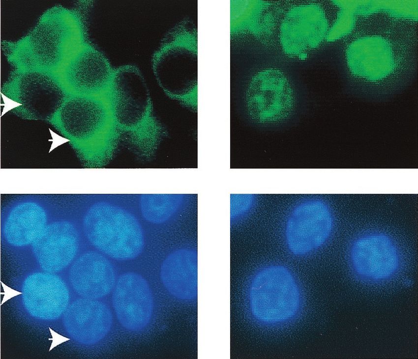
293T cells. As shown in Fig. 7B, most of the IRF-7 when
expressed alone was localized in the cytoplasm; however, when
LMP-1 was present, most of the IRF-7 localized in the nucleus
with a punctate appearance. In type III cells, endogenous
FIG. 6. IRF-7A and full-length LMP-1 are required for the efficient
activation of endogenous Tap-2. (A) RPA was performed with Tap-2 IRF-7 is also mainly localized in the nucleus (data not shown).
and GAPDH probes. Lanes 1 and 2, yeast RNA and RNA from the Therefore, LMP-1 augments the phosphorylation and nuclear
T-2 cell line; lanes 3 to 7, RNAs from transfected Akata cells. Plasmid translocation of IRF-7.
pcLMP-1 was transfected with pcDNA-IRF-7A (lane 3), IRF-7B (lane
4), IRF-7C (lane 5), IRF-7DN (lane 6), IRF-2 (lane 7), or pcDNA3
(lane 8). Lanes 9 to 12, pcDNA-IRF-7A was transfected with pcDNA3 DISCUSSION
(lane 9), full-length pcLMP-1 (lane 10), LMP CTAR-1 (lane 11), or
CTAR-2 (lane 12). Bottom panel for lanes 9 to 12, short exposure for The signal transduction pathway of LMP-1 has been exten-
GAPDH-protected area. Specific protections are shown. A represen- sively studied. The involvement of tumor necrosis factor re-
tative experiment is shown. (B) Relative Tap-2 levels from panel A. ceptor-associated factors and NF-B in the immediate steps of
Data were obtained by normalizing Tap-2 RNA levels to GAPDH
RNA levels with the use of a PhosphorImager. The column numbers the signal transduction pathway has been established conclu-
match the lanes in panel A. sively (9, 17, 25, 26, 28, 50). While induction of some genes by
LMP-1 may result from the direct activation of NF-B, other
genes may need a secondary mediator(s). Here we have shown
LMP-1 could activate the endogenous Tap-2 gene (lane 3). that the induction of Tap-2 requires IRF-7 as a secondary
These data are in agreement with the facts that IRF-7A is a mediator. First, Tap-2 expression is associated with both IRF-7
major form of IRF-7 and that LMP-1 primarily stimulates the and LMP-1 expression (Fig. 1) (46, 66). Second, LMP-1 in-
expression of IRF-7A (66, 67). duces Tap-2 expression in DG75, BJAB, and BL41 cells, in
To dissect the domain requirement of LMP-1 for the activation which IRF-7 can be stimulated (Fig. 4) (46) (data not shown).
of Tap-2, LMP-1 mutants were tested along with IRF-7A for their Also, ectopic expression of IRF-7 enhances the induction of
ability to increase endogenous Tap-2 RNA in Akata cells. Cur- Tap-2 by LMP-1 in DG75 cells (Fig. 4). Third, LMP-1 could
rently, two major functional domains of LMP-1 have been dis- not induce Tap-2 in Akata cells, in which IRF-7 could not be
sected, namely, CTAR-1 and CTAR-2. These two domains can induced by LMP-1. However, addition of ectopic IRF-7, which
activate different signaling molecules (16, 29). As shown in Fig. 6 artificially restores the defect, could activate the expression of
(lanes 9 to 12), full-length LMP-1 is most efficient for the induc- Tap-2 in Akata cells (Fig. 5). Fourth, the Tap-2 promoter
tion of Tap-2 with IRF-7A (lane 2). construct could be activated by IRF-7 and further enhanced by
LMP-1 enhances phosphorylation and nuclear transloca- LMP-1 specifically. Also, the activation of the Tap-2 promoter
tion of IRF-7. In order to understand the mechanism whereby was dependent on the intact ISRE sequence (Fig. 3). Fifth,348 ZHANG AND PAGANO J. VIROL.
Downloaded from http://jvi.asm.org/ on December 23, 2020 by guest
FIG. 7. LMP-1 facilitates the phosphorylation and nuclear translocation of IRF-7. (A) LMP-1 facilitates the phosphorylation of IRF-7. 293T
cells were transfected with IRF-7A with (lanes 2 and 4) or without (lanes 1 and 3) LMP-1 expression plasmid. At 24 h after transfection, cells were
labeled with [32P]orthophosphate, immunoprecipitates were separated in SDS-PAGE and transferred to a membrane, and Western blotting with
IRF-7 antibody was done after autoradiography for detection of phospho-IRF-7. Lanes 5 and 6, DG75 and Jijoye cells were labeled with
[32P]orthophosphate, respectively. More DG75 cell lysate was needed for immunoprecipitation in order to get equal amounts of total IRF-7. (B)
LMP-1 facilitates the nuclear translocation of IRF-7. 293T cells were transfected with EGFP-IRF-7A with or without the LMP-1 expression
plasmid. The subcellular localization of IRF-7 was examined 24 h after transfection. Several cells were not transfected with the EGFP-IRF-7A
plasmid. Arrows indicate corresponding cells. Magnification, ⫻400.
IRF-7 could bind specifically to the ISRE in the Tap-2 pro- LMP-1 regulates IRF-7 in a posttranslational manner (Fig. 7).
moter in vitro and to the Tap-2 promoter under physiological Therefore, LMP-1 has an intimate relationship with both the
conditions in vivo (Fig. 2). Sixth and finally, LMP-1 facilitates expression and activation of IRF-7.
the phosphorylation and nuclear translocation of IRF-7 (Fig. 7). Considering all the existing data, we propose an LMP-1-
It is apparent that IRF-7 is the most relevant IRF member triggered signal transduction pathway that leads to stimulation
for the activation of Tap-2 by LMP-1. First, both IRF-7 and of expression of IRF-7. LMP-1 further activates IRF-7 by
IRF-2 are associated with EBV type III latency, in which levels phosphorylation and nuclear translocation of the protein. Fi-
of Tap-2 are high. IRF-2 could also bind to the Tap-2 ISRE, as nally, activated IRF-7 mediates the activation of the Tap-2
determined by EMSA (data not shown). However, neither gene (Fig. 8).
IRF-2 nor LMP-1 plus IRF-2 could activate Tap-2 expression. The domain analysis of activation of Tap-2 by IRF-7 showed
Also, LMP-1 cannot induce the expression of IRF-2 (66). Sec- that full-length IRF-7A is required both for activation of the
ond, LMP-1 could specifically enhance the activation of the Tap-2 promoter construct and for the increase in endogenous
Tap-2 promoter by IRF-7 but not by IRF-1, although IRF-1 Tap-2 RNA (Fig. 3 and 6). These data suggest that the IRF-7A
can activate the Tap-2 promoter reporter (Fig. 3C). Third, activation domain, at least in the B-cell lines tested, is located
other IRFs tested, such as IRF-1, IRF-3, and ICSBP, are not in the C terminus beyond aa 227, because IRF-7B, which lacks
associated with type III latency (34, 66, 67). Fourth and finally, aa 227 to 255 in IRF-7A, could not activate Tap-2. This resultVOL. 75, 2001 LMP-1 AND IRF-7 ACTIVATE Tap-2 349
the survival both of the host and of the virus, a life-long coex-
istence may thus be maintained between the human host and
EBV. In support of such a notion, the LMP-1-positive immu-
noblastic B-cell lymphomas of the immunosuppressed are
highly susceptible to recovered patients’ cytolytic T cells or to
adoptive CTL therapy (7, 8, 38, 45).
In summary, our data provide direct evidence that IRF-7 is
involved in regulation of a crucial immune system gene and is
a secondary mediator for the LMP-1 viral protein in modulat-
ing the normal functions of the immune and inflammatory
responses.
FIG. 8. Model for induction of the Tap-2 gene by LMP-1. (Step 1) ACKNOWLEDGMENTS
Downloaded from http://jvi.asm.org/ on December 23, 2020 by guest
LMP-1 induces the expression of IRF-7. (Step 2) LMP-1 facilitates the We thank Nancy Raab-Traub, Paula Pitha, Ben Levi, Keiko Ozato,
phosphorylation and nuclear translocation of IRF-7. (Step 3). The Peter Howley, Tom Maniatis, Ho-sun Park, and Peter van Endert for
activated IRF-7 mediates the activation of the Tap-2 gene. providing valuable reagents for this work. We also thank Lihong Wu
for technical help, Shannon Kenney and Jenny Ting for critical reading
of the manuscript, and the UNC sequencing facility.
This work was supported in part by grants from the National Insti-
seems to contradict a previously reported activation domain tute of Allergy and Infectious Diseases (AI 42372-01) and from the
(corresponding to aa 107 to 224 of IRF-7A) obtained in the National Cancer Institute (CA 19014).
L929 mouse fibroblast line (1). The different cell lines that
were used for the study may contribute to this discrepancy. In REFERENCES
support of such a notion, IRF-7B could efficiently activate the 1. Au, W. C., P. A. Moore, D. W. LaFleur, B. Tombal, and P. M. Pitha. 1998.
Characterization of the interferon regulatory factor-7 and its potential role
Tap-2 promoter construct in FaDuHyg, an epithelial cell line in the transcription activation of interferon A genes. J. Biol. Chem. 273:
(data not shown). However, considering that IRF-7 is primarily 29210–29217.
a lymphoid factor, a conclusion based on B cells may be more 2. Beck, S., A. Kelly, E. Radley, F. Khurshid, R. P. Alderton, and J. Trowsdale.
1992. DNA sequence analysis of 66 kb of the human MHC class II region
relevant to its biological function. encoding a cluster of genes for antigen processing. J. Mol. Biol. 228:433–441.
IRF-7 appears to be a secondary mediator for the repression 3. Belich, M. P., and J. Trowsdale. 1995. Proteasome and class I antigen
of the EBV latency promoter (Qp) by LMP-1 (66). In this processing and presentation. Mol. Biol. Rep. 21:53–56.
4. Ben-Bassat, H., N. Goldblum, S. Mitrani, T. Goldblum, J. M. Yoffey, M. M.
paper, we have shown that IRF-7 is a secondary mediator for Cohen, Z. Bentwith, B. Ramot, E. Klein, and G. Klein. 1977. Establishment
Tap-2 activation. The domain requirements of IRF-7 for these in continuous culture of a new type of lymphocyte from a “Burkitt-like”
malignant lymphoma (line D.G.-75). Int. J. Cancer 19:27–33.
two biological effects are quite different. For Qp repression, 5. Calender, A., M. Billaud, J. P. Aubry, J. Banchereau, M. Vuillaume, and
the N-terminal DNA-binding domain is sufficient (67); how- G. M. Lenoir. 1987. Epstein-Barr virus (EBV) induces expression of B-cell
ever for Tap-2 activation, both the N- and C-terminal domains activation markers on in vitro infection of EBV-negative B-lymphoma cells.
Proc. Natl. Acad. Sci. USA 84:8060–8064.
of IRF-7 are required (Fig. 3 to 6). Whether IRF-7 is involved 6. Chen, F., J.-Z. Zou, L. DiRenzo, G. Winberg, L.-F. Hu, E. Klein, G. Klein,
in the induction of more LMP-1-regulated genes needs to be and I. Ernberg. 1995. A subpopulation of normal B cells latently infected
addressed. Since HLA 1, Tap-1, and Tap-2 are often induced with Epstein-Barr Virus resembles Burkitt lymphoma cells in expressing
EBNA-1 but not EBNA-2 or LMP-1. J. Virol. 69:3752–3758.
simultaneously for antigen processing (e.g., by treatment with 7. Crawford, D. H., and J. M. Edwards. 1982. Immunity to Epstein-Barr virus
IFN-␣, IFN-␥, or lipopolysaccharide), it is tempting to specu- in cyclosporin A-treated renal allograft recipients. Lancet i:1469–1470.
8. Crawford, D. H., P. Sweny, J. M. Edwards, G. Janossy, and A. V. Hoffbrand.
late that IRF-7 may also be involved in the regulation of the 1981. Long-term T-cell-mediated immunity to Epstein-Barr virus in renal-
Tap-1 and HLA I genes, both of which have been shown to be allograft recipients receiving cyclosporin A. Lancet i:10–12.
regulated by IRF-1 (39, 62). 9. Devergne, O., E. Hatzivassiliou, K. Izumi, K. Kaye, M. Kleijnen, E. Kieff,
and G. Mosialos. 1996. Association of TRAF1, TRAF2, and TRAF3 with an
What advantage does EBV gain by inducing Tap-2 and other Epstein-Barr virus LMP-1 domain important for B lymphocyte transforma-
HLA I-related genes? Type III EBV latency, an LMP-1-ex- tion: role in NF-B activation. Mol. Cell. Biol. 16:7098–7108.
pressing latency state, is established transiently in primary in- 10. Donato, L., H. de la Salle, D. Hanau, M. M. Tongio, M. Oswald, A. Vande-
venne, and J. Geisert. 1995. Association of HLA class I antigen deficiency
fection of human B cells in vivo (reviewed in references 19 and related to a TAP2 gene mutation with familial bronchiectasis. J. Pediatr.
44). Type III cells have enhanced growth, survival, and invasive 127:895–900.
11. Floettmann, J. E., and M. Rowe. 1997. Epstein-Barr virus latent membrane
potential, which allow the EBV-infected cells to proliferate protein-1 (LMP1) C-terminus activation region 2 (CTAR2) maps to the far
quickly, thereby putting the human host at risk. This rapid C-terminus and requires oligomerization for NF-B activation. Oncogene
proliferative process is checked after the development of EBV- 15:1851–1858.
12. Fries, K. L., W. E. Miller, and N. Raab-Traub. 1996. Epstein-Barr virus
specific primary cytotoxic T lymphocytes (CTL), which elimi- latent membrane protein 1 blocks p53-mediated apoptosis through the in-
nate these type III latency cells because of the activation of duction of the A20 gene. J. Virol. 70:8653–8659.
Tap-2 and other HLA I-related genes by LMP-1, and ensure 13. Furnari, F. B., M. D. Adams, and J. S. Pagano. 1992. Regulation of the
Epstein-Barr virus DNA polymerase gene. J. Virol. 66:2837–2845.
the safety of the host. In X-linked immunoproliferative dis- 14. Gires, O., U. Zimber-Strobl, R. Gonnella, M. Ueffing, G. Marschall, R.
ease, in which T-cell activation is defective (51), EBV infection Zeidler, D. Pich, and W. Hammerschmidt. 1997. Latent membrane protein
1 of Epstein-Barr virus mimics a constitutively active receptor molecule.
is lethal. Interestingly, EBV in the normal host still survives the EMBO J. 16:6130–6140.
CTL attack by establishing a type I-like latency state, in which 15. Heresbach, D., M. Alizadeh, J. F. Bretagne, A. Dabadie, J. F. Colombel, M.
LMP-1 is not expressed (6, 30, 31, 40, 56). This type I-like Pagenault, L. Heresbach, N. Berre, B. Genetet, M. Gosselin, and G. Semana.
1997. TAP gene transporter polymorphism in inflammatory bowel diseases.
latency can escape host immune surveillance, which ensures Scand. J. Gastroenterol. 32:1022–1027.
the survival of the virus. Because the whole process may ensure 16. Huen, D. S., S. A. Henderson, S. Croom-Carter, and M. Rowe. 1995. The350 ZHANG AND PAGANO J. VIROL.
Epstein-Barr virus latent membrane protein-1 (LMP1) mediates activation Exp. Pathol. 3:449–456.
of NF-B and cell surface phenotype via two effector regions in its carboxy- 42. Ragona, G., I. Ernberg, and G. Klein. 1980. Induction and biological char-
terminal cytoplasmic domain. Oncogene 10:549–560. acterization of the Epstein-Barr virus carried by the Jijoye lymphoma cell
17. Kaye, K., O. Devergne, J. Harada, K. Izumi, R. Yalamanchili, E. Kieff, and line. Virology 101:553–557.
G. Mosialos. 1996. Tumor necrosis factor receptor associated factor 2 is a 43. Reiss, M., T. Munoz-Antonia, J. M. Cowan, P. C. Wilkins, Z. L. Zhou, and
mediator of NF-B activation by latent infection membrane protein 1, the V. F. Vellucci. 1993. Resistance of human squamous carcinoma cells to
Epstein-Barr virus transforming protein. Proc. Natl. Acad. Sci. USA 93: transforming growth factor beta 1 is a recessive trait. Proc. Natl. Acad. Sci.
11085–11090. USA 90:6280–6284.
18. Kaye, K. M., K. M. Izumi, and E. Kieff. 1993. Epstein-Barr virus latent 44. Rickinson, A. B., and E. Kieff. 1996. Epstein-Barr virus, p. 2397–2446. In
membrane protein 1 is essential for B-lymphocyte growth transformation. B. N. Fields, D. M. Knipe, and P. M. Howley (ed.), Virology, 3rd ed.
Proc. Natl. Acad. Sci. USA 90:9150–9154. Lippincott-Raven Publishers, Philadelphia, Pa.
19. Kieff, E. 1996. Epstein-Barr virus and its replication, p. 2343–2396. In B. N. 45. Rooney, C. M., M. A. Roskrow, C. A. Smith, M. K. Brenner, and H. E.
Fields, D. M. Knipe, and P. M. Howley (ed.), Virology, 3rd ed. Lippincott- Heslop. 1998. Immunotherapy for Epstein-Barr virus-associated cancers.
Raven Publishers, Philadelphia, Pa. J. Natl. Cancer Inst. Monogr. 23:89–93.
20. Klein, G. 1994. Epstein-Barr virus strategy in normal and neoplastic B cells. 46. Rowe, M., R. Khanna, C. A. Jacob, V. Argaet, A. Kelly, S. Powis, M. Belich,
Cell 77:791–793. D. Croom-Carter, S. Lee, S. R. Burrows, J. Trowsdale, D. J. Moss, and A. B.
21. Kumagai, S., S. Kanagawa, A. Morinobu, M. Takada, K. Nakamura, S. Rickinson. 1995. Restoration of endogenous antigen processing in Burkitt’s
Sugai, E. Maruya, and H. Saji. 1997. Association of a new allele of the TAP2 lymphoma cells by Epstein-Barr virus latent membrane protein-1: coordinate
Downloaded from http://jvi.asm.org/ on December 23, 2020 by guest
gene, TAP2*Bky2 (Val577), with susceptibility to Sjogren’s syndrome. Ar- up-regulation of peptide transporters and HLA-class I antigen expression.
thritis Rheum. 40:1685–1692. Eur. J. Immunol. 25:1374–1384.
22. Laimins, L. A., P. Gruss, R. Pozzatti, and G. G. Khoury. 1984. Character- 47. Salter, R. D., and P. Cresswell. 1986. Impaired assembly and transport of
ization of enhancer elements in the long terminal repeat of Moloney murine HLA-A and -B antigens in a mutant T⫻B cell hybrid. EMBO J. 5:943–949.
sarcoma virus. J. Virol. 49:183–189. 48. Salter, R. D., D. N. Howell, and P. Cresswell. 1985. Genes regulating HLA
23. Liebowitz, D., D. Wang, and E. Kieff. 1986. Orientation and patching of the class I antigen expression in T-B lymphoblast hybrids. Immunogenetics 21:
latent infection membrane protein encoded by Epstein-Barr virus. J. Virol. 235–246.
58:233–237. 49. Sambrook, J. E., E. F. Fritsch, and T. Maniatis. 1989. Molecular cloning: a
24. Marie, I., J. E. Durbin, and D. E. Levy. 1998. Differential viral induction of laboratory manual, 2nd ed, vol. 3. Cold Spring Harbor Laboratory, Cold
distinct interferon-alpha genes by positive feedback through interferon reg- Spring Harbor, N.Y.
ulatory factor-7. EMBO J. 17:6660–6669. 50. Sandberg, M., W. Hammerschmidt, and B. Sugden. 1997. Characterization of
25. Miller, W., J. Cheshire, and N. Raab-Traub. 1998. Interaction of tumor LMP1’s association with TRAF1, TRAF2, and TRAF3. J. Virol. 71:4649–4656.
necrosis factor receptor-associated factor signaling proteins with the latent 51. Sayos, J., C. Wu, M. Morra, N. Wang, X. Zhang, D. Allen, S. van Schaik, L.
membrane protein 1 PXQXT motif is essential for induction of epidermal Notarangelo, R. Geha, M. G. Roncarolo, H. Oettgen, J. E. De Vries, G.
growth factor receptor expression. Mol. Cell. Biol. 18:2835–2844. Aversa, and C. Terhorst. 1998. The X-linked lymphoproliferative-disease
26. Miller, W. E., J. L. Cheshire, A. S. Baldwin, and N. Raab-Traub. 1997. The gene product SAP regulates signals induced through the co-receptor SLAM.
NPC derived C15 LMP1 protein confers enhanced activation of NF-B and Nature 395:462–469.
induction of the EGFR in epithelial cells. Oncogene 16:1869–1877. 52. Silvennoinen-Kassinen, S., I. Ikaheimo, and A. Tiilikainen. 1997. TAP1 and
27. Miller, W. E., H. S. Earp, and N. Raab-Traub. 1995. The Epstein-Barr virus TAP2 genes in nickel allergy. Int. Arch. Allergy Immunol. 114:94–96.
latent membrane protein 1 induces expression of the epidermal growth 53. Takada, K. 1984. Cross-linking of cell surface immunoglobulins induces
factor receptor. J. Virol. 69:4390–4398. Epstein-Barr virus in Burkitt’s lymphoma lines. Int. J. Cancer 33:27–32.
28. Miller, W. E., G. Mosialos, E. Kieff, and N. Raab-Traub. 1997. Epstein-Barr 54. Tanaka, K. 1994. Role of proteasomes modified by interferon-gamma in
virus LMP1 induction of the epidermal growth factor receptor is mediated antigen processing. J. Leukocyte Biol. 56:571–575.
through a TRAF signaling pathway distinct from NF-B activation. J. Virol. 55. Teisserenc, H., W. Schmitt, N. Blake, R. Dunbar, S. Gadola, W. L. Gross, A.
71:586–594. Exley, and V. Cerundolo. 1997. A case of primary immunodeficiency due to
29. Mitchell, T., and B. Sugden. 1995. Stimulation of NF-B-mediated transcrip- a defect of the major histocompatibility gene complex class I processing and
tion by mutant derivatives of the latent membrane protein of Epstein-Barr presentation pathway. Immunol. Lett. 57:183–187.
virus. J. Virol. 69:2968–2976. 56. Tierney, R., N. Steven, L. Young, and A. Rickinson. 1994. Epstein-Barr virus
30. Miyashita, E. M., B. Yang, G. J. Babcock, and D. A. Thorley-Lawson. 1997. latency in blood mononuclear cells: analysis of viral gene transcription dur-
Identification of the site of Epstein-Barr virus persistence in vivo as a resting ing primary infection and in the carrier state. J. Virol. 68:7374–7378.
B cell. J. Virol. 71:4882–4891. 57. Townsend, A., and J. Trowsdale. 1993. The transporters associated with
31. Miyashita, E. M., B. Yang, K. M. C. Lam, D. H. Crawford, and D. A. antigen presentation. Semin. Cell Biol. 4:53–61.
Thorley-Lawson. 1995. A novel form of Epstein-Barr virus latency in normal 58. van Endert, P. M., R. Tampe, T. H. Meyer, R. Tisch, J. F. Bach, and H. O.
B cells in vivo. Cell 80:593–601. McDevitt. 1994. A sequential model for peptide binding and transport by the
32. Monaco, J. J., and D. Nandi. 1995. The genetics of proteasomes and antigen transporters associated with antigen processing. Immunity 1:491–500.
processing. Annu. Rev. Genet. 29:729–754. 59. Wang, D., D. Leibowitz, and E. Kieff. 1985. An EBV membrane protein
33. Nguyen, H., J. Hiscott, and P. M. Pitha. 1997. The growing family of inter- expressed in immortalized lymphocytes transforms established rodent cells.
feron regulatory factors. Cytokine Growth Factor Rev. 8:293–312. Cell 43:831–840.
34. Nonkwelo, C., I. K. Ruf, and J. Sample. 1997. Interferon-independent and 60. Wang, F., C. Gregory, C. Sample, M. Rowe, D. Liebowitz, R. Murray, A.
-induced regulation of Epstein-Barr virus EBNA-1 gene transcription in Rickinson, and E. Kieff. 1990. Epstein-Barr virus latent membrane protein
Burkitt lymphoma. J. Virol. 71:6887–6897. (LMP1) and nuclear proteins 2 and 3C are effectors of phenotypic changes
35. Nonkwelo, C., J. Skinner, A. Bell, A. Rickinson, and J. Sample. 1996. Tran- in B lymphocytes: EBNA-2 and LMP1 cooperatively induce CD23. J. Virol.
scription start sites downstream of the Epstein-Barr virus (EBV) Fp pro- 64:2309–2318.
moter in early-passage Burkitt lymphoma cells define a fourth promoter for 61. Wathelet, M. G., C. H. Lin, B. S. Parekh, L. V. Ronco, P. M. Howley, and T.
expression of the EBV EBNA-1 protein. J. Virol. 70:623–627. Maniatis. 1998. Virus infection induces the assembly of coordinately activated
36. Orlando, V., H. Strutt, and R. Paro. 1997. Analysis of chromatin structure by transcription factors on the IFN-beta enhancer in vivo. Mol. Cell 1:507–518.
in vivo formaldehyde cross-linking. Methods 11:205–214. 62. White, L. C., K. L. Wright, N. J. Felix, H. Ruffner, L. F. Reis, R. Pine, and
37. Pagano, J. S. 1987. Pathogenesis and treatment of Epstein-Barr virus infec- J. P.-Y. Ting. 1996. Regulation of LMP2 and TAP1 genes by IRF-1 explains
tion. J. Exp. Pathol. 3:441–448. the paucity of CD8⫹ T cells in IRF-1⫺/⫺ mice. Immunity 5:365–367.
38. Papadopoulos, E. B., M. Ladanyi, D. Emanuel, S. Mackinnon, F. Boulad, 63. York, I. A., A. L. Goldberg, X. Y. Mo, and K. L. Rock. 1999. Proteolysis and
M. H. Carabasi, H. Castro-Malaspina, B. H. Childs, A. P. Gillio, T. N. Small, class I major histocompatibility complex antigen presentation. Immunol.
et al. 1994. Infusions of donor leukocytes to treat Epstein-Barr virus-asso- Rev. 172:49–66.
ciated lymphoproliferative disorders after allogeneic bone marrow trans- 64. Yoshizaki, T., H. Sato, M. Furukawa, and J. S. Pagano. 1998. The expression
plantation. N. Engl. J. Med. 330:1185–1191. of matrix metalloproteinase 9 is enhanced by Epstein-Barr virus latent mem-
39. Penninger, J. M., C. Sirard, H. W. Mittrucker, A. Chidgey, I. Kozieradzki, brane protein 1. Proc. Natl. Acad. Sci. USA 95:3621–3626.
M. Nghiem, A. Hakem, T. Kimura, E. Timms, R. Boyd, T. Taniguchi, T. 65. Zhang, L., and J. S. Pagano. 1999. Interferon regulatory factor 2 represses
Matsuyama, and T. W. Mak. 1997. The interferon regulatory transcription the Epstein-Barr virus BamHI Q latency promoter in type III latency. Mol.
factor IRF-1 controls positive and negative selection of CD8⫹ thymocytes. Cell. Biol. 19:3216–3223.
Immunity 7:243–254. 66. Zhang, L., and J. S. Pagano. 2000. Interferon regulatory factor 7 is induced
40. Qu, L., and D. T. Rowe. 1992. Epstein-Barr virus latent gene expression in by Epstein-Barr virus latent membrane protein 1. J. Virol. 74:5748–5757.
uncultured peripheral blood lymphocytes. J. Virol. 66:3715–3724. 67. Zhang, L., and J. S. Pagano. 1997. IRF-7, a new interferon regulatory
41. Raab-Traub, N., K. Flynn, C. Klein, G. Pizza, C. De-Vinci, L. Occhiuzzi, G. factor associated with Epstein-Barr virus latency. Mol. Cell. Biol. 17:
Farneti, U. Caliceti, and E. Pirodda. 1987. EBV associated malignancies. J. 5748–5757.You can also read