Selective inhibition of JAK3 signaling is sufficient to reverse alopecia areata
←
→
Page content transcription
If your browser does not render page correctly, please read the page content below
Selective inhibition of JAK3 signaling is sufficient to reverse
alopecia areata
Zhenpeng Dai, … , Yuqian Chang, Angela M. Christiano
JCI Insight. 2021;6(7):e142205. https://doi.org/10.1172/jci.insight.142205.

 Research Article   Inflammation

The Janus kinase/signal transducers and activators of transcription (JAK/STAT) are key intracellular mediators in the
signal transduction of many cytokines and growth factors. Common γ chain cytokines and interferon-γ that use the
JAK/STAT pathway to induce biological responses have been implicated in the pathogenesis of alopecia areata (AA), a T
cell–mediated autoimmune disease of the hair follicle. We previously showed that therapeutic targeting of JAK/STAT
pathways using the first-generation JAK1/2 inhibitor, ruxolitinib, and the pan-JAK inhibitor, tofacitinib, was highly effective
in the treatment of human AA, as well as prevention and reversal of AA in the C3H/HeJ mouse model. To better define the
role of individual JAKs in the pathogenesis of AA, in this study, we tested and compared the efficacy of several next-
generation JAK-selective inhibitors in the C3H/HeJ mouse model of AA, using both systemic and topical delivery. We
found that JAK1-selective inhibitors as well as JAK3-selective inhibitors robustly induced hair regrowth and decreased AA-
associated inflammation, whereas several JAK2-selective inhibitors failed to restore hair growth in treated C3H/HeJ mice
with AA. Unlike JAK1, which is broadly expressed in many tissues, JAK3 expression is largely restricted to hematopoietic
cells. Our study demonstrates inhibiting JAK3 signaling is sufficient to prevent and reverse disease in the preclinical
model of AA.

Find the latest version:
https://jci.me/142205/pdf
RESEARCH ARTICLE

                                           Selective inhibition of JAK3 signaling
                                           is sufficient to reverse alopecia areata
                                           Zhenpeng Dai,1 James Chen,1 Yuqian Chang,1 and Angela M. Christiano1,2
                                           Department of Dermatology and 2Department of Genetics and Development, Columbia University, Vagelos College of
                                           1

                                           Physicians and Surgeons, New York, New York, USA.

                                                   The Janus kinase/signal transducers and activators of transcription (JAK/STAT) are key intracellular
                                                   mediators in the signal transduction of many cytokines and growth factors. Common γ chain
                                                   cytokines and interferon-γ that use the JAK/STAT pathway to induce biological responses have been
                                                   implicated in the pathogenesis of alopecia areata (AA), a T cell–mediated autoimmune disease
                                                   of the hair follicle. We previously showed that therapeutic targeting of JAK/STAT pathways using
                                                   the first-generation JAK1/2 inhibitor, ruxolitinib, and the pan-JAK inhibitor, tofacitinib, was highly
                                                   effective in the treatment of human AA, as well as prevention and reversal of AA in the C3H/HeJ
                                                   mouse model. To better define the role of individual JAKs in the pathogenesis of AA, in this study,
                                                   we tested and compared the efficacy of several next-generation JAK-selective inhibitors in the C3H/
                                                   HeJ mouse model of AA, using both systemic and topical delivery. We found that JAK1-selective
                                                   inhibitors as well as JAK3-selective inhibitors robustly induced hair regrowth and decreased AA-
                                                   associated inflammation, whereas several JAK2-selective inhibitors failed to restore hair growth
                                                   in treated C3H/HeJ mice with AA. Unlike JAK1, which is broadly expressed in many tissues, JAK3
                                                   expression is largely restricted to hematopoietic cells. Our study demonstrates inhibiting JAK3
                                                   signaling is sufficient to prevent and reverse disease in the preclinical model of AA.

                                           Introduction
                                           Alopecia areata (AA) is an autoimmune disease of the hair follicle (HF) that ranges in presentation from
                                           circular patches on the scalp to complete hair loss and is associated with an enormous psychological bur-
                                           den to patients (1, 2). The etiology of AA is not completely understood but likely involves a combination
                                           of genetic predisposition and environmental triggers (3). We previously showed that cytotoxic natural kill-
                                           er group 2 member D–positive (NKG2D+), CD8+ T cells accumulate in the skin and contribute to HF
                                           destruction (4, 5). The pathogenesis of AA is also associated with the overexpression of proinflammatory
Conflict of interest: Columbia             cytokines, such as interferon-γ (IFN-γ) and common γ chain (γc) cytokines, which break down HF immune
University has licensed patents            privilege and promote the survival and function of cytotoxic T lymphocytes in affected skin (6, 7). Notably,
(patent title: Methods for treating        these proinflammatory cytokines signal through their receptors via the family of Janus kinase/signal trans-
hair loss disorders; patent number:
                                           ducers and activators of transcription (JAK/STAT).
US9198911B2) on the use of JAK
                                                JAK/STAT pathways play an essential role in both innate and adaptive immunity as well as hema-
inhibitors in alopecia areata to Aclaris
Therapeutics, Inc. AMC is a consultant     topoiesis. Unrestrained activation of the JAK/STAT pathways contributes to a number of autoimmune
to Aclaris Therapeutics, Almirall, and     diseases and proliferative disorders, making JAKs an attractive target for pharmacologic manipulation in
Janssen and a shareholder of Aclaris       the treatment of such conditions (8, 9). Indeed, small molecule JAK inhibitors (JAKi) demonstrated clin-
Therapeutics.                              ical efficacy in the treatment of rheumatoid arthritis and myelofibrosis, as well as other autoimmune and
Copyright: © 2021, Dai et al. This is      malignant proliferative disorders (10–12). AA is characterized by dysregulation of JAK/STAT activity, in
an open access article published under     particular, the γc cytokine and IFN-γ signaling pathway (3, 4). Our lab recently pioneered the use of the
the terms of the Creative Commons          JAK1/2 inhibitor ruxolitinib and baricitinib, as well as the pan-JAK inhibitor, tofacitinib, in the treatment
Attribution 4.0 International License.     of human AA (3, 13–15). However, the relative contribution of JAK1, JAK2, and JAK3 inhibition to the
Submitted: July 13, 2020                   therapeutic benefit of ruxolitinib, baricitinib, and tofacitinib in AA has not been investigated.
Accepted: February 25, 2021                     Recently, a number of JAK-selective inhibitors have entered clinical trials for the treatment of various
Published: April 8, 2021                   malignancies and inflammatory diseases. For example, INCB039110, a JAK1-selective inhibitor, showed
                                           efficacy in phase II trials of chronic plaque psoriasis and myelofibrosis (16, 17). The JAK2-selective inhib-
Reference information: JCI Insight.
2021;6(7):e142205.                         itor CEP-33779 appeared to be efficacious in mouse models of systemic lupus erythematosus (18). Fedra-
https://doi.org/10.1172/jci.               tinib and pacritinib are additional JAK2-selective inhibitors that showed therapeutic efficacy in a murine
insight.142205.                            model of myeloproliferative disease as well as myeloid and lymphoid malignancies, respectively (19, 20).

                                                                                                                                                            1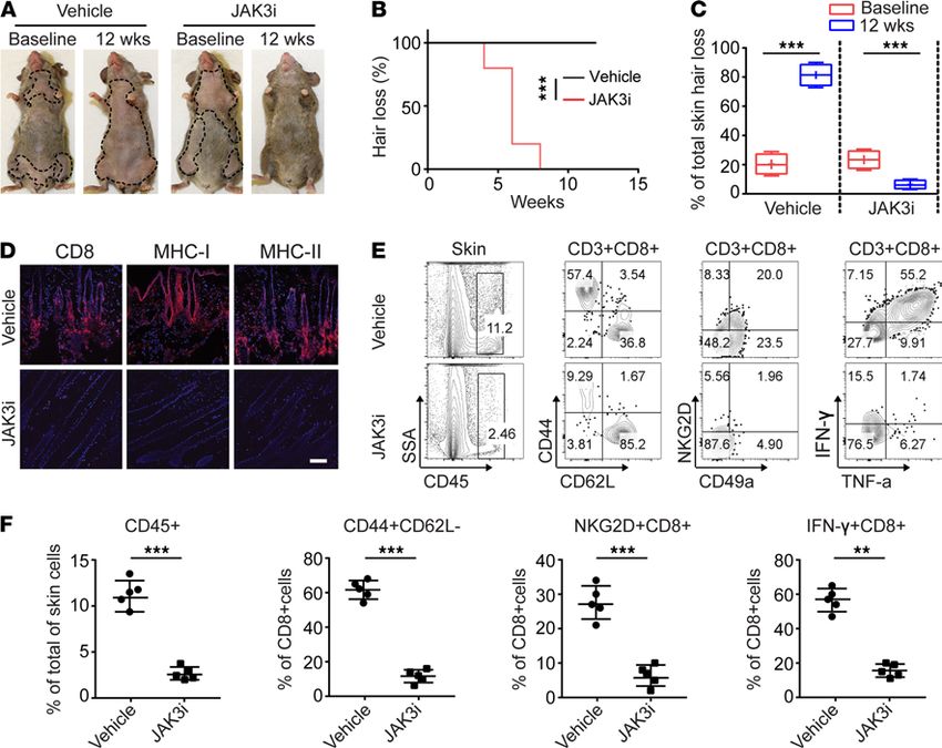
RESEARCH ARTICLE
The irreversible covalent JAK3-selective inhibitor PF-06651600 was shown to be effective in rodent models
of arthritis and mouse models of multiple sclerosis (21).
Our previous gene expression studies suggested a clear role for JAK1 and JAK3 in AA disease patho-
genesis, but notably, not for JAK2 signaling (4, 13–15). Due to the essential role of JAK2 in hematopoiesis,
JAK2 inhibition is believed to be the source of several known side effects in other diseases (19). Therefore,
highly selective inhibition of JAK1 or JAK3, with no off-target activity against other JAKs, could potentially
enhance efficacy and reduce the risk of undesirable side effects. However, it is not known whether selective
inhibition of JAK1 or JAK3 alone is sufficient to disrupt AA cytokine signaling and ameliorate the inflam-
matory processes. The availability of selective JAKi now enables the pharmacological investigation of IFN-γ
and γc cytokine signaling, since JAK2-selective inhibitors target IFN-γ signaling, JAK3-selective inhibitors
target γc cytokine signaling, and JAK1-selective inhibitors target both cytokine signaling pathways.
To interrogate the role of individual JAKs in AA, we used a panel of JAK-selective inhibitors to
treat C3H/HeJ mice with AA. We found that simultaneous IFN-γ and γc cytokine signaling blockade by
JAK1-selective inhibitors, as well as inhibition of γc signaling alone with JAK3-selective inhibitors, potently
induced hair growth in C3H/HeJ mice with AA. In contrast, JAK2-selective inhibitors failed to restore hair
regrowth, indicating that inhibition of IFN-γ alone is not sufficient for treatment of AA in the C3H/HeJ
mouse model of AA. Our results establish that γc cytokine signaling pathways are the primary therapeutic
targets in AA treatment and that reversal can be achieved with either JAK1 inhibition or JAK3 inhibition.
Results
Inhibition of cytokine-dependent signaling by JAK-selective inhibitors. To investigate the specificity of JAKi on cyto-
kine-dependent STAT phosphorylation in vitro using different primary mouse cell types, first, we stimulated
murine splenocytes pretreated with INCB039110 (JAK1i), CEP-33779 (JAK2i), or PF-06651600 (JAK3i)
with various cytokines (Supplemental Table 1; supplemental material available online with this article;
https://doi.org/10.1172/jci.insight.142205DS1). We tested IL-7 and IL-15, which signal through JAK1/3
and lead to STAT5 tyrosine phosphorylation, and found that INCB039110 and PF-06651600 (but not CEP-
33779) robustly inhibited this response (Figure 1, A and B). Next, we used IL-10 to induce STAT3 tyrosine
phosphorylation via JAK1/TYK2 and found that it was specifically inhibited by INCB039110 (Figure 1C).
In primary mouse macrophages, we observed that only CEP-33779 (but not INCB039110 or PF-06651600)
inhibited JAK2-dependent GM-CSF signaling (Figure 1D). IFN-γ signaling through JAK1/2-induced STAT1
tyrosine phosphorylation, which was inhibited by INCB039110 or CEP-33779 (but not by PF-06651600)
(Figure 1E). Finally, in mouse HF dermal sheath cells, we found that INCB039110 or CEP-33779 inhib-
ited IFN-γ–induced STAT1 tyrosine phosphorylation (Figure 1F). Together, these data demonstrated that
INCB039110, CEP-33779, and PF-06651600 selectively inhibited their respective JAKs in vitro.
Induction of NKG2D+CD8+ T cells was blocked by JAK1- or JAK3-selective inhibitor. We previously showed
that cytotoxic NKG2D+CD8+ T cells are both necessary and sufficient to induce AA and that IL-15 is a
critical γc cytokine for NKG2D+CD8+ T cell induction in AA (4). To investigate the role of JAK signaling
in NKG2D+CD8+ T cell induction, we stimulated naive CD8+ T cells with IL-15 in the presence of individ-
ual JAKi. IL-15 robustly induced NKG2D+CD8+ T cells, and both INCB039110 and PF-06651600 abol-
ished this effect (Figure 1G). IL-15 also augmented the cytotoxic function of CD8+ T cells by increasing
the production of granzymes and perforin. This effect was markedly reduced in CD8+ T cells treated with
INCB039110 or PF-06651600 compared with CEP-33779 (Figure 1H). Together, these data demonstrate
that JAK1- or JAK3-selective inhibitors selectively inhibited γc cytokine signaling.
Skin infiltrating CD8+ T cells were responsive to γc cytokine stimulation. Our previous RNA-Seq analysis
showed that γc cytokines and their receptors, including IL-7Rα (CD127) and IL-15Rβ (CD122), were
upregulated in alopecic skin from both human patients and C3H/HeJ mice with AA (4). We examined the
expression of CD122 and CD127 in skin infiltrating CD8+ T cells at the protein level. We observed similar
levels of CD122 or CD127 on CD8+ T cells between lymphoid organs and skin (Supplemental Figure 2A).
Furthermore, IL-7 and IL-15 induced comparable levels of STAT5 tyrosine phosphorylation in CD8+ T
cells between lymphoid organs and skin (Supplemental Figure 2B). These data indicate that skin infiltrating
CD8+ T cells are likely dependent on γc cytokines for their survival and function.
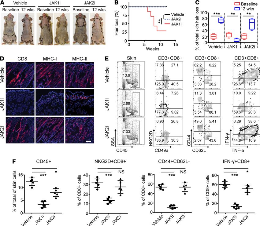
Systemic treatment with JAK1-selective inhibitor reversed AA. We previously showed that the JAK1/2 inhib-
itors ruxolitinib and baricitinib, which block both IFN-γ and γc cytokine signaling, showed therapeutic effi-
cacy in the treatment of AA in humans and mice (4, 14). However, the relative contribution of JAK1 versus
JCI Insight 2021;6(7):e142205 https://doi.org/10.1172/jci.insight.142205 2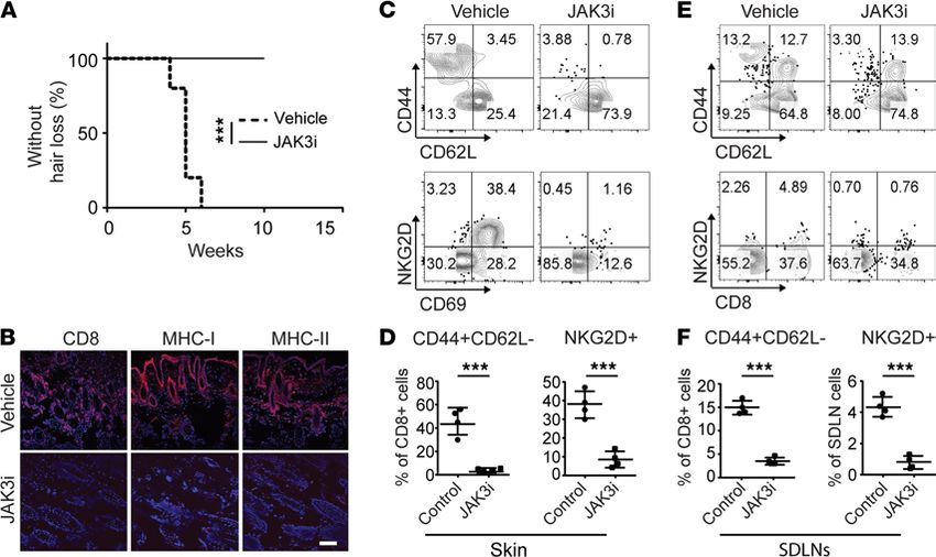
RESEARCH ARTICLE

                                 Figure 1. Inhibition of cytokine-dependent signaling by JAK-selective inhibitors. (A–E) C3H/HeJ mouse splenocytes
                                 were pretreated with 1 μM of INCB039110 (JAK1i), CEP-33779 (JAK2i), or PF-06651600 (JAK3i) or vehicle control for 1 hour
                                 at 37°C. The treated cells were stimulated with IL-7 (20 ng/mL), IL-15 (40 ng/mL), IL-10 (50 ng/mL), GM-CSF (50 ng/mL),
                                 or IFN-γ (50 ng/mL) for 20 minutes at 37°C. Phosphorylated STAT (p-STAT) expression in indicated cell subsets was
                                 presented as representative plots and mean fluorescence intensity (MFI). CD3+CD8+ cells (A and B), CD19+ cells (C), CD11b+
                                 cells (D), and CD3+ cells (E) were gated for p-STAT expression. (F) C3H/HeJ mouse HF dermal sheath cells were pretreated
                                 with 1 μM of INCB039110, CEP-33779, or vehicle control for 1 hour at 37°C. The treated cells were then stimulated with
                                 IFN-γ (50 ng/mL) for indicated time points at 37°C and analyzed by Western blotting for p-STAT1, total STAT1, and the
                                 housekeeping protein β-actin. (G and H) C3H/HeJ mouse splenocytes were pretreated with 500 nM of indicated JAKi or
                                 vehicle control for 1 hour at 37°C. The treated cells from C3H/HeJ mice were then stimulated with 20 ng/mL IL-15 and
                                 250 nM of indicated JAKi at 37°C for 72 hours. (G) Expression of NKG2D and (H) expression of Granzyme B (GZMB) and
                                 Perforin 1 (PRF1) by CD8+ T cells presented as representative plots and MFI. The data shown are from 1 representative
                                 experiment out of 2 replicates. W/O, without treatment.

                                 JAK2 in the pathogenesis of AA is not clear. To define the role of JAK1 and JAK2 in AA, we took advantage
                                 of the next generation of JAK1- and JAK2-selective inhibitors to block JAK1 or JAK2 signaling. C3H/HeJ
                                 AA mice were treated systemically with INCB039110 (JAK1i), CEP-33779 (JAK2i), or vehicle for 12 weeks.
                                 We observed that the mice treated with JAK1i showed robust hair regrowth compared with vehicle-treated
                                 mice, which displayed hair loss (Figure 2, A–C). In contrast, CEP-33779 had no effect in restoring hair
                                 regrowth and did not prevent further progressive hair loss in all treated mice (Figure 2, A–C). Similar to the
                                 effect of CEP-33779 on AA, 2 additional JAK2i (fedratinib and pacritinib) also failed to reverse the disease
                                 (Supplemental Table 1, Supplemental Figure 3, and Supplemental Figure 4). Consistent with hair regrowth,
                                 immunohistological analysis of skin revealed that JAK1i-treated mice showed substantially reduced histo-
                                 logical markers of the disease (CD8, MHC class I, and MHC class II) (Figure 2D). In contrast, the skin of
                                 the JAK2i-treated mice displayed massive CD8+ infiltrates and increased expression of MHC class I and
                                 MHC class II (Figure 2D and Supplemental Figure 3C), similar to vehicle treated.
                                      We next determined the composition of skin infiltrating immune cells after JAKi treatment. We observed
                                 a significantly decreased frequency of CD45+ immune cells, CD44+CD62L–CD8+ effector/memory T cells
                                 (CD8+ TE/M), and IFN-γ–producing CD8+ T cells, in the skin of JAKi1-treated mice, and to a lesser extent,
                                 in the skin of JAK2i-treated mice compared with controls (Figure 2, E and F, and Supplemental Figure 3D).

JCI Insight 2021;6(7):e142205 https://doi.org/10.1172/jci.insight.142205                                                                                  3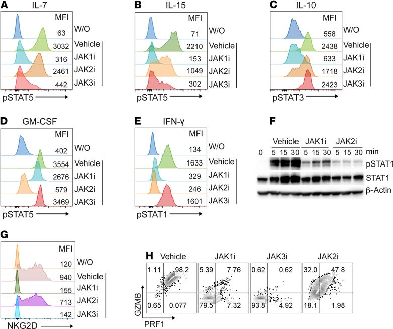
RESEARCH ARTICLE

Figure 2. JAK1-selective inhibitor treatment reversed AA. Five C3H/HeJ AA mice per group were treated systemically with INCB039110 (JAK1i) or CEP-33779
(JAK2i) at a dosage of 50 mg/kg for 12 weeks. (A) Representative images of individual JAK3i or vehicle-treated C3H/HeJ mice before or after 12 weeks’ treatment.
(B) Time course of hair regrowth shown as weeks after treatment. **P < 0.01, log-rank test. (C) Percentage of total skin hair loss or regrowth shown before and
after treatment. The box plots depict the minimum and maximum values (whiskers), the upper and lower quartiles, and the median. The length of the box
represents the interquartile range. **P < 0.01, ***P < 0.001 (unpaired Student’s t test). (D) Representative immunofluorescence images of skin sections from
JAKi- or control-treated mice, stained with anti-CD8, anti–MHC class I, or anti–MHC class II mAbs. Scale bar: 200 μm. (E) and (F) Percentages of skin infiltrating
CD45+ leukocytes, CD44+CD62L–CD8+ T cells, NKG2D+CD8+ T cells, as well as IFN-γ–producing CD8+ T cells within indicated populations within the skin after JAK3i
treatment. *P < 0.05, ***P < 0.001 (1-way ANOVA). Two replicate experiments were performed for a total of 10 mice per group.

                                    Further, treatment with JAK1i significantly reduced the absolute numbers and frequencies of CD8+ TE/M,
                                    CD8+NKG2D+ T cells, and IFN-γ–producing CD8+ T cells within skin draining lymph nodes (SDLNs)
                                    compared with controls and JAK2i-treated mice (Supplemental Figure 5). Since JAK1i inhibits both γc
                                    cytokine and IFN-γ signaling, and JAK2i inhibits IFN-γ signaling, these data indicate that inhibition of γc
                                    cytokine signaling may be sufficient for reversal of AA.
                                         Systemic treatment with JAK3-selective inhibitor prevented the onset of AA in C3H/HeJ grafted mice. We next
                                    postulated whether inhibition of γc cytokine signaling alone was sufficient to prevent the onset of AA
                                    using JAK3-selective inhibitor. C3H/HeJ skin grafted mice were treated with the JAK3-selective inhibitor
                                    PF-06651600 (JAK3i) or vehicle for 4 weeks. The mice were scored for signs of hair loss. As expected, all
                                    control mice developed AA by week 7 after skin grafting (Figure 3A). In contrast, the mice that were treated
                                    with systemic JAK3i showed no signs of hair loss during the time of observation (Figure 3A). Image analysis
                                    of skin revealed that JAK3i-treated mice showed substantially reduced AA-associated skin inflammation, as
                                    shown by staining of histological markers of the disease (Figure 3B). Flow cytometric analysis of CD45+ sub-
                                    set composition in the skin showed that JAK3i treatment significantly reduced the frequencies of CD8+ TE/M,

JCI Insight 2021;6(7):e142205 https://doi.org/10.1172/jci.insight.142205                                                                                        4
RESEARCH ARTICLE
Figure 3. JAK3-selective inhibitor treatment prevented the onset of AA. C3H/HeJ grafted mice were given PF-06651600 (JAK3i) at a dosage of 30 mg/kg for
4 weeks. (A) Survival curve analysis depicts the hair loss between JAK3i- and control-treated mice. ***P < 0.01, log-rank test. (B) Representative immunoflu-
orescence images of skin sections from JAK3i- or control-treated mice, stained with anti-CD8, anti–MHC class I, or anti–MHC class II mAbs. Scale bar: 200 μm.
(C) Representative FACS plots of the skin single cell in the viable cell gate were acquired for each sample. (D) Summary graphs of the percentages of CD44+C-
D62L–CD8+ T cells as well as NKG2D+CD8+ T cells within the skin after treatment. ***P < 0.001 (unpaired Student’s t test). (E) Representative FACS plots of
SDLNs in the viable cell gate were acquired for each sample. (F) Summary graphs of the percentages of CD44+CD62L–CD8+ T cells as well as NKG2D+CD8+ T cells
within the SDLNs after treatment. ***P < 0.001 (unpaired Student’s t test). Two replicate experiments were performed for 10 mice per group.
as well as NKG2D+CD8+ T cells, compared with controls (Figure 3, C and D). We previously showed that
SDLNs are the primary sites for the development of alopecic T cells (4). Here, we observed that JAK3i sig-
nificantly reduced the percentages of CD8+ TE/M as well as NKG2D+CD8+ T cells within SDLNs compared
with controls (Figure 3, E and F). Taken together, our data demonstrate that the JAK3-selective inhibitor was
sufficient to block alopecic T cell proliferation and function and prevented the development of disease.
Systemic treatment with JAK3-selective inhibitor reversed AA. We next assessed the requirement for γc cyto-
kines in AA and the potential of JAK3i to regrow hair in AA mice. After 12 weeks of systemic treatment
with PF-06651600 (JAK3i), we observed robust hair regrowth in all treated mice compared with control
mice, which showed progressive hair loss (Figure 4, A–C). Consistent with hair regrowth, JAK3i-treated
mice showed substantially reduced AA-associated skin inflammation (Figure 4D). Flow cytometric anal-
ysis of the CD45+ subset composition in the skin revealed that JAK3i-treated mice showed a significantly
decreased frequency of CD8+ TE/M, as well as IFN-γ–producing CD8+ T cells, compared with control mice
(Figure 4, E and F). Further, treatment with JAK3i significantly reduced the absolute numbers and frequen-
cies of CD8+ TE/M, CD8+NKG2D+ T cells, as well as IFN-γ–producing CD8+ T cells within SDLNs com-
pared with controls (Supplemental Figure 6, A and B). Taken together, these results underscore the critical
role of γc cytokine signaling in established disease. We showed that blockade of γc cytokine signaling by
JAK3 inhibition alone suppressed alopecic T cell proliferation and function, and decreased their infiltration
into the skin, leading to the reversal of AA.
Molecular responses of C3H/HeJ AA mice treated with JAK-selective inhibitors. To define the molecular response
to JAKi treatment, we performed RNA-Seq analysis on a series of skin biopsies taken before and after sys-
temic treatment with INCB039110, CEP-33779, PF-06651600, ruxolitinib (JAK1/2i), tofacitinib (pan-JA-
Ki), or vehicle control (Supplemental Table 1). We previously defined biomarkers and molecular responses
in patients with AA treated with ruxolitinib or tofacitinib (13, 15). We performed a differential expression
analysis between each pre- and posttreatment cohort pair of INCB039110-, CEP-33779–, and PF-06651600–
treated skin RNA-Seq samples and performed unsupervised hierarchical clustering on the overlap of the gene
lists from all 3 treatments (Supplemental Table 3). The CEP-33779 treatment elicited no significant change
JCI Insight 2021;6(7):e142205 https://doi.org/10.1172/jci.insight.142205 5RESEARCH ARTICLE
Figure 4. JAK3-selective inhibitor treatment reversed AA. Five C3H/HeJ AA mice per group were treated systemically with PF-06651600 (JAK3i) at a dosage of
30 mg/kg for 12 weeks. (A) Representative images of individual JAK3i- or control-treated C3H/HeJ mice before or after 12 weeks’ treatment. (B) Time course of
hair regrowth shown as weeks after treatment. ***P < 0.001, log-rank test. (C) Percentage of total skin hair loss or regrowth shown before and after treatment.
The box plots depict the minimum and maximum values (whiskers), the upper and lower quartiles, and the median. The length of the box represents the inter-
quartile range. ***P < 0.001 (unpaired Student’s t test). (D) Representative immunofluorescence images of skin sections from JAK3i- or vehicle-treated mice,
stained with anti-CD8, anti–MHC class I, or anti–MHC class II mAbs. Scale bar: 200 μm. (E) and (F) Percentages of skin infiltrating CD45+ leukocytes, CD44+C-
D62L–CD8+ T cells, NKG2D+CD8+ T cells, as well as IFN-γ–producing CD8+ T cells within the skin after treatment. **P < 0.01, ***P < 0.001 (unpaired Student’s t
test). Two replicate experiments were performed for a total of 10 mice per group.
in differential expression at appropriate statistical thresholds compared with INCB039110-, PF-06651600–,
ruxolitinib-, or tofacitinib-treated mice. Molecular responses were overall consistent across all treatments that
resulted in responses (Supplemental Figure 7A). Notably, PF-06651600 treatment elicited the most robust
molecular response in terms of both the number of genes modulated (number of inversions in the heatmap),
as well as the degree to which they were differentially expressed (overall intensity of color in the heatmap)
(Supplemental Figure 7A). Concordantly, Alopecia Areata Disease Activity Index (ALADIN) scores and
gene expression profiles correlated with responses with treatment with INCB039110 and PF-06651600, as
well as tofacitinib or ruxolitinib (Supplemental Figure 7B).
Topical JAK1- or JAK3-selective inhibitor treatment restores hair growth in AA mice. Compared with systemic
therapy, topical formulations may offer decreased risk for adverse effects, including serious infections. Topical
therapies are an option for treatment of skin inflammatory diseases, including AA, and we previously showed
that topical administration of ruxolitinib and tofacitinib effectively reversed AA in C3H/HeJ AA mice (4).
We next investigated the effect of topical application of the individual JAK1-, JAK2-, and JAK3-selective
inhibitors in AA mice. Consistent with the results of systemic administration, INCB039110 and PF-06651600
robustly restored hair regrowth as early as 6 weeks of daily topical application (Figure 5, A and B). In con-
trast, treatment with JAK2i (CEP) for as long as 12 weeks had minimal effect on hair regrowth. Immuno-
fluorescence analysis showed that skin from INCB039110- and PF-06651600–treated mice had substantially
reduced AA-associated CD8+ infiltrates and markers of inflammation compared with JAK2i-treated mice
JCI Insight 2021;6(7):e142205 https://doi.org/10.1172/jci.insight.142205 6RESEARCH ARTICLE
Figure 5. Reversal of AA with topical
JAK1- or JAK3-selective inhibitor
treatment. C3H/HeJ mice with long
standing AA were treated topically
with INCB039110 (JAK1i), CEP-33779
(JAK2i), or PF-06651600 (JAK3i) or
control daily for 12 weeks, in cohorts of
4 mice per group. (A) Representative
images of individual JAK inhibitor– or
vehicle-treated C3H/HeJ mice before
or after 12 weeks’ treatment. (B) Per-
centage of dorsal hair loss or regrowth
is shown before and after treatment.
The box plots depict the minimum and
maximum values (whiskers), the upper
and lower quartiles, and the median.
The length of the box represents the
interquartile range. *P < 0.05, ***P <
0.001 (unpaired Student’s t test). (C)
Representative immunofluorescence
images of skin sections from JAKi- or
vehicle-treated mice, stained with
anti-CD8, anti–MHC class I, or anti–
MHC class II mAbs. Scale bar: 100 μm.
(D) Percentages of skin infiltrating
CD45+ leukocytes, NKG2D+CD8+ T cells,
CD44+CD62L–CD8+ T cells, CD103+C-
D69+CD8+ T cells, IFN-γ–producing CD8+
T cells, as well as GZMB- or PRF1-pro-
ducing CD8+ T cells within indicated
populations within the skin after JAK
inhibitor treatment. *P < 0.05, **P <
0.01, ***P < 0.001 (1-way ANOVA). Two
replicate experiments were performed
for a total of 8 mice per group.
and vehicle controls (Figure 5C). Flow cytometric analysis of skin infiltrating immune cells showed that both
INCB039110 and PF-06651600 markedly reduced the frequencies of CD45+ infiltrates in the skin, as well
as CD8+ T effectors (Figure 5D and Supplemental Figure 8). Similarly, topical application of 2 additional
JAK2i, fedratinib and pacritinib, showed no significant effect on hair regrowth (Supplemental Figure 9).
To further investigate whether these results were specific to this particular set of JAKi or could be rep-
licated using other JAK-selective inhibitors, we treated additional groups of mice with topical application
of an independent set of compounds including GLPG0634 (JAK1i), AZD-1480 (JAK2i), and VX-509
(JAK3i) (Supplemental Table 1). Both GLPG0634 and VX-509 robustly restored hair growth in AA mice,
whereas AZD-1480 failed to reverse AA (Supplemental Figure 10), consistent with our findings with the 3
other JAK2i (Figure 5 and Supplemental Figure 9). Our results support the finding that JAK2 inhibition is
dispensable for effective treatment of AA.
To exclude the possibility of systemic effects by topical JAKi treatment, we measured the number of CD8+
T effector cells in SDLNs. We found that numbers and frequencies of total CD8+ T cells, CD8+NKG2D+
T cells, and CD8+ TE/M cells within the SDLNs were not significantly changed by any of the JAK-selective
JCI Insight 2021;6(7):e142205 https://doi.org/10.1172/jci.insight.142205 7RESEARCH ARTICLE
inhibitors compared with control mice (Supplemental Figure 11), suggesting that systemic absorption of topi-
cally applied JAK inhibitors was minimal.
Discussion
This study was designed to interrogate the relative contribution of the γc pathway and IFN-γ pathway in the
pathogenesis of AA, using selective JAKi. JAK/STAT signaling plays a critical role in immune system regula-
tion; thus, pharmacological targeting of this pathway has shown promise in the treatment of various immune
disorders, including AA (3, 9, 19). Although we previously demonstrated the efficacy of the JAK1/2 inhibitor
ruxolitinib and the pan-JAK inhibitor tofacitinib in AA treatment in both C3H/HeJ mice and human patients (4,
13–15), no studies to date have examined the role of selectively inhibiting individual JAKs in the treatment of AA.
Here, we administered a wide range of selective JAKi both systemically and topically in C3H/HeJ AA mice
to block individual JAKs and their respective downstream signaling pathways. Our results uncovered a key role
of JAK1 and JAK3 signaling in AA, since the inhibition of either pathway by a selective JAK inhibitor was suffi-
cient for disease reversal. Indeed, in vivo exposure to INCB039110, a selective JAK1 inhibitor, or PF-06651600,
a selective JAK3 inhibitor, affected a variety of immune processes relevant to both the onset and progression of
AA, including the proliferation and activation of alopecic T cells in SDLNs, the secretion of proinflammatory
cytokines by skin-infiltrating mononuclear cells, and cytotoxic CD8+ T cell–mediated tissue destruction.
The importance of JAK1 and JAK3 signaling in AA pathogenesis is consistent with the central role of γc
cytokine signaling in the regulation of lymphocyte development, homeostasis, and function via receptors that
contain a unique γc cytokine receptor subunit (8, 9). The γc receptor subunit associates with JAK3 and func-
tions together with JAK1 to phosphorylate and activate STATs for downstream signaling. Previous studies sug-
gested that JAK1 may predominate over JAK3 in the transduction of γc cytokine signaling (20) and that the use
of selective JAK3 inhibitors alone might not produce a sufficient immunosuppressive effect to achieve efficacy
in the treatment of inflammatory diseases. However, more recent studies showed that JAK1 and JAK3 play
equal, albeit nonredundant, roles in propagating γc receptor signaling (22–24). Using a covalent JAK3 inhibi-
tor, it was recently shown that JAK3 is essential for a biphasic pattern of IL-2–induced STAT5 phosphorylation
in T cells in vitro (22), raising the possibility that selectively targeting JAK3 may be equally effective as targeting
JAK1 for inhibiting γc cytokine signaling (24, 25). Furthermore, the selective JAK3 inhibitor PF-06651600
demonstrated in vivo treatment efficacy in rodent models of adjuvant-induced arthritis and experimental auto-
immune encephalomyelitis (21), 2 T cell–mediated inflammatory disorders. In another study, both the covalent
JAK3 inhibitor Compound 2, as well as VX-509, a reversible JAK3 inhibitor, achieved partial efficacy in a rat
model of collagen-induced arthritis (25, 26), in which T cells are critical to the induction phase of the disease
but play a less substantial role in its chronic phase. Taken together, our studies support the hypothesis that
inhibiting JAK1 alone or JAK3 alone is sufficient in attenuating γc receptor signaling and reversing AA.
PF-06651600 is an irreversible JAK3-selective covalent inhibitor that potently inhibits JAK3 signaling, but
without activity against JAK1, JAK2, and TYK2 (21). This selectivity is achieved via covalent binding to a
unique cysteine residue (Cys909) in the catalytic domain of JAK3, which is not present in JAK1, JAK2, or
TYK2 (21). Tofacitinib was originally considered a JAK3-selective inhibitor; however, it was later shown that
many effects of tofacitinib can be achieved by blockade of JAK3-independent cytokines (27). The efficacy of
PF-06651600 in the C3H/HeJ AA mouse model reinforces the notion that γc cytokine signaling inhibition
plays a crucial role in reducing disease-related inflammation in AA. Although both JAK1 and JAK3 partici-
pate in signaling through γc-containing receptors, unlike JAK1 (which is broadly expressed in many tissues),
JAK3 expression is largely restricted to lymphocytes (24, 25).
Both proinflammatory cytokines (such as IFN-γ, IL-6, and γc cytokines) as well as antiinflammatory cyto-
kines (such as IL-10) signal through JAK1. IL-10 has been shown to play a regulatory role in AA (28); however,
IL-10–knockout C3H/HeJ mice were relatively resistant to the induction of AA (29). Although the role of
IL-10 in AA has yet to be defined, selective JAK3 inhibition would bypass the potential suppression of IL-10
and other antiinflammatory cytokines by JAK1 inhibition, leading to effective disease reversal. Nonetheless,
the therapeutic effects of JAK1 inhibition by INCB039110, despite its potential actions on IL-10, suggest that
JAK1 and JAK3 may play nonredundant roles in γc receptor signaling and AA pathogenesis. In line with this
idea, our gene expression analysis of AA mice before and after treatment with JAKi showed overall consistent
responses across all treatment conditions that led to disease reversal, including INCB039110 and PF-00651600.
In our study, treatment with the selective JAK2 inhibitor CEP-33779 failed to reverse disease in C3H/HeJ
mice. Furthermore, in our gene expression analyses, AA mouse skin that was treated with CEP-33779 clustered
JCI Insight 2021;6(7):e142205 https://doi.org/10.1172/jci.insight.142205 8RESEARCH ARTICLE
together with pretreatment mice skin and controls, supporting the lack of disease reversal using JAK2 inhibition.
To confirm this finding, we then used 3 additional JAK2-selective inhibitors, including fedratinib, pacritinib, and
AZD-1480, all of which showed similar results to CEP-33779. Inhibition with JAK2i had no effect on disease
reversal in C3H/HeJ mice, further supporting our hypothesis that JAK2 signaling does not play a significant role
in AA. JAK2 function is essential for the function of a number of cytokines, including erythropoietin, throm-
bopoietin, growth hormone, and GM-CSF (30). These cytokines are indispensable for hematopoietic stem cell
differentiation and proliferation; thus, potential significant side effects of JAK2 inhibitors may include anemia,
thrombocytopenia, and neutropenia. These types of adverse reactions may limit higher dosing of JAKi, since
our clinical trials and other studies have shown that high doses of ruxolitinib (JAK1/2 inhibitor) and tofacitinib
(pan-JAK inhibitor) were necessary for achieving optimal efficacy in AA (12–15).
IFN-γ signals through JAK1/2 and is a key cytokine implicated in the pathogenesis of AA. We showed in
vitro that the JAK1-selective and JAK2-selective inhibitors used in this study equally inhibited IFN-γ–mediated
signaling, as expected. However, the 4 JAK2-selective inhibitors in our study had little effect on AA disease
reversal. We and others previously showed that IFN-γ plays a critical role in disease induction (31), whereas
in chronic AA, numerous inflammatory cytokines (including IFN-γ) have been implicated in the disease (32).
Additionally, we and others previously showed that both type I and type II IFN (IFN-γ) signaling pathways are
active in the alopecia skin both from human patients with AA and C3H/HeJ mice with AA by gene microarray
assays (33). We further confirmed that both type I and type II IFN gene expression signatures were significantly
increased with alopecic mouse skin compared with normal-haired C3H/HeJ mice by quantitative PCR assays
(data not shown). Type I IFNs signal through JAK1 and TYK2, whereas type II IFNs signal through JAK1
and JAK2, and both type I and type II IFN induce CXCL9/10/11 and STAT1 production (34, 35). Therefore,
JAK2 inhibition alone might not inhibit type I IFN response genes such as CXCL9, CXCL11, and STAT1 that
can be induced by IFN-γ. However, we cannot formally exclude the possibility that JAK1 compensates in part
for JAK2 signaling inhibition in response to IFN-γ (36).
Our previous data showed that CD8+ T cells are the main drivers in AA, and these T cells are dependent
on γc cytokines for their function and survival (4). Consistent with our results, JAK2i have been shown in other
studies to have no significant effect on effector T cell reactivity in a mouse model of graft-versus-host disease,
indicating that JAK2 plays little (if any) role in effector T cell function (37). Accordingly, we showed that
targeting T cells by inhibiting the γc signaling pathway through JAK1 inhibition or JAK3 inhibition showed
therapeutic efficacy in the treatment of AA. Taken together, these findings suggest that the role of IFN-γ in AA
pathogenesis is likely to be secondary to γc signaling in the setting of established disease in AA.
Resident memory T cells (TRM) persist in peripheral tissues for long periods and play important roles in host
defense against infections and tumors. In the skin, pathogenic TRM are involved in a number of inflammatory
skin disorders, including psoriasis, vitiligo, and atopic dermatitis (38–42). Alopecic TRM have been reported
in patients with AA, but their role in AA remains undefined (43, 44). Most skin infiltrating CD8+ T cells in
C3H/HeJ AA mice also coexpressed markers characteristic for TRM (CD69+CD103+). We found that JAK1i or
JAK3i treatment significantly decreased the frequency of these TRM in the skin, but not JAK2i treatment. We
observed that a small number of TRM remained in the skin of mice even after hair regrowth following JAK1i or
JAK3i treatment. Notably, we have observed that many patients with AA began to relapse and lose hair after
successful ruxolitinib or tofacitinib treatment (13, 15). One explanation is that a small number of pathogenic
TRM may persist in the skin even after successful JAKi treatment, which may become reactivated after stimula-
tion by recovered HF autoantigens upon withdrawal of JAKi treatment, leading to disease relapse. Therefore,
a therapeutic strategy that not only inhibits the function of pathogenic TRM, but also eliminates them from
lesional skin, may produce durable disease remission in AA (39). Future studies will take advantage of the
accessibility of the skin by combinations of both topical and systemic JAKi treatment to eradicate the alopecic
TRM from lesional skin to achieve prolonged treatment efficacy and durable responses.
In summary, we identified crucial roles of JAK1 and JAK3 signaling in the pathogenesis of AA and showed
that blockade of γc signaling alone is sufficient to both reverse and prevent disease. We demonstrated treatment
efficacy when the JAKi drugs were administrated either topically or systemically. Our study also defines a mech-
anistic framework for the use of JAK1-selective or JAK3-selective inhibitors as potent, antiinflammatory agents
against AA. These data demonstrate that the selective inhibition of either JAK1 or JAK3 can effectively treat
AA, while avoiding the potential adverse effects associated with JAK2 inhibition and without sacrificing treat-
ment efficacy. Unlike JAK1, which is associated with multiple major cytokine receptor families, JAK3 is exclu-
sively associated with the γc cytokine receptor. Therefore, our results indicate that inhibiting JAK3 signaling is
JCI Insight 2021;6(7):e142205 https://doi.org/10.1172/jci.insight.142205 9RESEARCH ARTICLE
sufficient to prevent and reverse disease in the C3H/HeJ model of AA. Our findings invite future clinical trials
using novel JAKi that selectively target JAK1 and/or JAK3 for the treatment of AA.
Methods
Mice. C3H/HeJ mice (stock 000659, The Jackson Laboratory) in this study were maintained in a specific patho-
gen–free environment in a barrier facility, in accordance with the Institutional Animal Care and Use Committee
(IACUC) of Columbia University. Transfer of AA was performed using grafted alopecic C3H/HeJ skin or by
adoptive T cell transfer as described previously (4).
JAKi treatment. JAKi were obtained from different sources: INCB039110 (catalog HY-16997, Med-
ChemExpress), GLPG0634 (filgotinib) (catalog CT-GLPG, ChemieTek), CEP-33779 (catalog 406123,
Medkoo), fedratinib (catalog 202893, Medkoo), pacritinib (catalog 202571, Medkoo), AZD-1480 (catalog
A-1135, Active Biochem), PF-06651600 (catalog PZ0316, MilliporeSigma), decernotinib (VX-509) (catalog
S7541, Selleck), ruxolitinib (catalog S1378, Selleck), and tofacitinib (catalog 200811, Medkoo). For systemic
treatment, C3H/HeJ mice with AA were administered INCB039110 (50 mg/kg), CEP-33779 (50 mg/kg),
fedratinib (50 mg/kg), pacritinib (50 mg/kg), PF-06651600 (30 mg/kg), ruxolitinib (30 mg/kg), tofacitinib
(30 mg/kg), or vehicle control through an ALZET osmotic pump (model 1002, DURECT Corporation).
The JAKi were first dissolved in a small volume of DMSO (catalog D12345, Thermo Fisher Scientific) and
further were diluted with polyethylene glycol 300 (catalog 202371, MilliporeSigma). For topical treatment,
C3H/HeJ AA mice were topically treated with 2% (w/w) various JAKi in Aquaphor (Aquaphor) twice
daily, or vehicle control, as we described previously (4). Mice were scored weekly for signs of hair regrowth
and loss. Hair loss was monitored and scored according to our previously reported methods (4). Mice were
euthanized and organs were collected for analysis after treatment.
Preparation of tissue cell suspensions. To prepare skin single-cell suspensions, the skin was cleaned, defat-
ted, and digested in 0.25% trypsin (catalog 15050065, Thermo Fisher Scientific) for 20 minutes at 37°C.
Epidermis was separated from the dermis using forceps and scalpel blades. The dermis was finely minced
and digested for 45 minutes at 37°C with 2 mg/mL collagenase type IV (catalog CLS-4, Worthington
Biochemical Corporation) and 1 μg/mL DNase (catalog DN25, MilliporeSigma) in RPMI 1640 (catalog
61870127, Thermo Fisher Scientific) with 5% FBS (catalog 16000044, Thermo Fisher Scientific) in a shak-
er. The digested skin was then minced, passed over a 70 μm cell strainer (catalog 229483, CELLTREAT
Scientific), and washed before staining. Whole spleen or SDLNs were dissociated and filtered with a 70 μm
cell strainer. Splenocytes were depleted of erythrocytes by ACK Lysing Buffer (catalog A1049201, Thermo
Fisher Scientific) and washed before staining.
Antibodies and flow cytometry. Antibodies used for flow cytometry are listed in Supplemental Table 2. Cells
were stained with fixable viability stain LIVE/DEAD Fixable Blue (catalog L23105, Thermo Fisher Scientif-
ic) in Dulbecco’s PBS (catalog 14190250, Thermo Fisher Scientific) for 15 minutes at room temperature. Non-
specific antibody binding was blocked using TruStain FcX (catalog 101320, BioLegend). For surface marker
staining, cells were incubated with various combinations of fluorochrome-conjugated mAbs in Brilliant Stain
Buffer (catalog 563794, BD Biosciences) for 30 minutes at 4°C. The cells were fixed after surface marker
staining and then permeabilized using eBioscience Foxp3/Transcription Factor Staining Buffer Set (catalog
00-5523-00, Thermo Fisher Scientific) for detection of intracellular FoxP3, GZMB, PRF1, Ki67, TNF-α,
and IFN-γ according to the manufacturer’s instructions. Viable cell populations were gated based on forward
and side scatters and by Fixable Blue staining and acquired with an LSRII flow cytometer (BD Biosciences).
Analysis was carried out using FlowJo software (Tree Star). Gating strategies for flow cytometry experiments
are shown in Supplemental Figure 1.
In vitro culture and restimulation assays. For NKG2D+CD8+ T cell differentiation, splenic T cells were stim-
ulated with 100 ng/mL anti-CD3 in the presence of 10 ng/mL recombinant murine IL-15 (catalog 210-15,
PeproTech) and individual JAKi or vehicle control (DMSO) for 72 hours. For intracellular detection of IFN-γ
and TNF-α, single-cell suspensions were incubated in 10% FBS RPMI-1640 medium supplemented with Cell
Stimulation Cocktail (catalog 00-4970-03, 1:500, Thermo Fisher Scientific) at 37°C. After 1 hour, Brefeldin A
(catalog 555029, 1:1000, BD Biosciences) was added, followed by an additional 4-hour incubation at 37°C.
The cells were then fixed and permeabilized using the FoxP3 fixation/permeabilization kit and stained intra-
cellularly with anti–IFN-γ and anti–TNF-α for 30 minutes at 4°C.
STAT phosphorylation assays and Western blot. Single-cell suspensions of skin or splenocytes were pretreat-
ed with the indicated concentration of individual JAKi at 37°C for 60 minutes. The cytokines used were
JCI Insight 2021;6(7):e142205 https://doi.org/10.1172/jci.insight.142205 10RESEARCH ARTICLE
obtained from Peprotech. The treated cells were then incubated with IL-7 (catalog 217-17, 20 ng/mL),
IL-10 (catalog 210-10, 50 ng/mL), IL-15 (catalog 210-15, 50 ng/mL), IFN-γ (catalog 315-05, 50 ng/mL),
or GM-CSF (catalog 315-03, 50 ng/mL) at 37°C for 15 minutes. Following cytokine treatment, the cells
were fixed with 4% paraformaldehyde (catalog AAJ19943K2, Thermo Fisher Scientific) for 15 minutes at
room temperature. Fixed cells were then permeabilized with 90% ice-cold methanol (Thermo Fisher Sci-
entific) for 30 minutes on ice and were stained with cell surface markers and anti–p-STAT for 2 hours at
room temperature. The isolation of mouse dermal sheath cells was shown previously (4). The dermal sheath
cells were pretreated with individual JAKi (0.5 μM final) at 37°C for 60 minutes. The treated cells were
then incubated with IFN-γ (50 ng/mL) at 37°C for indicated time points at 37°C. Treated cells were then
treated for 20 minutes on ice with RIPA lysis buffer (catalog R0278, MilliporeSigma) supplemented with
protease inhibitors (catalog 11697498001, Roche Diagnostics) and phosphatase inhibitor cocktail (catalog
P5726, MilliporeSigma). Then 20 μg of total protein was resolved by a 10% SDS-PAGE (catalog 4561033,
Bio-Rad) and transferred to PVDF membranes (catalog IPFL85R, MilliporeSigma). After blocking for 60
minutes in 5% nonfat dry milk blocking buffer (catalog sc-2325, Santa Cruz Biotechnology), the membranes
were immunoblotted with rabbit anti–p-STAT1 (catalog 7649, 1:1000, Cell Signaling Technology) and rab-
bit anti-STAT1 (catalog 9172, 1:1000, Cell Signaling Technology), followed by secondary HRP-conjugated
anti-rabbit IgG (catalog 7074, 1:1000, Cell Signaling Technology), or anti–β-actin (catalog sc-47778 HRP,
1:5000, Santa Cruz Biotechnology), and chemiluminescent reagent (catalog WBKLS0100, MilliporeSigma).
The image was captured and analyzed using ChemiDoc MP Imaging System (Bio-Rad).
Immunofluorescence. Inflammatory skin infiltrates and markers were evaluated on frozen skin sections as
previously described (4). Acetone-fixed frozen skin sections were blocked in Dulbecco’s PBS + 5% goat serum
(catalog S-1000-20, Vector Laboratories) for 1 hour at room temperature. The fixed skin sections were incu-
bated with rat anti-CD8 (catalog 100702, 1:100, BioLegend) and rat anti–I-A/I-E (catalog 107602, 1:100, Bio-
Legend) overnight at 4°C, followed by incubation with Alexa Fluor 594–labeled secondary antibody (catalog
A-11007, 1:500, Thermo Fisher Scientific). The endogenous biotin was blocked using a streptavidin/biotin
blocking kit (catalog SP-2002, Vector Laboratories). Antifade Mountant with DAPI (catalog H-1200-10, Vec-
tor Laboratories) was used as the mounting medium. Immunofluorescence images were captured on a Zeiss
LSM 700 laser scanning confocal microscope.
RNA isolation, RNA-Seq, bioinformatics analysis, and data availability. Total cellular RNA was extracted using
RNeasy Plus Micro Kit (catalog 74034, QIAGEN) from skin homogenates of indicated JAKi-treated mice or
controls. RNA quality and quantity were determined using an Agilent BioAnalyzer (Agilent Technologies).
Libraries were constructed, pooled, and sequenced on an Illumina HiSeq 4000 at GENEWIZ. The hit counts
of each sample were normalized by the DESeq2 package in Bioconductor (45). Differential expression was
defined using a significance of FDR less than 0.05 comparing each treated cohort with the vehicle-treated
cohort. Unsupervised hierarchical clustering and gene adjacency matrices were generated using Multiple
Experiment Viewer. To visualize the difference in expression in the ALADIN genes, the z score–normalized
hit counts of the ALADIN genes were inputs for clustering (15). Clustering results in this study were done
naive to the treatment status of each sample and unblinded at the end. RNA-Seq data sets can be accessed
at the National Center for Biotechnology Information’s Gene Expression Omnibus database (https://www.
ncbi.nlm.nih.gov/geo/) under the accession code GSE167360.
Statistics. Statistical analyses were performed using GraphPad Prism 7.0 software (GraphPad Software
Inc.). Groups of data were compared using a 2-tailed Student’s t test. Log-rank tests were used to analyze
the hair loss or regrowth curves. One-way ANOVA was used for mean differences comparison from mul-
tiple groups. All the statistics were conducted using GraphPad Prism software. Data in bar and dot graphs
are means ± SEM. Statistically significant P values were indicated as follows: *P ≤ 0.05, **P ≤ 0.01, ***P ≤
0.001, and ****P ≤ 0.0001. Only significant differences (P < 0.05) are indicated in the figures. The investi-
gators were not blinded for the analyses.
Study approval. All animal experiments were performed in compliance with protocols approved by the
IACUC of Columbia University.
Author contributions
ZD and AMC conceived the study. ZD performed the experiments. JC and YC analyzed data and provided
critical review of the manuscript. ZD and AMC analyzed data and wrote the manuscript. AMC supervised
the study and provided funding.
JCI Insight 2021;6(7):e142205 https://doi.org/10.1172/jci.insight.142205 11RESEARCH ARTICLE
Acknowledgments
We thank Emily Chang, Jade Huang, and Ming Zhang for expert assistance in the laboratory. This study was
supported in part by Skin Disease Research Center in the Department of Dermatology (P30AR69632) at
Columbia University, NIH/National Institute of Arthritis and Musculoskeletal and Skin Diseases (NIAMS)
grant P50AR070588, Alopecia Areata Center for Research Translation (to AMC), and the Locks of Love
Foundation (to AMC). ZD is the recipient of a mentored young investigator grant award from National Alo-
pecia Areata Foundation and an NIH/NIAMS K01AR070291 award. We acknowledge the Columbia Uni-
versity Flow Cytometry Core supported in part by grants S10RR027050 and S10OD020056 from the NIH.
Address correspondence to: Angela M. Christiano, 1150 St. Nicholas Avenue, Room 307, New York, New York
10032, USA. Phone: 212.851.4850; Email: amc65@cumc.columbia.edu.
1. Papadopoulos AJ, et al. Alopecia areata. Pathogenesis, diagnosis, and therapy. Am J Clin Dermatol. 2000;1(2):101–105.
2. Gupta MA, Gupta AK. Depression and suicidal ideation in dermatology patients with acne, alopecia areata, atopic dermatitis
and psoriasis. Br J Dermatol. 1998;139(5):846–850.
3. Pratt CH, et al. Alopecia areata. Nat Rev Dis Primers. 2017;3:17011.
4. Xing L, et al. Alopecia areata is driven by cytotoxic T lymphocytes and is reversed by JAK inhibition. Nat Med. 2014;20(9):1043–1049.
5. Dai Z, et al. CXCR3 blockade inhibits T cell migration into the skin and prevents development of alopecia areata. J Immunol.
2016;197(4):1089–1099.
6. Paus R, et al. A ‘hairy’ privilege. Trends Immunol. 2005;26(1):32–40.
7. Gilhar A, et al. Lymphocytes, neuropeptides, and genes involved in alopecia areata. J Clin Invest. 2007;117(8):2019–2027.
8. O’Shea JJ, Plenge R. JAK and STAT signaling molecules in immunoregulation and immune-mediated disease. Immunity.
2012;36(4):542–550.
9. Welsch K, et al. Targeting JAK/STAT signalling in inflammatory skin diseases with small molecule inhibitors. Eur J Immunol.
2017;47(7):1096–1107.
10. Fleischmann R, et al. Placebo-controlled trial of tofacitinib monotherapy in rheumatoid arthritis. N Engl J Med. 2012;367(6):495–507.
11. Verstovsek S, et al. A double-blind, placebo-controlled trial of ruxolitinib for myelofibrosis. N Engl J Med. 2012;366(9):799–807.
12. Boy MG, et al. Double-blind, placebo-controlled, dose-escalation study to evaluate the pharmacologic effect of CP-690,550 in
patients with psoriasis. J Invest Dermatol. 2009;129(9):2299–2302.
13. Mackay-Wiggan J, et al. Oral ruxolitinib induces hair regrowth in patients with moderate-to-severe alopecia areata. JCI Insight.
2016;1(15):89790.
14. Jabbari A, et al. Reversal of alopecia areata following treatment with the JAK1/2 inhibitor baricitinib. EBioMedicine.
2015;2(4):351–355.
15. Jabbari A, et al. An open-label pilot study to evaluate the efficacy of tofacitinib in moderate to severe patch-type alopecia areata,
totalis, and universalis. J Invest Dermatol. 2018;138(7):1539–1545.
16. Bissonnette R, et al. A randomized, double-blind, placebo-controlled, dose-escalation study of the safety and efficacy of INCB039110,
an oral janus kinase 1 inhibitor, in patients with stable, chronic plaque psoriasis. J Dermatolog Treat. 2016;27(4):332–338.
17. Mascarenhas JO, et al. Primary analysis of a phase II open-label trial of INCB039110, a selective JAK1 inhibitor, in patients
with myelofibrosis. Haematologica. 2017;102(2):327–335.
18. Lu LD, et al. Depletion of autoreactive plasma cells and treatment of lupus nephritis in mice using CEP-33779, a novel, orally
active, selective inhibitor of JAK2. J Immunol. 2011;187(7):3840–3853.
19. O’Shea JJ, et al. Janus kinase inhibitors in autoimmune diseases. Ann Rheum Dis. 2013;72(2 suppl 2):ii111–ii115.
20. Haan C, et al. Jak1 has a dominant role over Jak3 in signal transduction through γc-containing cytokine receptors. Chem Biol.
2011;18(3):314–323.
21. Telliez JB, et al. Discovery of a JAK3-selective inhibitor: functional differentiation of JAK3-selective inhibition over pan-JAK or
JAK1-selective inhibition. ACS Chem Biol. 2016;11(12):3442–3451.
22. Smith GA, et al. Essential biphasic role for JAK3 catalytic activity in IL-2 receptor signaling. Nat Chem Biol. 2016;12(5):373–379.
23. Suzuki K, et al. Janus kinase 3 (Jak3) is essential for common cytokine receptor gamma chain (gamma(c))-dependent signaling:
comparative analysis of gamma(c), Jak3, and gamma(c) and Jak3 double-deficient mice. Int Immunol. 2000;12(2):123–132.
24. Thoma G, et al. Selective inhibitors of the Janus kinase Jak3--are they effective? Bioorg Med Chem Lett. 2014;24(19):4617–4621.
25. Elwood F, et al. Evaluation of JAK3 biology in autoimmune disease using a highly selective, irreversible JAK3 inhibitor. J Pharmacol
Exp Ther. 2017;361(2):229–244.
26. Mahajan S, et al. VX-509 (decernotinib) is a potent and selective janus kinase 3 inhibitor that attenuates inflammation in animal
models of autoimmune disease. J Pharmacol Exp Ther. 2015;353(2):405–414.
27. Schwartz DM, et al. JAK inhibition as a therapeutic strategy for immune and inflammatory diseases. Nat Rev Drug Discov.
2017;16(12):843–862.
28. McElwee KJ, et al. Resistance to alopecia areata in C3H/HeJ mice is associated with increased expression of regulatory cytokines
and a failure to recruit CD4+ and CD8+ cells. J Invest Dermatol. 2002;119(6):1426–1433.
29. Freyschmidt-Paul P, et al. Interleukin-10-deficient mice are less susceptible to the induction of alopecia areata. J Invest Dermatol.
2002;119(4):980–982.
30. Neubauer H, et al. Jak2 deficiency defines an essentialdevelopmental checkpoint in definitivehematopoiesis. Cell. 1998;93(3):397–409.
31. Mahalingam S, et al. Transcription of the interferon gamma (IFN-gamma )-inducible chemokine Mig in IFN-gamma-deficient
mice. J Biol Chem. 2001;276(10):7568–7574.
JCI Insight 2021;6(7):e142205 https://doi.org/10.1172/jci.insight.142205 12RESEARCH ARTICLE
32. Ito T, et al. Understanding the significance of cytokines and chemokines in the pathogenesis of alopecia areata. Exp Dermatol.
2020;29(8):726–732.
33. Ghoreishi M, et al. Type 1 interferon signature in the scalp lesions of alopecia areata. Br J Dermatol. 2010;163(1):57–62.
34. Groom JR, Luster AD. CXCR3 ligands: redundant, collaborative and antagonistic functions. Immunol Cell Biol. 2011;89(2):207–215.
35. Freyschmidt-Paul P, et al. Interferon-gamma-deficient mice are resistant to the development of alopecia areata. Br J Dermatol.
2006;155(3):515–521.
36. Majoros A, et al. Canonical and non-canonical aspects of JAK-STAT signaling: lessons from interferons for cytokine responses.
Front Immunol. 2017;8:29.
37. Perner F, et al. Specificity of JAK-kinase inhibition determines impact on human and murine T-cell function. Leukemia.
2016;30(4):991–995.
38. Clark RA. Resident memory T cells in human health and disease. Sci Transl Med. 2015;7(269):269rv1.
39. Pan Y, Kupper TS. Metabolic reprogramming and longevity of tissue-resident memory T cells. Front Immunol. 2018;9(1347):1347.
40. Cheuk S, et al. CD49a expression defines tissue-resident CD8+ T cells poised for cytotoxic function in human skin. Immunity.
2017;46(2):287–300.
41. Boniface K, et al. Vitiligo skin is imprinted with resident memory CD8 T cells expressing CXCR3. J Invest Dermatol.
2018;138(2):355–364.
42. Cheuk S, et al. Epidermal Th22 and Tc17 cells form a localized disease memory in clinically healed psoriasis. J Immunol.
2014;192(7):3111–3120.
43. Guo H, et al. The role of lymphocytes in the development and treatment of alopecia areata. Expert Rev Clin Immunol.
2015;11(12):1335–1351.
44. Yano S, et al. Analysis of the expression of cutaneous lymphocyte-associated antigen on the peripheral blood and cutaneous
lymphocytes of alopecia areata patients. Acta Derm Venereol. 2002;82(2):82–85.
45. Love MI, et al. Moderated estimation of fold change and dispersion for RNA-seq data with DESeq2. Genome Biol. 2014;15(12):550.
JCI Insight 2021;6(7):e142205 https://doi.org/10.1172/jci.insight.142205 13You can also read


