Original Article Alpha-synuclein oligomerization increases its effect on promoting NMDA receptor internalization
←
→
Page content transcription
If your browser does not render page correctly, please read the page content below
Int J Clin Exp Pathol 2019;12(1):87-100
www.ijcep.com /ISSN:1936-2625/IJCEP0087554

Original Article
Alpha-synuclein oligomerization increases its effect
on promoting NMDA receptor internalization
Wenjiao Yu1,2,3,4, Weiwei Yang1,2,3,4, Xuran Li1,2,3,4, Xin Li1,2,3,4, Shun Yu1,2,3,4,5
1
 Department of Neurobiology, Xuanwu Hospital of Capital Medical University, Beijing, China; 2Clinical Center for
Parkinson’s Disease, Capital Medical University, Beijing, China; 3National Clinical Research Center for Geriatric
Disorders, Beijing, China; 4Parkinson Disease Center of Beijing Institute for Brain Disorders, Beijing, China; 5Bei-
jing Key Laboratory for Parkinson’s Disease, Beijing, China
Received October 29, 2018; Accepted November 26, 2018; Epub January 1, 2019; Published January 15, 2019

Abstract: The internalization of NMDA receptors (NMDARs) is promoted by monomeric α-synuclein (α-syn). Because
of the pathogenic role of oligomeric α-syn, the effect of aggregated α-syn on this regulation deserves investiga-
tion. Several α-syn oligomers were prepared by incubating recombinant human α-syn in phosphate-buffered saline
(PBS), plasma of normal controls (NC) and patients with Parkinson’s disease (PD). The α-syn oligomers formed in
PBS are not phosphorylated and are different from the α-syn oligomers formed in the plasma of NC and PD that
are moderately and highly phosphorylated at serine 129, which is a key phosphorylation site of the α-syn molecule
in PD patients. After being added into the culture medium, the α-syn monomers and its oligomers formed in differ-
ent methods and rapidly entered into MES23.5 dopaminergic cells and induced an increase in the expression of
Rab5B, an endocytic protein that has been shown to regulate clathrin-mediated endocytosis of NMDARs. The levels
of surface GluN1, a subunit obligatory for the assembly of functional NMDAR, were reduced, but the total GluN1
changes didn’t show a parallel reduction of the surface of GluN1, indicating the internalization of GluN1. Compared
with the monomers, the oligomers, especially those formed in PD plasma, were more potent in promoting GluN1
internalization, and were abolished by clathrin inhibitor pitstop2. The above results suggest that α-syn oligomers,
especially those formed in PD plasma, increase the effect of α-syn in promoting the internalization of NMDAR GluN1
subunits, possibly through a clathrin-mediated endocytic mechanism.

Keywords: α-synuclein, NMDA receptor, endocytosis, parkinson’s disease, plasma

Introduction                                                          es a range of clinically relevant effects on brain
                                                                      function, involving local and distributed circuit-
The N-methyl-D-aspartate receptor (NMDAR) is                          ry. This may in turn underlie the observed cogni-
a subtype of the ionotropic glutamate receptor                        tive and behavioral disturbances in some of the
that plays critical roles in regulating synaptic                      neurodegenerative diseases such as PD and
plasticity and cognitive and motor functions                          AD [1, 6, 7].
[1-3]. The NMDAR complex constitutes hete-
rotetrametric transmembrane proteins, two
                                                                      Alpha-synuclein (α-syn) is a 140-amino acid
GluN1 and two GluN2 or GluN3 subunits, and
                                                                      protein enriched in the presynaptic terminals of
after it is activated, it allows the positively
charged ions to flow through the cell mem-                            neurons [8] that plays a role in synaptic plastic-
brane. A number of studies have demonstrated                          ity and neurotransmission [9]. Aberrant expres-
that the binding densities of the NMDAR sub-                          sion and aggregation of α-syn are thought to
units tend to decrease in the brains of patients                      cause neurodegeneration of the brain [10-14].
with neurodegenerative disorders such as                              Increased α-syn expression and aggregation
Parkinson’s disease (PD) and Alzheimer’s dis-                         are associated with cognitive and behavioral
ease (AD) [4, 5], leading to the hypofunction of                      deficiencies [11, 15, 16]. The fact that NMDARs
NMDARs. A progressive increase in the severity                        and α-syn are implicated in the cognitive and
of NMDAR hypofunction within the brain induc-                         behavioral deficiencies in the neurodegenera-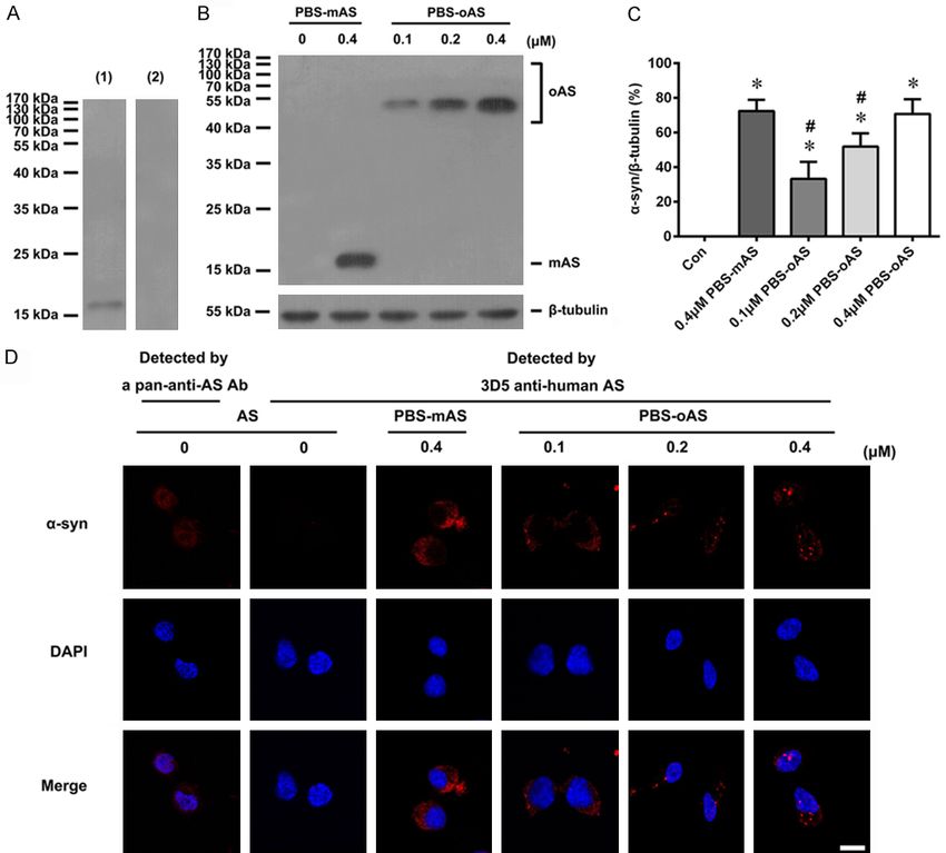
α-syn oligomers & NMDA receptor internalization

tive brain suggests that there may be a poten-       for idiopathic PD [32]. The subjects in the NC
tial link between the two proteins.                  group were recruited from the Physical
                                                     Examination Center of the hospital and were
The substantia nigra pars compacta (SNpc)            matched in age and gender with those of the
and ventral tegmental area (VTA) are the two         PD patients. All participants provided informed
brain areas that are involved in the pathogene-      consent, and underwent an evaluation that
sis of PD [17, 18]. Dopaminergic (DA) neurons        consisted of medical history, physical and neu-
from these two areas of the brain have projec-       rological examinations, laboratory tests, and
tions to the striatum, nucleus accumbens (ven-       neuropsychological assessments. The protocol
tral striatum), limbic systems, hippocampus          was approved by the Ethics Committee of the
and prefrontal cortex. These in turn regulate        Xuanwu Hospital. Blood was collected in EDTA-
voluntary movement control and cognitive func-       coated vacuum tubes, and the plasma was
tions such as emotions, motivation, rewards,         separated by centrifugation at 3,000×g for 20
and addictive behaviors [19-23]. In PD patients,     min. The plasma samples were aliquoted and
both SNpc and VTA displayed lesions by α-syn-        stored at -80°C until use.
containing Lewy pathology [24]. It has been
previously reported that the intracellular accu-     Cell culture
mulation of α-syn monomers, either due to
intracellular translocation of extracellular α-syn   The MES23.5 dopaminergic cells were obtained
protein or the intracellular overexpression of       as a generous gift from Dr Wei-Dong Le. The
the α-syn gene, promote the internalization of       cells were cultured and expanded as described
GluN1 subunits on the cell surface through a         previously [33] in a DMEM/F12 medium (Gibco,
clathrin-mediated endocytic mechanism by the         NY, USA) and supplemented with 5% fetal
participation of the endocytic protein Rab5B         bovine serum (Gibco, NY, USA), 100 U/100 ml
[25, 26]. Due to the pathogenic role of α-syn        penicillin/streptomycin, and Sato’s ingredients.
oligomers [27-30], the effect of oligomerized        All flasks were pre-coated with 0.01% poly-L-
α-syn on the expression of surface GluN1 in DA       lysine (Sigma-Aldrich, St. Louis, MO, USA).
neurons deserves further investigation.
                                                     Preparation of recombinant human α-syn
We have previously shown that recombinant
human α-syn incubated in plasma from PD              Recombinant human α-syn was prepared by
patients can aggregate into oligomers that dis-      the transformation of the plasmid pET-15b-
play increased cytotoxicity compared with            NACP into Escherichia coli BL21 cells and then
those formed in the PBS and plasma of normal         purified by sequential ion exchange chromatog-
health controls (NC) [31]. In the present study,     raphy, hydrophobic chromatography, and rev-
α-syn oligomers were prepared by incubating          erse phase chromatography [34]. The α-syn
recombinant human α-syn in either PBS or             proteins were examined by sodium dodecyl sul-
plasma from PD patients and NC, and sublethal        fate-polyacrylamide gel electrophoresis (SDS-
concentrations of the oligomers were used to         PAGE), and their identity was confirmed by
treat cultured dopaminergic cells to compare         western blotting using an anti-α-syn antibody.
their effects on the expressions of Rab5B and        Protein concentrations were determined using
surface GluN1. The potential mechanism and           a BCA Protein Assay Kit (Pierce Biotechnology,
pathological relevance for the regulation of         Rockford, IL, USA).
these were discussed.
                                                     Preparation of α-syn oligomers
Materials and methods
                                                     Individual PD or NC plasma was blended, and
Plasma samples                                       then removed the endogenous α-syn and
                                                     potential hemoglobin by affinity purification
Plasma samples were obtained from 20 clini-          using an overdose of antibodies against α-syn
cally diagnosed idiopathic PD patients and NC.       and hemoglobin. The blended PD or NC plasma
The PD patients were enrolled at the De-             was then diluted to 1/3 with PBS (pH 7.4). To
partment of Neurology, Xuanwu Hospital of            prepare the α-syn oligomers, 100 μM of recom-
Capital Medical University, and diagnosed by a       binant human α-syn was either dissolved in
consultant neurologist based on the UK               PBS or in diluted PD or NC plasma, and then
Parkinson’s Disease Society Brain Bank criteria      incubated at 37°C for 48 h with continuous

88                                                          Int J Clin Exp Pathol 2019;12(1):87-100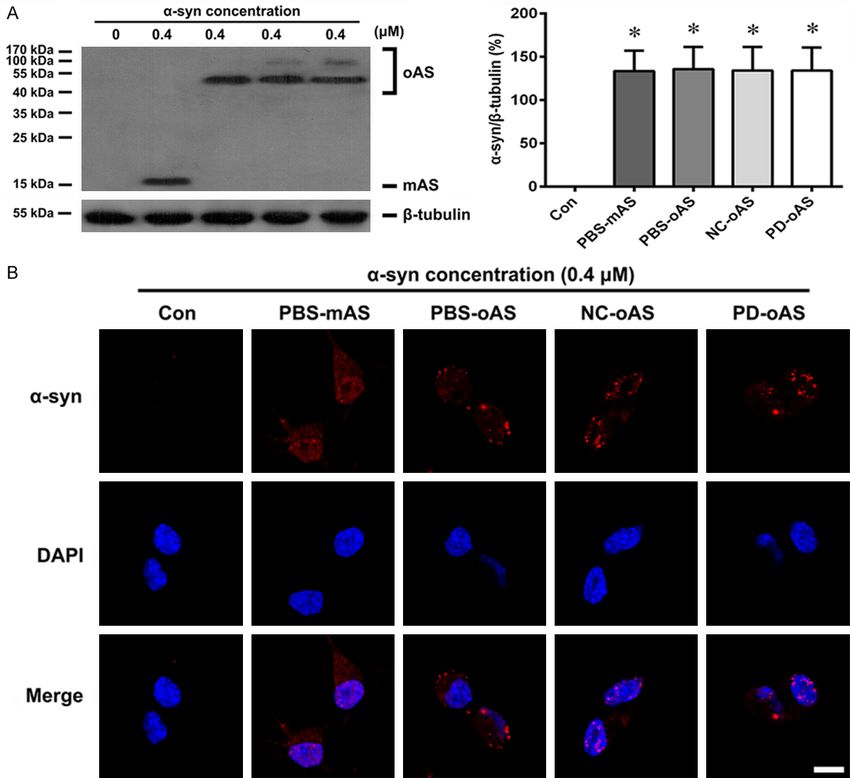
α-syn oligomers & NMDA receptor internalization

shaking (650 rpm) on an Eppendorf Ther-             samples on the grids were washed thrice with
momixer Comfort (Eppendorf AG 22331,                distilled water and stained with 2% uranyl ace-
Hamburg, Germany).                                  tate. Excess staining was removed by blotting
                                                    and air drying. The samples were then visual-
To obtain purified α-syn oligomers, the α-syn       ized under a JEM-2100 (Japan) transmission
molecules of various sizes were first isolated      electron microscope [38].
from the plasma according to the method that
was described previously [35]. Briefly, the plas-   Preparation of protein extracts
ma containing α-syn molecules was allowed to
pass through the CNBr-activated Sepharose 4B        The cells were washed thrice with ice-cold PBS
column (GE healthcare, Uppsala, Sweden) cou-        and lysed using a lysis buffer containing Tris-Cl
pled with anti-α-syn antibody. Then, the α-syn      (50 mM, pH 7.5), NaCl (150 mM), EGTA (5 mM),
molecules captured in the column were specifi-      EDTA (5 mM), SDS (2% w/v), and a protease
cally eluted by a glycine buffer (0.1 M, pH 2.5)    inhibitor cocktail. The lysates were centrifuged
followed by immediate neutralization with a         at 12,000×g for 30 min at 4°C, and the super-
Tris-HCl buffer (1 M, pH 9.0). The α-syn oligo-     natants were used as whole cell lysates [39].
mers in the eluates were separated by SDS-          The cell surface proteins were isolated accord-
PAGE from the monomers and dimers and then          ing to the method described before. In brief, the
were recovered using a Micro Protein Recovery       cells were washed with ice-cold (PBS), and the
Kit (Sangon, Biotech, Shanghai, China) [36].        cell surface proteins were biotinylated with 0.5
The α-syn oligomers formed in the PBS were          mg/mL EZ-Link-sulfo-NHS-LC-Biotin (Thermo
directly separated by SDS-PAGE and then             Scientific, Rockford, IL, USA) in PBS for 30 min
recovered using the Micro Protein Recovery Kit.     at 4°C. Then, the biotinylation reaction was ter-
Protein concentrations were determined using        minated by incubating the cells with 20 mmol/L
the BCA Protein Assay Kit as described above.       glycine. After being washed with ice cold PBS,
                                                    the cells were lysed using a RIPA buffer, fol-
Detection of oligomeric and phosphorylated          lowed by centrifugation at 5000×g for 5 min at
α-syn                                               4°C. After that the supernatant was collected
                                                    and incubated with avidin-conjugated agarose
α-Syn oligomers were measured using an
                                                    beads for 2 h at 4°C. The cell surface proteins
enzyme-linked immunosorbent assay (ELISA)
                                                    captured by avidin-coupled beads were ana-
as described initially by El-Agnaf and his col-
                                                    lyzed by western blotting [40].
leagues [37]. Briefly, the non-biotinylated and
biotinylated 3D5 anti-α-syn monoclonal anti-        Western blot analysis
bodies were used for capturing and detection,
respectively. After completion of the immunore-     Western blot analysis was performed as
action, the contents of each well of the ELISA      described before [25]. Samples (20 μg pro-
plate were incubated with ExtrAvidin alkaline       teins/lane) were separated by 10% SDS-PAGE,
phosphatase (Sigma-Aldrich, St. Louis, MO,          transferred onto a polyvinylidene difluoride
USA) followed by a reaction with enzyme sub-        (Millipore, Bedford, MA, USA) membrane, and
strate p-nitrophenyl phosphate (Sigma-Aldrich,      incubated at 4°C overnight with each primary
St. Louis, MO, USA). The absorbance was read        antibody against the following proteins: GluN1
at 405 nm using a microplate reader (Multiskan      (1:1000; BD PharmingenTM, Franklin Lakes, NJ,
MK3, Thermo Scientific, UT, USA).                   USA), Rab5B (1:10000; Santa Cruz Bio-
                                                    technology, Santa Cruz, CA, USA), α-syn [1:5000
To detect phosphorylated α-syn, an anti-pS129-
                                                    for 3D5 monoclonal antibody; 1:1000 for anti-
α-syn polyclonal antibody (Santa Cruz Bio-
technology, Santa Cruz, CA, USA) was used to        pan-α-α-syn antibody (Abcam, Cambridge, UK);
capture the antibody. The remaining steps were      1:1000 for anti-pS129-α-syn polyclonal anti-
the same as those for the detection of α-syn        body (1:5000; Santa Cruz Biotechnology, Santa
oligomers.                                          Cruz, CA, USA)]; β-tubulin (1:10000; Abcam,
                                                    Cambridge, UK) and calnexin (1:10000; Abcam,
Transmission electron microscopy                    Cambridge, UK), followed by 1 h reaction at
                                                    room temperature using horseradish peroxi-
Purified oligomeric α-syn samples were placed       dase-conjugated anti-mouse or anti-rabbit IgG
on the copper grids coated with Formvar. The        (1:5000; Vector Laboratories, Inc., CA, USA).

89                                                         Int J Clin Exp Pathol 2019;12(1):87-100
α-syn oligomers & NMDA receptor internalization Figure 1. Oligomerization and phosphorylation of α-syn in different incubating conditions. 100 μM of recombinant human α-syn was incubated in PBS, NC, and PD plasma, respectively. After incubation, the levels of oligomeric (A) and phosphorylated (B) α-syn in the samples were measured by ELISA. The α-syn proteins in the incubating solu- tions were purified using immunoaffinity chromatography, which were then examined by Coomassie brilliant blue (CBB) staining (C) and western blot analysis using 3D5 anti-α-syn antibody (D) and anti-pS129 α-syn antibody (E). Lanes 1-4 show the samples before affinity purification (1), the non-specific eluates (2, 3), and the specific α-syn eluates (4). (F-H) The α-syn oligomers formed in PBS (lane 1) and purified from NC (lane 2) and PD (lane 3) plasma 90 Int J Clin Exp Pathol 2019;12(1):87-100
α-syn oligomers & NMDA receptor internalization

were analyzed by western blot before and after separation from the monomers and dimers. (F) Non-separated α-syn
samples were detected by a 3D5 anti-α-syn antibody. (G) Separated α-syn oligomers detected by a 3D5 anti-α-syn
antibody. (H) Separated α-syn oligomers were detected by an anti-pS129 α-syn antibody. (I) The purified α-syn oligo-
mers were further analyzed by transmission electron microscope. Data are expressed as the means ± SD. *P < 0.01
vs. PBS group, #P < 0.01 vs. NC plasma group; n = 20. PBS-oAS: oligomeric α-syn formed in PBS; NC-oAS: oligomeric
α-syn formed in NC plasma; PD-oAS: oligomeric α-syn formed in PD plasma. Bar = 100 nm.

The immunoreactivity was visualized by an                      that was specific for oligomeric and phosphory-
enhanced chemiluminescence reagent (Prom-                      lated α-syn. The high and moderate levels of
ega, Madison, WI, USA).                                        the α-syn oligomers were quantified in the plas-
                                                               ma of both PD and NC, respectively, which
Immunofluorescence staining                                    were highly and moderately phosphorylated.
                                                               No phosphorylated α-syn was detected in PBS,
Cells were cultured for 24 h before treatment                  but the levels of the α-syn oligomers were lower
with α-syn for 60 min. The cells were then fixed               than those in the plasma of PD and NC (Figure
at 4°C for 30 min with 4% (w/v) paraformalde-                  1A, 1B).
hyde, treated for 10 min with copper sulfate to
reduce cellular autofluorescence [41], and then                To obtain pure α-syn oligomers, the α-syn pro-
blocked for 30 min with 3% (w/v) bovine serum                  teins in the NC and PD plasma were first iso-
albumin. To observe the intracellular transloca-               lated from the plasma using immunoaffinity
tion of α-syn and total GluN1 expression, the                  chromatography and then analyzed by Coo-
cells were initially permeabilized with 0.3% (v/v)             massie brilliant blue (CBB) staining (Figure 1C)
Triton X-100 for 30 min at room temperature,                   and western blot using antibodies against non-
incubated overnight at 4°C with 3D5 anti-α-syn                 phosphorylated (Figure 1D) and phosphorylat-
antibody (1:1000) or mouse monoclonal anti-                    ed α-syn (Figure 1E). In the specific eluates,
GluN1 antibody (1:1000), followed by 2 h reac-                 the monomers, dimers, trimers, and large-
tion at room temperature with Alexa Fluor 594                  sized polymers of α-syn were detected by the
goat anti-mouse IgG (1:1000; Invitrogen,                       anti-non-phosphorylated α-syn antibody (lane
Carlsbad, CA, USA). For detection of the GluN1                 4, Figure 1D). However, only the trimers and
(1:1000) cell surface, the cells were directly                 large-sized polymers of α-syn were revealed by
incubated overnight at 4°C with anti-GluN1                     the anti-pS129 α-syn antibody (lane 4, Figure
antibody without permeabilization with Triton                  1E). This indicated that the phosphorylated
X-100, followed by 2 h reaction with Alexa Fluor               α-syn was more prone to aggregate into oligo-
594 goat anti-mouse IgG. The cells were then                   mers. The α-syn oligomers in the specific elu-
counterstained with DAPI before being ob-                      ates and in PBS were separated by SDS-PAGE
served under a confocal laser microscope                       and recovered using a Micro Protein Recovery
(Leica TCS-SP8, Heidelberg, Germany).                          Kit to purify the oligomers. After purification,
                                                               only the oligomers were detected in the recov-
Statistical analysis
                                                               ered solutions (Figure 1G), which was in con-
Data are expressed as the means ± standard                     trast to the non-purified samples (Figure 1F).
deviation (SD). Statistical analyses were per-                 In addition, the α-syn oligomers purified from
formed using SPSS 22.0. A one-way ANOVA fol-                   the PD and NC plasma were differentially phos-
lowed by Tukey’s multiple comparison test were                 phorylated, but were absent from those oligo-
performed to evaluate the differences between                  mers purified from PBS (Figure 1H).
the groups. P < 0.05 was considered to be sta-
                                                               The α-syn aggregates were further examined
tistically significant.
                                                               under a transmission electron microscope
Results                                                        (TEM). Under TEM, the α-syn aggregates
                                                               appeared granular in shape and touched each
Purification and characterization of α-syn                     other. The α-syn granules formed in the NC/PD
oligomers                                                      plasma appeared bigger than those formed in
                                                               PBS. In addition, the assemblies in the PD
After incubation, the α-syn proteins in the PBS                plasma were bigger than those in the NC plas-
and NC/PD plasma were analyzed with ELISA                      ma gathered from (Figure 1I).

91                                                                     Int J Clin Exp Pathol 2019;12(1):87-100
α-syn oligomers & NMDA receptor internalization
Figure 2. Endogenous α-syn expression and concentration-dependent intracellular translocation of α-syn oligomers.
Endogenous α-syn was analyzed by western blotting (A). Low levels of endogenous monomeric α-syn were detected
by an anti-pan-α-syn antibody (lane 1), but not by the 3D5 anti-human α-syn antibody (lane 2). Purified α-syn mono-
mers (0.4 μM) and oligomers (0.1 to 0.4 μM) formed in PBS were added to the culture medium of MES23.5 dopa-
minergic cells. After 1 h incubation, the levels of α-syn monomers and oligomers in the whole cell lysates were mea-
sured (B) and quantified (C) by western blotting using a 3D5 anti-human α-syn antibody. In cells treated with α-syn
monomers, only monomeric α-syn was detected. In cells treated with α-syn oligomers, only oligomeric α-syn was
identified, which increased in the amount as the concentrations of extracellular α-syn oligomers were augmented.
The cells were also labeled by immunofluorescence staining for α-syn (red) and counterstained for nuclei (blue)
using DAPI (D). In the untreated cells, the anti-pan-α-syn antibody revealed faint α-syn-positive signals (red), which
could not be detected by the 3D5 anti-human α-syn antibody. However, in the α-syn-treated cells, positive α-syn
signals could be detected by the anti-human α-syn antibody, which presented either a diffused (in the monomers-
treated cells) or granular (in the oligomers-treated cells) appearance. Data are expressed as the means ± SD; n =
4. *P < 0.05 vs. control, #P < 0.05 vs. 0.4 μM α-syn monomers in PBS. Con: PBS control; AS: α-syn; PBS-mAS: α-syn
monomers in PBS; PBS-oAS: α-syn oligomers formed in PBS; NC-oAS: α-syn oligomers formed in NC plasma; PD-oAS:
α-syn oligomers formed in PD plasma. Bar = 10 μm.
Intracellular translocation and accumulation of monomeric α-syn to penetrate the cell mem-
of extracellularly added α-syn oligomers brane has been confirmed by several studies
[25, 26, 44-47]. To establish a cell model with
According to Ahn et al., the extracellular α-syn an intracellular accumulation of α-syn oligo-
can rapidly enter into living cells in a non-endo- mers, the α-syn oligomers that were initially
cytic manner [42] without being degraded by formed in the PBS were added to the culture
the cellular proteolytic systems [43]. The ability medium of MES23.5 cells and then underwent
92 Int J Clin Exp Pathol 2019;12(1):87-100α-syn oligomers & NMDA receptor internalization Figure 3. Intracellular translocation of α-syn oligomers formed in different conditions. The α-syn monomers (0.4 μM) and oligomers (0.4 μM) formed in PBS, NC and PD plasma were added to the culture medium of MES23.5 dopaminergic cells. After 1 h incubation, the levels of α-syn monomers and oligomers in the whole cell lysates were measured and quantified by western blotting using 3D5 anti-human α-syn antibody (A). In cells treated with α-syn monomers, only monomeric α-syn was detected. In cells treated with different α-syn oligomers, only oligomeric α-syn was identified. The cells were also labeled immunofluorescently for α-syn (red) and counterstained for nuclei (blue) using DAPI (B). In the untreated cells, no α-syn-positive signals were detected by the 3D5 anti-human α-syn antibody. However, in the α-syn-treated cells, positive α-syn signals were detected by the anti-human α-syn antibody, presenting in an either diffused (in the monomers-treated cells) or in granular (in the oligomers-treated cells) ap- pearance. Con: PBS control; PBS-mAS: α-syn monomers in PBS; PBS-oAS: α-syn oligomers formed in PBS; NC-oAS: α-syn oligomers formed in NC plasma; PD-oAS: α-syn oligomers formed in PD plasma. Bar = 10 μm. examination of their intracellular translocation monomers or trimers, respectively (Figure 2B). after 1 h of incubation. Western blot analysis In addition, augmenting concentrations of using an anti-pan-α-syn showed that only low extracellular α-syn oligomers led to the increase levels of endogenous α-syn were detected in of α-syn trimers in the cells (Figure 2C). In the untreated control cells, which was not rec- agreement with the western blot results, immu- ognized by 3D5 anti-human α-syn (Figure 2A). nofluorescence staining using 3D5 anti-human In contrast, in the cells treated with α-syn α-syn revealed granular α-syn-positive struc- monomers or oligomers, the 3D5 anti-human tures in the oligomer-treated cells, which were α-syn revealed a single band at 18 kD or 54 kD in contrast to the diffused staining of α-syn in that was similar to the molecular size of α-syn the monomer-treated cells. In the untreated 93 Int J Clin Exp Pathol 2019;12(1):87-100
α-syn oligomers & NMDA receptor internalization

cells, the endogenous α-syn could be detected         plasma showed a moderate reduction of sur-
using the anti-pan-α-syn, but not by 3D5 anti-        face GluN1 expression (Figure 4A-C). The α-syn
human α-syn (Figure 2D). Therefore, in the fol-       oligomers formed in PBS and α-syn monomers
lowing experiments, we used the 3D5 anti-             induced only a mild reduction of surface GluN1
human antibody to detect the exogenous                expression. In all α-syn-treated cells, the levels
recombinant human α-syn. The above results            of total GluN1 were stable, indicating an inter-
indicated that α-syn oligomers could enter the        nalization of GluN1 from the cell surface.
MES23.5 cells as its monomers without any
obvious degradation.                                  The α-syn-induced internalization of surface
                                                      GluN1 could be observed by immunofluores-
We next compared the intracellular transloca-         cence staining. In cells not permeabilized with
tions of α-syn oligomers formed in different          Triton X-100, the anti-GluN1 antibody could
conditions. For this purpose, the α-syn oligo-        only bind to the surface GluN1. The results of
mers formed in PBS and NC/PD plasma were              immunofluorescent staining revealed that the
added to the culture medium. After 1 h of incu-       most intensive GluN1 signals were found on the
bation, the cells were lysed for western blot         surfaces of untreated cells, diminishing suc-
analysis or fixed for immunofluorescence stain-       cessively in cells treated with α-syn monomers,
ing. The western blot results showed that the         α-syn oligomers formed in PBS, and α-syn oligo-
oligomeric α-syn was detected in the cells treat-     mers formed in NC and PD plasma (Figure 4B).
ed with different α-syn oligomers (Figure 3A),        In contrast, in cells permeabilized with Triton
indicating that the α-syn oligomers entered into      X-100, such as the anti-GluN1 antibody, could
the cells. Compared with cells treated with           also bind to the intracellular GluN1, and both
α-syn oligomers formed in PBS, wherein only           surface and cytoplasmic GluN1 could be
α-syn trimers at 54 kD were detected, the cells       stained and demonstrated similar GluN1 signal
treated with α-syn oligomers formed in NC/PD          intensities among various α-syn-treated cells
plasma showed two bands, where one at 54 kD           (Figure 4C).
corresponded to the α-syn trimers, and another
at 108 kD was identical to the α-syn hexamers         Effects of α-syn oligomers on Rab5B expres-
(Figure 3A). Again, there was only a single band      sion
at 18 kD to be detected in the cells treated with
α-syn monomers, which was absent in the               Rab5B is a small GTPase that is involved in
untreated cells if 3D5 anti-human α-syn was           clathrin-mediated endocytosis [48]. that has
used (Figure 3A). As illustrated by immunofluo-       been shown to participate in α-syn-induced
rescence staining, the cells treated with differ-     NMDAR internalization [25, 26]. The differential
ent α-syn oligomers presented granular α-syn-         effects of various α-syn oligomers on surface
positive structures in the cells. The cells treated   GluN1 expression suggested that they may
with α-syn monomers exhibited the diffused            affect the expression of Rab5B differently.
staining of α-syn (Figure 3B).                        Indeed, when α-syn monomers and different
                                                      oligomers were used to treat the cells, they
Effects of different α-syn oligomers on surface       induced different levels of increase in Rab5B
GluN1 internalization                                 expression. The strongest effect was observed
                                                      for the oligomers formed in PD plasma, fol-
As shown in the above results, the extracellular      lowed by the oligomers formed in the NC plas-
α-syn oligomers could enter into the MES23.5          ma and PBS (Figure 5).
dopaminergic cells, leading to the accumula-
tion of α-syn oligomers in the cells. After thatα-    Inhibition of α-syn-induced GluN1 internaliza-
syn monomers and different α-syn oligomers            tion by pitstop2
were added to the culture medium of the
MES23.5 cells, we observed their effects on           Rab5B has been reported to participate in the
GluN1 expression. After 1 h of incubation, the        regulation of the abundance of cell surface
cells were either lysed for western blot analysis     NMDA receptors through clathrin-mediated
or fixed for immunofluorescent staining. The          endocytosis [49-51]. If α-syn-induced GluN1
most significant reduction in the levels of sur-      internalization was also mediated by clathrin-
face GluN1 was observed in cells treated with         mediated endocytosis, then the clathrin inhibi-
α-syn oligomers formed in PD plasma. Also,            tor could block this process. To test this possi-
cells treated with α-syn oligomers formed in NC       bility, some of the cells were exposed to

94                                                           Int J Clin Exp Pathol 2019;12(1):87-100
α-syn oligomers & NMDA receptor internalization 95 Int J Clin Exp Pathol 2019;12(1):87-100
α-syn oligomers & NMDA receptor internalization
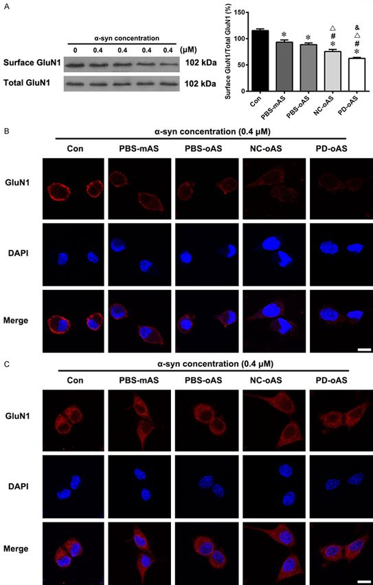
Figure 4. Effects of α-syn oligomers on GluN1 expression. The α-syn monomers (0.4 μM) and oligomers (0.4 μM)
formed in PBS, NC and PD plasma were added to the culture medium of MES23.5 dopaminergic cells. After 1 h
incubation, the levels of surface and total GluN1 were measured and quantified by western blotting (A). The levels
of surface GluN1 were decreased differently in the cells treated with α-syn monomers and various oligomers, while
the levels of total GluN1 remained stable. The cells without treatment with Triton X-100 were immunofluorescently
stained for GluN1 (red) and counterstained for nuclei (blue) using DAPI (B). Only the surface GluN1 was detected,
which decreased differentially in the cells treated with α-syn monomers and various oligomers. Immunofluorescent
staining of the Triton X-100-treated cells was also performed for GluN1 (C). In these cells, both the surface and cyto-
plasmic GluN1 could be stained and the staining intensity showed no apparent differences. Data are expressed as
the means ± SD; n = 4. *P < 0.05 vs. control, #P < 0.05 vs. α-syn monomers in PBS, ΔP < 0.05 vs. α-syn oligomers
formed in PBS, &P < 0.05 vs. α-syn oligomers formed in NC plasma; Con: PBS control; PBS-mAS: α-syn monomers in
PBS; PBS-oAS: α-syn oligomers formed in PBS; NC-oAS: α-syn oligomers formed in NC plasma; PD-oAS: α-syn oligo-
mers formed in PD plasma. Bar = 10 μm.
nalization, induced by either α-syn monomers
or different oligomers, involved a clathrin-medi-
ated endocytic mechanism.
Discussion
In the present study, we prepared different
α-syn oligomers by incubating recombinant α-
syn in PBS, NC, or PD plasma. We found that
the α-syn oligomers formed in the NC and PD
plasma were phosphorylated at serine 129.
This phosphorylation was much greater in the
PD plasma compared to the NC plasma. The
discrepancy for the α-syn aggregates formed in
different conditions was also reflected in the
images of TEM. The granular α-syn aggregates
formed in NC/PD plasma were bigger than
those in PBS. In addition, they touched each
other to form irregular, large assemblies, which
were bigger in PD plasma than in NC plasma.
Figure 5. Effects of α-syn oligomers on Rab5B expres- The above results indicated that the α-syn
sion. The levels of Rab5B were measured by western oligomers formed under different conditions
blotting 1 h after the cells were treated by 0.4 μM of have different conformations. This discrepancy
α-syn monomers and purified oligomers were formed may be due to their differential phosphorylation
in various incubating conditions. β-tubulin: loading
control for whole-cell lysates. Data are expressed as and other potential factors present in the
means ± SD; n = 4. *P < 0.05 vs. control, #P < 0.05 plasma.
vs. α-syn monomers, ΔP < 0.05 vs. α-syn oligomers
formed in PBS, &P < 0.05 vs. α-syn oligomers formed As mentioned before, extracellular α-syn rapid-
in NC plasma. Con: PBS control; PBS-mAS: α-syn ly enters into the living cells in a non-endocytic
monomers in PBS; PBS-oAS: α-syn oligomers formed manner without being degraded by cellular pro-
in PBS; NC-oAS: α-syn oligomers formed in NC plas-
ma; PD-oAS: α-syn oligomers formed in PD plasma. teolytic systems [42, 43]. We have previously
demonstrated that the extracellular addition of
monomeric α-syn can enter into MES23.5
pitstop2, a selective inhibitor of clathrin-medi- dopaminergic cells and lead to the intracellular
ated endocytosis [52], before incubation with accumulation of α-syn monomers [25, 44],
α-syn monomers or various oligomers. In the inducing surface GluN1 internalization [25].
absence of pitstop2, α-syn monomers and vari- The effect of extracellular α-syn on surface
ous oligomers induced different levels of reduc- GluN1 was similar to that of intracellular overex-
tions in the expression of surface GluN1. In the pression of α-syn [25], indicating that the extra-
presence of pitstop2, the reductions of surface cellular addition of α-syn exerted this effect
GluN1 expression were abolished (Figure 6). after entering into the cells. To investigate the
This suggested the possibility that GluN1 inter- effects of α-syn oligomers on surface GluN1,
96 Int J Clin Exp Pathol 2019;12(1):87-100α-syn oligomers & NMDA receptor internalization
strongly suggest that Rab5B
may play a role in this regula-
tion. Rab5B is a small ATPase
molecule that functions as a
regulator in the early endocyt-
ic pathway [53]. It has been
suggested that Rab5B partici-
pates in the regulation of cell
surface NMDA receptors thr-
ough clathrin-mediated endo-
cytosis [49-51]. Our previous
studies showed that the GluN1
internalization induced by the
intracellular accumulation of
α-syn monomers was accom-
panied by the upregulation of
Rab5B expression, which was
abolished by silencing Rab5B
expression [25]. In the pres-
ent study, we found that the
intracellular accumulation of
α-syn oligomers was also ac-
companied by an increase in
the expression of Rab5B. Be-
sides, the levels of Rab5B
Figure 6. Effect of the clathrin inhibitor on α-syn-induced surface GluN1 were closely related to the
reduction. A portion of the cells were pre-incubated with pitstop2 (15 μM) surface expressions of GluN1
for 10 min before treatment with α-syn monomers and purified oligomers
formed in various incubating conditions. One hour later, the levels of surface in the cells treated with differ-
and total GluN1 were measured by western blotting. Data are expressed as ent α-syn oligomers. For ex-
the means ± SD; n = 4. *P < 0.05 vs. control, #P < 0.05 vs. α-syn monomers, ample, the most prominent in-
Δ
P < 0.05 vs. α-syn oligomers formed in PBS, &P < 0.05 vs. α-syn oligomers crease in the levels of Rab5B
formed in NC plasma. Con: PBS control; P2: Pitstop2; PBS-mAS: α-syn mono- was observed in the cells
mers in PBS; PBS-oAS: α-syn oligomers formed in PBS; NC-oAS: α-syn oligo-
mers formed in NC plasma; PD-oAS: α-syn oligomers formed in PD plasma. treated with α-syn oligomers
formed in PD plasma, and was
associated with the most sig-
we added various purified α-syn oligomers to nificant reduction of surface GluN1 expression
the culture medium of MES23.5 dopaminergic in the same cells. Accordingly, mild and moder-
cells and then observed their intracellular tra- ate upregulations of Rab5B in the cells treat-
nslocation. As demonstrated by western blot- ed with α-syn oligomers formed in NC plasma
ting and immunofluorescent staining, all kinds and PBS were associated with mild and moder-
of α-syn oligomers as well as α-syn monomers ate downregulations of surface GluN1 in the
entered into the cells. Simultaneously, different corresponding cells. Taken together, the above
levels of the reduction of surface GluN1 were results suggest that the differential regulations
detected. As the levels of total GluN1 were un- of surface GluN1 expression by different α-syn
changed and the surface GluN1 was reduced, oligomers depend on their different regulations
this indicated an internalization of GluN1 on the of Rab5B. Since Rab5B participates in the reg-
cell surface. This effect was more prominent for ulation of cell surface NMDA receptors throu-
the oligomers than the monomers. In addition, gh clathrin-mediated endocytosis [49-51], we
the oligomers formed in PD plasma were more speculated that the α-syn oligomers-induced
potent than those formed in NC plasma/PBS in GluN1 internalization, which was accompanied
promoting GluN1 internalization. by increased Rab5B expression, might be also
mediated by clathrin. To demonstrate the role
The underlying mechanism regarding the effe- of clathrin in this regulation, we applied clathrin
ct of α-syn oligomers on GluN1 internalization specific inhibitor pistop2 before the addition
remains to be clarified, and the available data of α-syn oligomers to the culture medium. We
97 Int J Clin Exp Pathol 2019;12(1):87-100α-syn oligomers & NMDA receptor internalization
observed a complete inhibition of GluN1 inter- References
nalization. Our previous study suggested that
α-syn monomers-induced GluN1 internalization [1] Newcomer JW and Krystal JH. NMDA receptor
was mediated by clathrin-mediated endocyto- regulation of memory and behavior in humans.
sis [25, 26]. This result indicates that the clath- Hippocampus 2001; 11: 529-542.
rin-mediated endocytosis may also participate [2] Ossowska K, Wolfarth S, Schulze G, Wardas J,
in the α-syn oligomers-induced GluN1 inter- Pietraszek M, Lorenc-Koci E, Smialowska M
and Coper H. Decline in motor functions in ag-
nalization.
ing is related to the loss of NMDA receptors.
The present study provided evidence for the Brain Res 2001; 907: 71-83.
first time that α-syn oligomers can promote [3] Zweifel LS, Argilli E, Bonci A and Palmiter RD.
Role of NMDA receptors in dopamine neurons
NMDAR GluN1 subunit internalization. In par-
for plasticity and addictive behaviors. Neuron
ticular, the present study demonstrated that
2008; 59: 486-496.
the α-syn oligomers formed in PD plasma, are [4] Xu Y, Yan J, Zhou P, Li J, Gao H, Xia Y and Wang
highly phosphorylated, and exhibit a more Q. Neurotransmitter receptors and cognitive
potent effect on GluN1 internalization. Because dysfunction in alzheimer’s disease and parkin-
GluN1 is an obligatory subunit that is essential son’s disease. Prog Neurobiol 2012; 97: 1-13.
for the assembly of functional NMDAR [54], its [5] Avila J, Llorens-Martin M, Pallas-Bazarra N, Bo-
reduction on the cell surface indicated the los M, Perea JR, Rodriguez-Matellan A and Her-
impairment of NMDAR function. Most studies nandez F. Cognitive decline in neuronal aging
support that α-syn oligomerization is toxic to and alzheimer’s disease: role of nmda recep-
neurons [10, 27, 55, 56]. The present study tors and associated proteins. Front Neurosci
suggests that one of the potential mechanisms 2017; 11: 626.
for the neurotoxicity of α-syn oligomer is its det- [6] Mota SI, Ferreira IL and Rego AC. Dysfunction-
rimental effect of NMDARs. al synapse in alzheimer’s disease - a focus on
NMDA receptors. Neuropharmacology 2014;
Acknowledgements 76: 16-26.
[7] Ding W, Ding LJ, Li FF, Han Y and Mu L. Neuro-
This work was supported by grants from the degeneration and cognition in parkinson’s dis-
Natural Science Foundation of China (816- ease: a review. Eur Rev Med Pharmacol Sci
71244, 81371200, and 81401042), the Beijing 2015; 19: 2275-2281.
Municipal Science & Technology Commission [8] Jakes R, Spillantini MG and Goedert M. Identi-
(Z161100005116011, Z171100000117013), fication of two distinct synucleins from human
brain. FEBS Lett 1994; 345: 27-32.
the Beijing Municipal Commission of Health
[9] Ghiglieri V, Calabrese V and Calabresi P. Alpha-
and Family Planning (PXM2017_026283_00-
synuclein: from early synaptic dysfunction to
0002), and the Beijing Nova Program (Z18- neurodegeneration. Front Neurol 2018; 9:
1100006218052, xx2018096). 295.
[10] Paleologou KE, Kragh CL, Mann DM, Salem
Disclosure of conflict of interest
SA, Al-Shami R, Allsop D, Hassan AH, Jensen
PH and El-Agnaf OM. Detection of elevated lev-
None.
els of soluble alpha-synuclein oligomers in
Abbreviations post-mortem brain extracts from patients with
dementia with lewy bodies. Brain 2009; 132:
1093-1101.
PD, Parkinson’s disease; AD, Alzheimer’s dis-
[11] Clinton LK, Blurton-Jones M, Myczek K, Tro-
ease; NMDAR, N-methyl-D-aspartate receptor;
janowski JQ and LaFerla FM. Synergistic inter-
α-syn, Alpha-synuclein; PBS, Phosphate-buff-
actions between abeta, tau, and alpha-synu-
ered saline; SDS, Sodium dodecyl sulfate; clein: acceleration of neuropathology and
ELISA, Enzyme-linked immunosorbent assay; cognitive decline. J Neurosci 2010; 30: 7281-
EDTA, Ethylene diamine tetraacetic Acid; EGTA, 7289.
Ethylene glycol tetraacetic acid; KF, potassium [12] Bandopadhyay R. Sequential extraction of sol-
fluoride; DTT, dithiothreitol. uble and insoluble alpha-synuclein from par-
kinsonian brains. J Vis Exp 2016.
Address correspondence to: Shun Yu, Department [13] Chen M, Liu J, Lu YQ, Duan CL, Lu LL, Gao G,
of Neurobiology, Xuanwu Hospital of Capital Medical Chan P, Yu S and Yang H. Age-dependent al-
University, Beijing, China. Tel: +86-13911103956; pha-synuclein accumulation is correlated with
E-mail: yushun103@163.com elevation of mitochondrial TRPC3 in the brains
98 Int J Clin Exp Pathol 2019;12(1):87-100α-syn oligomers & NMDA receptor internalization
of monkeys and mice. J Neural Transm 2017; sociated with reduced inward current and
124: 441-453. Ca(2+) influx upon NMDA stimulation. Neuro-
[14] Li X, Yang W, Li X, Chen M, Liu C and Yu S. Age- science 2015; 300: 297-306.
dependent elevations of oligomeric and phos- [27] Winner B, Jappelli R, Maji SK, Desplats PA,
phorylated alpha-synuclein synchronously oc- Boyer L, Aigner S, Hetzer C, Loher T, Vilar M,
curs in the brain and gastrointestinal tract of Campioni S, Tzitzilonis C, Soragni A, Jessberg-
cynomolgus monkeys. Neurosci Lett 2018; er S, Mira H, Consiglio A, Pham E, Masliah E,
662: 276-282. Gage FH and Riek R. In vivo demonstration
[15] Freichel C, Neumann M, Ballard T, Muller V, that alpha-synuclein oligomers are toxic. Proc
Woolley M, Ozmen L, Borroni E, Kretzschmar Natl Acad Sci U S A 2011; 108: 4194-4199.
HA, Haass C, Spooren W and Kahle PJ. Age- [28] Kaufmann TJ, Harrison PM, Richardson MJ,
dependent cognitive decline and amygdala pa- Pinheiro TJ and Wall MJ. Intracellular soluble
thology in alpha-synuclein transgenic mice. alpha-synuclein oligomers reduce pyramidal
Neurobiol Aging 2007; 28: 1421-1435. cell excitability. J Physiol 2016; 594: 2751-
[16] Luk KC, Kehm V, Carroll J, Zhang B, O’Brien P, 2772.
Trojanowski JQ and Lee VM. Pathological al- [29] Bourdenx M, Koulakiotis NS, Sanoudou D,
pha-synuclein transmission initiates Parkin- Bezard E, Dehay B and Tsarbopoulos A. Protein
son-like neurodegeneration in nontransgenic aggregation and neurodegeneration in proto-
mice. Science 2012; 338: 949-953. typical neurodegenerative diseases: examples
[17] Brichta L and Greengard P. Molecular determi- of amyloidopathies, tauopathies and synucle-
nants of selective dopaminergic vulnerability inopathies. Prog Neurobiol 2017; 155: 171-
in parkinson’s disease: an update. Front Neu- 193.
roanat 2014; 8: 152. [30] Ono K. The oligomer hypothesis in alpha-synu-
[18] Alberico SL, Cassell MD and Narayanan NS. cleinopathy. Neurochem Res 2017; 42: 3362-
The vulnerable ventral tegmental area in par- 3371.
kinson’s disease. Basal Ganglia 2015; 5: 51- [31] Wang P, Li X, Li X, Yang W and Yu S. Blood plas-
55. ma of patients with parkinson’s disease in-
[19] Hosp JA and Luft AR. Dopaminergic meso-cor- creases alpha-synuclein aggregation and neu-
tical projections to m1: role in motor learning rotoxicity. Parkinsons Dis 2016; 2016:
and motor cortex plasticity. Front Neurol 2013; 7596482.
4: 145. [32] Hughes AJ, Daniel SE, Kilford L and Lees AJ.
[20] Puig MV, Antzoulatos EG and Miller EK. Pre- Accuracy of clinical diagnosis of idiopathic Par-
frontal dopamine in associative learning and kinson’s disease: a clinico-pathological study
memory. Neuroscience 2014; 282: 217-229. of 100 cases. J Neurol Neurosurg Psychiatry
[21] Luo SX and Huang EJ. Dopaminergic neurons 1992; 55: 181-184.
and brain reward pathways: from neurogene- [33] Yu S, Zuo X, Li Y, Zhang C, Zhou M, Zhang YA,
sis to circuit assembly. Am J Pathol 2016; 186: Ueda K and Chan P. Inhibition of tyrosine hy-
478-488. droxylase expression in alpha-synuclein-trans-
[22] Howard CD, Li H, Geddes CE and Jin X. Dynam- fected dopaminergic neuronal cells. Neurosci
ic nigrostriatal dopamine biases action selec- Lett 2004; 367: 34-39.
tion. Neuron 2017; 93: 1436-1450. [34] Hossain S, Alim A, Takeda K, Kaji H, Shinoda T
[23] Edelmann E and Lessmann V. Dopaminergic and Ueda K. Limited proteolysis of NACP/al-
innervation and modulation of hippocampal pha-synuclein. J Alzheimers Dis 2001; 3: 577-
networks. Cell Tissue Res 2018; 373: 711-727. 584.
[24] Seidel K, Mahlke J, Siswanto S, Kruger R, Hein- [35] Mousavi Hosseini K and Nasiri S. Preparation
sen H, Auburger G, Bouzrou M, Grinberg LT, of factor VII concentrate using CNBr-activated
Wicht H, Korf HW, den Dunnen W and Rub U. sepharose 4B immunoaffinity chromatogra-
The brainstem pathologies of Parkinson’s dis- phy. Med J Islam Repub Iran 2015; 29: 170.
ease and dementia with Lewy bodies. Brain [36] Liu G, Chen M, Mi N, Yang W, Li X, Wang P, Yin
Pathol 2015; 25: 121-135. N, Li Y, Yue F, Chan P and Yu S. Increased oligo-
[25] Cheng F, Li X, Li Y, Wang C, Wang T, Liu G, merization and phosphorylation of alpha-synu-
Baskys A, Ueda K, Chan P and Yu S. Alpha- clein are associated with decreased activity of
synuclein promotes clathrin-mediated NMDA glucocerebrosidase and protein phosphatase
receptor endocytosis and attenuates NMDA- 2A in aging monkey brains. Neurobiol Aging
induced dopaminergic cell death. J Neuro- 2015; 36: 2649-2659.
chem 2011; 119: 815-825. [37] El-Agnaf OM, Salem SA, Paleologou KE, Curran
[26] Chen Y, Yang W, Li X, Li X, Yang H, Xu Z and Yu MD, Gibson MJ, Court JA, Schlossmacher MG
S. Alpha-synuclein-induced internalization of and Allsop D. Detection of oligomeric forms of
NMDA receptors in hippocampal neurons is as- alpha-synuclein protein in human plasma as a
99 Int J Clin Exp Pathol 2019;12(1):87-100α-syn oligomers & NMDA receptor internalization
potential biomarker for parkinson’s disease. [49] Blaabjerg M, Baskys A, Zimmer J and Vawter
FASEB J 2006; 20: 419-425. MP. Changes in hippocampal gene expression
[38] Bharathi, Indi SS and Rao KS. Copper- and after neuroprotective activation of group I
iron-induced differential fibril formation in al- metabotropic glutamate receptors. Brain Res
pha-synuclein: TEM study. Neurosci Lett 2007; Mol Brain Res 2003; 117: 196-205.
424: 78-82. [50] Arnett AL, Bayazitov I, Blaabjerg M, Fang L,
[39] Jiang J, Yang W, Huang P, Bu X, Zhang N and Li Zimmer J and Baskys A. Antisense oligonucle-
J. Increased phosphorylation of Ets-like tran- otide against GTPase Rab5b inhibits metabo-
scription factor-1 in neurons of hypoxic precon- tropic agonist DHPG-induced neuroprotection.
ditioned mice. Neurochem Res 2009; 34: Brain Res 2004; 1028: 59-65.
1443-1450. [51] Baskys A, Fang L and Bayazitov I. Activation of
[40] Snyder EM, Nong Y, Almeida CG, Paul S, Moran neuroprotective pathways by metabotropic
T, Choi EY, Nairn AC, Salter MW, Lombroso PJ, group I glutamate receptors: a potential target
Gouras GK and Greengard P. Regulation of for drug discovery? Ann N Y Acad Sci 2005;
NMDA receptor trafficking by amyloid-beta. Nat 1053: 55-73.
Neurosci 2005; 8: 1051-1058. [52] von Kleist L, Stahlschmidt W, Bulut H, Gromo-
[41] Potter KA, Simon JS, Velagapudi B and Capa- va K, Puchkov D, Robertson MJ, MacGregor
dona JR. Reduction of autofluorescence at the KA, Tomilin N, Pechstein A, Chau N, Chircop M,
microelectrode-cortical tissue interface im- Sakoff J, von Kries JP, Saenger W, Krausslich
proves antibody detection. J Neurosci Methods HG, Shupliakov O, Robinson PJ, McCluskey A
2012; 203: 96-105. and Haucke V. Role of the clathrin terminal do-
[42] Ahn KJ, Paik SR, Chung KC and Kim J. Amino main in regulating coated pit dynamics re-
acid sequence motifs and mechanistic fea- vealed by small molecule inhibition. Cell 2011;
tures of the membrane translocation of alpha- 146: 471-484.
synuclein. J Neurochem 2006; 97: 265-279. [53] Bucci C, Lutcke A, Steele-Mortimer O, Olk-
[43] Lee HJ, Suk JE, Bae EJ and Lee SJ. Clearance konen VM, Dupree P, Chiariello M, Bruni CB,
and deposition of extracellular alpha-synuclein Simons K and Zerial M. Co-operative regula-
aggregates in microglia. Biochem Biophys Res tion of endocytosis by three Rab5 isoforms.
Commun 2008; 372: 423-428. FEBS Lett 1995; 366: 65-71.
[44] Yin J, Han J, Zhang C, Ma QL, Li X, Cheng F, Liu [54] Ishii T, Moriyoshi K, Sugihara H, Sakurada K,
G, Li Y, Ueda K, Chan P and Yu S. C-terminal Kadotani H, Yokoi M, Akazawa C, Shigemoto R,
part of alpha-synuclein mediates its activity in Mizuno N, Masu M, et al. Molecular character-
promoting proliferation of dopaminergic cells. J ization of the family of the N-methyl-D-aspar-
Neural Transm (Vienna) 2011; 118: 1155- tate receptor subunits. J Biol Chem 1993; 268:
1164. 2836-2843.
[45] Cavaliere F, Cerf L, Dehay B, Ramos-Gonzalez [55] Colla E, Jensen PH, Pletnikova O, Troncoso JC,
P, De Giorgi F, Bourdenx M, Bessede A, Obeso Glabe C and Lee MK. Accumulation of toxic al-
JA, Matute C, Ichas F and Bezard E. In vitro al- pha-synuclein oligomer within endoplasmic
pha-synuclein neurotoxicity and spreading reticulum occurs in alpha-synucleinopathy in
among neurons and astrocytes using lewy vivo. J Neurosci 2012; 32: 3301-3305.
body extracts from Parkinson disease brains. [56] Rockenstein E, Nuber S, Overk CR, Ubhi K,
Neurobiol Dis 2017; 103: 101-112. Mante M, Patrick C, Adame A, Trejo-Morales M,
[46] Tofaris GK, Goedert M and Spillantini MG. The Gerez J, Picotti P, Jensen PH, Campioni S, Riek
transcellular propagation and intracellular traf- R, Winkler J, Gage FH, Winner B and Masliah E.
ficking of alpha-synuclein. Cold Spring Harb Accumulation of oligomer-prone alpha-synu-
Perspect Med 2017; 7. clein exacerbates synaptic and neuronal de-
[47] Valdinocci D, Radford RA, Siow SM, Chung RS generation in vivo. Brain 2014; 137: 1496-
and Pountney DL. Potential modes of intercel- 1513.
lular alpha-synuclein transmission. Int J Mol
Sci 2017; 18.
[48] Bucci C, Parton RG, Mather IH, Stunnenberg H,
Simons K, Hoflack B and Zerial M. The small
gtpase Rab5 functions as a regulatory factor in
the early endocytic pathway. Cell 1992; 70:
715-728.
100 Int J Clin Exp Pathol 2019;12(1):87-100You can also read