Optimization of heparin monitoring with anti-FXa assays and impact of dextran sulfate for measuring all in-vivo drug activity
←
→
Page content transcription
If your browser does not render page correctly, please read the page content below
Preprints (www.preprints.org) | NOT PEER-REVIEWED | Posted: 4 March 2021                         doi:10.20944/preprints202103.0171.v1

           Article

     Optimization of heparin monitoring with anti-FXa assays and impact

     of dextran sulfate for measuring all in-vivo drug activity

     Jean AMIRAL1, Cédric AMIRAL2, Claire DUNOIS2

                                   1 Affiliation 1; SH-Consulting, 78570-ANDRESY (France)
                                   2 Affiliation 2; HYPHEN BioMed, 95000 NEUVILLE sur OISE (France)
                                   * Correspondence: jean.amiral@scientific-hemostasis.com; Tel.: JA +33614581765

                                   Abstract:

                                   Heparins, Unfractionated or Low Molecular Weight, are permanently at the spotlight of both clin-
                                   ical indications and laboratory monitoring. An accurate drug dosage is necessary for an efficient
                                   and safe therapy. The one-stage anti-FXa kinetics’ assays are the most widely and universally used
                                   with full automation for large series, without needing exogenous Antithrombin. WHO interna-
                                   tional standards are available for UFH and LMWH, but external quality assessment surveys still
                                   report a high inter-assay variability. This heterogeneity results from: assay formulation, designed
                                   without or with dextran sulfate to measure all heparin in blood circulation; calibrators for testing
                                   UFH or LMWH with the same curve; and automation parameters. The various factors which im-
                                   pact heparin measurements are reviewed, and we share our experience to optimize assays for
                                   completely testing plasma heparin. Evidence is provided on the usefulness of low molecular
                                   weight dextran sulfate to mobilize all drug present in blood circulation. Other key factors concern
                                   adjustment of assay conditions to obtain fully superimposable calibration curves for UFH and
                                   LMWH, and automation parameters. The study is illustrated by the performances of the various
                                   anti-FXa assays used for testing heparin on UFH or LMWH treated patients’ plasmas and obtained
                                   using citrate or CTAD anticoagulants. Comparable results are obtained only when CTAD antico-
                                   agulant is used. Using citrate UFH is underestimated in the absence of dextran sulfate. Heparin
                                   calibrators, adjustment of automation parameters and data treatment contribute to other smaller
                                   differences.

                                   Keywords: heparins; anti-FXa assays; automation; calibration curves superimposition; dextran
                                   sulfate

                                   1. Background

                                   Heparin therapy and its monitoring: Heparin and its derivatives, including Unfraction-
                                   ated Heparin (UFH), Low Molecular Weight Heparin (LMWH) and Fondaparinux, re-
                                   main a major group of anticoagulants with multiple indications in various clinical situa-
                                   tions associated with thrombosis or its risk of occurrence [1-3]. Since its discovery, more
                                   than 1 century ago [4, 5], and its introduction, heparin is used for preventing or managing
                                   thrombotic diseases, and the prognosis of these complications has been totally reversed
                                   in many pathological contexts like traumatology, hip or knee replacement, post-surgery,
                                   or cardiology [6-9]. A close monitoring is required for therapy adjustment, especially for
                                   UFH, to obtain an efficient anticoagulant effect and avoiding bleeding risk [10, 11]. Blood
                                   heparin concentration is not always predictable as some patients can present an impaired
                                   clearance, especially when there is a deficient kidney function [12, 13], or they can face
                                   heparin resistance, in presence of netosis, high circulating histone concentrations or am-
                                   yloidosis [14-17]. In addition, rare and severe side effects can develop with heparin in-
                                   duced thrombocytopenia, a life-threatening antibody-dependent complication, which
                                   requires an immediate heparin withdrawal and use of a different anticoagulant [18, 19].
                                   Heparin remains however the anticoagulant of choice in many critical circumstances, due

            © 2021 by the author(s). Distributed under a Creative Commons CC BY license.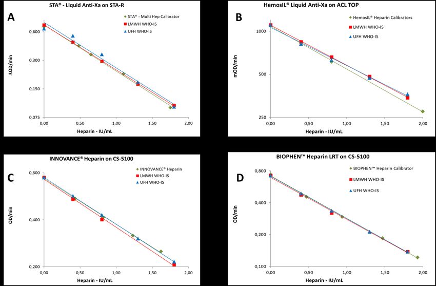
Preprints (www.preprints.org) | NOT PEER-REVIEWED | Posted: 4 March 2021                doi:10.20944/preprints202103.0171.v1

                                                                                                                        2 of 17

                                 to its rapid anticoagulant response and efficacy, but also to its other beneficial activities,
                                 anti-inflammatory and antiproliferative [1, 8, 20-22]. Furthermore, heparin can produce
                                 blood anticoagulation through additional mechanisms than inhibition of coagulation
                                 serine esterases, especially through the release of Tissue Factor Pathway Inhibitor (TFPI)
                                 from endothelium, process which is more effective at the onset of therapy, and depend-
                                 ent on heparin sulfation grade and molecular weight [23-2].

                                 The UFH and LMWH drug-dosage needs to be accurately adjusted for each treated pa-
                                 tient according to the clinical pattern and physiological status, which can impact drug
                                 clearance [2, 6, 13, 26, 27]. If drug concentration is not enough in blood circulation,
                                 thrombotic diseases is not correctly controlled, and conversely, if it is too high, due to
                                 overdosage or an impaired clearance, patient can bleed. Both situations can lead to a fatal
                                 outcome, highlighting the criticism of drug monitoring. Many assays have been devel-
                                 oped over time for testing heparin in blood circulation, during open heart surgery, or in
                                 plasma [11, 28, 29]. The first assays proposed for evaluating its anticoagulant potential
                                 were based on the prolongation of clotting time, and later the activated clotting time
                                 (ACT) was introduced for testing high concentrations in cardiology patients, especially in
                                 intensive care units [28-30]. However, in clinical settings most of the heparin treated pa-
                                 tients have been monitored with the Activated Partial Thromboplastin Time (APTT),
                                 performed on citrated plasma, for a long time [31,32]. This clotting method is still the first
                                 line laboratory assay in many countries, despite its limitations [33, 34]. The availability of
                                 chromogenic assays, introduced about 40 years ago, has permitted the progressive de-
                                 velopment of more specific methods for testing heparin concentrations in plasma [10, 30,
                                 33, 35-38]. Specific thrombin or FXa chromogenic substrates are used for enzyme inhibi-
                                 tion methods. This lead to develop first 2-stage assays, then anti-FXa kinetics assays, fully
                                 automated. These latter are now the most widely used with the various available coagu-
                                 lation instruments. Heparin measurements are much more accurate when monitored
                                 with chromogenic assays than when tested with APTT or ACT [33, 35, 38], as these clot-
                                 ting methods present many interferences, especially in severely ill patients. They can
                                 result from high Factor VIII concentration [31], inappropriate citrate content in blood
                                 samples obtained in insufficiently filled tubes, blood activation during collection, or low
                                 hematocrit.

                                 Mode of action of heparin: Heparin is an indirect catalytic inhibitor and requires An-
                                 ti-Thrombine (AT) for inhibiting coagulation serine esterases, mainly thrombin, also
                                 named activated Factor II (FIIa), and FXa, and in a lesser extend FIXa, FXIa and FVIIa [39,
                                 40]. In the absence of heparin, AT is a progressive inhibitor of thrombin and FXa. When
                                 present, heparin binds to AT through an irregular pentasaccharide sequence, in a mole-
                                 cule-to-molecule complex. AT becomes then a fast-acting inhibitor of thrombin and FXa
                                 and forms finally a stable irreversible complex with these serine esterases, whilst heparin
                                 is released from the complex and becomes available for activating a new AT molecule [41,
                                 42]. The limiting factor for the anticoagulant action of heparin, in addition to its concen-
                                 tration, is then the concentration of AT, and the drug turn-over for inhibiting serine es-
                                 terases. The turn-over of heparin for AT activation, and therefore its anticoagulant po-
                                 tential, depends on its characteristics, especially the pentasaccharide sequences’ density
                                 and its molecular weight (MW) or polysaccharide length [4, 5, 43]. In body or in the assay
                                 system, heparin anticoagulant activity is dependent on AT concentration only if that one
                                 is too low. When AT is present at an enough concentration, anticoagulant activity is then
                                 heparin dose dependent. Other characteristics of heparin, like the global electronegative
                                 charge and the sulfate groups density, affect better its non-anticoagulant biological effects
                                 [8, 9, 21, 2]. Heparin is an electronegatively charged molecule which can interact with
                                 many blood proteins and bind to various blood cells through exposed surface proteins,
                                 especially to endothelial cells and platelets [44, 45]. UFH has a higher affinity for blood
                                 proteins and cells than LMWH. Proteins which can impact heparin activity in blood or
Preprints (www.preprints.org) | NOT PEER-REVIEWED | Posted: 4 March 2021               doi:10.20944/preprints202103.0171.v1

                                                                                                                       3 of 17

                                 plasma are first platelet factor 4 (PF4), a protein released from platelet α-granules and
                                 which has the highest affinity and can neutralize this drug at stoichiometric concentra-
                                 tions [4], then Histidine-Rich-Glyco-Protein (HRGP), a protein involved in fibrinolysis for
                                 the regulation of plasminogen binding to fibrin [47, 48]. But other proteins can also bind
                                 to heparin with a lower affinity, like vitronectin, β2-Glycoprotein, but their incidence on
                                 heparin activity is negligible.

                                 Chromogenic assays for heparin monitoring: The first heparin chromogenic assays in-
                                 troduced were the 2-stage assays, based on the inhibition of a constant amount of FIIa or
                                 FXa. Diluted tested specimen is mixed with a constant concentration of purified AT and
                                 FIIa or FXa for a fixed time, in a first step, followed by the addition of the chromogenic
                                 substrate, which reacts with the non-inhibited FIIa or FXa, in the second step [49, 50].
                                 An inverse dose-response curve is obtained between heparin concentrations and ab-
                                 sorbance, measured at 405 nM. The assays must be calibrated with the same type of
                                 heparin measured, in a like-to-like manner. Calibrators are prepared by spiking the as-
                                 sayed drug in normal citrated plasma or in the assay buffer for obtaining the reference
                                 range. Performing these laboratory methods requires a high level of technical expertise,
                                 and the assay conditions need to be strictly adhered to. Each stage is critical, and the
                                 timing must be respected exactly. High quality biochemicals, including AT, FIIa or FXa,
                                 and chromogenic substrates, are required. These assays are extremely sensitive, with
                                 ranges from ≤ 0.10 IU/mL for anti-FXa or ≤ 0.05 IU/ml for anti-FIIa methods. Samples
                                 containing heparin must be highly diluted before testing. When heparin is assayed in
                                 plasma, a platelet depleted plasma with a low PF4 content (< 10 ng/mL) is required for
                                 preparing calibrators. For the value assignment of heparin drugs, a reference range is
                                 prepared in the assay buffer containing Bovine Serum Albumin (BSA) or
                                 Poly-Ethylene-Glycol (PEG 6000) as carrier substances. The exact conditions for per-
                                 forming these assays are documented in Pharmacopeias (EP, USP, JP). These assays’
                                 constraints have limited the use and automation for these methods, especially since the
                                 introduction of automated instruments, which face limitations for managing exactly the 2
                                 exact incubation times required. The 2-stage assays however remain the reference
                                 methods for testing heparin and its derivatives by pharmaceutical industry in association
                                 with like-to-like drug reference materials [51].

                                  Automated one-stage anti-FXa kinetics methods have been developed for the current
                                 laboratory monitoring of heparin therapy, along with plasma calibrators for UFH,
                                 LMWH or Fondaparinux. These assays can be automated on any of the coagulation in-
                                 struments now available in laboratories and an assay precalibration is currently used [30,
                                 52]. A new calibration is only required from time to time, the permanence of measure-
                                 ment performances being verified daily with control plasmas. No exogenous AT is
                                 needed for kinetics anti-FXa assays, and endogenous assayed plasma AT is enough in
                                 when ≥ 50%. Cautions are required for testing plasmas from pediatric patients, or from
                                 patients with a low AT (< 50%). For performing the assay, the tested plasma, undiluted or
                                 slightly diluted with physiological saline or assay buffer, is automatically pipetted into
                                 the instrument reactive cuvette, and is mixed at 37°C with the FXa specific chromogenic
                                 substrate at an optimized concentration; when the temperature is equilibrated at 37°C, 1
                                 to 2 minutes later, a constant and in excess concentration of FXa, prewarmed at 37°C, is
                                 added and the reaction starts. There is a competition of FXa for the AT-heparin com-
                                 plexes and its chromogenic substrate. Higher is the heparin concentration and lesser FXa
                                 is available for cleaving the substrate. The change in absorbance, measured at 405 nm, is
                                 an indirect relationship of heparin concentration. The assay calibrator is obtained with
                                 heparin spiked in plasma at various concentrations, covering the dynamic range. WHO
                                 International Standards (IS) are available for UFH and LMWH and allow standardization
                                 and traceability of calibrators proposed by each heparin diagnostic device manufacturer
                                 [53, 54]. As each heparin type has a specific inhibition kinetics for FXa, plasma calibrators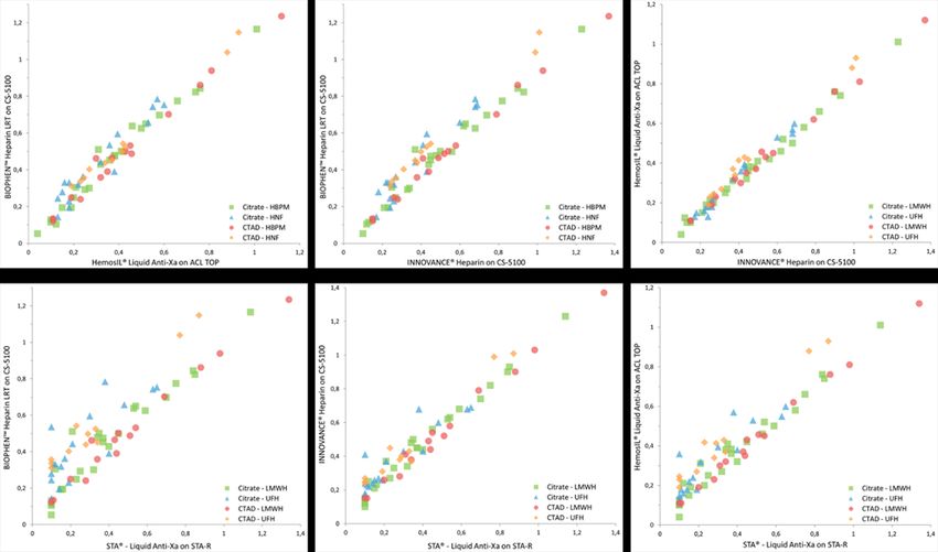
Preprints (www.preprints.org) | NOT PEER-REVIEWED | Posted: 4 March 2021               doi:10.20944/preprints202103.0171.v1

                                                                                                                      4 of 17

                                 prepared with the same heparin tested must be used. However, there is a strong market
                                 request to use a single heparin calibration for all heparin types, whether UFH or LMWH.
                                 Most manufacturers now propose a single heparin calibration curve, hybrid, for testing
                                 all heparins. This goal is achieved correctly fully superimposable UFH and LMWH cali-
                                 bration are obtained. Today, the current practice for monitoring any type of heparin
                                 therapy is to use the one-stage anti-FXa chromogenic kinetics assay, fully automated,
                                 with only one precalibrated curve, associated with UFH or LMWH control plasmas.

                                 Variability of heparin measurements: Although important efforts have been performed
                                 to standardize, automate, and optimize heparin testing, with availability of ISs and of
                                 guidelines issued by scientific societies or regulatory bodies, many differences in meas-
                                 ured plasma heparin concentrations are still observed when the various branded heparin
                                 anti-FXa chromogenic assays are used [55-58]. This is illustrated by the external quality
                                 assessment programs, like ECAT, which show a remaining significant reagent to reagent
                                 and laboratory to laboratory variability, more especially for UFH in the low range [57).
                                 The debate on which anti-FXa method generates the right results has been recently open
                                 again, with the extended indications of heparin treatments, using either UFH or LMWH,
                                 in Covid-19 patients, as thrombosis is a frequent disease complication [55, 59-61].

                                 Indeed, heparin measurement is an assay which concerns a catalytic indirect inhibitor,
                                 and many parameters impact its kinetics. The design of assay conditions is essential for
                                 its performances. With the same assay principle, the presence of multiple proteins bind-
                                 ing to heparin in plasma produce significant differences depending on the reagent con-
                                 cept, its formulation and the calibration used. Many years ago, the use of low molecular
                                 weight dextran sulfate (DS) was introduced for improving the heparin anti-FXa assays. It
                                 was claimed that presence of this component allowed measuring the full heparin activity
                                 in plasma, by limiting the impact of ex-vivo neutralization, especially by platelet released
                                 products [62-64). Now, many heparin diagnostic device manufacturers use this compo-
                                 nent, which is indicated on the instructions for use, whilst others do not yet [57,61].

                                 Another important incidence on measured heparin concentrations results from the cali-
                                 bration used. Heparin can be often tested in emergency conditions. Clinical laboratories
                                 do not always know which heparin brand or type is used for patients’ treatments. There
                                 is then a high expectation to use a single heparin calibration for any heparin type to be
                                 measured. Attempts have been done for reaching this objective. One approach is to de-
                                 velop assay conditions, which permit obtaining the same dose-response curve for UFH
                                 and LMWH [62, 63]. Calibration curves for UFH and LMWH are then fully superim-
                                 posable. Another approach is to build a hybrid curve by mixing or combining UFH and
                                 LMWH for plasma calibrators to get a median curve, between that of UFH and that of
                                 LMWH [65]. In this report we show the impact of DS for measuring the various heparin
                                 types, and its contribution to the exactness and accuracy of heparin measurement on
                                 plasma. Reagents and reference material from the various manufacturers are compared
                                 for the measurement of UFH or LMWH on citrate or CTAD anticoagulated plasmas from
                                 heparin treated patients [66]. Assays are calibrated with the manufacturers’ proposed
                                 heparin calibrators comparatively to the WHO UFH or LMWH International Standards.
                                 We then discuss the factors which are responsible for the variations of measured heparin
                                 concentrations and the assays’ biases.

                                 2. Materials and Methods

                                 Patients and normal plasmas: citrated normal plasmas and plasma pool were supplied
                                 frozen by Precision Biologic Inc. (Halifax, Canada), and stored at < -70°C until use.
Preprints (www.preprints.org) | NOT PEER-REVIEWED | Posted: 4 March 2021 doi:10.20944/preprints202103.0171.v1
5 of 17
Plasmas from hospitalized patients with heparin therapy for post-surgery thrombosis
prevention, using either UFH or LMWH, were obtained from Beaujon University Hos-
pital (Clichy, France), as the left-over residual plasma, and obtained according to CLSI.
Blood was collected either on 0.109 citrate or CTAD (Cit-
rate-Theophylline-Adenosine-Dipyridamole) anticoagulant from heparin treated patients
(UFH or LMWH), and plasma was decanted following 20 minutes centrifugation at 2,000
g, at Room Temperature (RT), then stored frozen atPreprints (www.preprints.org) | NOT PEER-REVIEWED | Posted: 4 March 2021 doi:10.20944/preprints202103.0171.v1
6 of 17
tions for use for each assay, and plasma diluent was either Owren Veronal Buffer (rea-
gents A and C), or 0.15 M sodium chloride (reagents C and D).
Verification of dose-response curves for UFH and LMWH: the citrate plasma pool sup-
plemented with either UFH or LMWH ISs was assayed for each reagent-instrument
combination (A, B, C and D), parallelly with the manufacturers’ calibrators.
Correlation studies: all plasmas, from UFH or LMWH treated patients, and whether cit-
rate or CTAD anticoagulated, were tested with the 4 anti-FXa assay combinations and
correlation diagrams were established. Sub-analysis was then performed for the various
groups, plasmas from UFH or LMWH treated patients, obtained using citrate or CTAD
anticoagulant.
Heparin characteristics of plasma calibrators: heparin calibrators from the various
manufacturers were tested with the 2-stage anti-FXa or anti-FIIa assays with the CS-2400
instrument and calibrated with the UFH or LMWH WHO-ISs spiked in plasma. This
measurement allowed analyzing the content of each plasma calibrator by establishing the
anti-FXa/Anti-FIIa ratios: UFH has a ratio of 1.00, whilst depending on the branded ma-
terial LMWH has a ratio from 1.6 to 9.7 [4, 43].
Calibration curves analysis: Heparin calibrators proposed by each manufacturer for its
anti-FXa kinetics assay were evaluated comparatively to UFH and LMWH WHO-ISs.
Each proposed manufacturer’s heparin calibrator and the UFH or LMWH WHO-ISs
spiked in plasma, with a concentration range from 0.00 to 1.80 IU/ml, as described before,
were tested with each anti-FXa reagent-instrument combination, as described here above
(A, B, C and D). For each combination, the 3 calibration curves obtained (heparin assay
manufacturer’s calibrator, UFH IS and LMWH IS) are compared.
Statistics were performed using the analyse-it software.
3. Results
Calibration curves for the various assays:
The various calibration curves obtained with each anti-FXa combination for the manu-
facturer’s calibrator and the UFH or LMWH WHO-ISs are shown on figure 1. Superim-
position between the manufacturer calibration curve and those obtained with the WHO
International UFH or LMWH Standards is globally good, although some slight deviation
can be seen depending on the system used. In combination A, UFH-IS calibration lacks
linearity, especially in the low range, and absorbances measured are above the manufac-
turers’ calibration, which can result in underestimation of UFH concentrations, especially
for low heparin concentrations. Superimposition is better in the high range. In combina-
tion B, UFH and LMWH ISs calibrations have an acceptable superimposition, and manu-
facturer’s calibration appears to deviate below ISs curves, which can underestimate UFH
or LMWH concentrations. In combination C, superimposition is also acceptable, with the
assay calibration like that of UFH-IS but slightly above that of LMWH-IS, which can tend
to slightly underestimate LMWH; superimposition for all the curves is also obtained for
combination D.
Deviations are higher for UFH, especially for low concentrations, when DS is not used in
the assay system. A better accuracy and exactness are also obtained when heparin plasma
calibrator concentrations are regularly distributed over the dynamic range, than concen-
trated in the lower part, as for combination B.Preprints (www.preprints.org) | NOT PEER-REVIEWED | Posted: 4 March 2021 doi:10.20944/preprints202103.0171.v1
7 of 17
Figure 1. Comparison of calibration curves for each anti-FXa assay used with the manufacturer’s coagulation instrument
as compared to the International Standards for UFH or LMWH. Heparin concentrations are on abscissae and change in
absorbance per minute (OD/min) on ordinates.
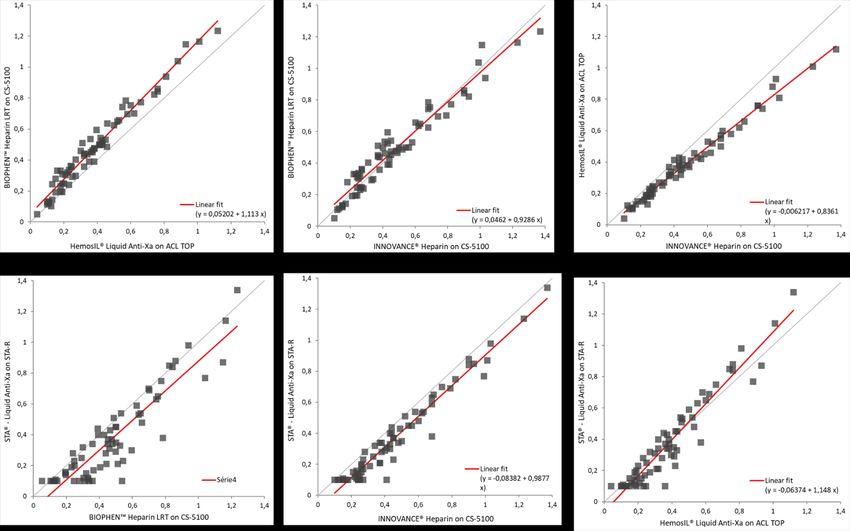
Correlation studies:
Correlation studies are performed on the global patients’ plasma group, obtained
from blood samples from UFH or LMWH treated patients and collected on citrate or
CTAD anticoagulants. Figure 2 shows the various correlation diagrams, for each manu-
facturer’s device compared to the others: B vs D; C vs D; C vs B; D vs A; C vs A; B vs A.
The reagents containing DS (B, C and D) present acceptable correlations between them,
whilst there is a higher dispersion when these reagents are compared with reagent A,
designed without DS.
The differences are higher for UFH samples than for LMWH. The correlation line
tendency for A and B is to underestimate heparin concentrations as compared to C and D,
as expected from the calibration curve analysis.
The mean values for the various subgroups of plasmas tested (UFH or LMWH with
citrate anticoagulant or with CTAD anticoagulant) are shown on table 1. Mean heparin
concentrations are lower when measured with reagents A and B than with reagents C and
D. Differences are partly due to the use of dextran sulfate for the assay formulation, and
partly to the calibration used.Preprints (www.preprints.org) | NOT PEER-REVIEWED | Posted: 4 March 2021 doi:10.20944/preprints202103.0171.v1
8 of 17
Figure 2. shows the global crossed correlations between the 4 different branded anti-FXa assays for all the tested
plasma samples from heparin treated patients (either with UFH or LMWH), and anticoagulated with citrate or
CTAD. Assays B, C and D contain dextran sulfate, whilst assay A does not. The global correlation is better when
assays containing dextran sulfate are compared between them.
Table 1: Mean heparin concentrations measured on the 68 plasmas from UFH or LMWH treated patients, using
the 4 various anti-FXa kinetics assays (Stago, Werfen-IL, Siemens and HYPHEN BioMed); blood was collected
either on citrate or CTAD anticoagulant, and plasma decanted following centrifugation.
N=68 Mean SD Minimum Median Maximum
STA® - Liquid Anti-Xa (A) 0.376 0.282 0.10 0.315 1.34
HemosIL® Liquid Anti-Xa (B) 0.383 0.235 0.04 0.355 1.12
INNOVANCE® Heparin (C) 0.466 0.278 0.10 0.410 1.37
BIOPHEN™ Heparin LRT (D) 0.479 0.266 0.05 0.452 1.24
Impact of anticoagulant:Preprints (www.preprints.org) | NOT PEER-REVIEWED | Posted: 4 March 2021 doi:10.20944/preprints202103.0171.v1
9 of 17
To understand and illustrate which factors are responsible for heparin concentration
differences between assays, the various correlation diagrams were drawn by identifying
each patients’ plasma group. Figure 3 shows for each combination the correlation dia-
grams with the separate identification of each subgroup: UFH-Citrate; LMWH-Citrate;
UFH-CTAD; LMWH-CTAD. This diagram shows obviously that the differences are
mainly due to citrate plasma samples containing UFH, and in a lesser extend to citrate
samples containing LMWH. When CTAD is used as anticoagulant, a much better co-
herence of heparin concentrations measured is obtained between all assays.
Figure 3. Crossed correlations for the comparison of the various tested subgroups (UFH-Citrate: blue triangles;
LMWH-Citrate: green squares; UFH-CTAD: orange dots; LMWH-citrate: orange diamonds) with the various re-
agent-instrument combinations A, B, C and D. The highest differences are observed for UFH-citrate, and in a
lesser extend for LMWH-citrate, plasma samples, especially when anti-FXa reagents with (B, C and D) or without
(A) dextran sulfate are compared.
To confirm the factors explaining the heparin concentration differences measured with
the various reagents, especially when designed with or without DS, correlations were
analyzed separately for each group of plasma samples, obtained from blood collected on
citrate or CTAD anticoagulant as shown on the correlation diagrams presented on figure
4. Results are shown for UFH or LMWH plasmas with the 2 anticoagulants, citrate or
CTAD, only for the comparison between reagents A and D. However, similar correlations
are obtained for A when compared to reagents B or C (data not shown).
The highest dispersion of results between reagents A and D concerns UFH samples
collected on citrate anticoagulant. When the same samples are collected on CTAD anti-
coagulant a much better correlation is obtained. The same comments can be done when
comparison is made between reagent A and reagents B or C, whilst correlations are ac-Preprints (www.preprints.org) | NOT PEER-REVIEWED | Posted: 4 March 2021 doi:10.20944/preprints202103.0171.v1
10 of 17
ceptable when reagents B, C and D are compared between whether on UFH and LMWH
plasmas, anticoagulated with citrate or CTAD.
These data suggest that UFH is partially inhibited ex-vivo and its concentration is
underestimated when reagent A is used. Presence of DS prevents from this inhibition.
Figure 4. correlation diagrams between the anti-FXa reagent designed without dextran sulfate (A) and another one
with (D) for the various subgroups of tested samples: citrate-UFH; citrate-LMWH; CTAD-UFH; CTAD-LMWH. The
correlation is poor for citrate anticoagulated samples, especially for UFH, whilst it is acceptable for CTAD antico-
agulated plasmas, containing either UFH or LMWH.
The mean heparin concentrations measured with the 4 anti-FXa assays combinations
were analyzed for each of the subgroups treated with either UFH or LMWH, and antico-
agulated with citrate or CTAD. Table 2 shows the values obtained for each subgroup,
underlining the important impact of the anticoagulant used and assay design without DS,
on the heparin concentrations measured especially for the low concentration range. Other
differences observed with the various assays and the various groups can be explained by
the calibration curves biases, when compared with the UFH or LMWH reference curves
obtained with the ISs. This has an additional impact on reagent B in the low UFH range,
and in a lesser extent on reagent C.
Table 2: mean heparin concentrations, in IU/mL, measured with the 4 different anti-FXa reagents on the various
subgroups: Citrate-UFH; Citrate-LMWH; CTAD-UFH and CTAD-LMWH).Preprints (www.preprints.org) | NOT PEER-REVIEWED | Posted: 4 March 2021 doi:10.20944/preprints202103.0171.v1
11 of 17
STA® LIQUID HEMOSIL® INNOVANCE® BIOPHEN™
ANTI-XA LIQUID ANTI-XA HEPARIN HEPARIN LRT
Citrate Mean 0.25 0.31 0.37 0.43
UFH N=17 Min-Max 0.10-0.65 0.13-0.60 0.17-0.69 0.14-0.79
(IU/ML) CTAD Mean 0.33 0.43 0.46 0.55
N=11 Min-Max 0.10-0.87 0.19-0.93 0.24-1.01 0.31-1.15
Citrate Mean 0.40 0.38 0.48 0.46
LMWH
N=25 Min-Max 0.10-1.14 0.04-1.01 0.10-1.23 0.05-1.17
(IU/ML)
CTAD Mean 0.51 0.44 0.55 0.51
N=15 Min-Max 0.10-1.34 0.11-1.12 0.15-1.37 0.12-1.24
Composition of the various heparin calibrators:
As heparin anti-FXa reagents are indicated for testing all heparin types, manufacturers
proposed superimposed curves or hybrid curves which can be used irrelevantly for test-
ing UFH or LMWH with the same heparin calibrator. We evaluated the specific antico-
agulant activity of each heparin plasma calibrator to FXa and FIIa, with the 2-stages as-
says. The specific anti-FXa to anti-FIIa ratios were calculated for each calibrator. Results
are presented on table 1.
UFH has an anti-FXa/Anti-FIIa ratio of 1.00 and the various LMWH have ratios rang-
ing from 1.6 to 9.7, partly dependent on the MW size distribution, and on the pentasac-
charide density. From these data it can be deduced that Stago heparin calibrator set con-
tains 2 calibrators (calibrators 2 and 4) obtained by supplementing plasma with UFH and
2 with LMWH (calibrators 3 and 5), whilst all the IL HemosIL heparin calibrators contain
a mixture of UFH with some LMWH. Siemens and HYPHEN BioMed heparin calibrators
are homogenous and prepared with only LMWH added to plasma. The anti-FXa to An-
ti-FIIa ratios show that different LMWH are used: this ratio (mean of 2.10) is lower for
the Siemens calibrators, like that of certoparin, and higher for HYPHEN BioMed (mean of
4.02), like that of enoxaparin. The WHO International Standard for LMWH 11/176 has an
Anti-FXa/FIIa ratio of 3.12 (1068 IU for anti-FXa and 342 IU for anti-FIIa).
The appropriateness for the use of a single heparin calibration curve for measuring
UFH or LMWH depends first on the accuracy of the superimposition of both curves ob-
tained with the corresponding ISs. Both WHO standards were proposed as each heparin
type, UFH or LMWH, present different characteristics for inhibition kinetics.Preprints (www.preprints.org) | NOT PEER-REVIEWED | Posted: 4 March 2021 doi:10.20944/preprints202103.0171.v1
12 of 17
Table3: Analysis of the various heparin calibrators from the different manufacturers by testing their anti-FXa
and anti-FIIa activities (IU/mL) as compared with the manufacturers’ claimed concentrations for the used heparin
calibrator from the lots used, and anti-FXa/anti-FIIa ratios.
Heparin Anti-FXa/Anti-IIa Manufacturer's Target
Brand Anti-IIa IU/ml Anti-FXa IU/ml
Calibrators ratio Value IU/ml
1 0.00 0.02 0.00 0.00
IL 2 0.59 0.77 1.32 0.80
3 1.68 2.32 1.38 2.00
1 0.00 0.02 0.00 0.00
2 0.59 0.57 0.98 0.40
Diagnostica Stago 3 0.38 0.82 2.08 0.68
4 1.12 1.08 0.95 0.98
5 1.06 2.18 2.06 1.79
1 0.00 0.00 0.00 0.00
2 0.18 0.37 2.06 0.43
Siemens 3 0.38 0.79 2.08 0.85
4 0.56 1.19 2.13 1.27
5 0.74 1.58 2.14 1.67
1 0.00 0.00 0.00 0.00
2 0.11 0.44 4.00 0.45
HYPHEN BioMed 3 0.23 0.92 4.00 0.92
4 0.33 1.35 4.09 1.36
5 0.48 1.92 4.00 1.80
4. Discussion
Recent articles have pointed out the variability of heparin measurements using the var-
ious commercially available Anti-FXa assays. This debate has been reactivated with the
extended use of heparin therapy in Covid-19 patients, and the detection in some patients
of high sensitivity, when drug clearance is decreased, or resistance, when strong in-
flammation, Nets and histones are present (14, 15, 67, 68). Some recent studies suggest
that there is an overestimation of measured heparin concentrations, especially for UFH,
when DS is used for the anti-FXa assay formulation, whilst other reports support this
technical choice as providing the most accurate estimation of circulating heparin antico-
agulant activity [55, 59, 60]. Especially, this debate questions which is the right residual
heparin concentration following neutralization with protamine sulfate at the end of ex-
tra-corporeal circulation, and when the rebound effect is observed [59, 69-71]. Studies
using heparinase or heparinase showed that the measured residual heparin does not al-
ways match with the anticoagulant activity measured [72-74], and presence of DS can
provide a better estimation. As a developer of heparin testing reagents, we analyzed
these different reports and anti-FXa assays’ performances through our experience.
In this study we have evaluated various factors impacting the measurement of heparin
concentrations on plasma from UFH or LMWH treated patients using the 4 major com-
mercially available anti-FXa assays. We have investigated the incidence of assays’ for-
mulations, and of the manufacturers’ calibration curves proposed. Heparin calibrators
have been tested by comparison with the UFH or LMWH WHO International Standards,
spiked in a normal platelet poor plasma pool. Three of the anti-FXa assays (reagents
B/IL-Werfen, C/Siemens-Innovance and D/ HYPHEN BioMed-Biophen) are designed
with dextran sulfate, a component which was reported to make available for testing all
mobilizable heparin with anticoagulant activity, as present in the sample [62, 63], whilstPreprints (www.preprints.org) | NOT PEER-REVIEWED | Posted: 4 March 2021 doi:10.20944/preprints202103.0171.v1
13 of 17
one (reagent A/Stago-STA Liquid Anti-Xa) does not contain this component. Tested
plasma samples were provided by Beaujon University’ Hospital (Clichy, France) and
were obtained from UFH or LMWH treated patients, collected on citrate or on CTAD
anticoagulant. CTAD tends to be less and less used nowadays, as laboratories wish to use
only 1 anticoagulant tube type for standardizing hemostasis testing. CTAD was devel-
oped to increase blood and plasma stability, especially for heparin testing [66, 75]. This
anticoagulant formulation prevents from platelet activation and release of heparin neu-
tralization proteins. The assay-to-assay comparison was performed on the global group
including 68 plasmas and analyzed for each subgroup concerning the heparin type and
the anticoagulant used. Four groups were then obtained: UFH-citrate (N=17);
LMWH-citrate (N=25); UFH-CTAD (N=11); LMWH-CTAD (N=15). Globally, on the total
group, correlations were acceptable between reagents B, C and D, and there was a higher
dispersion of values and a poorer correlation between reagent A and the 3 others, espe-
cially in the low range. When subgroups were analyzed separately, it was obvious that
major deviations were observed for plasmas from UFH treated patients and anticoagu-
lated with citrate. A better homogeneity was obtained when samples were collected us-
ing CTAD anticoagulant, thus preventing from heparin neutralization ex-vivo. Correla-
tions between assays are much better when plasma samples are collected on CTAD.
These results strongly suggest that UFH is partly neutralized ex-vivo by platelet released
proteins, and that this inhibition is prevented using CTAD anticoagulant. When reagents
contain DS, this inhibition is prevented, and heparin concentrations measured are ex-
pected to match better with those present in blood circulation, and this observation is
supported by various studies [60, 62, 63, 74].
When comparing the mean heparin concentrations and standard deviations, the lowest
values were obtained with reagent A, then B, especially for UFH, and the highest ones
with reagents C, then D. Despite reagents B, C and D all contain DS in their formulation,
some differences were observed between the mean concentrations measured, especially
for reagent B as compared to reagents C and D. However, SDs are similar between rea-
gents. The choice of the calibration curve for measuring irrelevantly plasma samples
containing UFH or LMWH contributes to explain these differences. Clinical laboratories
need a 24/24 h and 7/7 d available anti-FXa assay for measuring heparin and monitoring
treated patients, and some of the analysis are requested in emergency. The type of hepa-
rin used is not always known, and therefore using a single calibration curve is necessary.
This approach needs to be carefully established and validated to give accurate results for
all types of heparin used. WHO proposes nevertheless 2 separate International Standards
for UFH or LMWH, and assay manufacturers need to establish anti-FXa assay conditions
to obtain fully superimposable calibration curves for all heparin types. When the right
conditions are fulfilled, calibration curves obtained with UFH or LMWH in plasma can
be used without any difference. The heparin calibrations proposed by the various man-
ufacturers differ significantly, and this can have an important impact on exactness and
accuracy of measured heparin concentrations. Reagents C and D use a calibration curve
prepared with only LMWH spiked in a platelet depleted plasma pool. However, LMWH
used by both manufacturers C and D are not the same as demonstrated by the different
anti-FXa/Anti-FIIa ratios. LMWH is like certoparin for reagent C and like enoxaparin for
reagent D. Conversely, reagents A and B propose a hybrid calibration curve obtained by
mixing or combining UFH and LMWH in plasma. The anti-FXa/anti-FIIa ratios suggest
that calibrators used for reagent B are obtained using a mixture of UFH and LMWH, the
UFH concentration being predominant as shown by the low ratio obtained. In contrast,
reagent A uses an alternate combination of plasmas containing UFH (calibrators 2 and 4)
or LMWH (calibrators 3 and 5), with an anti-FXa/anti-FIIa ratio like that of certoparin. If
UFH and LMWH calibration curves are not strictly superimposable, then the hybrid
calibration curve is an intermediate curve which introduces some biases for both UFHPreprints (www.preprints.org) | NOT PEER-REVIEWED | Posted: 4 March 2021 doi:10.20944/preprints202103.0171.v1
14 of 17
and LMWH samples. UFH concentrations tend then to be underestimated, as observed
with reagents A and B, especially in the low range.
Lastly, the various anti-FXa reagents were used along with each manufacturer proposed
instrument for reagents A, B and C, and have been used by adhering strictly to the in-
structions for use. Reagent D is proposed as a multiplatform reagent, with applications
specifically developed for all the major instruments available. In this study, reagent D
was combined with the Sysmex CS-5100 instrument.
When using homogenous (same manufacturer) reagent-instrument systems, reagent
weaknesses can be masked by the assay software adaptation, or by introducing algo-
rithms for optimizing the assay apparent performances. This approach is used for ad-
justing the intrinsic low anti-FXa activity present in all plasma samples, and which can be
variable from plasma to plasma. In the absence of heparin, this intrinsic anti-FXa activity
can account for 0 to 0.05 IU/ml in normal plasmas, and more rarely up to 0.10 IU. This
background activity is likely due to the anti-FXa activity of TFPI, Protein S or the β-AT
form. The anti-FXa heparin assay is an inverse relationship between absorbance change
measured at 405 nM, and heparin concentration. Therefore, normal plasmas, whilst they
are expected to have all the same basic absorbance in the absence of heparin, do not al-
ways show a zero anti-FXa activity. Assay systems can manage this variability by mask-
ing that effect and “starting to measure” the change in absorbance only from a threshold
value, corresponding to plasmas with the highest anti-FXa intrinsic activity. The apparent
heparin concentrations in all plasmas are then of 0 in the absence of heparin, but low
concentrations of heparin, in the range 0 to 0.10 IU/mL, or even up to 0.15 IU/ml, can be
missed, which contributes to the underestimation in the low range. This approach is of
course not possible when the reagent is a multiplatform one, and no adjustment assay
software can be used. Heparin concentrations measured in plasma are then obtained
without any data treatment.
5. Conclusions
In this report we provide evidence on the usefulness of dextran sulfate for the an-
ti-FXa assays used for measuring plasma concentrations of UFH or LMWH, as shown by
the good correlation between all assays, designed with or without dextran sulfate, when
plasma is obtained from CTAD collected blood, thus preventing from platelet activation
and release of heparin neutralizing proteins, but not when blood is collected on citrate.
Assay variability can also result from the heparin calibration type used, the exactness of
UFH and LMWH superimposition of calibration curves and the assay software for
treatment of assay raw data. Analyzing these factors can help for a better understanding
of differences reported in many studies on heparin concentrations when measured with
the various anti-FXa reagents.
Funding: This research received no external funding
Institutional Review Board Statement: Not applicable.
Informed Consent Statement: Not applicable.
Acknowledgments: In this section, you can acknowledge any support given which is not covered
by the author contribution or funding sections. This may include administrative and technical
support, or donations in kind (e.g., materials used for experiments).
Conflicts of Interest: Jean AMIRAL is scientific consultant for HYPHEN BioMed (France) and
Sysmex Corp. (Japan); Cédric AMIRAL and Claire DUNOIS are employees of HYPHEN BioMed.
References
1. Onishi A, St Ange K, Dordick JS, Linhardt RJ. Heparin and anticoagulation. Front Biosci (Landmark Ed). 2016 Jun 1;21:1372-9Preprints (www.preprints.org) | NOT PEER-REVIEWED | Posted: 4 March 2021 doi:10.20944/preprints202103.0171.v1
15 of 17
2. Parker CR, Kataria V. Monitoring Unfractionated Heparin: A Review of Activated Partial Thromboplastin Time Versus Anti-
factor Xa. AACN Adv Crit Care. 2019 Dec 15;30(4):305-312.
3. Hao C, Xu H, Yu L, Zhang L. Heparin: An essential drug for modern medicine. Prog Mol Biol Transl Sci. 2019;163:1-19
4. Barrowcliffe TW. History of heparin. Handb Exp Pharmacol. 2012;(207):3-22.
5. Hemker HC. A century of heparin: past, present and future. J Thromb Haemost. 2016 Dec;14(12):2329-2338.
6. Kakkar VV, Corrigan T, Spindler J, Fossard DP, Flute PT, Crellin RQ, Wessler S, Yin ET. Efficacy of low doses of heparin in
prevention of deep-vein thrombosis after major surgery. A double-blind, randomised trial. Lancet. 1972 Jul 15;2(7768):101-6
7. Lima M, Rudd T, Yates E. New Applications of Heparin and Other Glycosaminoglycans. Molecules. 2017 May 6;22(5):749.
8. Beurskens DMH, Huckriede JP, Schrijver R, Hemker HC, Reutelingsperger CP, Nicolaes GAF. The Anticoagulant and
Nonanticoagulant Properties of Heparin. Thromb Haemost. 2020 Oct;120(10):1371-1383.
9. Drouet L, Harenberg J, Torri G. The Multiple Faces of Heparin: Opportunities in COVID-19 Infection and Beyond. Thromb
Haemost. 2020 Oct;120(10):1347-1350.
10. Kitchen S. Problems in laboratory monitoring of heparin dosage. Br J Haematol. 2000 Nov;111(2):397-406.
11. Raivio P, Kuitunen A, Petäjä J, Ilveskero S, Lassila R. Monitoring high-dose heparinization during cardiopulmonary by-pass--a
comparison between prothrombinase-induced clotting time (PiCT) and two chromogenic anti-factor Xa activity assays.
Thromb Haemost. 2008 Feb;99(2):427-34.
12. Atiq F, van den Bemt PM, Leebeek FW, van Gelder T, Versmissen J. A systematic review on the accumulation of prophylactic
dosages of low-molecular-weight heparins (LMWHs) in patients with renal insufficiency. Eur J Clin Pharmacol. 2015
Aug;71(8):921-9
13. Sciascia S, Radin M, Schreiber K, Fenoglio R, Baldovino S, Roccatello D. Chronic kidney disease and anticoagulation: from
vitamin K antagonists and heparins to direct oral anticoagulant agents. Intern Emerg Med. 2017 Dec;12(8):1101-1108
14. Longstaff C, Hogwood J, Gray E, Komorowicz E, Varjú I, Varga Z, Kolev K. Neutralisation of the anti-coagulant effects of
heparin by histones in blood plasma and purified systems. Thromb Haemost. 2016 Mar;115(3):591-9.
15. Zuo Y, Yalavarthi S, Shi H, Gockman K, Zuo M, Madison JA, Blair C, Weber A, Barnes BJ, Egeblad M, Woods RJ, Kanthi Y,
Knight JS. Neutrophil extracellular traps (NETs) as markers of disease severity in COVID-19. medRxiv [Preprint]. 2020 Apr
14:2020.04.09.20059626.
16. Streng AS, Delnoij TSR, Mulder MMG, Sels JWEM, Wetzels RJH, Verhezen PWM, Olie RH, Kooman JP, van Kuijk SMJ, Brandts
L, Ten Cate H, Lorusso R, van der Horst ICC, van Bussel BCT, Henskens YMC. Monitoring of Unfractionated Heparin in Se-
vere COVID-19: An Observational Study of Patients on CRRT and ECMO. TH Open. 2020 Nov 19;4(4):e365-e375.
17. White D, MacDonald S, Bull T, Hayman M, de Monteverde-Robb R, Sapsford D, Lavinio A, Varley J, Johnston A, Besser M,
Thomas W. Heparin resistance in COVID-19 patients in the intensive care unit. J Thromb Thrombolysis. 2020
Aug;50(2):287-291.
18. Greinacher A. Heparin-induced thrombocytopenia. J Thromb Haemost. 2009 Jul;7 Suppl 1:9-12
19. Amiral J, Seghatchian J. An update on evidence based diagnostic and confirmatory testing strategies for heparin induced
thrombocytopenia using combined immunological and functional assays. Transfus Apher Sci. 2018 Dec;57(6):804-81
20. Hajjar DP, Boyd DB, Harpel PC, Nachman RL. Histidine-rich glycoprotein inhibits the antiproliferative effect of heparin on
smooth muscle cells. J Exp Med. 1987 Mar 1;165(3):908-13
21. Brown RA, Lever R, Jones NA, Page CP. Effects of heparin and related molecules upon neutrophil aggregation and elastase
release in vitro. Br J Pharmacol. 2003 Jun;139(4):845-53
22. Ouyang Y, Yu Y, Zhang F, Chen J, Han X, Xia K, Yao Y, Zhang Z, Linhardt RJ. Non-Anticoagulant Low Molecular Weight
Heparins for Pharmaceutical Applications. J Med Chem. 2019 Jan 24;62(2):1067-1073
23. Kaiser B, Glusa E, Hoppensteadt DA, Breddin HK, Amiral J, Fareed J. A supersulfated low-molecular-weight heparin (IK-SSH)
increases plasma levels of free and total tissue factor pathway inhibitor after intravenous and subcutaneous administration in
humans. Blood Coagul Fibrinolysis. 1998 Sep;9(6):517-23
24. Falkon L, Garí M, Barbanoj M, Amiral J, Fontcuberta J. Tissue factor pathway inhibitor and anti-FXa kinetic profiles of a new
low-molecular-mass heparin, Bemiparin, at therapeutic subcutaneous doses. Blood Coagul Fibrinolysis. 1998 Mar;9(2):137-41
25. Ma Q, Tobu M, Schultz C, Jeske W, Hoppensteadt D, Walenga J, Cornelli U, Lee J, Linhardt R, Hanin I, Fareed J. Molecular
weight dependent tissue factor pathway inhibitor release by heparin and heparin oligosaccharides. Thromb Res.
2007;119(5):653-61
26. Baglin T, Barrowcliffe TW, Cohen A, Greaves M; British Committee for Standards in Haematology. Guidelines on the use and
monitoring of heparin. Br J Haematol. 2006 Apr;133(1):19-34.
27. Kalkana C, Conventional Anticoagulant Therapy, Hospital Chronicles, 2015, 10(4): 210–222
28. Despotis GJ, Summerfield AL, Joist JH, Goodnough LT, Santoro SA, Spitznagel E, Cox JL, Lappas DG. Comparison of activated
coagulation time and whole blood heparin measurements with laboratory plasma anti-Xa heparin concentration in patients
having cardiac operations. J Thorac Cardiovasc Surg. 1994 Dec;108(6):1076-82.
29. Bowers J, Ferguson JJ 3rd. The use of activated clotting times to monitor heparin therapy during and after interventional pro-
cedures. Clin Cardiol. 1994 Jul;17(7):357-61
30. Newall F. Anti-factor Xa (anti-Xa) assay. Methods Mol Biol. 2013;992:265-72. doi: 10.1007/978-1-62703-339-8_19
31. Mehmedagić A, Skrbo S, Softić D, Haracić M. In vitro modeling of the influence of FVIII activity and heparin induced prolon-
gation of APTT. Bosn J Basic Med Sci. 2005 Aug;5(3):26-9Preprints (www.preprints.org) | NOT PEER-REVIEWED | Posted: 4 March 2021 doi:10.20944/preprints202103.0171.v1
16 of 17
32. Marlar RA, Clement B, Gausman J. Activated Partial Thromboplastin Time Monitoring of Unfractionated Heparin Therapy:
Issues and Recommendations. Semin Thromb Hemost. 2017 Apr;43(3):253-260
33. McGlasson DL, Kaczor DA, Krasuski RA, Campbell CL, Kostur MR, Adinaro JT. Effects of pre-analytical variables on the an-
ti-activated factor X chromogenic assay when monitoring unfractionated heparin and low molecular weight heparin antico-
agulation. Blood Coagul Fibrinolysis. 2005 Apr;16(3):173-6.
34. Eikelboom JW, Hirsh J. Monitoring unfractionated heparin with the aPTT: time for a fresh look. Thromb Haemost. 2006
Nov;96(5):547-52
35. Byun JH, Jang IS, Kim JW, Koh EH. Establishing the heparin therapeutic range using aPTT and anti-Xa measurements for
monitoring unfractionated heparin therapy. Blood Res. 2016 Sep;51(3):171-174.
36. Whitman-Purves E, Coons JC, Miller T, DiNella JV, Althouse A, Schmidhofer M, Smith RE. Performance of Anti-Factor Xa
Versus Activated Partial Thromboplastin Time for Heparin Monitoring Using Multiple Nomograms. Clin Appl Thromb He-
most. 2018 Mar;24(2):310-316.
37. Arachchillage DRJ, Kamani F, Deplano S, Banya W, Laffan M. Should we abandon the APTT for monitoring unfractionated
heparin? Thromb Res. 2017 Sep;157:157-161
38. Bürki S, Brand B, Escher R, Wuillemin WA, Nagler M. Accuracy, reproducibility and costs of different laboratory assays for the
monitoring of unfractionated heparin in clinical practice: a prospective evaluation study and survey among Swiss institutions.
BMJ Open. 2018 Jun 9;8(6):e022943.
39. Béguin S, Lindhout T, Hemker HC. The mode of action of heparin in plasma. Thromb Haemost. 1988 Dec 22;60(3):457-62
40. Amiral J, Seghatchian J. Revisiting antithrombin in health and disease, congenital deficiencies and genetic variants, and labor-
atory studies on α and β forms. Transfus Apher Sci. 2018 Apr;57(2):291-297
41. Rezaie AR. Heparin chain-length dependence of factor Xa inhibition by antithrombin in plasma. Thromb Res. 2007;119(4):481-8
42. Olson ST, Richard B, Izaguirre G, Schedin-Weiss S, Gettins PG. Molecular mechanisms of antithrombin-heparin regulation of
blood clotting proteinases. A paradigm for understanding proteinase regulation by serpin family protein proteinase inhibitors.
Biochimie. 2010 Nov;92(11):1587-96
43. Gray E, Mulloy B, Barrowcliffe TW. Heparin and low-molecular-weight heparin. Thromb Haemost. 2008 May;99(5):807-18.
44. Dawes J. Interactions of heparins in the vascular environment. Haemostasis. 1993 Mar;23 Suppl 1:212-9
45. Peysselon F, Ricard-Blum S. Heparin-protein interactions: from affinity and kinetics to biological roles. Application to an in-
teraction network regulating angiogenesis. Matrix Biol. 2014 Apr;35:73-81
46. Denton J, Lane DA, Thunberg L, Slater AM, Lindahl U. Binding of platelet factor 4 to heparin oligosaccharides. Biochem J. 1983
Feb 1;209(2):455-60
47. Lijnen HR, Hoylaerts M, Collen D. Heparin binding properties of human histidine-rich glycoprotein. Mechanism and role in
the neutralization of heparin in plasma. J Biol Chem. 1983 Mar 25;258(6):3803-8
48. Preissner KT, Müller-Berghaus G. Neutralization and binding of heparin by S protein/vitronectin in the inhibition of factor Xa
by antithrombin III. Involvement of an inducible heparin-binding domain of S protein/vitronectin. J Biol Chem. 1987 Sep
5;262(25):12247-53
49. Teien AN, Lie M, Abildgaard U. Assay of heparin in plasma using a chromogenic substrate for activated factor X. Thromb Res.
1976 Mar;8(3):413-6
50. Larsen ML, Abildgaard U, Teien AN, Gjesdal K. Assay of plasma heparin using thrombin and the chromogenic substrate
H-D-Phe-Pip-Arg-pNA (S-2238). Thromb Res. 1978 Aug;13(2):285-8
51. Tan X, Cui H. Comparative studies on biological activity of generic and branded enoxaparin in vivo and vitro. Blood Coagul
Fibrinolysis. 2015 Oct;26(7):805-10
52. ten Cate H, Lamping RJ, Henny CP, Prins A, ten Cate JW. Automated amidolytic method for determining heparin, a heparin-
oid, and a low-Mr heparin fragment, based on their anti-Xa activity. Clin Chem. 1984 Jun;30(6):860-4
53. Terao E, Daas A. Collaborative study for the calibration of replacement batches for the heparin low-molecular-mass for assay
biological reference preparation. Pharmeur Bio Sci Notes. 2016;2015:35-47
54. Gray E. Standardisation of unfractionated and low-molecular-weight heparin. Handb Exp Pharmacol. 2012;(207):65-76
55. Hardy M, Douxfils J, Bareille M, Lessire S, Gouin-Thibault I, Fontana P, Lecompte T, Mullier F. Studies on hemostasis in
COVID-19 deserve careful reporting of the laboratory methods, their significance, and their limitations. J Thromb Haemost.
2020 Nov;18(11):3121-3124.
56. Smahi M, De Pooter N, Hollestelle MJ, Toulon P. Monitoring unfractionated heparin therapy: Lack of standardization of an-
ti-Xa activity reagents. J Thromb Haemost. 2020 Oct;18(10):2613-2621.
57. Hollestelle MJ, van der Meer FJM, Meijer P. Quality performance for indirect Xa inhibitor monitoring in patients using inter-
national external quality data. Clin Chem Lab Med. 2020 Oct 25;58(11):1921-1930
58. Toulon P, Smahi M, De Pooter N. APTT therapeutic range for monitoring unfractionated heparin therapy. Significant impact of
the anti-Xa reagent used for correlation. J Thromb Haemost. 2021 Feb 8
59. Mouton C, Calderon J, Janvier G, Vergnes MC. Dextran sulfate included in factor Xa assay reagent overestimates heparin ac-
tivity in patients after heparin reversal by protamine. Thromb Res. 2003;111(4-5):273-9
60. Nougier C, Benoit R, Dargaud Y. Response to "Studies on hemostasis in COVID-19 deserve careful reporting of the laboratory
methods, their significance and their limitation": Don't throw the baby out with the bathwater. J Thromb Haemost. 2020
Nov;18(11):3124-3126.Preprints (www.preprints.org) | NOT PEER-REVIEWED | Posted: 4 March 2021 doi:10.20944/preprints202103.0171.v1
17 of 17
61. Hollestelle MJ, van der Meer FJM, Meijer P. Quality performance for indirect Xa inhibitor monitoring in patients using inter-
national external quality data. Clin Chem Lab Med. 2020 Oct 25;58(11):1921-1930
62. Lyon SG, Lasser EC, Stein R. Modification of an amidolytic heparin assay to express protein-bound heparin and to correct for
the effect of antithrombin III concentration. Thromb Haemost. 1987 Oct 28;58(3):884-7
63. Wagenvoord RJ, Hendrix HH, Kolde HJ, Hemker HC. Development of a rapid and sensitive chromogenic heparin assay for
clinical use. Haemostasis. 1993;23(1):26-37.
64. Ignjatovic V, Summerhayes R, Gan A, Than J, Chan A, Cochrane A, Bennett M, Horton S, Shann F, Lane G, Ross-Smith M,
Monagle P. Monitoring Unfractionated Heparin (UFH) therapy: which Anti-Factor Xa assay is appropriate? Thromb Res.
2007;120(3):347-51
65. David L. McGlasson, MS, Using a Single Calibration Curve With the Anti-Xa Chromogenic Assay for Monitoring Heparin
Anticoagulation, Laboratory Medicine, Volume 36, Issue 5, May 2005, Pages 297–299,
66. Contant G, Gouault-Heilmann M, Martinoli JL. Heparin inactivation during blood storage: its prevention by blood collection in
citric acid, theophylline, adenosine, dipyridamole-C.T.A.D. mixture. Thromb Res. 1983 Jul 15;31(2):365-74
67. White D, MacDonald S, Bull T, Hayman M, de Monteverde-Robb R, Sapsford D, Lavinio A, Varley J, Johnston A, Besser M,
Thomas W. Heparin resistance in COVID-19 patients in the intensive care unit. J Thromb Thrombolysis. 2020
Aug;50(2):287-291.
68. Magro G. COVID-19: Review on latest available drugs and therapies against SARS-CoV-2. Coagulation and inflammation
cross-talking. Virus Res. 2020 Sep;286:198070.
69. Dellagrammaticas D, Lewis SC, Gough MJ; GALA Trial Collaborators. Is heparin reversal with protamine after carotid
endarterectomy dangerous? Eur J Vasc Endovasc Surg. 2008 Jul;36(1):41-4.
70. van Veen JJ, Maclean RM, Hampton KK, Laidlaw S, Kitchen S, Toth P, Makris M. Protamine reversal of low molecular weight
heparin: clinically effective? Blood Coagul Fibrinolysis. 2011 Oct;22(7):565-70
71. Jia Z, Tian G, Ren Y, Sun Z, Lu W, Hou X. Pharmacokinetic model of unfractionated heparin during and after cardiopulmonary
bypass in cardiac surgery. J Transl Med. 2015 Feb 1;13:45.
72. Nasser NJ, Sarig G, Brenner B, Nevo E, Goldshmidt O, Zcharia E, Li JP, Vlodavsky I. Heparanase neutralizes the anticoagula-
tion properties of heparin and low-molecular-weight heparin. J Thromb Haemost. 2006 Mar;4(3):560-5.
73. Nasser NJ, Sarig G, Brenner B, Nevo E, Goldshmidt O, Zcharia E, Li JP, Vlodavsky I. Heparanase neutralizes the anticoagula-
tion properties of heparin and low-molecular-weight heparin. J Thromb Haemost. 2006 Mar;4(3):560-5
74. Galeone A, Rotunno C, Guida P, Bisceglie A, Rubino G, Schinosa Lde L, Paparella D. Monitoring incomplete heparin reversal
and heparin rebound after cardiac surgery. J Cardiothorac Vasc Anesth. 2013 Oct;27(5):853-8.
75. Toulon P, Abecassis L, Smahi M, Ternisien C. Monitoring treatments with unfractionated heparin: CTAD must be used instead
of citrate as the anticoagulant solution when using partial-draw collection tubes. Results of a multicenter evaluation. Thromb
Res. 2010 Dec;126(6):536-42.You can also read