BML-265 and Tyrphostin AG1478 Disperse the Golgi Apparatus and Abolish Protein Transport in Human Cells - Frontiers
←
→
Page content transcription
If your browser does not render page correctly, please read the page content below
ORIGINAL RESEARCH
                                                                                                                                         published: 11 October 2019
                                                                                                                                       doi: 10.3389/fcell.2019.00232

                                            BML-265 and Tyrphostin AG1478
                                            Disperse the Golgi Apparatus and
                                            Abolish Protein Transport in Human
                                            Cells
                                            Gaelle Boncompain 1 , Nelly Gareil 1 , Sarah Tessier 2 , Aurianne Lescure 2 , Thouis R. Jones 2 ,
                                            Oliver Kepp 3,4 , Guido Kroemer 3,4,5,6,7 , Elaine Del Nery 2 and Franck Perez 1*
                                            1
                                              Dynamics of Intracellular Organization Laboratory, Institut Curie, PSL Research University, Sorbonne Université, Centre
                                            National de la Recherche Scientifique, UMR 144, Paris, France, 2 BioPhenics High-Content Screening Laboratory, Cell and
                                            Tissue Imaging Facility (PICT-IBiSA), Institut Curie, PSL Research University, Translational Research Department, Paris,
                                            France, 3 Equipe Labellisée par la Ligue Contre le Cancer, Université de Paris, Sorbonne Université, INSERM U1138, Centre
                                            de Recherche des Cordeliers, Paris, France, 4 Metabolomics and Cell Biology Platforms, Gustave Roussy, Villejuif, France,
                           Edited by:       5
                                              Suzhou Institute for Systems Medicine, Chinese Academy of Medical Sciences, Suzhou, China, 6 Pôle de Biologie, Hôpital
                    Vladimir Lupashin,
                                            Européen Georges Pompidou, AP-HP, Paris, France, 7 Department of Women’s and Children’s Health, Karolinska University
    University of Arkansas for Medical
                                            Hospital, Karolinska Institute, Stockholm, Sweden
              Sciences, United States
                       Reviewed by:
                       Elizabeth Sztul,     The steady-state localization of Golgi-resident glycosylation enzymes in the Golgi
University of Alabama at Birmingham,        apparatus depends on a balance between anterograde and retrograde transport. Using
                        United States
                  Nobuhiro Nakamura,
                                            the Retention Using Selective Hooks (RUSH) assay and high-content screening, we
     Kyoto Sangyo University, Japan         identified small molecules that perturb the localization of Mannosidase II (ManII) used as
                         Martin Lowe,
                                            a model cargo for Golgi resident enzymes. In particular, we found that two compounds
        The University of Manchester,
                     United Kingdom         known as EGFR tyrosine kinase inhibitors, namely BML-265 and Tyrphostin AG1478
                  *Correspondence:          disrupt Golgi integrity and abolish secretory protein transport of diverse cargos, thus
                         Franck Perez       inducing brefeldin A-like effects. Interestingly, BML-265 and Tyrphostin AG1478 affect
                 franck.perez@curie.fr
                                            Golgi integrity and transport in human cells but not in rodent cells. The effects of
                    Specialty section:      BML-265 are reversible since Golgi integrity and protein transport are quickly restored
         This article was submitted to      upon washout of the compounds. BML-265 and Tyrphostin AG1478 do not lead to
                     Membrane Traffic,
                a section of the journal
                                            endosomal tubulation suggesting that, contrary to brefeldin A, they do not target the
  Frontiers in Cell and Developmental       trans-Golgi ARF GEF BIG1 and BIG2. They quickly induce COPI dissociation from Golgi
                                Biology
                                            membranes suggesting that, in addition to EGFR kinase, the cis-Golgi ARF GEF GBF1
            Received: 11 July 2019
                                            might also be a target of these molecules. Accordingly, overexpression of GBF1 prevents
      Accepted: 27 September 2019
        Published: 11 October 2019          the effects of BML-265 and Tyrphostin AG1478 on Golgi integrity.
                            Citation:       Keywords: golgi, membrane trafficking, high-content screening, EGFR kinase inhibitor, GBF1
            Boncompain G, Gareil N,
     Tessier S, Lescure A, Jones TR,
 Kepp O, Kroemer G, Del Nery E and
            Perez F (2019) BML-265
                                           INTRODUCTION
   and Tyrphostin AG1478 Disperse
    the Golgi Apparatus and Abolish
                                           The Golgi apparatus lies at the center of the secretory pathway of eukaryotic cells. This organelle
   Protein Transport in Human Cells.       receives neo-synthesized secretory proteins from the ER and sorts them to their destination
         Front. Cell Dev. Biol. 7:232.     compartments, such as the endosomes or the plasma membrane (Boncompain and Weigel,
      doi: 10.3389/fcell.2019.00232        2018). In the Golgi complex, cargos are often processed and modified through proteolysis and

Frontiers in Cell and Developmental Biology | www.frontiersin.org                 1                                             October 2019 | Volume 7 | Article 232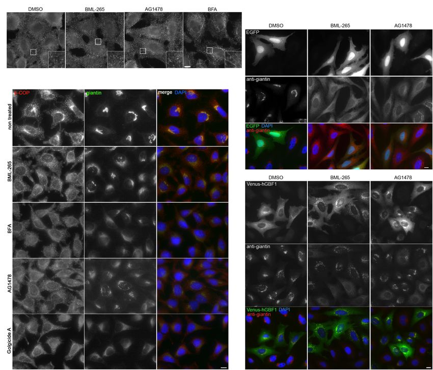
Boncompain et al.                                                                                           BML-265 Disrupts Golgi and Trafficking

glycosylation. Defects in the function and/or localization of             MATERIALS AND METHODS
Golgi associated proteins regulating trafficking and especially
of glycosylation enzymes could lead to diverse diseases (Zappa            Screening Procedure
et al., 2018). Golgi-resident glycosylation enzymes are type              Cell Seeding
II transmembrane proteins transported from the ER to the                  HeLa cells stably expressing Streptavidin-KDEL_ManII-SBP-
Golgi in a COPII-dependent manner (Ward et al., 2001).                    EGFP (Boncompain et al., 2012) were counted using T4
The Golgi apparatus is a dynamic polarized organelle the                  Cellometer (Nexcelom) and seeded in 384-well plates (ViewPlate-
composition of which is controlled by a balance of anterograde            384 Black Perkin Elmer, catalog number 6007460) at 2,000
and retrograde fluxes of proteins. In particular, the steady-state        cells/well using a Multidrop Combi (Thermo Fisher Scientific) in
intra-Golgi localization of Golgi-resident glycosylation enzymes          40 µl of cell media. The screen was performed at same early cell
is ensured by constant recycling from cisterna to cisterna                passages in two replicate experiments.
up to the ER (Storrie et al., 1998). Retrograde transport of
glycosylation enzymes occurs via COPI vesicles either by direct           Compound Libraries
binding to coatomer (Liu et al., 2018) or through interaction             A FDA-approved drug library, comprised of 640 compounds
with GOLPH3 (Tu et al., 2008; Ali et al., 2012). COPI coat                diluted in Dimethyl Sulfoxide (DMSO) was purchased from
recruitment is modulated by ADP-ribosylation factors (ARF),               Enzo Life Sciences (BML-2842) together with 80 known kinase
which cycle from an inactive state (GDP-bound) to an active               inhibitors of well-defined activity (Screen-Well Kinase Inhibitor
state (GTP-bound) (Serafini et al., 1991; Donaldson et al., 1992a).       Library, catalog number BML-2832), 33 phosphatase inhibitors
ARFs are activated by guanine nucleotide exchange factors                 (Phosphatase Inhibitor library, catalog number BML-2834)
(GEF). At the cis-Golgi, the Golgi brefeldin A-resistant factor           and 53 protease inhibitors (Screen-WellTM Protease Inhibitor
1 (GBF1) plays a pivotal role in regulating organelle structure           Library, catalog number BML-2833). 24 h after seeding, 10 µL
and vesicle trafficking by catalyzing the activation of ARF1              of compounds was transferred to 384-well plates using the
leading to COPI assembly and recruitment to Golgi membranes               MultiChannel ArmTM 384 (MCA 384) (TECAN) to the cells, to
(Presley et al., 2002).                                                   a final concentration of 10 µM and 0.5% of DMSO. Nocodazole
    Because anterograde transport of cargos, especially of Golgi-         (2.5 µM final dilution) and brefeldin A (10 µM final dilution)
resident glycosylation enzymes, is constantly counterbalanced             were added as biologically relevant phenotypic controls in a single
by retrograde transport (Cole et al., 1996; Storrie et al., 1998;         control plate. All chemical compounds were diluted in dimethyl
Sengupta et al., 2015), it is difficult to distinguish between            sulfoxide (DMSO) as 10 mM stock solution and robotically
defects in the anterograde or the retrograde transport when               reformatted in-house into 384-well source plates.
a Golgi enzyme localization is perturbed. The development
of the Retention Using Selective Hooks (RUSH) assay enables               Compound Treatment and Biotin Pulse
to overcome this difficulty through synchronization of fluxes,            After 90 min of incubation with the compounds at 37◦ C and 5%
enabling quantitative analysis of anterograde transport of cargos         CO2 , cells were treated with 40 µM of biotin for 30 min at 37◦ C.
(Boncompain et al., 2012). The RUSH assay uses physiological              The screening was performed in three biological replicates. After
conditions for mammalian cells and is induced by simple                   biotin treatment, cells were processed for immunofluorescence
addition of the non-toxic vitamin biotin. We have previously              performed as follows: cells were fixed with a fresh solution of 3%
shown that the RUSH assay is amenable to high-content                     paraformaldehyde for 15 min using the MultiChannel ArmTM
phenotypic screening (Boncompain et al., 2012, 2019; Liu                  384 (MCA 384) (TECAN). Cells were quenched with 50 mM
et al., 2017; Zhao et al., 2018). To identify small molecules             NH4 Cl in phosphate buffered saline (PBS) solution for 5 min,
that regulate the trafficking of Golgi-resident glycosylation             and then incubated for 1 h at room temperature (RT) with a
enzymes, we combined the RUSH assay to high content                       primary mouse anti-GM130 antibody (1:500, BD Biosciences,
screening of chemical libraries using Mannosidase II (ManII)              catalog number 610822) diluted in 1% BSA-0.2% saponin. Cells
as a model Golgi enzyme. We identified several molecules                  were further washed with PBS and co-incubated for 1h RT with a
either inhibiting or accelerating ManII transport from ER                 secondary anti-mouse Alexa 647 antibody (1:400, Thermo Fisher
to Golgi. Supervised classification enabled the discovery of              Scientific, catalog number A-31571) and Hoechst 33242 (DNA)
two small molecules displaying BFA-like activity on which                 (1:500, Sigma, catalog number 14533).
we focused the present study. We further showed that these                   For dose-response experiments, cells were treated 24 h after
molecules, namely BML-265 and Tyrphostin AG1478, described                seeding with compounds (20 µL of media/DMSO solution) and
as EGFR kinase inhibitors have additional intracellular biological        vehicle DMSO 0.5%, titrated in a 8- point, three-fold dilution
effects. Both compounds induced reversible Golgi disruption               starting at a concentration of 10 µM. The assay plates were
and inhibition of the secretory transport in human cells, but             then incubated 24 h at 37◦ C at 5% CO2 and processed as
not in rodent cells. The analysis of their effects on endosomal           described above.
tubulation and dissociation of COPI from Golgi membranes
suggested the cis-Golgi ARF GEF GBF1 to be the target of                  Image Acquisition and Analysis
BML-265. This was further supported by overexpression of                  Image acquisition was performed using the INCell 2000
human GBF1, which prevents the effects of BML-265 and                     automated widefield system (GE Healthcare, United States) at a
Tyrphostin AG1478.                                                        20X magnification (Nikon 20X/0.45, Plan Apo, CFI/60), using the

Frontiers in Cell and Developmental Biology | www.frontiersin.org     2                                       October 2019 | Volume 7 | Article 232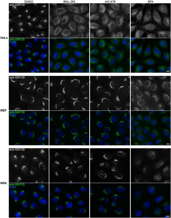
Boncompain et al.                                                                                          BML-265 Disrupts Golgi and Trafficking

same exposure time for all plates in the experiment and across            Antibodies and Immunofluorescence
replicate experiments. Plates were loaded onto the microscope             Monoclonal mouse anti-GM130 was purchased from BD
system with a Kinedx robotic arm (PAA, United Kingdom).                   Biosciences (catalog number 610823, used at 1:1000) and rabbit
Images of four different positions in each well were acquired,            polyclonal anti-GM130 from Abcam (catalog number ab52649,
each containing channels for Hoechst 33242, ManII-SBP-EGFP                dilution 1:2000). Anti-GFP was purchased from Merck (catalog
and GM130. The total number of cells measured in a well                   number 11814460001, use at 1:1000). Anti giantin (TA10) was
was typically around 300. Computational image processing                  obtained from the recombinant antibody platform of the Institut
operations were performed using the dual area object analysis in          Curie (dilution 1:100). Anti betaCOP (mAD) and anti-transferrin
the INCell Analyzer Workstation 3.7 software (GE Healthcare).             receptor (OKT9) were a gift from T. Kreis, University of Geneva,
Nuclei were defined based on DNA staining and cell region was             dilution 1:200 and 1:1000 respectively. The anti-betaCOP
segmented using top-hat and collar segmentation, respectively.            antibody requires cell fixation using methanol. The antibody
ManII-SBP-EGFP in the Golgi area was identified as inclusions             directed to GalT (B4GalT1) used in Supplementary Figure S2
in the collar cell area using multiscale top-hat segmentation,            was purchased from Abnova (catalog number PAB20512, dilution
and quantified by the average intensity of pixels within the              1:1000) and the one used in Supplementary Figure S3 was
defined inclusion region. Individual cells were then gated to             obtained from CellMAb (catalog number CB02, dilution 1:100).
single phenotypes using CellProfiler Analyst software (Jones              The anti-TGN46 was purchased from BioRad (catalog number
et al., 2008). Briefly, cell samples from all replicate experiments       AHP500G, dilution 1:2000). The antibody directed to Sec24C
were sorted into four-defined classes (1. Golgi-disrupted, 2.             was a kind gift from David Stephens (University of Bristol,
ER + Golgi, 3. ER-retained and 4. Golgi) using DMSO,                      United Kingdom) (dilution 1:500). The anti-EEA1 antibody was
brefeldin A and nocodazole-treated wells as a training set for the        purchased from BD biosciences (catalog number 610456, dilution
classifier active learning module. Obtained data was normalized           1:2000). The anti-LAMP1 was purchased from Merck (catalog
using the robust Z-score method under the assumption that                 number L1418, dilution 1:3000).
most compounds are inactive and can serve as controls (Malo                  Alexa488, Cy3 and Cy5 conjugated secondary antibodies were
et al., 2006; Birmingham et al., 2009). Plate positional effects          purchased from Jackson Immunoresearch and used at 1:400.
were corrected using median polishing (Mosteller and Tukey,                  Immunostaining of intracellular targets was performed after
1977; Birmingham et al., 2009) applied to each phenotypic                 fixation with paraformaldehyde 3% for 15 min at room
class. Hits for each compound were identified as follows:                 temperature. After 3 washes with PBS, cells were permeabilized
sample median and median absolute deviation (MAD) were                    with PBS supplemented with BSA 2 g/l and saponin 0.5 g/l for
calculated for each replicate from the population of screening            5 min at room temperature. Antibodies were diluted in PBS
data points (named as sample) and used to compute Robust                  supplemented with BSA 2 g/l and saponin 0.5 g/l and incubated
Z-scores [RZ-scores (Iglewicz and Hoaglin, 1993)] according to            with cells for 45 min. Coverslips were mounted in Mowiol
the formula: RZscore = (Perturbator value-median (reference               containing DAPI.
sample))/(1.4826 × MAD). A compound was identified as a ‘hit’,               Immunostaining to monitor the presence of EGFP-tagged
if the RZ-score was 2.                                            cargos at the cell surface was performed on living cells seeded
                                                                          on glass coverslips. Cells were washed with ice cold PBS and
Cells, Plasmids, Transfection                                             incubated on ice with the anti-GFP antibody diluted in PBS for
Hela wildtype and stably expressing Str-KDEL_ManII-SBP-EGFP               45 min. Cells were then washed 3 times with ice cold PBS and
(Boncompain et al., 2012) cells, mouse embryonic fibroblasts              fixed with paraformaldehyde 2% for 15 min at room temperature.
(MEF) and normal rat kidney (NRK) cells were cultured in                  After 3 washes with PBS, the cells were incubated with the
Dubelcco’s modified Eagle medium (DMEM) (Thermo Fisher                    secondary antibodies for 40 min at room temp.
Scientific) supplemented with 10% Fetal Calf Serum (FCS, GE                  After 3 washes with PBS, coverslips were in Mowiol.
Healthcare), 1 mM sodium pyruvate and 100 µg/ml penicillin                Observation and acquisition of pictures of fixed samples were
and streptomycin (Thermo Fisher Scientific).                              performed using an epifluorescence microscope (Leica) equipped
   RUSH plasmids were previously described TNF-SBP-EGFP,                  with a Coolsnap camera (Roper Scientific) using the software
SBP-EGP-GPI (Boncompain et al., 2012), SBP-EGFP-EGFR                      Metamorph (Molecular Devices).
(Scharaw et al., 2016).
   The plasmid coding for Venus-GBF1 was a kind gift from C.
                                                                          EGFR Phosphorylation and Immunoblot
Jackson (Institut Jacques Monod, Paris, France).
                                                                          Cells were serum starved overnight. Cells were then pre-treated
   HeLa cells were transfected using calcium phosphate as
                                                                          with the indicated molecules at 10 µM for 1h. They were then
described previously (Jordan et al., 1996).
                                                                          stimulated with human EGF at 50 ng/ml final for 10 min.
                                                                             Cells were lysed in Laemmli 2,5 × buffer containing beta-
Chemicals                                                                 mercaptoethanol. Samples were then loaded on acrylamide
BML-265 was purchased from Enzo Life Sciences. Tyrphostin                 gels (Criterion TGX, BioRad) and transferred on nitrocellulose
AG1478 and Erlotinib were ordered from Cayman chemical.                   membrane (GE Healthcare) using Power blotter Semi dry from
DMSO, brefeldin A, Golgicide A and D-biotin were                          Thermo Scientific. Blocking and incubation of antibodies were
purchased Sigma-Aldrich.                                                  performed in PBS supplemented with 0.1% Tween and 5% milk.

Frontiers in Cell and Developmental Biology | www.frontiersin.org     3                                     October 2019 | Volume 7 | Article 232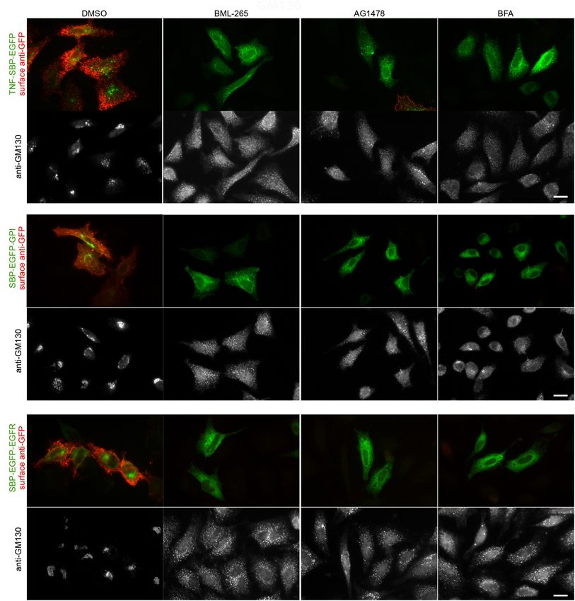
Boncompain et al. BML-265 Disrupts Golgi and Trafficking
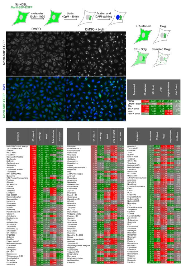
Detection was performed using SuperSignal West Pico Substrate from ER to Golgi. Cells were then fixed and nuclei were stained
from Thermo and Chemidoc machine (BioRad). using DAPI. The Golgi disrupting agent, brefeldin A (BFA) and
Human EGF was purchased from Sigma-Aldrich the microtubule-depolymerizing drug, nocodazole (Noco) were
(catalog number E9644). used as positive phenotypic controls for ManII ER-retention and
Antibodies to detect phosphorylated EGFR and total EGFR Golgi disruption, respectively. In addition, wells not incubated
were purchased from Cell Signaling Technology (PhosphoPlus with biotin were included in order to prevent ManII-SBP-EGFP
EGFR (Tyr1068) Antibody Duet, catalog number 11862S, exit from the ER. 0.5% of DMSO was used as solvent control.
dilution 1:1000). Anti-actin (clone AC40) used as loading After image acquisition and segmentation, several features
control was purchased from Sigma Aldrich (catalog number were measured (see Materials and Methods section for details).
A3853, dilution 1:1000). Secondary anti-mouse and anti-rabbit Samples were classified into four phenotypical classes (1. ER-
poly-HRP antibodies were purchased from Thermo Scientific retained, 2. ER + Golgi, 3. Golgi disrupted and 4. Golgi) using
(dilution 1:10000). above-mentioned controls as training set using Cell Profiler
Analyst (Figure 1C). A cell count was also used to evaluate toxic
Real Time Imaging effects of the compounds.
HeLa cells stably expressing Streptavidin-KDEL_ManII-SBP- Robust z-score for each phenotypic class was calculated
EGFP were grown on 25 mm glass coverslips. Cells were (see Materials and Methods) allowing identification of outliers.
maintained in presence of biotin at 40 µM to allow stable For instance, the ER-retained robust Z-score of the condition
localization of ManII-SBP-EGFP in the Golgi apparatus. “DMSO” is very different (20.46) from the one of the condition
Coverslips were transferred to an L-shape tubing equipped “DMSO + biotin” (−2.19) (Figure 1D). “DMSO + biotin”
Chamlide chamber (Live Cell Instrument). Pre-warmed Leibovitz corresponds to the control condition, for which ER to Golgi
medium (Life Technologies) supplemented with 40 µM of transport of ManII occurred normally while “DMSO” (i.e.,
biotin was used. BML-265 diluted at 10 µM in Leibovitz absence of biotin) is exemplifying the robust Z-score obtained
supplemented with biotin was added at time 0. After the indicated by a hit preventing normal transport and inducing ER-retention.
time, BML-265 was washed thanks to several washes with Please note that the score for Golgi-disruption of cells incubated
pre-warmed Leibovitz. For the recovery period, medium was with nocodazole and biotin (‘Noco + biotin’) is higher than
replaced by pre-warmed Leibovitz containing biotin. Imaging the one of cells treated with nocodazole only (‘Noco’) because
was performed at 37◦ C in a thermostat controlled chamber using our analysis uses ManII-SBP-EGFP signal to detect Golgi
an Eclipse 80i microscope (Nikon) equipped with a spinning disk elements and not an independent Golgi marker. In consequence,
confocal head (Perkin) and an Ultra897 iXon camera (Andor). the accumulation of ManII-SBP-EGFP in Golgi mini-stacks
Image acquisition was performed using MetaMorph software due to biotin addition leads to a better detection of Golgi-
(Molecular Devices). Maximum intensity projections of several disruption (Figure 1D).
Z-slices are shown. As we were primarily looking for molecules inhibiting ER
to Golgi transport of ManII, the results were sorted using
ER-retained score (Figures 1D,E). The two top hit molecules
identified as inhibitors of ManII ER to Golgi transport were BML-
RESULTS
265 and Tyrphostin AG1478. Their score for the 4 phenotypic
classes was similar to the controls DMSO [No biotin], BFA and
High-Content Phenotypic Screen Led to BFA + biotin confirming that they inhibit ManII transport to the
Identification of Molecules Regulating Golgi (Figures 1D,E).
the Trafficking of ManII We then performed a dose-response analysis, in triplicates,
Using the previously described HeLa cell line stably expressing using serial dilutions (10 doses from 30 µM to 1.52 nM) of
Streptavidin-KDEL_ManII-SBP-EGFP (Boncompain et al., 50 selected molecules either inhibiting or accelerating ER to
2012), we designed a phenotypic screen to identify small Golgi transport. The same quantitative analysis that was done
molecules modulating the trafficking of the Golgi enzyme in the primary screen was carried out here. Four families of
Mannosidase II (ManII) (Figure 1A). The cells co-express a compounds were identified based on the dose-response profile
non-fluorescent ER hook (Str-KDEL) with ManII fused to of ‘ER-retained’ and ‘Golgi disrupted’ scores (Supplementary
a Streptavidin Binding Peptide (SBP) and an EGFP (ManII- Figure S1). Some molecules inhibited ER to Golgi transport
SBP-EGFP) enabling RUSH control of ManII trafficking. at increasing doses without affecting the integrity of the
ManII-SBP-EGFP is retained in the ER in the absence of biotin Golgi apparatus. These molecules are suspected to affect cell
and is transported to the Golgi apparatus after incubation homeostasis or to be energy poisons as this family includes
with biotin (Figure 1B). This set-up was used to screen small ouabain (Na+ , K+ ATPase inhibitor). Based on the dose-
molecules from a library of FDA-approved molecules and response profiles, we identified two groups of molecules affecting
inhibitors of kinases, phosphatases and proteases (see Materials microtubules. A first group contained depolymerizing agents
and Methods section for details). Cells seeded in 384-well such as albendazole, while a second one was composed of
plates were incubated with small molecules, diluted at a final destabilizing agents such as vinca alkaloides (e.g., vinblastine).
concentration of 10 µM in DMSO, for 90 min. Biotin was then The dose-response profiles of ‘ER retained’ and ‘Golgi disrupted’
added for 30 min to induce the transport of ManII-SBP-EGFP scores showed that BML-265 and Tyrphostin AG1478 displayed
Frontiers in Cell and Developmental Biology | www.frontiersin.org 4 October 2019 | Volume 7 | Article 232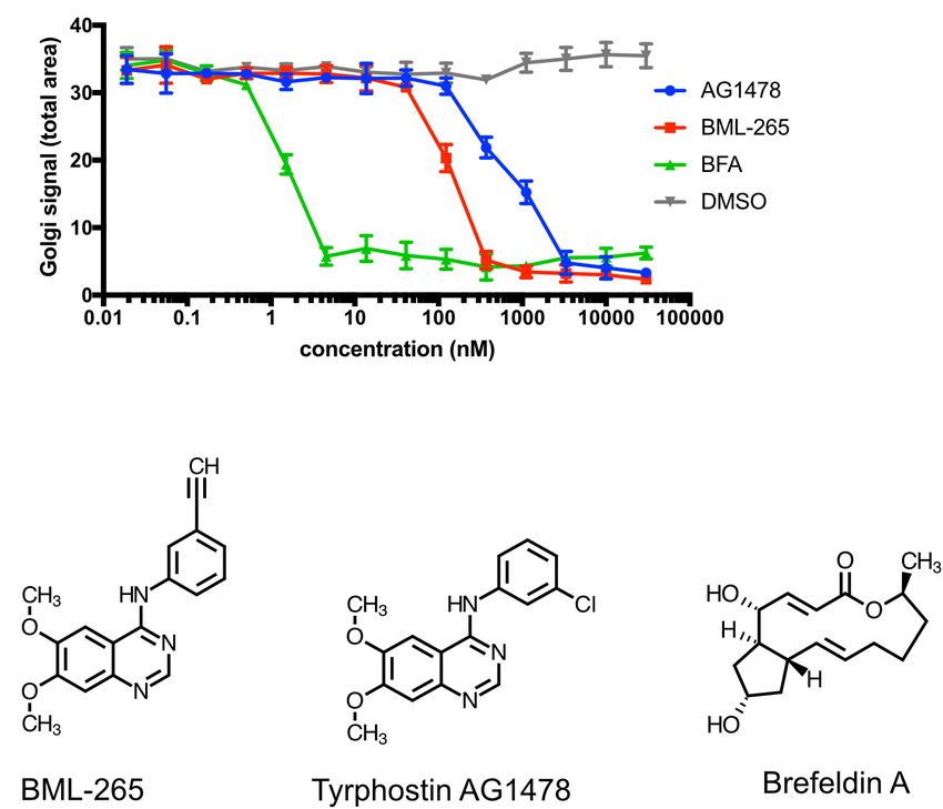
Boncompain et al.                                                                                                                BML-265 Disrupts Golgi and Trafficking

  FIGURE 1 | High-content screening for molecules regulating ER to Golgi transport of ManII. (A) Scheme of the screening processes. Cells stably expressing
  Str-KDEL_ManII-SBP-EGFP were incubated with each small molecule for 90 min. Synchronized transport of ManII was induced by addition of biotin. After 30 min,
  cells were then fixed and nuclei were stained with DAPI. (B) Pictures from screening plates depicting the controls DMSO (no traffic) and DMSO + biotin (transport to
  the Golgi). Scale bar: 10 µm. (C) Scheme of the four classes used for the classification of quantitative analysis of the pictures. (D,E) Scores obtained for the
  parameters ‘ER-retained,’ ‘ER + Golgi,’ ‘Golgi,’ ‘Golgi-disrupted,’ and cell count for each control (D) or small molecule compound (E).

Frontiers in Cell and Developmental Biology | www.frontiersin.org                  5                                               October 2019 | Volume 7 | Article 232
Boncompain et al.                                                                                            BML-265 Disrupts Golgi and Trafficking

similar effects (BFA-like). They inhibited the trafficking of              BML-265 Disperses the Golgi Apparatus
ManII to the Golgi starting from 123 – 370 nM and induced                  in Human Cells but Not in Rodent Cells
Golgi disruption on the same concentration range. Note that,
                                                                           Tyrphostin AG1478 was previously reported to affect Golgi
surprisingly, no Golgi disruption was detected at high doses of
                                                                           integrity in human but not in rodent cells. We thus analyzed
BML-265 and Tyrphostin AG1478. This is due to the way we
                                                                           the effects of BML-265 on the integrity of the Golgi complex
quantified Golgi organization. Indeed, at high doses, the Golgi
                                                                           by immunolabeling using an anti-GM130 antibody using a
apparatus was completely disassembled and no visible structures
                                                                           human cell line and two rodent cell lines. As observed
remained. In consequence, our analysis for Golgi disruption
                                                                           with BFA, incubation of the human epithelial cell line HeLa
did not detect any Golgi structures and scored it as “no Golgi
                                                                           with BML-265 and Tyrphostin AG1478 led to redistribution
disruption” (Supplementary Figure S1).
                                                                           of GM130, indicating Golgi disruption (Figure 4A). Similar
                                                                           results were obtained when staining for other Golgi markers,
The Effects of BML-265 and Tyrphostin                                      namely TGN46, GalT and giantin. As expected, the intracellular
AG1478 on Golgi Integrity and Trafficking                                  distribution of ER exit sites, stained with an anti-Sec24
                                                                           antibody, was also affected. In contrast, early endosomes
Indicate BFA-Like Activity                                                 and late endosomes/lysosomes organization was not perturbed
The above-mentioned effects of BML-265 and Tyrphostin
                                                                           after incubation with the molecules compared to DMSO
AG1478 were reminiscent of the ones obtained with BFA used
                                                                           control (Supplementary Figure S2). However, these molecules
as a control in our experiments. In addition, whereas BML-265
                                                                           did not affect Golgi localization and morphology of mouse
and Tyrphostin AG1478 are both annotated as EGFR kinase
                                                                           fibroblasts (MEF) and rat epithelial cells (NRK). In contrast, BFA
inhibitors, Tyrphostin AG1478 was described to target the cis-
                                                                           disrupted the Golgi complex of these two rodent cell models
Golgi ADP ribosylation factor guanine nucleotide exchange
                                                                           (Figures 4B,C).
factor (ARF GEF) named GBF1 (Pan et al., 2008). We thus
                                                                               BML-265 and Tyrphostin AG1478 are both annotated in
decided to compare the efficacy of BML-265, Tyrphostin
                                                                           the library as analogs of Erlotinib (Tarceva trade mark; OSI
AG1478 and BFA on the trafficking of ManII to the Golgi
                                                                           Pharmaceuticals, Genentech and Roche), one of the several
apparatus. HeLa stably expressing Str-KDEL_ManII-SBP-EGFP
                                                                           EGFR tyrosine kinase inhibitors, which has been largely studied
were incubated with serial dilutions of the molecules (from
                                                                           in clinical trials, with proven efficacy in humans. BML-265,
0.019 nM to 30 µM) for 90 min and biotin was then
                                                                           Tyrphostin AG1478 and Erlotinib have different structures but
added to induce the transport of ManII-SBP-EGFP. Our results
                                                                           share a quinazoline group (Figure 2B and Supplementary
show that BFA is more potent than BML-265 which is itself
                                                                           Figure S3). We verified that BML-265 was indeed able to
more potent than Tyrphostin AG1478 (Figure 2A). Whereas
                                                                           prevent EGFR phosphorylation upon stimulation with EGF.
IC50 of BFA is about 2 nM, IC50 of BML-265 is about
                                                                           Importantly, Erlotinib did not induce Golgi disruption even at
200 nM and of Tyrphostin AG1478 about 1 µM. Strikingly,
                                                                           high doses (Supplementary Figure S3). These results suggest that
the chemical structures of BML-265 and Tyrphostin AG1478
                                                                           effects of BML-265 and Tyrphostin AG1478 on Golgi integrity
are closely related while they are different from the one of
                                                                           and function are independent of their capability to inhibit
BFA (Figure 2B).
                                                                           EGFR phosphorylation.
   We then assessed the effects of BML-265 and Tyrphostin
AG1478 on the trafficking of secretory cargos addressed to the cell
surface. For this purpose, we used TNF (type II transmembrane              BML-265 Has Reversible Effects on Golgi
protein), GPI (GPI anchor) and EGFR (type I transmembrane                  Integrity
protein) as RUSH cargos (Boncompain et al., 2012; Fourriere                Treatment of HeLa cells for 1h with BML-265 or Tyrphostin
et al., 2016; Scharaw et al., 2016). In these fusion constructs,           AG1478 leads to a complete redistribution of Golgi complex
the EGFP is exposed to the extracellular space when the cargo              proteins throughout the cytoplasm (Figure 5A). However, the
reaches the plasma membrane. The effects of the molecules on               effects of both compounds are partially reversible. 1 h after
the anterograde transport of TNF, EGFP-GPI and EGFR were                   addition of the molecules, the medium was removed and
thus assessed by immunofluorescence on non-permeabilized cells             cells were washed before being incubated in normal medium.
using an anti-GFP antibody. The Golgi apparatus was stained                Cells were then fixed and the Golgi complex immunolabeled
using an anti-GM130 antibody. As observed with BFA, pre-                   using an anti-GM130 antibody. Forty-five min after washout,
treatment of the cells with BML-265 and Tyrphostin AG1478                  cells recovered a normal Golgi organization and localization
for 60 min prior to addition of biotin prevented transport of the          (Figure 5A). We next assessed by real-time imaging the re-
cargos to the cell surface (Figure 3). The EGFP-tagged cargos are          formation of the Golgi complex after washout of BML-265.
detected inside the cells, probably in the ER. Consistently with the       BML-265 was added to HeLa stably expressing ManII-SBP-
results obtained during the screening and IC50 experiments, the            EGFP. Quickly after addition of BML-265, transient ManII-
Golgi apparatus is disrupted upon incubation with BML-265 and              SBP-EGFP positive tubules were observed and ManII-SBP-EGFP
Tyrphostin AG1478.                                                         signal intensity at the Golgi decreases while increasing in the
   Altogether, our results indicate that BML-265 and                       whole cell corresponding to ER relocation of ManII-SBP-EGFP
Tyrphostin AG1478 display BFA-like effects on trafficking                  (Figure 5B and Supplementary Movie S1). Washout of BML-
and Golgi integrity.                                                       265 was then performed and cells imaged in real-time. The

Frontiers in Cell and Developmental Biology | www.frontiersin.org      6                                      October 2019 | Volume 7 | Article 232
Boncompain et al. BML-265 Disrupts Golgi and Trafficking
FIGURE 2 | BML-265 and Tyrphostin AG1478 display BFA-like effects. (A) HeLa cells stably expressing Str-KDEL_ManII-SBP-EGFP were incubated with serial
dilutions of DMSO, BML-265, Tyrphostin AG1478 (AG1478) and brefeldin A (BFA) for 90 min. Cell were then treated with biotin, fixed and stained using an
anti-GM130 antibody. ManII-SBP-EGFP signal at the Golgi was then quantified (a.u., arbitrary units) by fluorescence microscopy. 2 independent replicates were
performed. Error bars show standard deviation. (B) Schemes of the molecules BML-265, Tyrphostin AG1478, and brefeldin A (BFA).
Golgi complex visualized by ManII-SBP-EGFP redistributed to found in the Golgi area, probably associated to the Golgi complex
the perinuclear area. Signal intensity of ManII-SBP-EGFP at the and to vesicles, betaCOP is absent from the Golgi complex in
Golgi increased while ER signal decreased showing that ER to BML-265 treated cells even in cells still displaying a perinuclear
Golgi transport was restored. Golgi complex after this short treatment time (Figure 6B). As
expected, BFA also induced rapid dissociation of COPI coat from
BML-265 Might Target GBF1 Golgi membranes. We also detected dissociation of betaCOP
BFA inhibits Arf1 activation (Donaldson et al., 1992b; Helms from Golgi membranes for two molecules known as inhibitors
and Rothman, 1992) by targeting several ARF GEF: GBF1 at the of GBF1, Tyrphostin AG1478 and Golgicide A (GCA) (Pan et al.,
cis-Golgi and BIG1 and BIG2 at the TGN (Claude et al., 1999; 2008; Saenz et al., 2009) (Figure 6B). Overexpression of human
Yamaji et al., 2000). Due to its effects on the TGN ARF GEF, wild-type GBF1 prevented Golgi dispersal induced by incubation
BFA induces tubule formation at the TGN and on endosomes with BML-265 or Tyrphostin AG1478 (Figure 6C).
(Lippincott-Schwartz et al., 1991; Wood et al., 1991) as confirmed Altogether these results suggest that BML-265 targets
by immunostaining using an anti-Transferrin receptor (TfR) GBF1 causing Golgi dispersal and inhibiting secretory
antibody. In contrast, BML-265 and Tyrphostin AG1478 did not protein trafficking.
induce tubulation of TfR-positive compartments, suggesting that
they do not affect the TGN ARF GEF (Figure 6A). GBF1 is an
ARF GEF present at the cis-Golgi involved in the recruitment DISCUSSION
of the COPI coat on Golgi membranes. We next assessed the
distribution of COPI after treatment with BML-265. In HeLa In the past years, several image-based and high-throughput
cells treated with the molecules for 5 min, the distribution of the screens led to the identification of small molecules which
COPI coat was monitored using immunolabeling with an anti- affect membrane trafficking (Mishev et al., 2013). Among those,
betaCOP antibody and the Golgi complex was detected using an AMF-26/M-COPA, Golgicide A, Exo2, LG-186 and Tyrphostin
anti-Giantin antibody. Whereas in non-treated cells, betaCOP is AG1478 were identified as molecules displaying effects similar
Frontiers in Cell and Developmental Biology | www.frontiersin.org 7 October 2019 | Volume 7 | Article 232Boncompain et al. BML-265 Disrupts Golgi and Trafficking FIGURE 3 | BML-265 and Tyrphostin AG1478 inhibit the transport of secretory proteins. HeLa cells transiently expressing Str-KDEL_TNF-SBP-EGFP (A), SBP-EGFP-GPI (B) or SBP-EGFP-EGFR (C) were pre-treated with the indicated molecules at 10 µM for 1 h. Trafficking of the reporters was then induced by incubation with biotin for 1 h. The presence of the GFP-tagged reporters (green, upper panel) at the plasma membrane was then detected using an anti-GFP antibody on non-permeabilized cells (red, upper panel). The Golgi apparatus was visualized using immunostaining against GM130 (bottom panel). Scale bar: 10 µm. to BFA, but being more specific because they specifically target of FDA-approved molecules as well as inhibitors of proteases, the ARF GEF GBF1 (Pan et al., 2008; Spooner et al., 2008; Saenz phosphatases and kinases revealed several compounds able to et al., 2009; Boal et al., 2010; Ohashi et al., 2012). In the present modulate ManII trafficking, either by inhibiting or accelerating study, we initially intended to search for compounds that regulate ER to Golgi transport. Further studies will be necessary to the ER to Golgi transport of the Golgi-resident glycosylation clarify the biological activity of these molecules on ER to Golgi enzyme ManII. Our study using a compound library composed transport and more largely on secretory protein trafficking. We Frontiers in Cell and Developmental Biology | www.frontiersin.org 8 October 2019 | Volume 7 | Article 232
Boncompain et al.                                                                                                               BML-265 Disrupts Golgi and Trafficking

  FIGURE 4 | BML-265 affects Golgi integrity in human cells but not in rodent cells. HeLa cells (A), mouse embryonic fibroblasts (MEF) (B) or normal rat kidney (NRK)
  cells (C) were incubated with the indicated molecules at 10 µM final for 1h30. Cells were then fixed and the Golgi apparatus was stained using an anti-GM130
  antibody (green on merge). Nuclei were stained using DAPI (blue on merge). Scale bar: 10 µm.

focused our attention on the previously studied Tyrphostin                             protein transport. BML-265 and Tyrphostin AG1478 display
AG1478 as well as BML-265, which both prevent ManII transport                          a very close structure and are both annotated as Erlotinib
to the Golgi, induce Golgi disassembly and prevent secretory                           analogs, being inhibitors of EGFR kinase activity. They share a

Frontiers in Cell and Developmental Biology | www.frontiersin.org                  9                                              October 2019 | Volume 7 | Article 232
Boncompain et al.                                                                                                             BML-265 Disrupts Golgi and Trafficking

  FIGURE 5 | The effects of BML-265 on Golgi integrity are reversible. (A) HeLa cells were treated with the indicated molecules at 10 µM for 1h and molecules were
  removed by extensive washings. Cells were then incubated in normal medium for 45 min, fixed and the Golgi apparatus was stained using an anti-GM130 antibody.
  Scale bar: 10 µm. (B) HeLa cells stably expressing Str-KDEL_ManII-SBP-EGFP were cultivated in presence of biotin 40 µM to allow stable Golgi localization of
  ManII-SBP-EGFP and imaged by spinning disk confocal microscopy. Under the microscope, BML-265 was added at 10 µM final at time 0 min. After 50 min,
  BML-265 was washed out and pictures were acquired every 30 s. Maximum projection of 11 z-slices is shown. Scale bar: 10 µm.

quinazoline moiety with Erlotinib and other described inhibitors                      is given to patients sometimes in combination with cetuximab
of receptor kinases (Fry et al., 1994; Ward et al., 1994; Stamos                      for the treatments of some cancers. Considering that mouse
et al., 2002). Erlotinib as well as Gefitinib, Lapatinib and several                  cells are sensitive only to the EGFR kinase inhibition activity
Tyrphostins were present in our library but were not scored                           of BML-265 and Tyrphostin AG1478, in vivo tests in mouse
as molecules inhibiting the transport of ManII and/or affecting                       models would underestimate their toxic effects and fail to early
Golgi integrity. The similarity between BML-265 and Tyrphostin                        capture clinically significant secondary effects that might arise in
AG1478 in terms of structure and effects, and the absence                             humans. The suggested target for these compounds is the cis-
of effects of other Erlotinib analogs, suggest that they both                         Golgi ARF GEF GBF1 since they exert BFA-like effects, but do
act on Golgi function independently of their ability to inhibit                       not induce endosome tubulation. Nucleotide exchange activity of
EGFR phosphorylation. Interestingly, other molecules known to                         GBF1 is mediated by its catalytic Sec7 domain (Renault et al.,
target GBF1, such as Golgicide A, do not bear a quinazoline                           2003). The Sec7 domains of human and mouse GBF1 display
moiety (Saenz et al., 2009) and were not reported to inhibit                          98% of similarity in their amino acid sequence. Even though
EGFR phosphorylation.                                                                 the catalytic domain of GBF1 is highly conserved in human
   Our results for BML-265 and Tyrphostin AG1478 as well                              and mouse cells, this difference might be sufficient to modify
as previous published work on Tyrphostin AG1478 (Pan et al.,                          the putative binding sites of BML-265 and Tyrphostin AG1478.
2008) show that these small molecules induce Golgi disruption                         In the present study, we showed that overexpression of human
in human cells, but not in rodent cells. The differential effects                     GBF1 prevents the effects of BML-265 on Golgi integrity. This
of BML-265 and Tyrphostin AG1478 on human versus rodent                               result strongly suggests that GBF1 might be a target of BML-
cells are interesting in the point of view of their clinical use.                     265. However, we cannot exclude indirect effects of the molecules
EGFR is activated due to mutation and/or overexpression in                            on GBF1 neither the existence of other cellular upstream targets.
diverse epithelial tumors and is associated with poor prognosis                       The mode of interaction of these molecules with human GBF1
(Nicholson et al., 2001). Consequently, inactivation of EGFR                          as well as the mechanisms of inactivation of GBF1 require
signaling pathway is a target for cancer treatment. Erlotinib                         further investigation.

Frontiers in Cell and Developmental Biology | www.frontiersin.org                10                                            October 2019 | Volume 7 | Article 232
Boncompain et al.                                                                                                                 BML-265 Disrupts Golgi and Trafficking

  FIGURE 6 | BML-265 exerts its effects on Golgi integrity and trafficking through targeting GBF1. (A) HeLa cells were incubated with the indicated molecules at
  10 µM for 1 h. After fixation, endosomes were stained using an anti-transferrin receptor (TfR) antibody. Scale bar: 10 µm. The inset displayed an enlarged view of the
  boxed area. (B) HeLa cells were incubated with the indicated molecules at 10 µM for 5 min and were then fixed using methanol. COPI coat was stained using an
  anti-betaCOP antibody, the Golgi apparatus using anti-giantin antibody and nuclei using DAPI. Scale bar: 10 µm. (C) HeLa cells transiently expressing human GBF1
  tagged with Venus (Venus-hGBF1) or cytoplasmic EGFP as a transfection control were incubated with the indicated molecules at 10 µM for 1 h. After fixation, the
  Golgi apparatus was immunolabeled using an anti-giantin antibody. Scale bar: 10 µm.

DATA AVAILABILITY STATEMENT                                                             FUNDING
All datasets generated for this study are included in the                               Work performed in the F. Perez laboratory was funded by
manuscript/Supplementary Files.                                                         Centre Nationale de la Recherche Scientifique, the Fondation
                                                                                        pour la Recherche Medicale (FRM DEQ20120323723), the Labex
                                                                                        CellTisPhyBio, and the Agence Nationale de la Recherche
AUTHOR CONTRIBUTIONS                                                                    (ANR-12-BSV2-0003-01). The lab of F. Perez is part of
                                                                                        Labex CelTisPhyBio (11-LBX-0038) and Idex Paris Sciences
GB carried out the experiments and analyzed the data. NG                                et Lettres (ANR-10-IDEX-0001-02 PSL). GK is supported
carried out the experiments. ST and AL carried out high content                         by the Ligue contre le Cancer (équipe labellisée); Agence
screens. TJ and ED analyzed the high-content screening data. ED                         Nationale de la Recherche (ANR) – Projets blancs; ANR under
supervised the high content-screening. OK and GK helped in the                          the frame of E-Rare-2, the ERA-Net for Research on Rare
analysis of the high content screening data. GB, FP, and ED wrote                       Diseases; Association pour la recherche sur le cancer (ARC);
the manuscript. GB and FP designed the study.                                           Cancéropôle Ile-de-France; Chancellerie des Universités de Paris

Frontiers in Cell and Developmental Biology | www.frontiersin.org                  11                                              October 2019 | Volume 7 | Article 232
Boncompain et al. BML-265 Disrupts Golgi and Trafficking
(Legs Poix), Fondation pour la Recherche Médicale (FRM); FIGURE S1 | A dose-response analysis of 50 molecules led to the identification
a donation by Elior; European Research Area Network on of 4 classes of molecules. HeLa cells stably expressing
Str-KDEL_ManII-SBP-EGFP were incubated with serial dilutions of 50 selected
Cardiovascular Diseases (ERA-CVD, MINOTAUR); Gustave
molecules. Trafficking was induced by addition of biotin and phenotypic analysis
Roussy Odyssea, the European Union Horizon 2020 Project was carried out using the approach developed for the screen. ‘ER-retained’ (A)
Oncobiome; Fondation Carrefour; High-end Foreign Expert and ‘Golgi-disrupted’ (B) scores were calculated and displayed on the graphs.
Program in China (GDW20171100085 and GDW20181100051), Three independent replicates were performed and reported here
Institut National du Cancer (INCa); Inserm (HTE); Institut (colored curves).
Universitaire de France; LeDucq Foundation; the LabEx FIGURE S2 | Effects of BML-265, AG1478 on different markers of intracellular
Immuno-Oncology; the RHU Torino Lumière; the Seerave compartments. (A) HeLa cells were incubated with the indicated molecules at
Foundation; the SIRIC Stratified Oncology Cell DNA Repair and 10 µM final for 1 h. After fixation, immunostaining using anti-TGN46 (left),
anti-Sec24 (middle) or anti-GalT (right) antibodies was performed. Scale bar:
Tumor Immune Elimination (SOCRATE); and the SIRIC Cancer
10 µm. (B) HeLa cells were incubated with the indicated molecules
Research and Personalized Medicine (CARPEM). at 10 µM final for 1 h. Immunolabeling of early endosomes using an anti-EEA1
antibody, of the Golgi apparatus using an anti-Giantin antibody and of
late/endosomes/lysosomes using an anti-LAMP1 antibody was performed.
Scale bar: 10 µm.
ACKNOWLEDGMENTS
FIGURE S3 | BML-265 behaves as Erlotinib analog and inhibits EGFR
The authors acknowledge the Cell and Tissue Imaging Facility phosphorylation but Erlotinib does not affect Golgi integrity. (A) HeLa cells were
serum starved overnight and were treated with the indicated molecules at 10 µM
(PICT-IBiSA), Institut Curie, a member of the French National
for 90 min. Cells were then incubated with EGF at 50 ng/ml for 10 min.
Research Infrastructure, and France-BioImaging (ANR10-INBS- Phosphorylated EGFR (P-EGFR), total EGFR and actin (used as a loading
04). The authors also thank the recombinant antibody platform control) were detected by immunoblot. (B) Scheme of the Erlotinib molecule.
of the Institut Curie. (C) HeLa cells were incubated with Erlotinib for 90 min at the indicated
concentrations. The Golgi apparatus was stained using 3 different
antibodies: anti-GalT (green), anti-GM130 (rouge), and anti-giantin (bleu).
Scale bar: 10 µm.
SUPPLEMENTARY MATERIAL MOVIE S1 | The effects of BML-265 are reversible. HeLa cells stably expressing
Str-KDEL_ManII-SBP-EGFP maintained in presence of biotin 40 µM to allow
The Supplementary Material for this article can be found online stable Golgi localization of ManII-SBP-EGFP. BML-265 at 10 µM was added at
at: https://www.frontiersin.org/articles/10.3389/fcell.2019.00232/ time 0. Time is indicated in min:sec. After 50 min, BML-265 was washed. Pictures
full#supplementary-material were acquired every 30 s. Maximum projection of 11 z-slices is shown.
REFERENCES coatomer protein beta-COP to Golgi membranes. Proc. Natl. Acad. Sci. U.S.A.
89, 6408–6412. doi: 10.1073/pnas.89.14.6408
Ali, M. F., Chachadi, V. B., Petrosyan, A., and Cheng, P. W. (2012). Golgi Donaldson, J. G., Finazzi, D., and Klausner, R. D. (1992b). Brefeldin A inhibits
phosphoprotein 3 determines cell binding properties under dynamic flow by Golgi membrane-catalysed exchange of guanine nucleotide onto ARF protein.
controlling Golgi localization of core 2 N-acetylglucosaminyltransferase 1. Nature 360, 350–352. doi: 10.1038/360350a0
J. Biol. Chem. 287, 39564–39577. doi: 10.1074/jbc.M112.346528 Fourriere, L., Divoux, S., Roceri, M., Perez, F., and Boncompain, G. (2016).
Birmingham, A., Selfors, L. M., Forster, T., Wrobel, D., Kennedy, C. J., Shanks, Microtubule-independent secretion requires functional maturation of Golgi
E., et al. (2009). Statistical methods for analysis of high-throughput RNA elements. J. Cell Sci. 129, 3238–3250. doi: 10.1242/jcs.188870
interference screens. Nat. Methods 6, 569–575. doi: 10.1038/nmeth.1351 Fry, D. W., Kraker, A. J., McMichael, A., Ambroso, L. A., Nelson, J. M., Leopold,
Boal, F., Guetzoyan, L., Sessions, R. B., Zeghouf, M., Spooner, R. A., Lord, W. R., et al. (1994). A specific inhibitor of the epidermal growth factor
J. M., et al. (2010). LG186: an inhibitor of GBF1 function that causes Golgi receptor tyrosine kinase. Science 265, 1093–1095. doi: 10.1126/science.806
disassembly in human and canine cells. Traffic 11, 1537–1551. doi: 10.1111/j. 6447
1600-0854.2010.01122.x Helms, J. B., and Rothman, J. E. (1992). Inhibition by brefeldin A of a Golgi
Boncompain, G., Divoux, S., Gareil, N., de Forges, H., Lescure, A., Latreche, L., membrane enzyme that catalyses exchange of guanine nucleotide bound to
et al. (2012). Synchronization of secretory protein traffic in populations of cells. ARF. Nature 360, 352–354. doi: 10.1038/360352a0
Nat. Methods 9, 493–498. doi: 10.1038/nmeth.1928 Iglewicz, B., and Hoaglin, D. (1993). How to Detect and Handle Outliers. Milwaukee
Boncompain, G., Herit, F., Tessier, S., Lescure, A., del Nery, E., Gestraud, P., et al. WI: American Society for Quality Control.
(2019). Targeting CCR5 trafficking to inhibit HIV-1 infection. Sci. Adv. Jones, T. R., Kang, I. H., Wheeler, D. B., Lindquist, R. A., Papallo, A., Sabatini,
Boncompain, G., and Weigel, A. V. (2018). Transport and sorting in the Golgi D. M., et al. (2008). CellProfiler analyst: data exploration and analysis software
complex: multiple mechanisms sort diverse cargo. Curr. Opin. Cell Biol. 50, for complex image-based screens. BMC Bioinform. 9:482. doi: 10.1186/1471-
94–101. doi: 10.1016/j.ceb.2018.03.002 2105-9-482
Claude, A., Zhao, B. P., Kuziemsky, C. E., Dahan, S., Berger, S. J., Yan, J. P., Jordan, M., Schallhorn, A., and Wurm, F. M. (1996). Transfecting mammalian
et al. (1999). GBF1: a novel Golgi-associated BFA-resistant guanine nucleotide cells: optimization of critical parameters affecting calcium-phosphate
exchange factor that displays specificity for ADP-ribosylation factor 5. J. Cell precipitate formation. Nucleic Acids Res. 24, 596–601. doi: 10.1093/nar/24.4.596
Biol. 146, 71–84. doi: 10.1083/jcb.146.999.71 Lippincott-Schwartz, J., Yuan, L., Tipper, C., Amherdt, M., Orci, L., and Klausner,
Cole, N. B., Sciaky, N., Marotta, A., Song, J., and Lippincott-Schwartz, J. (1996). R. D. (1991). Brefeldin A’s effects on endosomes, lysosomes, and the TGN
Golgi dispersal during microtubule disruption: regeneration of Golgi stacks at suggest a general mechanism for regulating organelle structure and membrane
peripheral endoplasmic reticulum exit sites. Mol. Biol. Cell 7, 631–650. doi: traffic. Cell 67, 601–616. doi: 10.1016/0092-8674(91)90534-6
10.1091/mbc.7.4.631 Liu, L., Doray, B., and Kornfeld, S. (2018). Recycling of Golgi glycosyltransferases
Donaldson, J. G., Cassel, D., Kahn, R. A., and Klausner, R. D. (1992a). ADP- requires direct binding to coatomer. Proc. Natl. Acad. Sci. U.S.A. 115, 8984–
ribosylation factor, a small GTP-binding protein, is required for binding of the 8989. doi: 10.1073/pnas.1810291115
Frontiers in Cell and Developmental Biology | www.frontiersin.org 12 October 2019 | Volume 7 | Article 232Boncompain et al. BML-265 Disrupts Golgi and Trafficking Liu, P., Zhao, L., Loos, F., Iribarren, K., Lachkar, S., Zhou, H., et al. (2017). Stamos, J., Sliwkowski, M. X., and Eigenbrot, C. (2002). Structure of the epidermal Identification of pharmacological agents that induce HMGB1 release. Sci. Rep. growth factor receptor kinase domain alone and in complex with a 4- 7:14915. doi: 10.1038/s41598-017-14848-1 anilinoquinazoline inhibitor. J. Biol. Chem. 277, 46265–46272. doi: 10.1074/ Malo, N., Hanley, J. A., Cerquozzi, S., Pelletier, J., and Nadon, R. (2006). Statistical jbc.m207135200 practice in high-throughput screening data analysis. Nat. Biotechnol. 24, 167– Storrie, B., White, J., Rottger, S., Stelzer, E. H., Suganuma, T., and Nilsson, T. (1998). 175. doi: 10.1038/nbt1186 Recycling of golgi-resident glycosyltransferases through the ER reveals a novel Mishev, K., Dejonghe, W., and Russinova, E. (2013). Small molecules for dissecting pathway and provides an explanation for nocodazole-induced Golgi scattering. endomembrane trafficking: a cross-systems view. Chem. Biol. 20, 475–486. doi: J. Cell Biol. 143, 1505–1521. doi: 10.1083/jcb.143.6.1505 10.1016/j.chembiol.2013.03.009 Tu, L., Tai, W. C., Chen, L., and Banfield, D. K. (2008). Signal-mediated dynamic Mosteller, M., and Tukey, J. (1977). Data Analysis and Regression. Boston, MA: retention of glycosyltransferases in the Golgi. Science 321, 404–407. doi: 10. Addison-Wsley. 1126/science.1159411 Nicholson, R. I., Gee, J. M., and Harper, M. E. (2001). EGFR and cancer prognosis. Ward, T. H., Polishchuk, R. S., Caplan, S., Hirschberg, K., and Lippincott-Schwartz, Eur. J. Cancer 37(Suppl. 4), S9–S15. J. (2001). Maintenance of Golgi structure and function depends on the integrity Ohashi, Y., Iijima, H., Yamaotsu, N., Yamazaki, K., Sato, S., Okamura, M., of ER export. J. Cell Biol. 155, 557–570. doi: 10.1083/jcb.200107045 et al. (2012). AMF-26, a novel inhibitor of the Golgi system, targeting ADP- Ward, W. H., Cook, P. N., Slater, A. M., Davies, D. H., Holdgate, G. A., and Green, ribosylation factor 1 (Arf1) with potential for cancer therapy. J. Biol. Chem. 287, L. R. (1994). Epidermal growth factor receptor tyrosine kinase. Investigation 3885–3897. doi: 10.1074/jbc.M111.316125 of catalytic mechanism, structure-based searching and discovery of a potent Pan, H., Yu, J., Zhang, L., Carpenter, A., Zhu, H., Li, L., et al. (2008). A inhibitor. Biochem. Pharmacol. 48, 659–666. novel small molecule regulator of guanine nucleotide exchange activity of the Wood, S. A., Park, J. E., and Brown, W. J. (1991). Brefeldin A causes a microtubule- ADP-ribosylation factor and golgi membrane trafficking. J. Biol. Chem. 283, mediated fusion of the trans-Golgi network and early endosomes. Cell 67, 31087–31096. doi: 10.1074/jbc.M806592200 591–600. doi: 10.1016/0092-8674(91)90533-5 Presley, J. F., Ward, T. H., Pfeifer, A. C., Siggia, E. D., Phair, R. D., and Lippincott- Yamaji, R., Adamik, R., Takeda, K., Togawa, A., Pacheco-Rodriguez, G., Ferrans, Schwartz, J. (2002). Dissection of COPI and Arf1 dynamics in vivo and role in V. J., et al. (2000). Identification and localization of two brefeldin A-inhibited Golgi membrane transport. Nature 417, 187–193. doi: 10.1038/417187a guanine nucleotide-exchange proteins for ADP-ribosylation factors in a Renault, L., Guibert, B., and Cherfils, J. (2003). Structural snapshots of the macromolecular complex. Proc. Natl. Acad. Sci. U.S.A. 97, 2567–2572. doi: mechanism and inhibition of a guanine nucleotide exchange factor. Nature 426, 10.1073/pnas.97.6.2567 525–530. doi: 10.1038/nature02197 Zappa, F., Failli, M., and De Matteis, M. A. (2018). The Golgi complex in disease Saenz, J. B., Sun, W. J., Chang, J. W., Li, J., Bursulaya, B., Gray, N. S., et al. (2009). and therapy. Curr. Opin. Cell Biol. 50, 102–116. doi: 10.1016/j.ceb.2018.03.005 Golgicide A reveals essential roles for GBF1 in Golgi assembly and function. Zhao, L., Liu, P., Boncompain, G., Loos, F., Lachkar, S., Bezu, L., et al. (2018). Nat. Chem. Biol. 5, 157–165. doi: 10.1038/nchembio.144 Identification of pharmacological inhibitors of conventional protein secretion. Scharaw, S., Iskar, M., Ori, A., Boncompain, G., Laketa, V., Poser, I., et al. (2016). Sci. Rep. 8:14966. doi: 10.1038/s41598-018-33378-y The endosomal transcriptional regulator RNF11 integrates degradation and transport of EGFR. J. Cell Biol. 215, 543–558. doi: 10.1083/jcb.201601090 Conflict of Interest: OK and GK are cofounders of Samsara Therapeutics. Sengupta, P., Satpute-Krishnan, P., Seo, A. Y., Burnette, D. T., Patterson, G. H., and Lippincott-Schwartz, J. (2015). ER trapping reveals Golgi enzymes continually The remaining authors declare that the research was conducted in the absence of revisit the ER through a recycling pathway that controls Golgi organization. any commercial or financial relationships that could be construed as a potential Proc. Natl. Acad. Sci. U.S.A. 112, E6752–E6761. doi: 10.1073/pnas.1520957112 conflict of interest. Serafini, T., Orci, L., Amherdt, M., Brunner, M., Kahn, R. A., and Rothman, J. E. (1991). ADP-ribosylation factor is a subunit of the coat of Golgi-derived COP- Copyright © 2019 Boncompain, Gareil, Tessier, Lescure, Jones, Kepp, Kroemer, coated vesicles: a novel role for a GTP-binding protein. Cell 67, 239–253. doi: Del Nery and Perez. This is an open-access article distributed under the terms 10.1016/0092-8674(91)90176-y of the Creative Commons Attribution License (CC BY). The use, distribution or Spooner, R. A., Watson, P., Smith, D. C., Boal, F., Amessou, M., Johannes, L., reproduction in other forums is permitted, provided the original author(s) and the et al. (2008). The secretion inhibitor Exo2 perturbs trafficking of Shiga toxin copyright owner(s) are credited and that the original publication in this journal between endosomes and the trans-Golgi network. Biochem. J. 414, 471–484. is cited, in accordance with accepted academic practice. No use, distribution or doi: 10.1042/BJ20080149 reproduction is permitted which does not comply with these terms. Frontiers in Cell and Developmental Biology | www.frontiersin.org 13 October 2019 | Volume 7 | Article 232
You can also read


