Drug Repurposing of Pantoprazole and Vitamin C Targeting Tumor Microenvironment Conditions Improves Anticancer Effect in Metastatic ...
←
→
Page content transcription
If your browser does not render page correctly, please read the page content below
ORIGINAL RESEARCH
                                                                                                                                                 published: 07 July 2021
                                                                                                                                         doi: 10.3389/fonc.2021.660320

                                              Drug Repurposing of Pantoprazole
                                              and Vitamin C Targeting Tumor
                                              Microenvironment Conditions
                                              Improves Anticancer Effect in
                            Edited by:
               Eduardo López-Urrutia,
                 National Autonomous
                                              Metastatic Castration-Resistant
          University of Mexico, Mexico        Prostate Cancer
                         Reviewed by:
            Marianna Kruithof-de Julio,
                                              Zhoulei Li 1†, Peng He 1†, Yali Long 1†, Gang Yuan 2, Wanqing Shen 1, Zhifeng Chen 1,
         University of Bern, Switzerland
                César López-Camarillo,
                                              Bing Zhang 1, Yue Wang 1, Dianchao Yue 1, Christof Seidl 3*‡ and Xiangsong Zhang 1*‡
          Universidad Autónoma de la         1 Department of Nuclear Medicine, The First Affiliated Hospital of Sun Yat-Sen University, Guangzhou, China, 2 Department of
            Ciudad de México, Mexico
                                              Geriatrics, The First Affiliated Hospital of Sun Yat-Sen University, Guangzhou, China, 3 Department of Nuclear Medicine,
                   *Correspondence:           Klinikumrechts der Isar, Technical University Munich, Munich, Germany
                      Xiangsong Zhang
            zhxiangs@mail.sysu.edu.cn
                           Christof Seidl     The effective and economical therapeutic strategy for metastatic castration-resistant
              christof.seidl@mri.tum.de       prostate cancer (mCRPC) is still requested from patients, who are not available for Lu-
     †
      These authors have contributed          177 or Ra-223 treatment. Drug repurposing as a cost-effective and time-saving alternative
             equally to this work and
                share first authorship         to traditional drug development has been increasingly discussed. Proton pump inhibitors
     ‡
      These authors have contributed          (PPIs) such as pantroprazole, which are commonly used as antacids, have also been
             equally to this work and         shown to be effective in cancer chemoprevention via induction of apoptosis in multiple
                share last authorship
                                              cancer cell lines. Vitamin C is an essential micronutrient for human body, has been
                 Specialty section:
                                              proposed as a potential anti-cancer agent. In this context, have we investigated the
       This article was submitted to          combination of vitamin C and pantoprazole for the management of metastatic castration-
    Molecular and Cellular Oncology,
                                              resistant prostate cancer (mCRPC). Six chosen human adenocarcinoma cell lines were
              a section of the journal
               Frontiers in Oncology          used to investigate the influence of pantoprazole on the microenvironment of cancer cells
          Received: 29 January 2021           (extracellular pH and production of exosomes). Tumor growth and tumor 18F-FDG uptake
            Accepted: 22 June 2021            in PC3 xenografts were analyzed following varied treatment. Our in vitro Results have
            Published: 07 July 2021
                                              suggested that pantoprazole enhanced the cytotoxic activity of vitamin C by regulating pH
                             Citation:
          Li Z, He P, Long Y, Yuan G,
                                              values and production of exosomes in cancer cells. Moreover, the synergistic effect of
           Shen W, Chen Z, Zhang B,           pantoprazole and vitamin C was pH-dependent since pantoprazole was more effective at
               Wang Y, Yue D, Seidl C
                                              a slightly acidic pH. In vivo, the combined treatment using pantoprazole and vitamin C
             and Zhang X (2021) Drug
        Repurposing of Pantoprazole           produced better therapeutic outcomes than treatment with vitamin C or pantoprazole
              and Vitamin C Targeting         alone, as demonstrated via tumor growth and uptake of 18F-FDG. Therefore, we suggest
             Tumor Microenvironment
     Conditions Improves Anticancer
                                              that pantoprazole combined with vitamin C could be as a possible strategy to
      Effect in Metastatic Castration-        manage mCRPC.
           Resistant Prostate Cancer.
             Front. Oncol. 11:660320.         Keywords: drug repurposing, proton pump inhibitor pantoprazole, pH value, metastatic castration-resistant
     doi: 10.3389/fonc.2021.660320            prostate cancer, 18F-FDG PET/CT

Frontiers in Oncology | www.frontiersin.org                                          1                                           July 2021 | Volume 11 | Article 660320
Li et al.                                                                                                                         Drug Repurposing for mCRPC

INTRODUCTION                                                                           resistant cancer cells in various cancers (18). Reactive oxygen
                                                                                       species (ROS), which are constantly formed metabolic products
Prostate cancer is the second most common cancer in men                                in mammals, can induce concentration-dependent apoptotic cell
worldwide and the sixth most common cancer in men in China                             death (19, 20). Vitamin C has been reported to induce apoptosis
(1, 2). In early stage, when the disease is limited to the prostate,                   of cancer cells through the generation of ROS, including
surgical and/or medical castration is the most preferred therapy,                      superoxide (O2-) and H2O2 (21–23). Furthermore, results of
but more than 90% of patients develop castration-resistant                             our study suggest that the pH-value of the extracellular
prostate cancer (CRPC) (1). Although, radiotherapy is an                               environment could be an important contributor to the
alternative therapeutic choice for patients with CRPC (3), the                         anticancer effect of vitamin C (24). PPIs have been reported to
relapse presented following radiation therapy in 30–50% patients                       enhance anticancer effects on melanoma cells through the
still (4, 5).One new and very effective strategy involves the                          regulation of extracellular pH, induction of apoptosis and the
administration of radio-ligands that target prostate-specific                           accumulation of ROS (25, 26). In the current study, we highlight
membrane antigen (PSMA), which is overexpressed in prostate                            the regulatory effects of anticancer treatment with a combination
cancer cells. For this purpose, both the beta-emitter Lu-177 and                       of vitamin C and pantoprazole on the pH value, the production
the alpha-emitter Ac-225 are coupled to PSMA-617 or PSMA-                              of exosomes in the tumor microenvironment, and ROS
I&T targeting metastatic prostate cancer cells (6, 7).However,                         production. In addition, the results of the present study suggest
these therapies are not promoted by the China Food and Drug                            that vitamin C in combination with pantoprazole could be
Administration (CFDA). Therefore, the development of an                                repurposed for patients suffering from mCRPC.
effective, alternative therapeutic strategy for CRPC is still
requested. Drug repurposing allows a quicker, cheaper, and
probably more efficient translation from the laboratory to the                          METHODS AND MATERIALS
clinic than the development of new drugs (8, 9). In a previous
study, we could demonstrate that sulfasalazine, which is used for                      Cell Lines
the treatment of inflammatory arthritis and inflammatory bowel                           The human adenocarcinoma cell lines PC3, DU145, MCF7,
diseases, improves the anticancer effect of pharmacological                            SKBR3, OVCAR3 and SKOV3 were purchased from the Cell
vitamin C in mCRPC cells (10).                                                         Bank of the Chinese Academy of Sciences. The cells were
    Proton pump inhibitors (PPIs) such as pantoprozole,                                cultured in RPMI 1640 medium (PC3, OVCAR3 and SKOV3)
esomeprazole and omeprazole, which are commonly used as                                (Gibco, Grand Island, NY, USA) or MEM (DU145, MCF7 and
antacids, have also been shown to be effective in cancer                               SKBR3) (Gibco) containing 10% FBS (Gibco) and 1% penicillin/
chemoprevention via induction of apoptosis in multiple cancer                          streptomycin (MRC, Changzhou, China). All cell lines were
cell lines (11, 12). Because of their wide availability and low cost,                  cultured at 5% CO2 and 37°C.
PPIs are promising candidates for drug repurposing (13). PPIs
e x e r t a n t i c a n c e r e ff e c t s b y t a r g e t i n g t h e t u m o r       Drugs
microenvironment, is specifically characterized by acidification                         Vitamin C (Sigma-Aldrich, St. Louis, MS, USA) was solubilized
and hypoxia (14). An acidic extracellular environment induces                          in phosphate buffered solution (PBS, MRC, Changzhou, China)
tissue damage and stimulates the destruction of enzymes in the                         to prepare a 40 mM stock solution and stored at 4°C. For the in
extracellular matrix (ECM), thus potentiating metastasis and                           vitro study, vitamin C was diluted to concentrations of 1, 2, 4, 8
multidrug resistance (MDR) cell phenotypes (15–17). Therefore,                         and 16 mM. Chelators that inhibit redox cycling of iron (i.e.,
targeting the pH value of the tumor microenvironment is                                desferrioxamine (DFO, Sigma-Aldrich) and diethylenetriamine-
considered as an effective strategy for the treatment of cancer.                       pentaacetic acid, (DTPA, Sigma-Aldrich) were solubilized in PBS
PPIs are commonly used to treat acid-related diseases through                          to prepare 2 mM and 10 mM stock solutions, respectively, and
disruption of pH homeostasis in tumor cells by targeting                               stored at 25°C. Vitamin C was diluted to concentrations between
V-ATPase (11, 16).                                                                     0 and 8 mM in cell culture medium at pH 6.5 and 7.5 to detect
    Intravenous administration of a pharmacological dose of                            the influence of the cell culture pH value on the therapeutic effect
vitamin C has been shown to promote the death of therapy-                              of drugs. The cell culture medium was titrated to different pH
                                                                                       values with hydrochloric acid and sodium hydroxide. DFO and
Abbreviations: BSA, bovine serum albumin; CRPC, castration-resistant prostate          DTPA were diluted to 200 mM and 1 mM, respectively, with
cancer; DBA, dolichosbifows agglutinin; DFO, desferrioxamine; DHA,                     medium. Pantoprazole (MCE, Monmouth Junction, NJ, USA)
dehydroascorbate; DTPA, diethylenetriamine-pentaacetic acid; ECM,                      was solubilized in distilled sterile water to prepare a 10 mM stock
extracellular matrix; FACS, fluorescence-activated cell sorting; FDG,
fluorodeoxyglucose; GLUTs, glucose transporters; GBM, glioblastoma
                                                                                       solution and stored at -20°C.
multiforme; LNCaP, androgen-sensitive human prostate adenocarcinoma; LIP,
labile iron pool; MDR, metastasis and multidrug resistance; mCRPC, metastatic          WST-8 Assay
castration-resistant prostate cancer; NSCLC, non-small cell lung cancer; NSAID,        The WST-8 assay was carried out according to the manufacturer’s
non-steroidal anti-inflammatory drug; PBS, phosphate buffered solution; PET,            instructions (DOJINDO Laboratories, Kumamoto, Japan) to
positron emission tomography; PI, propidium iodide; PPI, proton pump inhibitor;
RCTs, randomized controlled clinical trials; ROIs, regions of interest; ROS,
                                                                                       detect the effects of different treatments on cell viability. Briefly,
reactive oxygen species; SVCTs, sodium-dependent vitamin C transporters;               1×10⁴ cells per well of a 96-well plate were incubated in 100 mL at
TBR, tumor to background ratio; VC, vitamin C.                                         37°C, pre-treated with or without pantoprazole (100 mM) for 24 h

Frontiers in Oncology | www.frontiersin.org                                        2                                     July 2021 | Volume 11 | Article 660320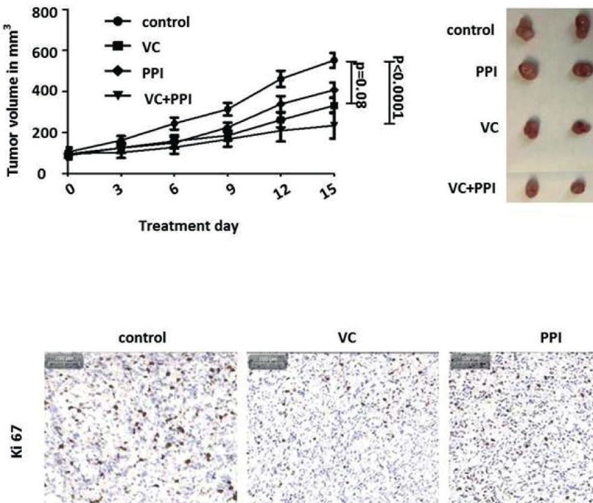
Li et al.                                                                                                         Drug Repurposing for mCRPC

and then treated with vitamin C (4 mM) for 4 h. Then, 100 µL of          Purification of Exosomes From Cell
WST-8 reagent was added per well, and the cells were incubated           Culture Supernatants
for 1-2 h. Absorbance was measured at 450 nm using a Multiskan           PC3 and DU145 cells were cultured with cell culture medium
FC instrument (Thermo Fisher Scientific, Waltham, MA, USA).               with a pH between 6.5 and 7.5 for two weeks, and then
Cell viability was calculated according to: absorbance of the            1.5 – 2.0×10⁶ cells were incubated in 75 cm2 flasks until they
sample/absorbance of the control (24).                                   reached approximately 60–70% confluence. Subsequently, the
                                                                         cells were further incubated with exosome-free medium (Gibco)
Detection of Apoptotic Cells via Flow                                    for 24 h. Then cell culture media was collected and centrifugated
Cytometry                                                                at 300× g for 5 min. Following were supernatants centrifuged at
Adenocarcinoma cells (5×10⁵/well in 6-well plates in 3 mL) were          1200× g for 15 min, followed by 12,000× g for 30 min. In the end,
first incubated with or without pantoprazole (100 mM) for 24 h at         supernatants were centrifuged at 110,000× g for 1 h in a Sorvall
37°C. Then, vitamin C was administered at different                      WX Ultracentrifuge Series (ThermoFisher Scientific, Waltham,
concentration. After 16 h, the cells were detached using 0.05%           MA, USA) in order to pellet exosomes. After one wash in a large
trypsin solution, washed two times with PBS and centrifuged at           volume of phosphate-buffered saline (PBS), and centrifuged at
1,500 rpm for 5 min. Then, the cells were stained with fluorescein        110,000× g for another 1 h. At last, exosomes were resuspended
isothiocyanate (FITC)-labelled annexin V (BD Pharmingen, San             in PBS (50 µL) for further analysis (27).
Diego, CA, USA), counterstained with propidium iodide (PI; BD
Pharmingen), resuspended in binding solution (BD
Pharmingen), according to the manufacturer’s instructions
                                                                         Cellular Uptake of Vitamin C
                                                                         Cancer cells (4×10⁵ cells/well in 24-well plates) were seeded and
(BD Pharmingen), and finally analyzed by flow cytometry
                                                                         pre-cultured in conditioned medium with adjusted acidity-
(CytoFLEX S, Beckman Coulter, Pasadena, CA, USA).
                                                                         alkalinity (pH 6.5 or 7.5) with or without pantoprazole
                                                                         (100 mM) for 24 h. The acidity-alkalinity of the cell culture
Detection of Intracellular Reactive Oxygen
                                                                         medium was controlled and regulated four times throughout the
Species (ROS)                                                            24 h incubation. Subsequently, the culture medium was replaced
ROS assays were carried out according to the manufacturer’s
                                                                         by PBS of the same acidity-alkalinity (pH 6.5 or 7.5) containing
instructions (Sigma-Aldrich). Briefly, 5×10³ cells per well (96-
                                                                         0.1 µCi (3.7 kBq) L-[14C]-ascorbic acid (PerkinElmer, Boston,
well plate) were incubated at 37°C with or without pantoprazole
                                                                         MA, USA). After incubation at 37°C for 30 min with L-[14C]-
(100 mM) for 24 h. Then, vitamin C (4 mM) was added for the
                                                                         ascorbic acid, the culture media was aspirated and the cells were
cell culture for another 4 h. Finally, 100 mL of ROS detection
                                                                         washed three times with 1 ml of ice-cold PBS. Then 350 ml of 1 N
reagent solution diluted in assay buffer was added to each well,
                                                                         NaOH was used to lyse the cells and the lysed cell samples were
and the cells were incubated for 1 h. Florescence intensity
                                                                         collected and counted by MicroBeta2 liquid Scintillation detector
(lex=640/lem=675 nm) was measured using a SPECTRAmax
                                                                         (PerkinElmer). One hundred microliters of the cell lysate were
M5 instrument (Molecular Devices, San Jose, CA, USA). The
                                                                         used for determination of the protein concentration by modified
relative ROS signal was determined by calculating the ROS level
                                                                         Lowry protein assay (Thermo Scientific). Finally, the uptake
in the cells with regard to cell survival rate determined from the
                                                                         results were normalized as counts per minute (CPM) in
WST-8 assay and standardizing the value to the ROS signal of
                                                                         relation to 100 mg of protein content (24).
untreated controls.

Determination of Cellular pH Change via                                  Cellular Uptake
Flow Cytometry                                                           of 2-deoxy-2-[18F]fluoro-D-glucose
Adenocarcinoma cancer cells (5×10⁵/well in 6-well plates in 3            (18F-FDG)
                                                                         18
mL) were seeded and pre-cultured in conditioned medium with                 F-FDG was synthesized at the Department of Nuclear
adjusted acidity-alkalinity (pH 6.5 or 7.5) with or without              Medicine of the First Affiliated Hospital of Sun Yat-sen
pantoprazole (100 mM) for 24 h. After 24 hours of                        University. Cells (PC3 and DU145 cells) plated in 6-well plates
pantroprazole treatment, cells were collected and washed twice           (2-5×10⁵ cells per well, 3 ml) were incubated with 1 µCi (37 kBq)
                                                                         18
with PBS. Then, cells were incubated for 5 minutes at 37°C with            F-FDG in glucose-free culture medium for 30 min at 5% CO2
500 mL of pre-warmed PBS containing 1 mM LysoSensor probe.               and 37°C. After rapid washing twice with cold PBS, the cells were
The intracellular pH was detected using flow cytometry (11).              detached with trypsin, and cell-associated CPM was measured
                                                                         with a radiometric detector (PerkinElmer). Cellular uptake
Determination of the Change in the pH of                                 was expressed as the percentage of uptake per well relative to
the Cell Culture Medium With a pH Metre                                  that of the control group (no treatment with vitamin C
Cancer cells (4×10⁵ cells/well in 24-well plates) were seeded and        or pantoprazole).
pre-cultured in conditioned medium with adjusted acidity-
alkalinity (pH 6.5 or 7.5) with or without pantoprazole                  Animal Model: Tumor Volume and
(100 mM) for 4 h. The pH of the medium was determined in                 Therapeutic Regimens
triplicate using a SevenCompact™ev220 pH metre (METTLER                  Male Balb-c nude mice (4-6 weeks old, n=24) were purchased
TOLEDO, Columbus, OH, USA).                                              from the Model Animal Research Center of Nanjing University.

Frontiers in Oncology | www.frontiersin.org                          3                                   July 2021 | Volume 11 | Article 660320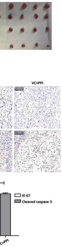
Li et al.                                                                                                            Drug Repurposing for mCRPC

For the induction of tumors, 5×10⁶ PC3 cells were suspended in             vivo studies, ≥3 technical replicates were used for each
sterile PBS (100 mL) and injected subcutaneously into the                  biological repeat.
flank region.
    Tumor diameters were measured every three days with a slide
caliper. Treatments were administered when the xenografts had
reached a diameter of approximately 6 mm. PC3-bearing
                                                                           RESULTS
animals were intraperitoneally injected with vitamin C (4 g/kg,            Pantoprazole Enhances the Cytotoxicity of
twice daily), pantoprazole (200 mg/kg, daily) or a combination of
                                                                           Vitamin C and Increases Intracellular ROS
vitamin C and pantoprazole. Pantoprazole was administered one
day before vitamin C injection.
                                                                           Accumulation in Prostate Cancer Cells in
    All mice were sacrificed 2 weeks after the initiation of                a pH- and Time-Dependent Manner
treatment. After sacrifice, the tumors were dissected for                   As determined by the WST-8 assay, in cell culture medium with
immunohistochemistry (IHC). No adverse effects were                        an acidic pH (6.5), the proliferation of most cancer cells except
observed in the animals.                                                   OVCAR3 cells was unaffected by the combination of vitamin C
                                                                           and pantoprazole when the compounds were administered
                                                                           simultaneously (Figure 1A and Supplement 1). However,
PET Imaging                                                                when cells were pretreated with pantoprazole for 24 h,
18
  F-FDG (see above for synthesis) was administered via tail vein           pantoprazole single treatment caused a reduction in the
injection (100 mL) at an activity dose of 100 µCi (3.7 MBq) per            viability of PC3 prostate cancer cells to 0.4 units and in DU145
mouse one day before and two weeks after initiation of                     prostate cancer cells to 0.7 units (Figure 1A). Moreover, the
treatment. Imaging was conducted using a micro-PET system                  reduction in cell viability was slightly more robust following
(Inveon, SIEMENS, Germany), and the radiotracer was allowed                combined administration of vitamin C and pantoprazole in both
to accumulate in the tumor for 45 min. The mice were then                  prostate cancer cell lines (Figure 1A). In contrast, at an alkaline
imaged for a 15 min static acquisition (28).                               pH (7.5), both vitamin C single treatment and in combination
                                                                           with pantoprazole resulted in significant reduction of the
PET Data Analysis                                                          viability in PC3 and DU145 cells. Compared to no
Tumor-to-background ratios (TBRs) were calculated to semi-                 pretreatment, pretreatment with pantoprazole (24 h) followed
quantitatively analyse18F-FDG uptake in the tumor. Circular                by combined administration of vitamin C and pantoprazole
three-dimensional regions of interest (ROIs) were delineated               caused an additional reduction in the viability of prostate
manually in the area with the highest tumor activity. The                  cancer cells (Figure 1A). Similar results were obtained for
diameter did not cover the entire tumor to avoid partial                   MCF7 and SKBR3 and SKOV3 cells. OVCAR3 showed
volume effects. For determination of background activity,                  somewhat different results (Supplement 1).
three-dimensional ROIs were delineated in the femoral muscle.                  Vitamin C and pantoprazole are reported to be important for
The TBR was calculated using the following quotient: mean                  ROS production (29–31). Our results have presented that in
tracer uptake in the tumor/mean tracer uptake in the muscle.               slightly acidic cell culture medium(pH=6.5), ROS content was
                                                                           increased to 2.1 relative units in PC3 and to 2.2 relative units in
Histologic and Immunohistochemical                                         DU145 cells following combined treatment after pretreatment
Analysis                                                                   with pantoprazole for 24 h. No increase was observed without
Tumor tissues were collected for IHC at the end of treatment.              pantoprazole pretreatment (Figure 1B). In cell culture medium
Apoptosis and proliferation were analysed based on staining with           with a slightly alkaline pH (7.5), the enhancement of ROS
antibodies targeting Ki-67 and cleaved caspase3 (Sevicebio, Palo           accumulation from pantoprazole was not so significant as
Alto, CA, USA) staining. Cells expressing Ki-67 or cleaved                 under acidic conditions (Figure 1B and Supplement 2).
caspase3 were quantified based on H-scores. H-scores are used
to assess the extent of nuclear immunoreactivity of steroid                Pantoprazole Enhances Apoptotic Cell
receptors. The H-score was calculated as follows:                          Death, Probably Due to the Increase in
   3 * the percentage of strongly stained nuclei + 2 * the                 Cellular Uptake of Vitamin C as Well as
percentage of moderately stained nuclei + the percentage of                the Inhibition of Exosome Production by
weakly stained nuclei. The range of H-scores is 0 to 300. IHC              Regulation of in Intra- and Extracellular pH
analysis was performed as reported previously (10).                        Values in Cancer Cells
                                                                           To characterize the cytotoxic mechanism of vitamin C and
Statistical Analysis                                                       pantoprazole in cancer cells, we first monitored apoptotic cell
For all analyses, p < 0.05 *, < 0.01 ** or < 0.001 *** was                 death using flow cytometric analysis (FACS). FACS analysis
considered as statistically significant. All analyses were                  revealed that pantoprazole enhanced vitamin C-induced
performed in GraphPad Prism (GraphPad Software, Inc.). Data                apoptotic cell death, as shown by a significant increase in the
are presented as the mean ± SD. For each experiment, n indicates           number ofannexin V/PI-positive cells as well as a marked
the number of individual biological repeats. For all in vitro and ex       decrease in the number of live, annexin V/PI-negative cells.

Frontiers in Oncology | www.frontiersin.org                            4                                    July 2021 | Volume 11 | Article 660320
Li et al.                                                                                                                                    Drug Repurposing for mCRPC

                        A

                        B

  FIGURE 1 | Pantoprazole in combination with vitamin C inhibits cell proliferation and induces ROS accumulation. A total of 1x10⁴ PC3 or DU145 cells per well
  (96-well plate) were incubated at 37°C with control (a), 4 mM vitamin C (b), 100 µM pantoprazole (c) or the combination of vitamin C and pantoprazole (d) for 4 h.
  Vitamin C was administered to cells with or without pretreatment with pantoprazole for 24 h (w: with pantoprazole pretreatment; w/o: without pantoprazole
  pretreatment). (A) Cell viability as assessed by the WST-8 assay and (B) ROS levels as detected based on fluorescence intensity 1 h to 2 h after the addition of
  diluted ROS detection reagent to the cell culture medium. The cell viability and ROS levels in the control group (a) were defined as 1.0. Any changes in cell viability
  and ROS levels following the different treatments are shown relative to the levels in the control group at two different pH values (pH 6.5 and 7.5, columns with
  different shades of grey). The bars represent the mean and SD of the mean of n≥3. N, not significant; *p < 0.05; **p < 0.01.

This was observed in PC3 and DU145 cells at a slightly acidic                             at both pH 6.5 and pH 7.5 (Figure 3A). Moreover, the
pH (6.5) (Figures 2A, C). For example, after 16 h of treatment                            intracellular pH of prostate and breast cancer cells was
with 4 mM vitamin C, 59% of PC3 cells and 58% of DU145 cells                              modified following alteration of the extracellular pH or
survived following treatment with vitamin C plus pretreatment                             following pantoprazole treatment (Figure 3B). This effect of
with pantoprazole (100 µM), whereas 74% of PC3 cells and 70%                              pantoprazole seemed to be stronger in acidic (pH 6.5) cell
of DU145 cells survived following treatment with vitamin C                                culture medium than in alkaline (pH 7.5) cell culture medium
only (Figures 2A, C). In cell culture medium with a pH of 7.5                             (Figure 3B). However, in SKOV3 cells, we did not observed a
(slightly alkaline), a similar effect was found in DU145 cells                            clear change in the intracellular pH in response to pantoprazole
following combined treatment. However, in PC3 cells,                                      treatment (Figure 3B). Furthermore, we noticed that in
particularly at vitamin C concentrations of 4, 8 and 16 mM,                               comparison with acidic pH (6.5), the alkaline p H
the elimination of tumors cells induced by the combined                                   (7.5) inhibited the production of exosomes significantly in
treatment regimen (vitamin C plus pretreatment with                                       both prostate cancer cell lines (Figure 4A). Moreover,
pantoprazole) was not superior to that with vitamin C only                                pantoprazole reduced the secretion of exosomes under acidic
(Figures 2B, D). FACS analysis of breast and ovarian cancer                               (6.5) but not alkaline conditions (Figure 4A). We also analyzed
cells also showed that the synergistic effect of pantoprazole on                          the cellular uptake of L-[14C]-ascorbic acid (vitamin C) after
cytotoxicity in slightly acidic (pH 6.5) cell culture medium was                          the addition of pantoprazole (100 µM for 6 h without the
stronger than that in alkaline (pH 7.5) cell culture medium                               administration of vitamin C) to the culture medium at a slightly
(Supplement 3).                                                                           acidic pH (6.5) and a slightly alkaline pH (7.5) (Figure 4B). In
    Our results also demonstrated that pantoprazole slightly                              DU145 cells incubated at pH 6.5 and 7.5 and in ovarian cancer
increased the extracellular pH of the cell culture medium                                 cells incubated at pH 6.5, pantoprazole induced a significant

Frontiers in Oncology | www.frontiersin.org                                           5                                            July 2021 | Volume 11 | Article 660320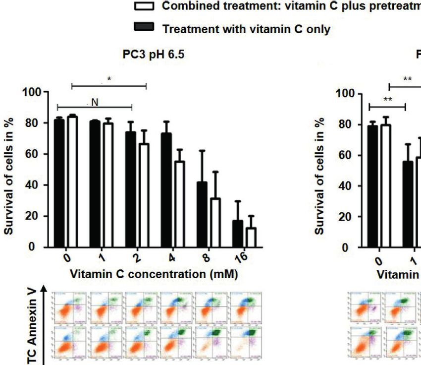
Li et al.                                                                                                                                        Drug Repurposing for mCRPC

                              A                                                            B

                              C                                                            D

  FIGURE 2 | Pantoprazole in combination with vitamin C induces apoptosis of prostate cancer cells. A total of 5x10⁵ PC3 or DU145 cells per well (6-well plate) were
  incubated at 37°C in slightly acidic (pH 6.5) or slightly alkaline (pH 7.5) cell culture medium with different concentrations of vitamin C for 16 h following with or without
  pretreatment with pantoprazole (100 µM) for 24 h: Cells were incubated in cell culture medium with a pH of 6.5 (A: PC3 cells; C: DU145 cells) or a pH of 7.5 (B: PC3
  cells; D: DU145 cells). Column diagram (upper panel): quantification of the FACS results. Colorized dot plot (bottom panel): FACS analysis data with increasing
  vitamin C concentrations as shown in the column diagrams (orange: surviving cells; green: PI- and AV- positive cells [apoptotic cells]; blue: PI-positive cells [necrotic
  cells]). Upper rows of the colorized dot plots: cells were treated with vitamin C only; bottom rows of the colorized dot plots: cells were treated with vitamin C and
  pantoprazole. The bars represent the mean and SD of the mean of n≥3. N, not significant; *p < 0.05, **p < 0.01.

increase in vitamin C uptake. However, in PC3 no difference in                             cycling of iron (DFO and DTPA) (22, 23). Interestingly, we
cellular vitamin C uptake was observed following addition of                               found that compared with control treatment, both intracellular
pantoprazole at pH 6.5 or pH 7.5. The same was true for MCF7                               and extracellular administration of chelators (DFO/DTPA)
and SKOV3 at pH 7.5 (Figure 4B).                                                           inhibited vitamin C-induced cytotoxicity in human prostate,
    We have also assayed the synergistic effect of redox-active                            breast and ovarian cancer cells (Supplement 4). Pantoprazole
iron with vitamin C treatment in prostate, breast and ovarian                              did not significantly influence the effect of chelators on the
cancer cells by performing the WTS-8 assay following combined                              toxicity of vitamin C, although pantoprazole could promote
treatment with vitamin C and chelators that inhibit the redox                              the cytotoxicity of vitamin C (Supplement 4).

Frontiers in Oncology | www.frontiersin.org                                            6                                              July 2021 | Volume 11 | Article 660320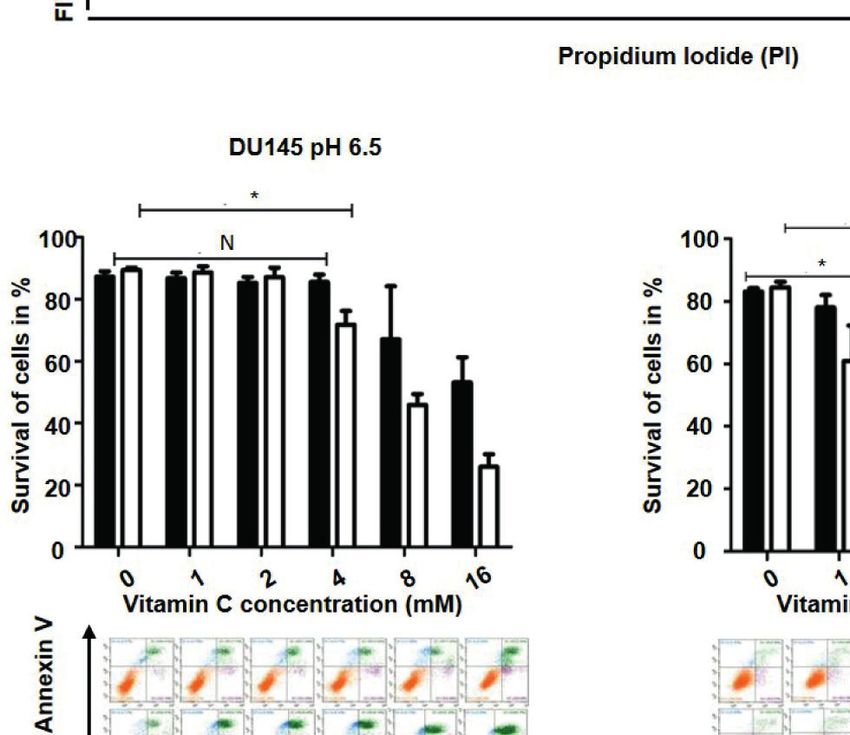
Li et al.                                                                                                                             Drug Repurposing for mCRPC

                         A

                         B

  FIGURE 3 | Pantoprazole regulates the extra- and intracellular pH of cancer cells. (A) Change in the extracellular pH of the cell culture medium of prostate
  (PC3, DU145), breast (MCF7) and ovarian cancer (SKOV3) cells following treatment with pantoprazole (grey columns) or without pantoprazole (controls, white
  columns) at a pH of 6.5 or 7.5. (B) Change in the intracellular pH of prostate (PC3 and DU145), breast (MCF7) and ovarian cancer (SKOV3) cells following
  treatment with or without 100 mM pantoprazole for 6 h at a pH of 6.5 or 7.5. Top panel: cells were incubated in cell culture medium of different pHs; middle
  panel: cells were incubated in cell culture medium with a pH of 6.5 with or without pantoprazole; bottom panel: cells were incubated in cell culture medium
  with a pH 7.5 with or without pantoprazole. Red pick: unstained cells; pink pick: cells incubated in cell culture medium with a pH of 7.5 containing
  pantoprazole; green pick: cells incubated in cell culture medium with a pH of 7.5 without pantoprazole; X-axis: FITC; Y-axis: cell count. The bars represent the
  mean and SD of the mean of n≥3.

Combined Treatment With Vitamin C and                                                (VC, PPI, VC + PPI) as soon as tumors had reached a volume of
Pantoprazole Significantly Inhibits Tumor                                             approximately 100 mm3(Figure 5A). While the control group
Growth of Prostate Cancer Xenografts                                                 received placebo only, the VC group was injected
In control animals that received placebo following subcutaneous                      intraperitoneally with 4 g/kg vitamin C twice daily, the PPI
inoculation of PC3 prostate cancer cells (5×10⁶), the doubling                       group received intraperitoneal injection of 200 mg/kg
time of the PC3 xenografts was 7 days (Figure 5A). Treatment                         pantoprazole daily, and the combined group (VC+PPI) was
was initiated in animals from the different treatment groups                         administered 200 mg/kg pantoprazole (daily) combined with 4

Frontiers in Oncology | www.frontiersin.org                                      7                                          July 2021 | Volume 11 | Article 660320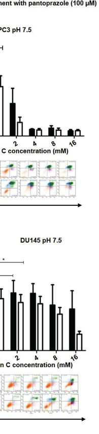
Li et al.                                                                                                                         Drug Repurposing for mCRPC

                             A

                             B

  FIGURE 4 | Pantoprazole significantly increases cellular vitamin C uptake and inhibits the production of exosomes depending on the pH value of the cell
  culture medium. (A) PC3 and DU145 cells were cultured in cell culture medium with a pH of 7.5 or a pH of 6.5 and treated with pantoprazole (100 mM) (PPI) or
  left untreated (control). The protein concentration was determined by the BCA protein assay, and exosomes were lysed using RIPA buffer. (B) Vitamin C
  uptake in prostate (PC3 and DU145), breast (MCF7) and ovarian cancer cells (SKOV3) following treatment with 100 mM pantoprazole for 6 h (grey columns) or
  without pantoprazole (control, white columns) in cell culture medium with a pH of 6.5 or 7.5. The bars represent the mean and SD of the mean of n≥3.
   *p < 0.05; **p < 0.01, ***p < 0.001.

g/kg vitamin C (twice daily). In the combined group,                               treatment regimen significantly decreased the percentage of
pantoprazole was administered one day before vitamin C.                            Ki67-positive cells from 38.5 to 20.5 (p = 0.004) (Figure 5B).
Combined treatment (VC+PPI) significantly suppressed tumor                          Treatment with vitamin C only induced a comparatively lower
growth compared to the untreated control group, as observed                        decrease in the percentage of Ki67-positive cells (p =
for PC3 xenografts 15 days after initiation of therapy (p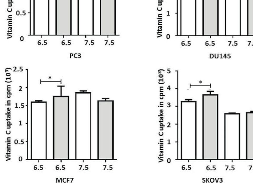
Li et al.                                                                                                                                 Drug Repurposing for mCRPC

                             A

                             B

  FIGURE 5 | Pantoprazole enhances the anticancer effect of vitamin C in mice bearing subcutaneous PC3 xenografts. (A) BALB/c nude mice (n=24; 4-6-week-old
  males) were subcutaneously injected with 5×10⁶ PC3 cells. When the tumor size had reached 100 mm3, pantoprazole (PPI, 200 mg/kg, daily) and vitamin C (VC, 4
  g/kg, twice daily) were injected intraperitoneally into the mice. After 15 days, all mice were euthanized, the remaining tumors were removed, and their volumes were
  measured. In the combined treatment group (VC + PPI), pantoprazole was given one day before injection of vitamin C. Left: tumor growth curves (day 0 – day 15)
  after the respective treatments; right: tumors prepared from the six different mice from each treatment group (control, PPI, VC, VC + PPI) after sacrifice (day 15).
  (B) Immunohistochemical (IHC) analysis of the proliferation marker Ki67 and the apoptosis marker cleaved caspase-3 in explanted tumors (upper panel). The grey
  bar in the upper left corner of each picture represents 100 µm. The lower panel shows semiquantitative analysis of the IHC results using H-scores as described in
  the Methods section. Quantification of the IHC results demonstrated a significant decrease (p = 0.004) in expression of the proliferation marker Ki67 and a significant
  increase (p < 0.0001) in expression of the apoptosis marker cleaved caspase 3 in VC + PPI-treated cells compared to controls, respectively. The bars represent the
  mean and SD of the mean of n≥3.

18
  F-FDG-PET monitoring, the mice were injected intravenously                           (Figure 6A). Additionally, combined treatment with VC + PPI in
with 3.7 MBq (100 µCi) 18F-FDG.As depicted in Figure 6A,                               vitro induced a stronger reduction in cellular 18F-FDGuptake
tumor growth was significantly inhibited by the combined                                (PC3: p=0.0007, DU145:p=0.002) in both prostate cancer cell
treatment. That is, 18F-FDG-PET imaging showed a significant                            lines than treatment with vitamin C alone (PC3: p=0.06, DU145:
reduction in18F-FDG uptake at 14 days after treatment initiation                       p=0.004) or pantoprazole alone (PC3: p=0.002, DU145:p=0.003),
(post therapy) compared with pretreatment values (pre therapy)                         indicating a reduced growth of tumor cells (Figure 6B).

Frontiers in Oncology | www.frontiersin.org                                        9                                           July 2021 | Volume 11 | Article 660320
Li et al.                                                                                                                                Drug Repurposing for mCRPC

                        A

                        B

  FIGURE 6 | Functional FDG-PET/CT imaging showing that pantoprazole enhances the anticancer effect of vitamin C in vivo. (A) Left panel: representative PET/CT
  scans showing the changes in tumor uptake of 18F-FDG (red arrows). 18F-FDG-PET/CT scans were carried out before (Pretherapy, day 0) and 14 days after the
  initiation of treatment (Posttherapy, day 14). The red arrows indicate the sites of subcutaneous injection of PC3 tumor cells and the accumulation of 18F-FDG. Right
  panel: uptake of 18F-FDG in tumor tissue (tumor to background ratio [TBR]) in control mice and mice treated with vitamin C (VC), pantoprazole (PPI) or both
  (VC+PPI). The TBR was calculated 14 days after the initiation of treatment and served as an indicator of tracer uptake. The mean TBR as detected via18F-FDG-PET/
  CT was significantly reduced following combined treatment (VC + PPI) compared with control treatment (p = 0.0003), indicating a significant reduction in tumor
  volume. Moreover, the reduction in TBR after combined VC + PPI treatment compared to treatment with vitamin C only was also significant (p = 0.005). (B) PC3 and
  DU145 prostate cancer cells were incubated with 18F-FDG for 30 min at 5% CO2 and 37°C. After washing twice with cold PBS, the cells were detached with trypsin,
  and cell-associated 18Fradioactivity was quantified with a g-counter. 18Fradioactivity is expressed as percent uptake per cell relative to the uptake of untreated
  controls. In both prostate cancer cell lines (PC3 and DU145), the combination treatment significantly reduced cellular uptake of 18F-FDG (p=0.0007 in PC3 and
  p=0.002 in DU145, compared to control, respectively. VC: treatment with vitamin C (4 mM); PPI: treatment with pantoprazole (100 µM); VC + PPI: combined
  treatment with vitamin C (4 mM) and pantoprazole (100 µM). The bars represent the mean and SD of the mean of n≥3.

DISCUSSION                                                                             of cancer cells such as: redox imbalance, epigenetic
                                                                                       reprogramming and oxygen-sensing regulation (34). They also
Previous studies have demonstrated that vitamin C triggers                             summarized clinical trials with regarding to this anticancer
cancer cell-selective cytotoxicity in vitro (18, 32, 33). Our                          feature of vitamin C in order to develop more effective
previous study has shown the impact of pH on the anticancer                            combination strategies and improve the overall clinical study
effect of vitamin C in PC3 and DU145 prostate cancer cells (24).                       design in the future (34).
Moreover, we could show that treatment of prostate cancer cells                           Proton pump inhibitors (PPIs) have shown to be beneficial
with vitamin C induces pH-dependent apoptosis through the                              for cancer chemoprevention by reduction of proliferation and
generation of ROS as well as a reduction in NADPH levels in                            induction of apoptosis in multiple cancer cell lines (11, 12, 35).
vitro (24). Additionally, Ngo et al. discussed how vitamin C is                        PPIs are commonly used to treat acid-related diseases, might
beneficial to anticancer therapy by targeting three vulnerabilities                     promote the disruption of pH homeostasis in tumor cells by

Frontiers in Oncology | www.frontiersin.org                                       10                                           July 2021 | Volume 11 | Article 660320
Li et al. Drug Repurposing for mCRPC
targeting V-ATPase (11, 16, 26). PPIs have also been reported to monitoring and predicting the therapeutic response to androgen
enhance a pH dependent anticancer effect on cancer cells deprivation therapy in patients with metastatic prostate cancer
through regulation of the production of exosomes and the (43, 44). PC3 cells isolated from metastatic prostate cancer
extracellular pH, which regulates the production of exosomes patients have been reported to be PSMA-negative and
involved in the pathogenesis of cancers (25, 27, 36). The identical castration-resistant (45, 46). In this study, we transplanted PC3
change of exosome production was found from our experiments cells into mice to mimic mCRPC, treated the mice with vitamin
(Figure 4A). In the current study, we have demonstrated that the C and/or pantoprazole and monitored the therapeutic effect of
PPI pantoprazole increases the cytotoxicity of vitamin C in the these drugs using 18F-FDG PET/CT imaging. We could identify
treatment of metastatic castration-resistant prostate cancer in the location of the mCRPC (PC3) xenografts. Furthermore, we
vitro and in vivo. Our results also highlight the regulation of pH have demonstrated that 18 F-FDGPET/CT imaging allows
in the tumor microenvironment (Figure 3), ROS accumulation monitoring of the therapeutic response following combined
(Figure 1 and Supplements 1, 2) and exosome production treatment with vitamin C and pantoprazole. As we could
(Figure 4A) following combined anticancer treatment with show, treatment induced a significant reduction in18F-FDG
vitamin C and pantoprazole in vitro. uptake in prostate cancer xenografts after two weeks
Furthermore, a series of clinical studies on the administration (Figure 6A). Therefore, our data support the further clinical
of PPIs to patients suffering from different cancers demonstrated investigation of 18F-FDG PET/CT imaging for the prediction of
that PPIs may be new effective agents for anticancer therapy therapeutic responses in patients with castration-resistant
(37–39). In addition, most patients suffering from CRPC metastatic prostate cancer following combined therapy with
undergo a long-term therapeutic regimen; thus low-income pantoprazole and vitamin C.
individuals may have unable to receive further adequate
anticancer treatment. Drug repurposing supplies a cheaper
and probably more efficient therapeutic possibility (9).
Previous studies showed that repurposing PPIs could enhance
CONCLUSION
the efficacy and safety of chemotherapy as well as improve the The drug repurposing of pantoprazole and vitamin C seems to be
therapy in solid tumors (40, 41). We have observed a stronger an alternative therapeutic strategy for patients suffering from
therapeutic effect when cancer cells were pretreated with mCRPC, since therapy using PSMA I&T or PSMA-617 labelled
pantoprazole for 24 h than after simultaneous treatment with Lu-177 or Ac-225 is not available worldwide. We have
vitamin C and pantoprazole simultaneously (Figure 1, 2 and shown that pantoprazole enhances the anticancer effect of
Supplements 1, 2). This could be explained with the fact that vitamin C in prostate cancer cells by increasing cellular
pantoprazole pretreatment significantly increased vitamin C vitamin C uptake, inhibiting exosome production and altering
uptake in DU145, MCF7 and SKOV3 cells (Figure 4B). the intracellular and extracellular pH. Moreover, 18F-FDG-PET
However, pantoprazole pretreatment did not significantly proved to be useful for monitoring the therapeutic response
increase the uptake of vitamin C in cells incubated in cell in CRPC.
culture medium with a pH of 7.5. This might due to that the
therapeutic effect of PPIs is pH dependent (27). Our results have
also shown that pantoprazole had a more beneficial anticancer
effect in a slightly acidic environment (Figure 1 and DATA AVAILABILITY STATEMENT
Supplements 1, 2). Pantoprazole significantly enhanced the
The original contributions presented in the study are included in
cellular uptake of vitamin C in cells incubated in slightly
the article/Supplementary Material. Further inquiries can be
acidic cell culture medium (pH 6.5). Furthermore, the toxicity
directed to the corresponding authors.
of vitamin C in NSCLC and GBM has been reported to depend
on redox-active labile iron (22). Likewise, we have demonstrated
that in prostate cancer cells (PC3, DU145), breast cancer cells
(MCF7) and ovarian cancer cells (SKOV3), the cytotoxicity of ETHICS STATEMENT
vitamin C depends on redox-active labile iron (Supplement 4).
Nevertheless, pantoprazole induces the enhancement of cellular The animal study was reviewed and approved by The
toxicity of vitamin C. However, pantoprazole has no additional Institutional Animal Care and Use Committee of The First
influence on iron redox cycling in cancer cell lines Affiliated Hospital of Sun Yat-sen University.
(Supplement 4).
Recent studies have demonstrated that PET/CT imaging
using PSMA ligands provides a higher sensitivity and AUTHOR CONTRIBUTIONS
specificity than other imaging methods for the evaluation of
advanced prostate cancer (6, 7, 42). However, in China, PET/CT Conceptualization, ZL and XZ. Data curation, ZL, PH, YL, CS,
imaging using PSMA ligands is still not an established approach and XZ. Formal analysis, ZL, PH, YL, WS, ZC, BZ, CS and XZ.
for the preclinical and clinical detection of prostate cancer. Other Funding acquisition, ZL, DY, and XZ. Investigation, ZL, PH,
Studies have suggested that 18F-FDG PET might be useful for YL, GY, ZC, BZ, YW, and XZ. Methodology, ZL, PH, YL, GY,
Frontiers in Oncology | www.frontiersin.org 11 July 2021 | Volume 11 | Article 660320Li et al. Drug Repurposing for mCRPC
WS, ZC, BZ, and XZ. Project administration, ZL and XZ. ACKNOWLEDGMENTS
Resources, ZL, DY, CS, and XZ. Supervision, ZL and XZ.
All authors contributed to the article and approved the We are grateful to all members of the XZ group for their
submitted version. contributions to this project. We thank Qiao Su, Wuguo Li
and all members of the animal care facility of the First Affiliated
Hospital of Sun Yat-Sen University for animal management. We
thank Yali Tang, Tong Zhang and QingqiangTu from the core
FUNDING facility for research equipment at Sun Yat-Sen University for
This research was funded by the National Science Foundation technical support for FACS analysis.
for Young Scientists of China [grant number 81901793], the
Young Teacher Foundation of Sun Yat-sen University (CN)
[grant number 19ykpy55], the Science and Technology SUPPLEMENTARY MATERIAL
Program of Guangzhou [grant number 201607010353] and the
Science and Technology Program of Guangzhou [grant number The Supplementary Material for this article can be found online
201707010110]. These funding programs finically supported at: https://www.frontiersin.org/articles/10.3389/fonc.2021.
our study. 660320/full#supplementary-material
13. Lu Z, Tian B, Guo X. Repositioning of Proton Pump Inhibitors in Cancer
REFERENCES Therapy. Cancer Chemoth Pharm (2017) 80:925–37. doi: 10.1007/s00280-017-
1. Zhu Y, Ye D. Chinese Expert Consensus on the Diagnosis and Treatment of 3426-2
Castration-Resistant Prostate Cancer (2019 Update). Cancer Manag Res 14. Asgharzadeh MR, Barar J, Pourseif MM, Eskandani M, Jafari Niya M,
(2020) 12:2127–40. doi: 10.2147/CMAR.S236879 Mashayekhi MR, et al. Molecular Machineries of pH Dysregulation in
2. Wang L, Pan S, Zhu B, Yu Z, Wang W. Comprehensive Analysis of Tumour Tumor Microenvironment: Potential Targets for Cancer Therapy.
Mutational Burden and its Clinical Significance in Prostate Cancer. BMC Urol BioImpacts (2017) 7:115–33. doi: 10.15171/bi.2017.15
(2021) 21:29. doi: 10.1186/s12894-021-00795-7 15. Vander Heiden MG, Cantley LC, Thompson CB. Understanding the Warburg
3. Hayden AJ, Catton C, Pickles T. Radiation Therapy in Prostate Cancer: A Effect: The Metabolic Requirements of Cell Proliferation. Science (2009)
Risk-Adapted Strategy. Curr Oncol (2010) 17:18–24. doi: 10.3747/ 324:1029–33. doi: 10.1126/science.1160809
co.v17i0.704 16. Chen M, Huang S, Zhang X, Zhang B, Zhu H, Yang VW, et al. Reversal Effects
4. Paller CJ, Antonarakis ES. Management of Biochemically Recurrent Prostate of Pantoprazole on Multidrug Resistance in Human Gastric Adenocarcinoma
Cancer After Local Therapy: Evolving Standards of Care and New Directions. Cells by Down-Regulating the V-ATPases/mTOR/HIF-1a/P-Gp and MRP1
Clin Adv Hematol Oncol (2013) 11:14–23. Signaling Pathway In Vitro and In Vivo. J Cell Biochem (2012) 113:2474–87.
5. Pu J, Li T, Liu N, Luo C, Quan Z, Li L, et al. PlCϵ Knockdown Enhances the doi: 10.1002/jcb.24122
Radiosensitivity of Castration−Resistant Prostate Cancer via the AR/PARP1/ 17. Maacha S, Bhat AA, Jimenez L, Raza A, Haris M, Uddin S, et al. Extracellular
DNA−PKcs Axis. Oncol Rep (2020) 43:1397–412. doi: 10.3892/or.2020.7520 Vesicles-Mediated Intercellular Communication: Roles in the Tumor
6. Kratochwil C, Bruchertseifer F, Rathke H, Hohenfellner M, Giesel FL, Microenvironment and Anti-Cancer Drug Resistance. Mol Cancer (2019)
Haberkorn U, et al. Targeted a-Therapy of Metastatic Castration-Resistant 18:55. doi: 10.1186/s12943-019-0965-7
Prostate Cancer With 225ac-PSMA-617: Swimmer-Plot Analysis Suggests 18. Ma Y, Chapman J, Levine M, Polireddy K, Drisko J, Chen Q. High-Dose
Efficacy Regarding Duration of Tumor Control. J Nucl Med (2018) 59:795– Parenteral Ascorbate Enhanced Chemosensitivity of Ovarian Cancer and
802. doi: 10.2967/jnumed.117.203539 Reduced Toxicity of Chemotherapy. Sci Transl Med (2014) 6:218r–22r.
7. Rathke H, Holland-Letz T, Mier W, Flechsig P, Mavriopoulou E, Röhrich M, doi: 10.1126/scitranslmed.3007154
et al. Response Prediction of 177Lu-PSMA-617 Radioligand Therapy Using 19. Circu ML, Aw TY. Reactive Oxygen Species, Cellular Redox Systems, and
Prostate-Specific Antigen, Chromogranin A, and Lactate Dehydrogenase. Apoptosis. Free Radical Bio Med (2010) 48:749–62. doi: 10.1016/
J Nucl Med (2020) 61:689–95. doi: 10.2967/jnumed.119.231431 j.freeradbiomed.2009.12.022
8. Duenas-Gonzalez A, Garcia-Lopez P, Herrera LA, Medina-Franco JL, Gonzalez- 20. Ormazabal V, Zuñiga FA, Escobar E, Aylwin C, Salas-Burgos A, Godoy A,
Fierro A, Candelaria M. The Prince and the Pauper. A Tale of Anticancer et al. Histidine Residues in the Na+-Coupled Ascorbic Acid Transporter-2
Targeted Agents. Mol Cancer (2008) 7:82. doi: 10.1186/1476-4598-7-82 (SVCT2) Are Central Regulators of SVCT2 Function, Modulating pH
9. Langedijk J, Mantel-Teeuwisse AK, Slijkerman DS, Schutjens MDB. Drug Sensitivity, Transporter Kinetics, Na+ Cooperativity, Conformational
Repositioning and Repurposing: Terminology and Definitions in Literature. Stability, and Subcellular Localization. J Biol Chem (2010) 285:36471–85.
Drug Discovery Today (2015) 20:1027–34. doi: 10.1016/j.drudis.2015.05.001 doi: 10.1074/jbc.M110.155630
10. Zheng Z, Luo G, Shi X, Long Y, Shen W, Li Z, et al. The Xc– Inhibitor 21. Halliwell B, Cross CE. Oxygen-Derived Species: Their Relation to Human
Sulfasalazine Improves the Anti-Cancer Effect of Pharmacological Vitamin C Disease and Environmental Stress. Environ Health Persp (1994) 102:5–12.
in Prostate Cancer Cells via a Glutathione-Dependent Mechanism. Cell Oncol doi: 10.1289/ehp.94102s105
(2020) 43:95–106. doi: 10.1007/s13402-019-00474-8 22. Schoenfeld JD, Sibenaller ZA, Mapuskar KA, Wagner BA, Cramer-Morales
11. Luciani F, Spada M, De Milito A, Molinari A, Rivoltini L, Montinaro A, et al. KL, Furqan M, et al. O2 ·– and H2O2 -Mediated Disruption of Fe Metabolism
Effect of Proton Pump Inhibitor Pretreatment on Resistance of Solid Tumors Causes the Differential Susceptibility of NSCLC and GBM Cancer Cells to
to Cytotoxic Drugs. JNCI J Natl Cancer Institute (2004) 96:1702–13. Pharmacological Ascorbate. Cancer Cell (2017) 31:487–500. doi: 10.1016/
doi: 10.1093/jnci/djh305 j.ccell.2017.02.018
12. Breedveld P, Zelcer N, Pluim D, Sonmezer O, Tibben MM, Beijnen JH, et al. 23. Lv H, Wang C, Fang T, Li T, Lv G, Han Q, et al. Vitamin C Preferentially Kills
Mechanism of the Pharmacokinetic Interaction Between Methotrexate and Cancer Stem Cells in Hepatocellular Carcinoma via SVCT-2. NPJ Precis Oncol
Benzimidazoles: Potential Role for Breast Cancer Resistance Protein in (2018) 2:1. doi: 10.1038/s41698-017-0044-8
Clinical Drug-Drug Interactions. Cancer Res (2004) 64:5804–11. 24. Li Z, He P, Luo G, Shi X, Yuan G, Zhang B, et al. Increased Tumoral
doi: 10.1158/0008-5472.CAN-03-4062 Microenvironmental pH Improves Cytotoxic Effect of Pharmacologic
Frontiers in Oncology | www.frontiersin.org 12 July 2021 | Volume 11 | Article 660320Li et al. Drug Repurposing for mCRPC
Ascorbic Acid in Castration-Resistant Prostate Cancer Cells. Front Pharmacol A Phase I/II Study in Companion Animals With Spontaneously Occurring
(2020) 11:570939. doi: 10.3389/fphar.2020.570939 Tumors. J Transl Med (2014) 12:225. doi: 10.1186/s12967-014-0225-y
25. Azzarito T, Venturi G, Cesolini A, Fais S. Lansoprazole Induces Sensitivity to 38. Wang B, Zhang J, Wang J, Sun S, Wang Z, Wang L, et al. Intermittent High
Suboptimal Doses of Paclitaxel in Human Melanoma. Cancer Lett (2015) Dose Proton Pump Inhibitor Enhances the Antitumor Effects of
356:697–703. doi: 10.1016/j.canlet.2014.10.017 Chemotherapy in Metastatic Breast Cancer. J Exp Clin Canc Res (2015)
26. Marino ML, Fais S, Djavaheri-Mergny M, Villa A, Meschini S, Lozupone F, 34:85. doi: 10.1186/s13046-015-0194-x
et al. Proton Pump Inhibition Induces Autophagy as a Survival Mechanism 39. Falcone R, Roberto M D, Antonio C, Romiti A, Milano A, Onesti CE, et al.
Following Oxidative Stress in Human Melanoma Cells. Cell Death Dis (2010) High-Doses of Proton Pump Inhibitors in Refractory Gastro-Intestinal
1:e87. doi: 10.1038/cddis.2010.67 Cancer: A Case Series and the State of Art. Digest Liver Dis (2016) 48:1503–
27. Logozzi M, Mizzoni D, Angelini D, Di Raimo R, Falchi M, Battistini L, et al. 5. doi: 10.1016/j.dld.2016.08.126
Microenvironmental pH and Exosome Levels Interplay in Human Cancer Cell 40. Ikemura K, Hiramatsu S, Okuda M. Drug Repositioning of Proton Pump
Lines of Different Histotypes. Cancers (2018) 10:370. doi: 10.3390/ Inhibitors for Enhanced Efficacy and Safety of Cancer Chemotherapy. Front
cancers10100370 Pharmacol (2017) 8:911. doi: 10.3389/fphar.2017.00911
28. Li Z, Herrmann K, Pirsig S, Philipp-Abbrederis K, Henninger M, Aichler M, 41. Patel KJ, Lee C, Tan Q, Tannock IF. Use of the Proton Pump Inhibitor
et al. Molecular Imaging for Early Prediction of Response to Sorafenib Pantoprazole to Modify the Distribution and Activity of Doxorubicin: A
Treatment in Sarcoma. Am J Nucl Med Mol Imaging (2013) 4:70–9. Potential Strategy to Improve the Therapy of Solid Tumors. Clin Cancer Res
29. Guaiquil VH, Vera JC, Golde DW. Mechanism of Vitamin C Inhibition of Cell (2013) 19:6766–76. doi: 10.1158/1078-0432.CCR-13-0128
Death Induced by Oxidative Stress in Glutathione-Depleted HL-60 Cells. 42. Creagan ET, Moertel CG, O’Fallon JR, Schutt AJ, O’Connell MJ, Rubin J, et al.
J Biol Chem (2001) 276:40955–61. doi: 10.1074/jbc.M106878200 Failure of High-Dose Vitamin C (Ascorbic Acid) Therapy to Benefit Patients
30. Rodriguez EA, Donath E, Waljee AK, Sussman DA. Value of Oral Proton With Advanced Cancer. A Controlled Trial. N Engl J Med (1979) 301:687–90.
Pump Inhibitors in Acute, Nonvariceal Upper Gastrointestinal Bleeding: A doi: 10.1056/NEJM197909273011303
Network Meta-Analysis. J Clin Gastroenterol (2017) 51:707–19. doi: 10.1097/ 43. Spratt DE, Gavane S, Tarlinton L, Fareedy SB, Doran MG, Zelefsky MJ, et al.
MCG.0000000000000625 Utility of FDG-PET in Clinical Neuroendocrine Prostate Cancer. Prostate
31. Seoane M, Esperanza M, Cid Á . Cytotoxic Effects of the Proton Pump (2014) 74:1153–9. doi: 10.1002/pros.22831
Inhibitor Omeprazole on the non-Target Marine Microalga Tetraselmis 44. Jadvar H, Velez EM, Desai B, Ji L, Colletti PM, Quinn DI. Prediction of Time
Suecica. Aquat Toxicol (2017) 191:62–72. doi: 10.1016/j.aquatox.2017.08.001 to Hormonal Treatment Failure in Metastatic Castration-Sensitive Prostate
32. Du J, Martin SM, Levine M, Wagner BA, Buettner GR, Wang S, et al. Cancer With18f-FDG PET/CT. J Nucl Med (2019) 60:1524–30. doi: 10.2967/
Mechanisms of Ascorbate-Induced Cytotoxicity in Pancreatic Cancer. Clin jnumed.118.223263
Cancer Res (2010) 16:509–20. doi: 10.1158/1078-0432.CCR-09-1713 45. Lescarbeau RM, Kaplan DL. Quantitative Analysis of Castration Resistant
33. Riordan NH, Riordan HD, Meng X, Li Y, Jackson JA. Intravenous Ascorbate Prostate Cancer Progression Through Phosphoproteome Signaling. BMC
as a Tumor Cytotoxic Chemotherapeutic Agent. Med Hypotheses (1995) Cancer (2014) 14:325. doi: 10.1186/1471-2407-14-325
44:207–13. doi: 10.1016/0306-9877(95)90137-X 46. Liu C, Hasegawa K, Russell SJ, Sadelain M, Peng K. Prostate-Specific
34. Ngo B, Van Riper JM, Cantley LC, Yun J. Targeting Cancer Vulnerabilities Membrane Antigen Retargeted Measles Virotherapy for the Treatment of
With High-Dose Vitamin C. Nat Rev Cancer (2019) 19:271–82. doi: 10.1038/ Prostate Cancer. Prostate (2009) 69:1128–41. doi: 10.1002/pros.20962
s41568-019-0135-7
35. Mirossay L, Mirossay A, Kocisova E, Radvakova I, Miskovsky P, Mojzis J. Conflict of Interest: The authors declare that the research was conducted in the
Hypericin-Induced Phototoxicity of Human Leukemic Cell Line HL-60 is absence of any commercial or financial relationships that could be construed as a
Potentiated by Omeprazole, an Inhibitor of H+K+-ATPase and 5’-(N,N- potential conflict of interest.
Dimethyl)-Amiloride, an Inhibitor of Na+/H+ Exchanger. Physiol Res (1999)
48:135–41. Copyright © 2021 Li, He, Long, Yuan, Shen, Chen, Zhang, Wang, Yue, Seidl and
36. Federici C, Lugini L, Marino ML, Carta F, Iessi E, Azzarito T, et al. Zhang. This is an open-access article distributed under the terms of the Creative
Lansoprazole and Carbonic Anhydrase IX Inhibitors Sinergize Against Commons Attribution License (CC BY). The use, distribution or reproduction in other
Human Melanoma Cells. J Enzym Inhib Med Ch (2016) 31:119–25. forums is permitted, provided the original author(s) and the copyright owner(s) are
doi: 10.1080/14756366.2016.1177525 credited and that the original publication in this journal is cited, in accordance with
37. Spugnini EP, Buglioni S, Carocci F, Francesco M, Vincenzi B, Fanciulli M, accepted academic practice. No use, distribution or reproduction is permitted which
et al. High Dose Lansoprazole Combined With Metronomic Chemotherapy: does not comply with these terms.
Frontiers in Oncology | www.frontiersin.org 13 July 2021 | Volume 11 | Article 660320You can also read