Valentino: a zebrafish gene required for normal hindbrain segmentation
←
→
Page content transcription
If your browser does not render page correctly, please read the page content below
Development 122, 3981-3990 (1996)                                                                                                3981
Printed in Great Britain © The Company of Biologists Limited 1996
DEV1128

valentino: a zebrafish gene required for normal hindbrain segmentation

Cecilia B. Moens1,*, Yi-Lin Yan1, Bruce Appel1, Allan G. Force1 and Charles B. Kimmel2
1Institute   of Neuroscience and 2Department of Biology, University of Oregon, Eugene, OR 97403-1254, USA
*Author for correspondence (e-mail: moens@uoneuro.uoregon.edu)

SUMMARY

Mutational analysis can serve both to identify new genes            development of rhombomeres 5 and 6, and propose that
essential for patterning embryonic development and to               valentino functions in the subdivision and expansion of a
determine their functions. Here we describe the identifica-         common precursor region in the presumptive hindbrain
tion and phenotypic characterization of alleles of valentino,       into the definitive rhombomeres 5 and 6. These results
which we recovered in a genetic screen that sought to               provide genetic evidence for a two-segment periodicity in
identify mutations in the zebrafish that disrupt region-            the hindbrain and suggest that this periodicity arises
specific gene expression patterns in the embryonic brain.           sequentially, through the specification and later subdivi-
valentino is required for normal hindbrain segmentation             sion of a two-rhombomere unit, or ‘protosegment’.
and the hindbrain of valentino mutant embryos is
shortened by the length of one rhombomere. We demon-                Key words: zebrafish, hindbrain, segmentation, valentino,
strate that valentino is required cell-autonomously in the          rhombomere

INTRODUCTION                                                        and Lumsden, 1991; Guthrie et al., 1993), and there are several
                                                                    genes that are expressed in alternate rhombomeres, at least one
The subdivision of a continuous embryonic field into reiterated     of which, Krox-20, is required for the development of rhom-
segments is a mechanism for the generation of regional              bomeres 3 and 5 (Wilkinson et al., 1989a; Schneider-Manoury
diversity that is used across animal phyla. Such a process is       et al., 1993; Swiatek and Gridley, 1993).
evident in the vertebrate hindbrain, whose complex organiz-            We have undertaken a genetic screen in the zebrafish to
ation is based upon the transient appearance of seven or eight      identify genes involved in brain regionalization, particularly in
segments, or rhombomeres, during embryogenesis (Vaage,              hindbrain segmentation. Screening by RNA in situ hybridiz-
1969). Rhombomeres serve to organize subsequent patterns of         ation for mutations that disrupt the normal regional patterns of
neuronal differentiation and neural crest migration in the          gene expression in the brain, we have identified three alleles
hindbrain, and thus determine the architecture, innervation and     of valentino (val), an essential gene required for segmentation
function of the vertebrate head (reviewed in Guthrie, 1995).        in the posterior hindbrain. Our analysis of valentino mutant
Neural crest cells leave the presumptive hindbrain at particu-      embryos and of genetic mosaics leads us to propose that
lar rhombomeric levels to contribute to the cranial ganglia and     valentino is required for the expansion and subdivision of a
pharyngeal arches, and motor neurons differentiating in par-        specified region of the presumptive hindbrain, which we term
ticular rhombomere pairs innervate the pharyngeal arches with       a ‘protosegment’, into the definitive rhombomeres 5 and 6. In
a 2:1 correspondence (Lumsden and Keynes, 1989; Lumsden             the absence of valentino function, this protosegment persists
et al., 1991). Cell lineage analysis has shown that rhom-           but, lacking a terminal rhombomere identity, it fails to form
bomeres constitute developmental compartments, since cells          boundaries with flanking rhombomeres. Our findings suggest
generally fail to cross rhombomere boundaries once they are         that hindbrain segmentation occurs sequentially, through the
formed (Birgbauer and Fraser, 1994).                                initial specification of protosegments that correspond to the
   Although the genetic mechanisms that bring about segmen-         two-segment units later defined by Hox gene expression, and
tation in the Drosophila embryo are well understood (Nüsslein-      their subsequent subdivision and expansion into the definitive
Volhard and Wieschaus, 1980; reviewed in Akam, 1987), less          rhombomeres.
is known about the genetic control of segmentation in the ver-
tebrate hindbrain. A number of lines of evidence have suggested
that rhombomeres have a two-segment periodicity, in which           MATERIALS AND METHODS
alternating odd and even identities are overlain by the             RNA in situ hybridization screening of haploid embryos
expression of Hox genes, which specify regional identity in         We screened haploid embryos (Streisinger et al., 1981) produced by
rhombomere pairs (Keynes and Krumlauf, 1994). Transplanta-          the F1 progeny of male fish that had been mutagenized with either N-
tion experiments in the chick have shown that cells in alternate    ethyl-N-nitrosourea (ENU; Solnica-Krezel et al., 1994) or γ-rays (C.
rhombomeres are more similar to one another in adhesive prop-       Walker and C. Kimmel, unpublished data). At 22 hours postfertiliza-
erties than they are to cells in adjacent rhombomeres (Guthrie      tion (h), 10 embryos from each clutch were dechorionated and fixed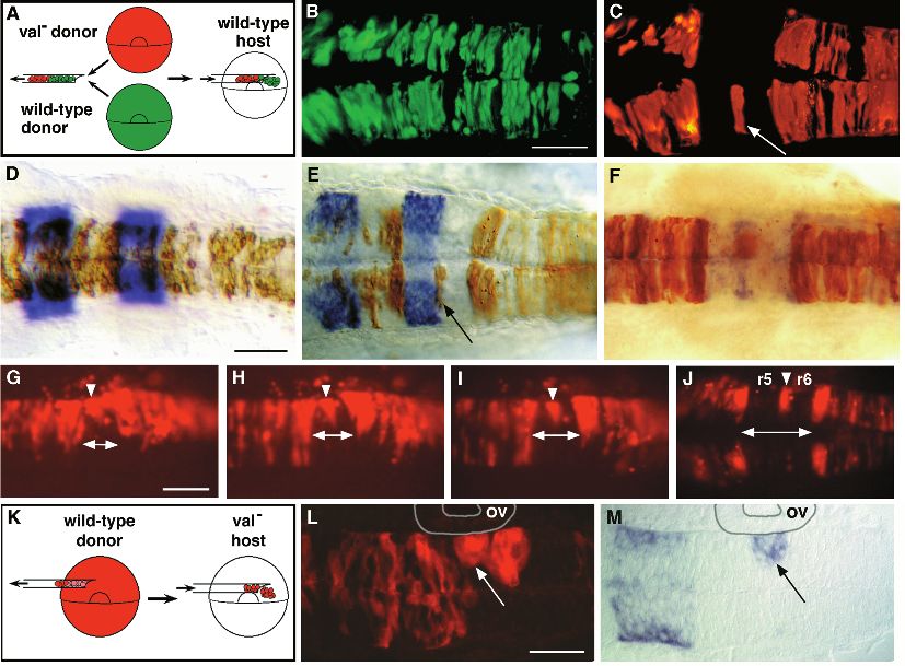
3982 C. B. Moens and others
in 4% paraformaldehyde (PFA) in phosphate-buffered saline (PBS). together, yielding wild-type and mutant embryos in a 3:1 ratio. In
Since we were interested in identifying mutations that subtly altered one set of experiments (schematized in Fig. 4A), embryos from such
the patterning of the central nervous system, we fixed embryos that a cross were labeled at the 1- to 2-cell stage with a mixture of
appeared morphologically normal under the dissecting microscope. lysinated tetramethyl rhodamine-dextran (LRD) and lysine-fixable
We screened simultaneously for mutations that disrupted the biotinylated dextran (Molecular Probes), and wild-type embryos
expression patterns of six genes: krox20 (Oxtoby and Jowett., 1993), from a cross between homozygous wild-type fish were labeled with
eng3 (Ekker et al., 1992), shh (Krauss et al., 1993), lim5 (Toyama et fluorescein-dextran. Using methods previously described (Ho and
al., 1995), myoD (Weinberg et al., 1996), and dlx-2 (Akimenko et al., Kane, 1990), cells from both types of donor were transplanted into
1994), after determining that the expression patterns of each of these the same unlabeled wild-type host embryo at the shield stage (6 h;
genes was essentially normal in wild-type haploid embryos. RNA in Kimmel et al., 1995). In some experiments cells were transplanted
situ hybridizations were performed essentially as described (Oxtoby heterochronically, from dome-stage (4.3 h) donors into shield-stage
and Jowett, 1993), using a 10×10 array of baskets constructed with hosts, with the same results. Cells were transplanted into the region
Beem capsules (size 00; Ted Pella) and silk mesh to transfer embryos of the host embryo that gives rise to the hindbrain (Woo and Fraser,
between washes. After colour development, embryos were washed in 1995), and host embryos were left to develop until 18-24 h, at which
PBS containing 0.1% Tween-20 (PBT) and were scored in PBT under time the distribution of labeled cells in the host hindbrain was deter-
a dissecting microscope. mined. The genotype of donor embryos was determined by visual
inspection of the hindbrain at 18 h. In a second set of experiments
PCR typing of valb361 embryos (schematized in Fig. 4K), cells from labeled wild-type donor
We identified PCR-based markers that are closely linked to valentino embryos were transplanted into unlabeled host embryos from a
using methods described previously (Postlethwait et al., 1994; C. valb337/val+ intercross. Host embryos were left to develop until 18
Moens and M. Giorgianni, unpublished results). The snail2 gene h, at which time they were genotyped and the distribution of wild-
(Thisse et al., 1995) maps to linkage group (LG) 23 (Gates and type cells was determined. The distribution of labeled cells in live
Postlethwait, personal communication) approximately 0.5 cM distal embryos was recorded using a Zeiss 310 confocal microscope and
to valb337. snail2 is deleted in valb361 and thus could be used to dis- images were pseudocoloured using Voxelview 3-dimensional
tinguish valb361 embryos from their wild-type siblings. The primers imaging software running on an Indigo 2XZ silicon graphics
used to amplify the snail2 gene were: 5′-CACTCCGAGGTGAA- computer. For the repeated observation of mosaic embryos over
GAAGTACC-3′ and 5′-GTGGAATCAAAACAGGCACC-3′, which time, transplanted cells were visualized using a low light-level
amplified a 175 bp fragment. As a control, we used primers that silicon-intensified camera (Videoscope) and images were obtained
amplify an unlinked (LG17) gene, nk2.2. Following RNA in situ using AxoVideo imaging software running on a Macintosh Quadra
hybridization, individual embryos were lysed in 50 µl Thermopol 950 computer.
buffer (New England Biolabs) and were treated with 1 mg/ml Pro- Host embryos were fixed in 4% PFA between 20 and 28 h, and
teinase K for 3 hours at 55°C followed by incubation at 98°C for 10 whole-mount RNA in situ hybridizations were performed as described
minutes. 8 µl of the resulting lysate was used for a single PCR above. In order to detect the donor-derived cells after RNA in situ
reaction. hybridization, host embryos were re-fixed overnight, then processed
for biotin detection using either avidin conjugated to horse-radish per-
Mosaic analysis oxidase (Fig. 4D-F; Vector Laboratories, Inc.) or avidin conjugated
valb337/valb337 embryos were produced by crossing valb337/val+ fish to Texas Red (Fig. 4L,M; Molecular Probes).
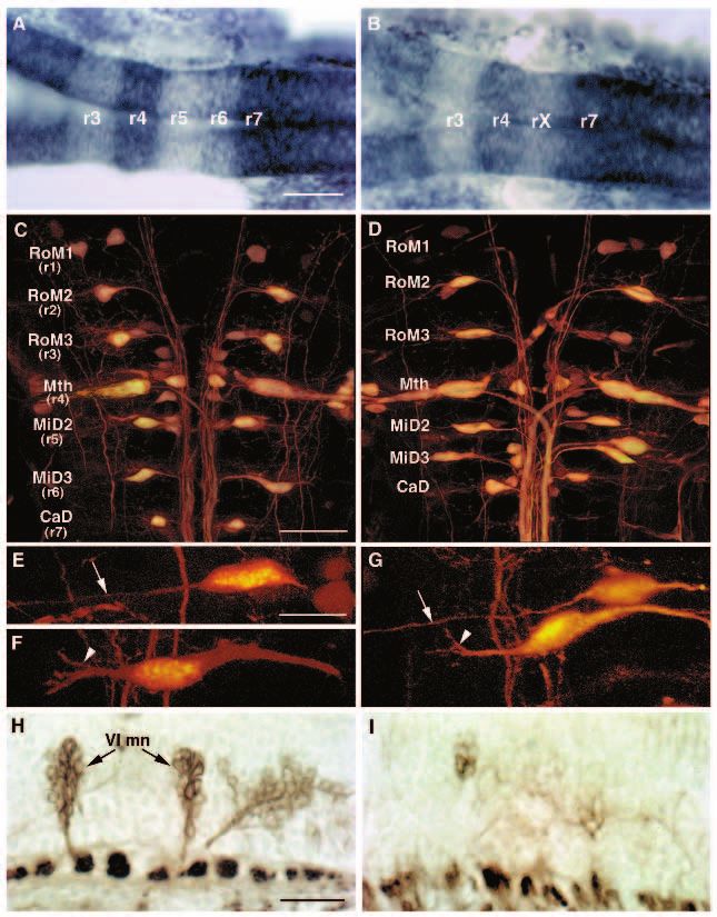
Fig. 1. krox20 expression
is disrupted in val−
embryos early during
hindbrain development.
We screened 472 ENU-
mutagenized and 741 γ-
ray-mutagenized haploid
genomes, and identified
three alleles of valentino,
one ENU-induced
(valb337) and two γ-ray-
induced (valb361 and
valb475).
(A-C) Whole-mount RNA
in situ hybridizations in
lateral view showing
expression of three genes,
shh, en3 and krox20, in 18
h wild-type (A), valb337
(B) and valb361 (C)
embryos. Anterior is to the left. In both alleles of valentino shown here, the r5 stripe of krox20 staining is reduced to a vestigial strip of
expression in the dorsal hindbrain at the position of the r4-r5 boundary (arrow). (D,E) Dorsal views of whole-mount RNA in situs showing
expression of krox20 in wild-type (D) and valb337 (E) embryos at the 2- to 3-somite stage (10J- 11 h). Anterior is to the top. In val− embryos,
krox20 expression is already disrupted in the presumptive r5. (F) Following RNA in situ hybridization at the 2- to 3-somite stage, embryos
from a cross between valb361/val+ individuals were sorted based on krox20 expression and then their genotype was determined by PCR using
snail2, which is deleted in valb361 (see Materials and Methods). 10/10 individuals scored as wild-type were in fact wild type as determined by
PCR (lanes 1-10) and 10/10 individuals scored as mutant were in fact mutant (lanes 11-20). Arrow: snail-2; arrowhead: nk2.2, an unlinked
gene that is amplified from both wild-type and mutant DNA. Scale bars, 50 µm.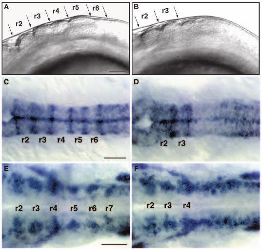
Hindbrain segmentation in valentino mutants 3983

Retrograde labeling of reticulospinal neurons                              similarly affected in val− embryos (data not shown); however,
5-day larvae were anesthetized and mounted in a drop of 1% agar            the r1- and r3-specific bands of rtk1, the r3-specific band of
made in Ringer’s solution. The tail was cut off at the level of the anus   krox20, as well as other markers of more anterior regions of
using spring scissors (Fine Science Tools) that had been dipped in a       the brain, are unaffected in val− embryos (Fig. 1 and data not
5% solution of LRD. Retrograde fills from this level of the spinal cord    shown).
are expected to result in labelling of the contralaterally projecting         valentino is required early in the segmentation of the pre-
MiD2c and MiD3c cells but not of the ipsilaterally projecting MiD2i        sumptive hindbrain, since the earliest known marker of seg-
or MiD3i cells (Metcalfe et al., 1986). Larvae were removed from the
agar and left to recover for 1 hour in Ringer’s solution before being
                                                                           mentation, krox20, is already disrupted in val− embryos at the
fixed overnight in 4% PFA. After fixation, the hindbrain was carefully     early somite stages. As early as krox20 expression is fully
removed, cleared stepwise in glycerol:PBS (50%, 70%, 90%), and             established in the presumptive r5, shortly after the end of gas-
mounted between 24×60 mm coverslips separated by the thickness of          trulation, it is reduced in 1/4 of the embryos produced in a cross
a single 22×22 mm coverslip. Images were obtained using a Zeiss 310        between heterozygotes (Fig. 1). To determine whether the
confocal microscope and were pseudocoloured using Voxelview 3-             embryos showing reduced krox20 expression in r5 at this stage
dimensional imaging software.                                              were indeed val−, embryos produced by crossing individuals
                                                                           heterozygous for the γ-ray-induced valb361 allele were sorted
Antibody staining
                                                                           based on their krox20 expression pattern at the 2- to 3-somite
16 µm cryostat sections were stained with the zn-5 antibody                stage (10J-11 h), and then were genotyped by PCR using the
(Trevarrow et al., 1990) using the indirect peroxidase anti-peroxidase
method (Hanneman et al., 1988).
                                                                           linked marker snail2, which is deleted in this deficiency (M.
                                                                           Gates and J. Postlethwait, personal communication; C. Moens
                                                                           and M. Giorgianni, unpublished results). We found that, at this
                                                                           stage, val− embryos could be reliably distinguished from their
RESULTS                                                                    wild-type siblings based on krox20 expression (Fig. 1F).

Identification of valentino
We performed an RNA in situ hybridization screen of haploid
zebrafish embryos to identify mutations that disrupt the region-
specific expression patterns of several marker genes in the
developing brain. Using this approach, we identified three
independent mutations in which the rhombomere 5 (r5)-
specific band of expression of krox20, a gene that is normally
expressed in r3 and r5 (Oxtoby and Jowett, 1993), was reduced
to a narrow strip of cells dorsal in the neural tube at the normal
position of the r4-5 boundary (Fig. 1). Complementation tests
and mapping showed that these mutations affect the same gene,
which we named valentino (val). The valb337 allele was iden-
tified among the haploid progeny of F1 females from N-ethyl-
N-nitrosourea (ENU)-mutagenized fish, and the valb361 and
valb475 alleles were identified among the haploid progeny of F1
females from γ-ray mutagenized fish. Both of the γ-ray-induced
mutations are deletions and at least one of them, valb475, con-
stitutes a valentino deficiency since it deletes genetic markers
on either side of valentino (C. Moens and M. Giorgianni,
unpublished results).
    All three val alleles are inherited in a Mendelian fashion as
recessive lethal traits. Embryos homozygous for the ENU-
induced allele die between 6 and 9 days after fertilization (d),
by which time they are edemic and have failed to form a swim               Fig. 2. val− embryos lack segment boundaries and segmental patterns
bladder. While the initial hindbrain defect caused by the γ-ray-           of neuronal differentiation posterior to rhombomere 4. (A,B) Lateral
induced mutations is identical to that caused by the ENU-                  view of live 18 h wild-type (A) and val− (B) embryos. Anterior is to
induced mutation (see below), embryos homozygous for the γ-                the left. In val− embryos, there are no visible rhombomere
ray-induced mutations die by 3 d. Since the phenotype of                   boundaries posterior to the r3-r4 boundary. (C,D) Dorsal views of
trans-heterozygous embryos (valb361/valb337 and valb475/valb337)           RNA in situ hybridizations of wild-type (C) and val− (D) embryos at
is identical to that of embryos homozygous for the ENU-induced             18 h showing expression of mariposa in the rhombomere boundaries.
allele, we infer that the ENU-induced allele is a null allele of           No expression is observed posterior to the r3-r4 boundary in val−
valentino, and that the earlier lethality caused by the γ-ray-             embryos. (E,F) Dorsal views of RNA in situ hybridizations of wild-
                                                                           type (E) and val− (F) embryos at 24 h showing expression of gap43
induced mutations is due to the deletion of other essential genes          in clusters of early differentiating neurons laterally in each
(see Materials and Methods). Except where specifically noted,              rhombomere. In val− embryos, this segmental pattern of gap43
the analysis presented below is of the ENU-induced allele.                 staining is lost posterior to r4. This disrupted pattern of neuronal
    All three val alleles were identified due to the disruption of         differentiation is also observed in val− embryos stained with the
the r5-specific band of krox20 expression described above. The             HNK-1 antibody (data not shown; Metcalfe et al., 1990; Trevarrow
r5-specific band of expression of rtk1 (Xu et al., 1994) is                et al., 1990). Scale bars, 50 µm.
3984 C. B. Moens and others

Earlier than this, val− embryos are more difficult to distinguish    localized cell death does not account for the observed reduction
from wild types because the initiation of r5-specific krox20         in hindbrain length in val− embryos, since we observe no dif-
expression in two lateral domains (Oxtoby and Jowett, 1993)          ference between wild-type and val− embryos that were treated
occurs to some extent in val− embryos.                               for the detection of programmed cell death at the 18-somite
                                                                     stage (data not shown; Gavrieli et al., 1992).
Hindbrain segmentation is disrupted in valentino                         We examined the valentino mutant phenotype at the single
mutants                                                              cell level by determining the positions of identifiable neurons
Although valentino was originally identified by RNA in               and neuronal cell types within the mutant hindbrain. The
situ hybridization, live mutant embryos have a transiently           reticulospinal neurons are a series of individually identifiable
visible phenotype during the period when rhombomeres are             neurons whose cell bodies form a ladder-like array corre-
visible. In wild-type embryos at the 18-somite stage (18 h), r2      sponding to the positions of the rhombomeres and which can
through r6 are visible as a series of prominent swellings, with      be visualized by the retrograde transport of lysinated
the otic vesicle lying lateral to r5. In val− embryos, the otic      rhodamine dextran from a spinal cord lesion (Kimmel et al.,
vesicle is reduced in size and no rhombomere boundaries are          1982; Metcalfe et al., 1986; Hanneman et al., 1988; see
visible posterior to the r3/r4 boundary, giving the posterior half   Materials and Methods). A subset of these (the ‘primary’
of the hindbrain a smooth appearance (Fig. 2A,B). The                reticulospinal neurons) undergo their final division before the
expression of mariposa, which is normally observed in rhom-          end of gastrulation (Mendelson, 1986) and transplantation
bomere boundaries (Y. Yan and J. Postlethwait, unpublished           experiments have revealed that they are committed to their par-
results), is altered in val− embryos in a manner that is consis-     ticular segmental identities well before rhombomeres are
tent with this loss of visible rhombomere boundaries. mariposa       visible (C. Moens, unpublished results). The most easily iden-
expression is normally observed in six stripes in the hindbrain      tifiable of the primary reticulospinal neurons are the large
at 18 h. In val− embryos, the three most posterior mariposa          Mauthner neurons, which differentiate bilaterally in r4. Other
stripes, corresponding to the r4/5, r5/6 and r6/7 boundaries, are    identifiable primary reticulospinal neurons differentiate in
absent (Fig. 2C,D).                                                  characteristic positions in r3 (the RoM3 cells), r5 (the MiD2
   The segmental pattern of neuronal differentiation normally        cells), r6 (the MiD3 cells) and r7 (the CaD cell; Fig. 3C,D).
observed during hindbrain development (Trevarrow et al.,             The MiD2cm cell is characterized by its medial position, its
1990) is also disrupted in val− embryos. Zebrafish gap43 is          rounded shape and its long, unbranched lateral dendrite (Fig.
expressed in a segmental pattern in the hindbrain at 24 h, in        3E). In contrast, the MiD3cm cell is characterized by its more
clusters of differentiating neurons that lie laterally in each       lateral position, its fusiform shape and its shorter, branched
rhombomere, as well as in the ganglia of the anterior and            lateral dendrite (Fig. 3F).
posterior lateral lines and trigeminal nerve (Fig. 2E; Reinhard          While the presence and position of these neurons are more
et al., 1994). While clusters of differentiating neurons are         variable in val− than in wild-type embryos, we identified
visible in r2, r3 and r4 of val− embryos, gap43-expressing cells     certain characteristic abnormalities (Fig. 3C-G; Table 1). The
posterior to r4 have lost their segmental organization (Fig. 2F).    distance from the Mauthner (r4) cell to the CaD (r7) cell, which
The disruption of segment boundaries and segmental patterns          is characterized by its dorsal and medial position, its rounded
of neuronal differentiation posterior to r4 in val− embryos          shape and the extensive arborization of its ventral dendrite (not
suggests that valentino affects not only r5 but also more            shown), is reduced on average to about 2/3 that observed in
posterior regions of the hindbrain.                                  wild-type siblings, again demonstrating that the posterior
                                                                     hindbrain is reduced by one rhombomere in val− embryos. Sur-
The hindbrain is reduced by the length of one                        prisingly, in light of the reduction of r5-specific gene
rhombomere in valentino mutants                                      expression, in most val− embryos both the MiD2cm and
We used region-specific RNA probes to further investigate the        MiD3cm cells are present and lie in the correct order. These
valentino mutant phenotype. In wild-type embryos, the
headache (hdc) gene is expressed in the spinal cord with a
anterior boundary of expression at the r6/7 boundary (A. Force,                                        Table 1
C. Dunn and J. Postlethwait, unpublished results), and the                                       Wild-type                       val−
g13.1 gene is expressed in r4 and anterior to the r2-3 boundary                           Avg. dist. ± SD              Avg. dist. ± SD
(Fig. 3A; B. Appel and J. Eisen, unpublished results). Double                                  (µm)           n*            (µm)           n
RNA in situ hybridizations using these probes show that the              RoM3-Mth             32±3.1          15           33±5.3          26
hindbrain is reduced by the length of approximately one rhom-            Mth-MiD2             26±2.7          16           27±6.1          23
bomere in val− embryos. In mutant embryos, the anterior                  Mth-MiD3             62±3.9          15           38±11.8         22
boundary of hdc expression is shifted towards the g13.1                  Mth-CaD              90±5.1          14           58±10.0         27
domain of expression in r4, leaving a region of one rhom-               *Refers to the number of unilateral measurements. Thus for one individual,
bomere’s length rather than two between r4 and r7. We term           n=2.
this single rhombomere-length unit ‘rX’ (Fig. 3B), and argue            The presence or absence of each class of reticulospinal neuron was
below that it corresponds to the domain that is normally sub-        determined in 45 mutant and 17 wild-type individuals. In 3/45 mutant
                                                                     embryos examined, we observed a unilaterally duplicated Mauthner neuron
divided and expanded into r5 and r6 in wild-type embryos. The        caudal to the normal Mauthner neuron. Such an event is observed in less than
expression boundaries of hdc and g13.1, which are normally           0.5% of wild-type embryos (Kimmel et al., 1978). In 11/45 mutant embryos
quite sharp, are diffuse where they border rX in val− embryos.       examined, we observed a unilateral loss of MiD2 neurons (0/17 wild-type),
This is consistent with the absence of rhombomere boundaries         and in 15/45 mutant embryos examined, we observed a unilateral loss of
in this region of the hindbrain in val− embryos. We note that        MiD3 neurons (0/17 wild-type).
Hindbrain segmentation in valentino mutants 3985
cells lie close together in the shortened interval between the did they undergo cell death, which is visible in mosaic embryos
CaD cell and the Mauthner cell (Fig. 3D,G). This interval cor- by the appearance of brightly labelled flecks of debris. Thus
responds to rX in Fig. 3B. val− cells appear to respond to the newly specified r5 and r6
The zn-5 antibody recognizes two clusters of efferent environments in a wild-type host by selective dispersal rather
neurons that lie ventrally and medially in r5 and r6. Based on than by selective cell death.
their position and axon projections, these neurons have been In the converse transplant experiment (schematized in Fig.
proposed to be the motor nuclei of the sixth (abducens) cranial 4K), wild-type cells contribute normally to the brain and spinal
nerve (Trevarrow et al., 1990). While these nuclei are easily cord of val− host embryos except when they lie in rX (Fig. 4L).
identifiable in sagittal sections of zn-5-stained wild-type Wild-type cells lying in rX do not extend from the pial surface
embryos at 3 days of development, they are rare or entirely to the ventricular surface of the hindbrain, and fail to divide
absent in val− embryos, as are their characteristic anterior-pro- across the midline, instead forming unilateral clumps of
jecting axons (Fig. 3H,I). The putative abducens motor nuclei rounded cells that appear to segregate away from the host cells.
differentiate relatively late during hindbrain development, This characteristic behaviour suggests that wild-type cells
since they first stain with zn-5 24 hours later than do the earlier lying in rX have a distinct identity from the surrounding mutant
differentiating hindbrain commissural neurons (Trevarrow et cells. Often clusters of wild-type cells come to lie at either end
al., 1990). Thus, although the primary reticulospinal neurons of rX, where rX borders r4 and r7. In these cases, clusters of
characteristic of r5 and r6 are usually present in val− embryos, wild-type cells lying ventral to the vestigial strip of krox20
at least one later-differentiating cell type characteristic of r5 expression that marks the anterior end of rX express krox20,
and r6 is absent. while the surrounding mutant cells do not (Fig. 4M). Thus, in
a mutant host embryo, wild-type cells respond to signals that
Mosaic analysis shows that valentino is required for specify r5 identity by autonomously expressing r5-specific
cells to contribute to r5 and r6 markers.
To determine which cells autonomously require valentino
function during hindbrain development, we transplanted cells
from labeled val− embryos into unlabeled wild-type hosts at DISCUSSION
the early gastrula stage (Fig. 4A; see Materials and Methods).
As an internal control, we also transplanted cells from a wild- We devised an RNA in situ hybridization screen in the
type donor labeled with a different fluorophor into the same zebrafish to identify mutations that disrupt the normal
wild-type host embryo. Both types of labeled cells were put segmental patterns of marker gene expression in the embryonic
into the region of the host gastrula that is fated to give rise to brain. Three alleles of valentino were identified in this screen
the hindbrain (Woo and Fraser, 1995), so that we could assess by their lack of all but a narrow dorsal strip of krox20
the distribution of mutant cells in the pharyngula-stage expression in r5. Mutations in valentino disrupt krox20
hindbrain (24 h). Wild-type cells contribute to the entire brain expression from the earliest time that it is established in r5 and
and spinal cord of wild-type hosts, where they form bilateral result in the absence of visible rhombomere boundaries and
groups of cells that extend from the ventricular to the pial boundary-specific gene expression posterior to the r3-4
surface (Fig. 4B). In contrast, we observed that val− cells trans- boundary. The normal segmental pattern of neuronal differen-
planted into the same wild-type host were specifically excluded tiation observed in the zebrafish hindbrain is also disrupted
from a sharply defined region in the hindbrain (Fig. 4C). posterior to r4 in val− embryos, consistent with experiments in
By identifying rhombomere boundaries in the mosaic the chick that showed that, in the absence of rhombomere
embryos by RNA in situ hybridization using krox20 or boundaries, cell mixing occurred between adjacent rhom-
mariposa and detecting the donor-derived cells immunohisto- bomeres (Guthrie et al., 1993). In as much as it disrupts the
chemically, we determined that the region from which val− process of segmentation itself, valentino is one of the relatively
cells are excluded in mosaic embryos corresponds precisely to small number of genes, including krox20, Hoxa-1 and kreisler,
r5 and r6 (Fig. 4D-F). Thus valentino is required cell- that have been shown to be required for hindbrain segmenta-
autonomously for cells to contribute to either r5 or r6, sug- tion in the mouse (Schneider-Maunoury et al., 1993; Swiatek
gesting that in the mutant itself there is no region with true r5 and Gridley, 1993; reviewed in Wright, 1993; also see below).
or r6 identity. We often observed mutant cells lying unilater- Based on our analysis of marker gene expression, of the
ally or bilaterally at the r5-r6 boundary in wild-type host positions of identified neurons and of genetic mosaics, we
embryos (Fig. 4C,E,F). propose that valentino is required cell-autonomously in a
In order to understand how val− cells come to be excluded process whereby a distinct region of the presumptive hindbrain
from r5 and r6 of a wild-type host, we followed their behaviour that we call a ‘protosegment’ corresponding to a two-rhom-
over time following transplantation. As early as the 7-somite bomere unit is subdivided and expanded into the definitive
stage (12.5 h), val− cells were observed to disperse from the rhombomeres 5 and 6 (Fig. 5).
presumptive r5 and r6, contributing instead to the flanking
rhombomeres (Fig. 4G). Mutant cells retracted first toward the ‘rX’: a distinct region in the val2 hindbrain that fails
lateral surface of the neural keel, and then gradually parted, to be subdivided and expanded into r5 and r6
leaving behind any cells that lay at the presumptive r5-6 The domains of marker gene expression and the positions of
boundary (Fig. 4H-J). Transplanted val− cells in the presump- the primary reticulospinal neurons indicate that the distance
tive r5 and r6 failed to complete a characteristic division across between r4 and r7 is reduced from two rhombomere lengths to
the midline that normally occurs at cell cycle 16 and generates one in val− embryos (summarized in Fig. 5). ‘rX’ is the region
bilateral pairs of sister cells (Kimmel et al., 1994), but neither that remains between, and fails to form boundaries with, r4 and3986 C. B. Moens and others Fig. 3. The hindbrain of val− embryos is reduced by the length of one rhombomere. (A,B) Dorsal view of 22 h wild-type (A) and val− (B) embryos showing expression of two genes: g13.1 in r4 and anterior to the r2-r3 boundary and hdc posterior to the r6-r7 boundary. Anterior is to the left. The distance between the posterior boundary of g13.1 expression in r4 and the anterior boundary of hdc expression is reduced by the length of approximately one rhombomere in the val− compared to the wild-type embryo. ‘rX’ refers to the region of that remains between r4 and r7. (C-G) Confocal images of 5-day old wild-type (C,E,F) and val− (D,G) embryos in which the hindbrain reticulospinal neurons are visualized by retrograde filling with lysinated rhodamine-dextran. Anterior is to the top. The names of individually identifiable neurons are indicated. In wild-type embryos, the RoM3 neurons lie in r3, Mauthner (Mth) in r4, MiD2 in r5, MiD3 in r6 and CaD in r7. In val− embryos, the average distance from Mth to CaD is reduced by the length of approximately one rhombomere, and the MiD2 and MiD3 neurons lie close together in the region of one rhombomere’s length between r4 and r7. (E- G) Higher power confocal images of MiD2 and MiD3 cells in wild-type (E,F) and mutant (G) embryos. Arrows indicate the long, unbranched lateral dendrite characteristic of the MiD2cm cell, while arrowheads indicate the shorter, branched lateral dendrite characteristic of the MiD3cm cell. Note that, although the MiD2cm and MiD3cm cells are immediately adjacent to one another in the mutant embryo shown in G, the MiD2cm cell is still anterior to the MiD3cm cell. (H,I) Sagittal sections of 56 h wild-type (H) and val− (I) embryos stained with the zn-5 antibody, which labels the putative motor nuclei of the abducens nerve (VI) in rhombomeres 5 and 6. Anterior is to the left. These motor nuclei are largely absent in the val− embryo. The putative abducens motor nuclei differentiate relatively late during hindbrain development, since they first stain with zn-5 24 hours later than do the earlier differentiating hindbrain commissural neurons (Trevarrow et al., 1990). Scale bars, (A,B) 50 µm; (C,D) 50 µm; (E-G) 20 µm; (H,I) 20 µm. r7. That this region is neither r5 nor r6, but has a distinct tities before rhombomeres are visible (C. Moens, unpublished identity is suggested by a number of lines of evidence. rX does results). not express r5-specific markers except in a narrow strip of The idea that rX of val− embryos is distinct from either r5 dorsal cells at the position where the r4-5 boundary would or r6 is strongly supported by our analysis of genetic mosaics normally form. Thus rX is not r5. However, the MiD2 reticu- (Fig. 4). When cells from a val− embryo at the early gastrula lospinal neuron, characteristic of r5, is generally found in rX stage are transplanted into a wild-type embryo at the same of val− embryos. As in the wild type, it lies anterior to the stage and the mosaic embryo is allowed to develop, mutant MiD3 reticulospinal neuron, which is characteristic of r6. We cells are specifically excluded from r5 and r6. Meanwhile, observe that later-differentiating r5- and r6-specific cell types wild-type cells transplanted into the same wild-type host con- are absent in val− embryos, as indicated by the absence of the tribute to the entire brain and spinal cord. Thus valentino is clusters of zn-5-positive cells which are the putative motor required cell-autonomously for cells to contribute to r5 and r6, nuclei of the sixth (abducens) cranial nerve. Thus rX has some suggesting that in the mutant embryo itself, there is no region but not all of the characteristics of both r5 and r6. That the with either r5 or r6 identity. Autonomy suggests that, in the MiD2 and MiD3 neurons are generally present and lie in the mutant embryo, cells fail to respond normally to regional dif- correct order along the anterior-posterior axis of val− embryos, ferences that are necessary for the specification of r5 and r6. if not in their correct positions, indicates that their specifica- That such regional differences do exist in val− embryos is tion is valentino-independent. It is even possible that they are supported by our analysis of genetic mosaics in which wild- specified before valentino functions: primary reticulospinal type cells are transplanted into a val− host. We observe that neurons are generated very early, before the end of gastrula- clusters of wild-type cells lying at the anterior end of rX in val− tion (Mendelson, 1986), and transplant experiments have embryos express the r5-specific marker krox20, but clusters shown that they are committed to their segment-specific iden- lying at the posterior end of rX do not. We predict that these
Hindbrain segmentation in valentino mutants 3987

Fig. 4. Mosaic analysis
demonstrates that valentino
is required for cells to
contribute to either r5 or r6.
(A) Schematic diagram of
the experimental approach
used for B-J. Rhodamine-
labeled cells from a val−
donor and fluorescein-
labeled cells from a wild-
type donor were transplanted
into the same unlabeled
wild-type host at the early
gastrula stage (see Materials
and Methods). The
distribution of labeled cells
was then observed at 24 h of
development.
(B,C) Confocal images taken
in dorsal view showing the
distribution of wild-type (B)
and val− cells (C) in the
hindbrain region of the same
wild-type host. val− cells are
specifically excluded from a
sharply defined region of the
hindbrain, but are otherwise
able to contribute to the
entire brain and spinal cord.
A single val− cell or small
cluster of val− cells is frequently observed at the center of this region (arrow in C). (D-F) krox20 staining (D,E) and mariposa staining (F) of
transplant recipients. Brown cells are donor-derived (see Materials and Methods). (D) The distribution of wild-type cells in the hindbrain of a
wild-type host. (E,F) The distribution of val− cells in a wild-type host, showing that mutant cells are specifically excluded from r5 and r6. The
embryo shown in E is the same embryo as is shown in B and C, and the cell noted in C is observed to lie at the r5-6 boundary (arrow in E). In
31 out of 39 genetically mosaic embryos analyzed in which transplanted cells had contributed to the hindbrain, labelled val− cells were
excluded from r5 and r6 although not necessarily from the boundary between them. In the remaining 8 mosaic embryos, val− cells were
observed in r5 and/or r6, but these cells were located ventrally, either in or very near the floor plate (not shown). In contrast, 94 out of 100
control embryos into which labeled wild-type cells had been transplanted had labeled cells distributed throughout the hindbrain, including r5
and r6. The remaining 6 control embryos had relatively few transplanted cells and these tended to be localized to rhombomere boundaries.
(G-J) A series of live images of rhodamine-labeled val− cells transplanted into a single wild-type host, shown in dorsal view. The time-points
shown are 7 somites (12 h; G), 10 somites (14 h; H), 14 somites (16 h; I) and 24 h (J). As early as the 7-somite stage, val− cells begin to be
excluded from the presumptive r5 and r6 of a wild-type host (indicated by a double arrow at the midline), with the exception of a few cells that
ultimately lie at the r5-6 boundary (arrowheads). (K) Schematic diagram of the experimental approach used for L and M. Rhodamine-labeled
cells from a wild-type donor were transplanted into a val− host embryo at the early gastrula stage. (L,M) Fluorescent and transmitted light
images of the same horizontal section of a mutant host embryo. Anterior is to the left. Donor-derived cells in this experiment are fluorescently
labelled. (L) Wild-type cells in a val− host form abnormal, unilateral clusters of rounded cells in rX, which is adjacent to the otic vesicle (ov),
while they otherwise contribute normally to the brain and spinal cord. (M) The more anterior of these clusters, which lies at the r4-rX
boundary, autonomously expresses krox20 although the surrounding mutant cells have failed to acquire r5 identity (arrow in L and M). Scale
bars: B,C, 100 µm; D-F, 50 µm; G-J, 100 µm; L,M, 50 µm.

posterior clusters instead express r6-specific markers. In these            chick that showed that boundary formation between adjacent
experiments, wild-type cells always segregate from the sur-                 rhombomeres requires an even-odd distinction (Guthrie and
rounding mutant cells in rX, indicating that their newly                    Lumsden, 1991), rX fails to form boundaries, either morpho-
acquired r5 or r6 identity is incompatible with that of rX.                 logically visible or as detected by mariposa expression, with
   What, then, is the identity of rX? We propose that during                r4 and r7.
normal development, a defined region or ‘protosegment’ in the                  Further analysis of the valentino gene will reveal more
presumptive hindbrain is subdivided and expanded into the                   precisely how it functions in the subdivision and expansion of
definitive rhombomeres r5 and r6 (Fig. 5A). In the absence of               r5 and r6 from their common precursor protosegment. Our
valentino, this protosegment fails to be subdivided into r5 and             results suggest that anterior-posterior differences exist within
r6, and fails to be expanded from one rhombomere’s length to                the protosegment independent of valentino, but that valentino
two, persisting instead as rX (Fig. 5B). We propose that rX                 is required within the protosegment for cells to respond to these
identity, although distinct, is earlier in a hierarchy of regional          differences by differentiating along r5- or r6-specific pathways.
identities than that of a definitive rhombomere. Thus rX is                 The valentino gene product could be required in the reception
neither ‘even’ nor ‘odd’. Consistent with experiments in the                or transduction of signals that specify these differences, or
3988 C. B. Moens and others

could act in concert with the products of genes that specify these        r6 of a wild-type host beginning at about the 7-somite stage,
differences, to drive r5- and r6-specific transcription. Alterna-         but valentino function is already required 1.5 hours earlier,
tively, valentino could be required for cells in the protosegment         when a disruption of the r5-specific expression of krox20 is
to become competent to respond to these differences.                      first detected in val− embryos. This difference may be arti-
                                                                          factual since earlier movement by some of the many val− cells
valentino mutant cells are excluded from wild-type                        initially present in the presumptive r5 and r6 may not have
r5 and r6 by selective dispersal, not selective cell                      been detected at our level of resolution. The difference may
death                                                                     also reflect a delay between the specification of rhombomere
The inability of val− cells to respond to regional differences            identity and the acquisition of cell surface properties that
that specify r5 and r6 results in their exclusion from r5 and r6          prevent cells with different regional identities from mixing.
in genetic mosaics. Beginning at approximately the 7-somite               It is also possible, however, that val− cells acquire r5 and r6
stage (12.5 h), val− cells begin to be excluded from the pre-             identity in a wild-type host but fail to maintain it, allowing
sumptive r5 and r6, migrating instead into the flanking rhom-             them to contribute transiently to the presumptive r5 and r6.
bomeres. Once there, they intercalate normally with the wild-             Indeed, valentino is not required for the initiation of r5-
type host cells, suggesting that valentino is not required for            specific krox20 expression, which occurs shortly before the
cells to take on r4 or r7 identity, and that val− cells remain            1-somite stage, since wild-type and val− embryos cannot be
uncommitted with respect to rhombomere identity until they                distinguished based on krox20 expression until the 2- to 3-
encounter signals to which they can respond. Thus the                     somite stage. This brief wave of valentino-independent r5 and
exclusion of val− cells from r5 and r6 involves selective cell            r6 identity in val− embryos may be sufficient to specify the
dispersal. These types of differential cell movements are rem-            MiD2 and MiD3 primary reticulospinal neurons, which have
iniscent of those observed when cells were transplanted                   undergone their final division by this time (Mendelson,
between odd- and even-numbered rhombomeres in the chick                   1986).
(Guthrie et al., 1993). These experiments suggested that dif-
ferential adhesiveness between cells from adjacent rhom-                  Similarities between valentino and kreisler mutants
bomeres could provide a mechanism for the maintenance of                  The val− phenotype is reminiscent of that of the mouse mutant,
rhombomere boundaries (Guthrie and Lumsden, 1991; Guthrie                 kreisler, first described by Hertwig (1944). Like valentino,
et al., 1993). However, we observe this selective dispersal of            kreisler mutant embryos lack morphological segmentation in
cells with different regional identities in our genetic mosaics           the hindbrain posterior to the r3-4 boundary (Deol, 1964). The
well before the appearance of visible rhombomere boundaries,              mouse kreisler gene has been cloned (Cordes and Barsh, 1994),
which occurs at about the 14-somite stage (16 h). Thus differ-            and we have evidence that valentino is its zebrafish homolog
ential adhesiveness between cells with different regional iden-           (C. Moens and S. Cordes, unpublished results). In spite of
tities may exist earlier than this mechanism has been proposed            striking similarities between the kreisler and valentino mutant
to account for the restriction of cell movement between rhom-             phenotypes, our interpretation of the valentino phenotype
bomeres in the chick (Guthrie and Lumsden, 1991; Guthrie et               differs from either of the current interpretations of the kreisler
al., 1993).                                                               phenotype (Cordes and Barsh, 1994; McKay et al., 1994). Both
                                                                          of these studies propose that the kreisler hindbrain consists of
Timing of valentino function                                              some combination of definitive rhombomeres. In contrast, our
Mutant cells appear to move out of the presumptive r5 and                 results suggest that in the valentino hindbrain r5 and r6 are

Fig. 5. Model for the function
of valentino in hindbrain                A) wild-type embryo                                   B) valentino mutant embryo
segmentation. In the wild-type
embryo (A), valentino is
                                                              AAA                                                   AAA
                                                              AAA                                                   AAA
required for the subdivision and

                                                              AAA                                                   AAA
expansion of a defined region
(or protosegment) in the
presumptive hindbrain (stippled                                     valentino                                           valentino
area), into rhombomeres 5 and
6, whose identities are distinct
                                                              AAA
                                                              AAAA
                                                               AAAAA                                                AAA
                                                              AAA
                                                               AAAA                                                 AAA
from that of the protosegment

                                                              AAA
                                                              AAAAAAA                                               AAA
(hatched rather than stippled)
and from each other (left
hatches versus right hatches). In            r1    r2    r3     r4     r5    r6 r7                 r1    r2   r3     r4     rX    r7
the mutant embryo (B), this                             g13.1                         hdc                    g13.1                         hdc
subdivision fails to happen and,                              krox20                                               krox20
as a result, the protosegment
persists as rX. In the genetic
mosaics, mutant cells are excluded from both r5 and r6 of a wild-type host because they are unable to make the transition from protosegment
identity to r5 or r6 identity, and, conversely, wild-type cells behave abnormally in rX of a mutant host because they have acquired an identity
(r5 or r6) which the surrounding mutant cells have failed to acquire. Coloured bars indicate the domains of expression of krox20 (yellow), hdc
(green) and g13.1 (red), and the blue vertical lines at the rhombomere boundaries indicate mariposa expression. The average positions of the
primary reticulospinal neurons are shown (black cells), as are the positions of the putative abducens motor nuclei (pink).
Hindbrain segmentation in valentino mutants 3989
replaced by a region of one rhombomere’s length that has a Program Long Term Fellowship. This work was supported by NIH
distinct and developmentally earlier identity than any defini- grants NS17963, NS23915, HD22486 and RR10715.
tive rhombomere.
Two-segment repeats REFERENCES
The patterns of neuronal differentiation, pharyngeal arch inner- Akam, M. (1987). The molecular basis for metameric pattern in the Drosophila
vation and neural crest migration observed in the chick embryo. Development 101, 1-22.
suggested the existence of a two-segment periodicity in the Akimenko, M. A., Ekker, M., Wegner, J., Lin, W. and Westerfield, M.
hindbrain (Lumsden and Keynes, 1989; Lumsden et al., 1991). (1994). Combinatorial expression of three zebrafish genes related to distal-
Transplantation experiments in the chick have supported this less: part of a homeobox gene code for the head. J. Neurosci. 14, 3475-3486.
Birgbauer, E. and Fraser, S. E. (1994). Violation of cell lineage restriction
hypothesis by showing that cells from alternate rhombomeres compartments in the chick hindbrain. Development 120, 1347-1356.
are more similar to each other in adhesive properties than they Cordes, S. P. and Barsh, G. S (1994). The mouse segmentation gene kr
are to cells from adjacent rhombomeres (Guthrie and Lumsden, encodes a novel basic domain-leucine zipper transcription factor. Cell 79,
1991; Guthrie et al., 1993). A two-segment periodicity is also 1025-1034.
Deol, M. S. (1964). The abnormalities of the inner ear in kreisler mice. J.
suggested by the observation that a number of genes, such as Embryol. Exp. Morph. 12, 475-490.
krox20, rtk1 and others, are expressed in alternate rhom- Ekker, M., Wegner, J., Akimenko, M. A. and Westerfield, M. (1992).
bomeres (Wilkinson et al., 1989a; Xu et al., 1994). Indeed, Coordinate embryonic expression of three zebrafish engrailed genes.
krox20 is required for the formation of two alternate rhom- Development 116, 1001-1010.
bomeres, r3 and r5, in the mouse (Schneider-Maunoury et al., Gavrieli, Y., Sherman, Y. and Ben-Sasson, S. A. (1992). Identification of
programmed cell death in situ via specific labeling of nuclear DNA
1993; Swiatek and Gridley, 1993). Furthermore, the anterior fragmentation. J. Cell. Biol. 119: 493-501.
boundaries of 3′-Hox gene expression largely conform to a Guthrie, S. and Lumsden, A. (1991). Formation and regeneration of
two-segment periodicity (Wilkinson et al., 1989b; reviewed in rhombomere boundaries in the developing chick hindbrain. Development
Keynes and Krumlauf, 1994). 112, 221-229.
Guthrie, S., Prince, V. and Lumsden, A. (1993). Selective dispersal of avian
Kimmel et al. (1985) noted a 2:1 correspondence between rhombomere cells in orthotopic and heterotopic grafts. Development 118,
the ‘segments’ defined by the positions of the cranial nerves 527-538.
and the segments defined by the reticulospinal neurons, and Guthrie, S. (1995). The status of the neural segment. Trends Neurosci. 18, 74-
suggested that neuromeres may originally have been 79.
assembled by a process of serial duplication (see also Stern, Hanneman, E., Trevarrow, B., Metcalfe, W. K., Kimmel, C. B. and
Westerfield, M. (1988). Segmental pattern of development of the hindbrain
1990). Our interpretation of the val− phenotype and of the and spinal cord of the zebrafish embryo. Development 103, 49-58.
results of our mosaic analysis support a model whereby seg- Hertwig, P. (1944). Die Genese der Hirn-und Gehörorganmißbildungen bei
mentation occurs hierarchically, with the hindbrain being rontgenmutierten Kreislermäusen. Z. KonstLehnre 28, 327-354.
divided into protosegments corresponding to two-rhombomere Ho., R. K. and Kane, D. A. (1990). Cell-autonomous action of zebrafish spt-1
mutation in specific mesodermal precursors. Nature 348, 728-730.
units which are then subdivided and expanded into the defini- Keynes, R. and Krumlauf, R. (1994). Hox genes and the regionalization of the
tive rhombomeres. The evidence presented here demonstrates nervous system. Annu. Rev. Neurosci. 17, 109-132.
such a process at work in the generation of rhombomeres 5 and Kimmel, C. B., Sessions, S. K. and Kimmel, R. J. (1978). Radiosensitivity
6, and it is possible that other, as-yet unidentified, genes and time of origin of Mauthner neuron in the zebrafish. Dev. Biol. 62, 526-
function in an analogous manner to valentino in other rhom- 529.
Kimmel, C. B., Powell, S. L. and Metcalfe, W. K. (1982). Brain neurons
bomere pairs. which project to the spinal cord in young larvae of the zebrafish. J. Comp.
Vaage (1969) noted that hindbrain segmentation in the chick Neurol. 205, 112-127.
occurred progressively, by the subdivision of three morpho- Kimmel, C. B., Metcalfe, W. K. and Schabtach, E. (1985). T reticular
logically visible ‘prorhombomeres’. However the two-rhom- interneurons: a class of serially repeating cells in the zebrafish hindbrain. J.
Comp. Neurol. 233, 365-376.
bomere unit in which valentino functions does not correspond Kimmel, C. B., Warga, R. M. and Kane, D. A. (1994). Cell cycles and clonal
either to Vaage’s prorhombomere B, which is subdivided into strings during formation of the zebrafish central nervous system.
r4 and r5, or prorhombomere C, which is subdivided into r6 Development 120, 265-276.
and r7. It may be that the timing of visible segmentation does Kimmel, C. B., Ballard, W. W., Kimmel, S. R., Ullmann, B. and Schilling,
not necessarily correspond to the timing of rhombomere spec- T. F. (1995). Stages of embryonic development of the zebrafish. Dev. Dyn.
203, 253-310.
ification. Interestingly, the two-rhombomere unit within which Krauss, S., Concordet, J. P. and Ingham, P. W. (1993). A functionally
valentino functions does correspond to the two-rhombomere conserved homolog of the Drosophila segment polarity gene hh is expressed
units defined by Hox gene expression (Keynes and Krumlauf, in tissues with polarizing activity in zebrafish embryos. Cell 75, 1431-1444.
1994). Mutations in valentino disrupt expression of krox20, Lumsden, A. and Keynes, R. (1989). Segmental patterns of neuronal
development in the chick hindbrain. Nature 337, 424-428.
which is known in turn to regulate Hox gene expression (Sham Lumsden, A., Sprawson, N. and Graham, A. (1991). Segmental origin and
et al., 1993; Nonchev et al., 1996). Taken together, these migration of neural crest cells in the hindbrain region of the chick embryo.
results suggest that valentino functions to regulate Hox gene Development 113, 1281-1291.
expression in the presumptive r5 and r6. McKay, I. J., Muchamore, I., Krumlauf, R., Maden, M., Lumsden, A. and
Lewis, J. (1994). The kreisler mouse: a hindbrain segmentation mutant that
We wish to thank Sharon Amacher for her help with the mosaic lacks two rhombomeres. Development 120, 2199-2211.
Mendelson, B. (1986). Development of reticulospinal neurons of the zebrafish.
analysis, and Will Talbot, Jim Langeland, Sharon Amacher, Bill
I. Time of Origin. J. Comp. Neurol. 251, 160-171.
Jackman, Bill Trevarrow, John Postlethwait, Judith Eisen and Victoria Metcalfe, W. K., Mendelson, B. and Kimmel, C. B. (1986). Segmental
Prince for their helpful comments on the manuscript. C. B. M. was homologies among reticulospinal neurons in the hindbrain of the zebrafish
supported by a Natural Sciences and Engineering Research Council larva. J. Comp. Neurol. 251, 147-159.
of Canada Postdoctoral Fellowship and a Human Frontier Science Metcalfe, W. K., Myers, P. Z., Trevarrow, B., Bass, M. B. and Kimmel, C.3990 C. B. Moens and others B. (1990). Primary neurons that express the L2/HNK-1 carbohydrate during Swiatek, P. J. and Gridley, T. (1993). Perinatal Lethality and defects in early development in the zebrafish. Development 110, 491-504. hindbrain development in mice homozygous for a targeted mutation of the Nonchev, S., Vesque, C., Maconochie, M., Seitanidou, T., Ariza- zinc finger gene Krox20. Genes Dev. 7, 2071-2084. McNaughton, L., Frain, M., Marshall, H., Sham, M. H., Krumlauf, R. Thisse, C., Thisse, B. and Postlethwait, J. H. (1995). Expression of snail2, a and Charnay, P. (1996). Segmental expression of Hoxa-2 in the hindbrain is second member of the zebrafish snail family, in cephalic mesendoderm and directly regulated by Krox-20. Development 122, 543-554. presumptive neural crest of wildtype and spadetail mutant embryos. Dev. Nüsslein-Volhard, C. and Wieschaus, E. (1980). Mutations affecting Biol. 172, 86-99. segment number and polarity in Drosophila. Nature 287, 795-801. Toyama, R., Curtiss, P. E., Otani, H., Kimura, M., Dawid, I. B. and Taira, Oxtoby, E. and Jowett, T. (1993). Cloning of the zebrafish krox-20 gene (krx- M. (1995). The LIM class homeobox gene lim5: implied role in CNS 20) and its expression during hindbrain development. Nucleic Acids Res. 21, patterning in Xenopus and zebrafish. Dev. Biol. 170, 583-593. 1087-1095. Trevarrow, B., Marks, D. L. and Kimmel, C. B. (1990). Organization of Postlethwait, J. H., Johnson, S. L., Midson, C. N., Talbot, W. S., Gates, M., hindbrain segments in the zebrafish embryo. Neuron 4, 669-679. Ballinger, E. W., Africa, D., Andrews, R., Carl, T., Eisen, J. S., et al. Vaage, S. (1969). The segmentation of the primitive neural tube in chick (1994). A genetic linkage map for the zebrafish. Science 264, 699-703. embryos (Gallus domesticus). Adv. Anat. Embryol. Cell Biol. 41, 1-88. Reinhard, E. Nedivi, E., Wegner, J., Skene, J. H. P. and Westerfield, M. Weinberg, E. S., Allende, M. L., Kelly, C. S., Abdelhamid, A., Murakami, (1994). Neural selective activation and temporal regulation of a mammalian T., Andermann, P., Doerre, O. G., Grunwald, D. J. and Riggleman, B. GAP-43 promoter in zebrafish. Development 120, 1767-1775. (1996). Developmental regulation of zebrafish MyoD in wildtype, no tail and Schneider-Maunoury, S., Topilko, P., Seitandou, T., Levi, G., Cohen- spadetail embryos. Development 122, 271-280. Tannoudji, M., Pournin, S., Babinet, C. and Charnay, P. (1993). Wilkinson, D. G., Bhatt, S., Cook, M., Boncinelli, E. and Krumlauf, R. Disruption of Krox-20 results in alteration of rhombomeres 3 and 5 in the (1989a). Segmental expression of Hox-2 homoeobox-containing genes in the developing hindbrain. Cell 75, 1199-1214. developing mouse hindbrain. Nature 341, 405-409. Sham, M. H., Vesque, C., Nonchev, S., Marshall, H., Frain, M., Gupta, R. Wilkinson, D. G., Bhatt, S., Chavrier, P., Bravo, R. and Charnay, P. D., Whiting, J., Wilkinson, D., Charnay, P. and Krumlauf, R. (1993). The (1989b). Segment-specific expression of a zinc-finger gene in the developing zinc finger gene Krox20 regulates HoxB2 (Hox2.8) during hindbrain nervous system of the mouse. Nature 337, 461-464. segmentation. Cell 72, 183-196. Woo, K. and Fraser, S. E. (1995). Order and coherence in the fate map of the Solnica-Krezel, L., Schier, A. F. and Driever, W. (1994). Efficient recovery zebrafish nervous system. Development 121, 2595-2609. of ENU-induced mutations from the zebrafish germline. Genetics 136, 1401- Wright, C. V. E. (1993). Hox genes and the hindbrain. Current Biology 3, 618- 1420. 621. Streisinger, G., Walker, C., Dower, N., Knauber, D. and Singer, F. (1981). Xu, Q., Holder, N., Patient, R. and Wilson, S. W. (1994). Spatially regulated Production of clones of homozygous diploid zebra fish (Brachydanio rerio). expression of three receptor tyrosine kinase genes during gastrulation in the Nature 291, 293-296. zebrafish. Development 120, 287-299. Stern, C. D. (1990). Two distinct mechanisms for segmentation? Seminars in Devel. Biol. 1, 109-116. (Accepted 4 September 1996)
You can also read