Immuno-PET imaging for non-invasive assessment of cetuximab accumulation in non-small cell lung cancer
←
→
Page content transcription
If your browser does not render page correctly, please read the page content below
Yamaguchi et al. BMC Cancer      (2019) 19:1000
https://doi.org/10.1186/s12885-019-6238-4

 RESEARCH ARTICLE                                                                                                                              Open Access

Immuno-PET imaging for non-invasive
assessment of cetuximab accumulation in
non-small cell lung cancer
Aiko Yamaguchi1,2†, Arifudin Achmad3,4,5†, Hirofumi Hanaoka1* , Yusri Dwi Heryanto3, Anu Bhattarai3, Ratianto3,
Erdene Khongorzul3, Rini Shintawati3,4, A. Adhipatria P. Kartamihardja3,4, Ayaka Kanai1, Yumi Sugo6,
Noriko S. Ishioka6, Tetsuya Higuchi3 and Yoshito Tsushima3

  Abstract
  Backgrounds: Overexpression of epidermal growth factor receptor (EGFR) has been established as a valid
  therapeutic target of non-small cell lung cancer (NSCLC). However, the clinical benefit of cetuximab as an EGFR-
  targeting drug is still controversial, partially due to the lack of effective means to identify suitable patients. This
  study aimed to investigate the potential of radiolabeled cetuximab as a non-invasive tool to predict cetuximab
  accumulation in NSCLC tumor xenografts with varying EGFR expression levels.
  Methods: The NSCLC tumors in model mice were subjected to in vivo biodistribution study and positron emission
  tomography (PET) imaging 48 h after injection of either 111In- or 64Cu-labeled cetuximab. The EGFR expression
  levels of NSCLC tumors were determined by ex vivo immunoblotting.
  Results: We found that tumors with high EGFR expression had significantly higher [111In]In-DOTA-cetuximab
  accumulation than tumors with moderate to low EGFR expression (P < 0.05). Strong correlations were found
  between [111In]In-DOTA-cetuximab tumor uptake and EGFR expression level (r = 0.893), and between [64Cu]Cu-
  DOTA-cetuximab tumor uptake with EGFR expression level (r = 0.915). PET imaging with [64Cu]Cu-DOTA-cetuximab
  allowed clear visualization of tumors.
  Conclusion: Our findings suggest that this immuno-PET imaging can be clinically translated as a tool to predict
  cetuximab accumulation in NSCLC cancer patients prior to cetuximab therapy.
                                                                                            64
  Keywords: Immuno-PET, Non-small cell lung cancer, Cetuximab,                                   Cu, Personalized medicine, EGFR

Background                                                                          mutation. However, acquired resistance to TKIs is common
Non-small cell lung cancer (NSCLC) remains a deadly can-                            and their modest effect in NSCLC patients without EGFR
cer worldwide, even with advances in treatment strategies                           mutation necessitates alternative therapeutic approaches
such as molecular targeted therapy and immunotherapy                                targeting EGFR [2].
[1]. Overexpression of epidermal growth factor receptor                                Cetuximab, a recombinant, human/mouse chimeric
(EGFR) plays a role in NSCLC, making anti-EGFR drugs an                             monoclonal antibody that specifically targets the extracellu-
attractive therapeutic option for this cancer. Tyrosine kin-                        lar domain of EGFR, has demonstrated favorable efficacy in
ase inhibitors (TKIs) targeting EGFR are currently recom-                           combination with platinum-based chemotherapies, but
mended as first-line therapy in patients with advanced                              identification of patients likely to benefit from these therap-
NSCLC harboring an EGFR tyrosine-kinase domain                                      ies remains challenging [3–5]. Studies suggest that strong
                                                                                    overexpression of EGFR rather than other factors including
                                                                                    KRAS mutation status is a determinant factor for the treat-
* Correspondence: hanaokah@gunma-u.ac.jp
†
 Aiko Yamaguchi and Arifudin Achmad contributed equally to this work.               ment efficacy of cetuximab in NSCLC patients. However, it
1
 Department of Bioimaging Information Analysis, Gunma University Graduate           is still unclear whether positivity in immunohistochemistry
School of Medicine, 3-39-22 Showa-machi, Maebashi 371-8511, Japan                   (IHC) or Fluorescent in situ Hybridisation (FISH) score
Full list of author information is available at the end of the article

                                      © The Author(s). 2019 Open Access This article is distributed under the terms of the Creative Commons Attribution 4.0
                                      International License (http://creativecommons.org/licenses/by/4.0/), which permits unrestricted use, distribution, and
                                      reproduction in any medium, provided you give appropriate credit to the original author(s) and the source, provide a link to
                                      the Creative Commons license, and indicate if changes were made. The Creative Commons Public Domain Dedication waiver
                                      (http://creativecommons.org/publicdomain/zero/1.0/) applies to the data made available in this article, unless otherwise stated.
Yamaguchi et al. BMC Cancer (2019) 19:1000 Page 2 of 8
and/or squamous histology can be reliably predictive, pre- CRL-5803), H1650 (adenocarcinoma; bronchoalveolar car-
sumably due to the heterogeneity of EGFR expression cinoma, ATCC CRL-5883), and HCC827 (adenocarcin-
within tumors and/or limitations related to biopsy-based oma, ATCC CRL-2868) were obtained from ATCC
assessment such as limited tissue sampling [6, 7]. Another (Manassas, VA, USA), and EBC1 (squamous cell lung car-
approach that could assess EGFR status within the entire cinoma, JCRB0820) was obtained from Japanese Collec-
tumor throughout the body could potentially provide more tion of Research Bioresources (Tokyo, Japan). All cell lines
comprehensive information to predict whether a patient were grown monolayers in RPMI 1640 medium (Wako,
will respond to cetuximab treatment. Osaka, Japan) supplemented with 10% heat-inactivated
Molecular imaging with radiolabeled antibodies, including FBS (Nichirei Bioscience, Tokyo, Japan) and 1% antibiotic
immuno-positron emission tomography (PET) imaging, can (0.1 mg/mL penicillin and 100 U/mL streptomycin,
provide quantitative information about antibody uptake at a Wako). The EGFR-null H520 cell line was used as a nega-
whole-body level in a non-invasive fashion [8]. Immuno- tive control to assess non-specific tumor uptake of radio-
PET has shown potential for the assessment of biomarker tracer. All cell lines were cultured in a humidified
expression status and/or prediction of clinical response [9, atmosphere comprising 5% CO2 and 95% air at 37 °C.
10]. Studies found a significant association between the Five-weeks-old female athymic Balb/c nude mice (17–20
tumor uptake of copper-64 (64Cu) labeled cetuximab g) were purchased from Japan CLEA (Tokyo, Japan) or
([64Cu]Cu-DOTA-cetuximab) and the expression levels of Japan SLC (Shizuoka, Japan) and allowed to acclimatize
EGFR protein in cervical cancer cell lines [11] and in xeno- for one week in the animal facility before any intervention
graft mouse models with various cancer types [12, 13]. By was initiated. Mice were socially housed (4–5 animals per
contrast, some studies have found disparity between the ex- cage) in cages in an air-conditioned room at 28 °C under a
pression levels of EGFR and tumor uptake of radiolabeled 12 h light/dark cycle with access to food and tap water ad
cetuximab in several tumor xenograft models from different libitum during all the experiment. Lung tumor xenografts
origins, implying the influence of other factors such as were prepared by subcutaneous injection of 5 × 10⁶ cells
pharmacokinetics and dynamics for cetuximab accumula- in 100 μL PBS suspension in the dorsal flank of the mice
tion in tumors [14, 15]. in awake. Mice were randomly assigned to each experi-
Considering the disease heterogeneity of NSCLC, the ap- ment when the tumor size become approximately 100–
plicability of [64Cu]Cu-DOTA-cetuximab for non-invasive 300 mm3 (2–4 week after the tumor implantation).
assessment of EGFR expression status in NSCLC warrants
further validation in pre-clinical models. In this study, we Westernblot analysis for EGFR expression
evaluated the usefulness of [64Cu]Cu-DOTA-cetuximab for Xenografts mice (n > 2 for each tumor) were euthanized
the selection of EGFR-overexpressing NSCLC tumors using by cervical dislocation and tumors were collected. The
xenograft mouse models with human NSCLC cell lines western blot analysis was performed according to the
having various EGFR protein expression levels. procedure previously described [12]. Anti-EGFR (#2232;
Cell Signaling, Beverly, MA, USA) or anti β-actin (clone
Methods AC-15; Sigma-Aldrich, Saint Louis, MO, USA) was used
Cetuximab was kindly provided by Merck KgaA (Darmstadt, as primary antibody. Membranes were visualized by
Germany). The bifunctional chelating agent p-SCN-Bn- scanning using the ImageQuant™ LAS 4010 imager (GE
DOTA, or 2-(4-isothiocyanatobenzyl)-1,4,7,10-tetraazacyclo- Healthcare, Piscataway, NJ, USA) and the obtained
dodecane-1,4,7,10-tetraacetic acid, was purchased from bands were densimetry analyzed using ImageJ 1.47 soft-
Macrocyclics (Dallas, TX, USA). Copper-64 (150–300 MBq) ware. The data were normalized over EGFR-null control
was produced on a biomedical cyclotron CYPRIS HM-18 H520. Based on the adjusted band density, the EGFR ex-
(Sumitomo Heavy Industries Ltd., Tokyo, Japan) at Gunma pression levels were classified in three categories; > 20:
University Hospital. Indium-111, in form of InCl3 (74 MBq/ highly-overexpressing, 10 to 20: high, 1 to 10: low to
mL) was obtained from Nihon Medi-Physics (Tokyo, Japan). moderate.
Cell lines and xenografts DOTA conjugation
The animal studies were performed in accordance with Cetuximab (2 mg/mL) was buffer-exchanged into borate-
our institutional guidelines and were approved by the buffered saline (0.1 M, pH 8.5) in Vivaspin (Sartorius Stedim
Local Animal Care Committee of Gunma University (ap- Biotech, Aubagne, France). To the concentrated cetuximab
proval number: 17–035). Human NSCLC cell lines H358 was added p-SCN-Bn-DOTA dissolved in N,N-dimethylfor-
(bronchioalveolar carcinoma, ATCC CRL-5807), H441 mamide (10:1 to cetuximab). The resulting mixture was in-
(papillary adenocarcinoma, ATCC HTB-174), H460 (large cubated overnight at 37 °C, and then unconjugated DOTA
cell lung cancer, ATCC HTB-177), H520 (squamous cell was removed by using size-exclusion column (Bio-spin 6
carcinoma, ATCC HTB-182), H1299 (carcinoma, ATCC Tris column, Bio-Rad Laboratories, Hercules, CA, USA) andYamaguchi et al. BMC Cancer (2019) 19:1000 Page 3 of 8
ultrafiltration (Vivaspin). The protein concentration of the Inveon, Siemens, Knoxville, TN, USA). To obtain images
resulting DOTA-cetuximab was determined by using a with equivalent qualities, the acquisition time was ad-
Nanodrop spectrophotometer (Thermo Scientific, Wilming- justed based on the activity dose. The acquisition time
ton, DE, USA). was 20 min, 60 min, or 120 min for 20 MBq, 4 MBq, or
The immunoreactivity of DOTA-cetuximab was evalu- 2–3 MBq activity dose, respectively. In all studies, PET
ated in a NSCLC cell line (H460) according to a method scanning was performed with animals over a heating pad
described previously [16]. No significant effect of heated at 37 °C. After the PET scan, mice were eutha-
DOTA-conjugation was observed. nized by cervical dislocation. The energy window was
set between 350 and 650 keV. The imaging data were re-
64 111
Preparation of Cu or In-labled DOTA-cetuximab constructed using a 3-D ordered-subsets expectation
For Cu labeling, DOTA-cetuximab (500 μg) was dis-
64
maximization algorithm. Attenuation correction and
solved in sodium acetate buffer (0.25 M, pH 5.5) and then scattering correction were not applied. All images were
added to the dried 64CuCl2 (150–300 MBq). The resulting quantified for tumor radiotracer uptake using the Inveon
mixture was incubated for 1 h at 40 °C. An aqueous solu- Research Workplace workstation (Siemens). Uptake of
tion of EDTA (100 mM, 5 μL) was added to quench the [64Cu]Cu-DOTA-cetuximab in the tumor was expressed
unconjugated 64Cu. Purification of [64Cu]Cu-DOTA- as average of standardized uptake values (SUVmean).
cetuximab was carried out using PD-10 desalting column The SUV was determined by using the following equa-
(GE Healthcare). [111In]In-DOTA-cetuximab was ob- tion: SUV = activity in a ROI (MBq/cc)/[injected dose
tained by a similar procedure. The radiochemical yield (MBq)/body weight (g)]. Region of interests (ROI) were
and radiochemical purity were determined using an in- manually drawn to contour the tumors three times each
stant TLC developed with saline. After purification, the by two investigators (HH, 10 years of experience, AK2, 1
radiochemical purities of both [64Cu]Cu-DOTA-cetuxi- year of experience). Tumor outlines were defined by the
mab and [111In]In-DOTA-cetuximab were more than pixel containing SUV higher than 0.6. There was no no-
99%. The specific activity of the final product per milli- ticeable difference in intra- and interobserver variability.
gram cetuximab was 2–3 MBq for 111In. Due to the
varying radiochemical yields, the specific activity of Statistical analysis
[64Cu]Cu-DOTA-cetuximab ranged 30–200 MBq/mg. The GraphPad Prism software (GraphPad Software, La
Jolla, CA, USA) was used for statistical analysis. Data are
Biodistribution study expressed as means ± SDs where appropriate. In the bio-
To detect and examine in vivo behavior of radiolabeled- distibution study with [111In]In-DOTA-cetuximab, com-
cetuximab in xenografts with various lung cancer cell lines, parison of means was performed using one-way
[111In]In-DOTA-cetuximab (protein dose: 20 μg) were intra- ANOVA followed by Tukey’s test. For the comparison
venously injected with 30 kBq via the tail vein in awake mice between tumor [111In]In-DOTA-cetuximab uptake,
(n ≥ 5 for each group). Mice were euthanized by decapita- [64Cu]Cu-DOTA-cetuximab uptake and adjusted EGFR
tion at 48 h after injection. Tissues of interest were collected band density, simple correlations between variables were
and weighed, and the radioactivity was measured using an analyzed using Pearson’s correlation coefficient. P-values
automated γ-counter ARC-7001 (Hitachi Aloka Medical, < 0.05 were considered statistically significant.
Tokyo, Japan). The radiotracer uptake was expressed as per-
centage of injected dose/g of tissue (%ID/g). Results
EGFR expression in NSCLC cell lines
PET imaging Immunoblot analysis of the xenograft tumors showed
To study PET usefulness for the assessment of EGFR ex- that the eight NSCLC cell lines express various levels of
pression level in vivo, xenografts of lung cancer cell lines EGFR (Fig. 1). Semi-quantitative confirmation of EGFR
(n = 2) were intravenously injected with 2–20 MBq expression showed that HCC827 surpasses the rest
[64Cu]Cu-DOTA-cetuximab via the tail vein in awake. (EGFR band density of 25.0, relative to EGFR-null
To minimize the influence of specific activity, protein H520), followed by H1650 and EBC-1 (13.5 and 10.9, re-
dose was fixed to 100 μg by the addition of non- spectively). Other EGFR-positive cell lines showed mod-
radiolabeled cetuximab. Considering the pharmacokinet- est expression levels ranging from 6.53 to 8.86.
ics of monoclonal antibody [8] and the physical half-life
of 64Cu (12.7 h), PET images were taken 48 h after injec- Biodistribution of [111In]in-DOTA-cetuximab
tion. After anesthetization by isoflurane inhalation (2.5% Figure 2 summarizes [111In]In-DOTA-cetuximab up-
in an air mixture), mice were imaged with a small- takes in major organs and tumors 48 h after injection in
animal PET scanner (Transaxial FOV: 10 cm, axial FOV: NSCLC tumor xenografts (n = 5 to 7/group). The tumor
12.7 cm, resolution at the center of FOV: 1.4 mm, uptake levels of [111In]In-DOTA-cetuximab in EGFR-Yamaguchi et al. BMC Cancer (2019) 19:1000 Page 4 of 8 Fig. 1 Epidermal growth factor receptor (EGFR) expression in eight non-small cell lung cancer (NSCLC) cell lines. Densitometric intensities of EGFR are presented as folds relative to H520 band density (1.0). β-actin was used as a loading control positive xenografts were significantly higher than that of uptakes of H358 (n = 6), H441 (n = 6), H460 (n = 6), and EGFR-null H520 tumors (P < 0.05). The uptake level of H1299 (n = 6) tumors were 15.1 ± 0.96%ID/g, 14.0 ± [111In]In-DOTA-cetuximab in the EGFR highly- 3.77%ID/g, 13.2 ± 5.56%ID/g, and 16.7 ± 3.46%ID/g, re- overexpressed HCC827 tumor (n = 7) was significantly spectively. Accumulation in normal organs was similar higher than those in other cell lines except for EBC-1 in all groups. (26.9 ± 3.10%ID/g, P < 0.05). Although large variation was observed, EBC-1 xenografts (n = 5), which had rela- PET imaging of [64Cu]cu-DOTA-cetuximab tively high EGFR expression levels, showed [111In]In- The whole-body distribution and tumor-targeting effi- DOTA-cetuximab uptake levels significantly higher than ciency of cetuximab was visualized non-invasively using xenografts with low to moderate EGFR expression levels small-animal PET imaging at 48 h after injection of (H358, H441, H460) (23.3 ± 7.63%ID/g, P < 0.05). H1650 [64Cu]Cu-DOTA-cetuximab in mice with NSCLC xeno- also showed a relatively high uptake level of [111In]In- grafts (n = 2 per group). As shown in Fig. 3, PET imaging DOTA-cetuximab (18.4 ± 3.59%ID/g, n = 5) although the clearly depicted the high uptake of [64Cu]Cu-DOTA- difference was not statistically significant compared to cetuximab in HCC827 xenografts, which highly overex- the cell lines with moderate to low EGFR expression press EGFR. Quantification of the PET images revealed levels (H358, H441, H460, and H1299). The other tu- that tumor uptakes (SUVmean of [64Cu]Cu-DOTA- mors derived from 4 NSCLC cell lines with moderate to cetuximab) in HCC827 tumors were exceptionally high low EGFR expression levels showed comparable uptake (3.17 and 4.41). Although H1650 showed relatively high levels of [111In]In-DOTA-cetuximab. The radioactivity SUVmean values (1.93 and 2.17), other xenografts of low Fig. 2 Biodistribution of [111In]In-DOTA-cetuximab at 48 h in mice xenograft models with non-small cell lung cancer (NSCLC) tumors with varying epidermal growth factor receptor (EGFR) expression levels. Each data point represents the mean ± SD of n = 5 to 7 per tumor model
Yamaguchi et al. BMC Cancer (2019) 19:1000 Page 5 of 8
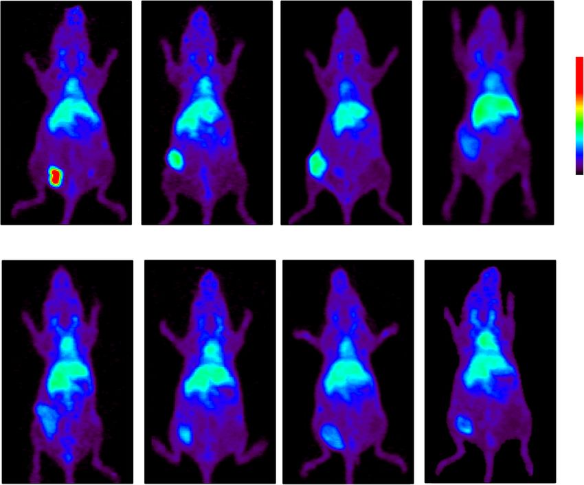
Fig. 3 Representative [64Cu]Cu-DOTA-cetuximab positron emission tomography (PET) images in mice xenograft models with non-small cell lung
cancer (NSCLC) tumors with varying epidermal growth factor receptor (EGFR) expression levels at 48 h. Arrows indicate the location of tumors
to moderate EGFR expression showed SUVmean values both graphs indicate the positive correlation between
comparable to those of EGFR-null H520 (range 0.87– variables. Tumor uptake of [111In]In-DOTA-cetuximab
1.66, number of mice analyzed: 10/10). uptake strongly correlated with the adjusted EGFR
band density (r = 0.893, p < 0.005). A strong correlation
Comparison of radiolabeled cetuximab tumor uptake was also found between tumor uptake of [64Cu]Cu-
with EGFR expression level DOTA-cetuximab and adjusted EGFR band density
Figure 4 shows the relationship between tumor (r = 0.915, p < 0.005). A moderate correlation was ob-
[111In]In-DOTA-cetuximab uptake or [64Cu]Cu- served between [111In]In-DOTA-cetuximab tumor up-
DOTA-cetuximab uptake and adjusted EGFR band take and [64Cu]Cu-DOTA-cetuximab tumor uptake
density. The linear relationships with a positive slope in (r = 0.694).
Fig. 4 Correlation between epidermal growth factor receptor (EGFR) expression and tumor uptake of radiolabeled cetuximab. (a) A significant
correlation of EGFR expression level and tumor uptake of [111In]In-DOTA-cetuximab was noted within tumor models of non-small cell lung
cancer. Each data represents the mean ± SD of n = 5 to 7 per tumor model. (b) Tumor uptake of [64Cu]Cu-DOTA-cetuximab in positron emission
tomography (PET) (mean standardized uptake values [SUV mean]) is correlated with the EGFR expression level. Each data point represents
SUVmean of each NSCLC tumor (8 cell lines, n = 2 per group) left panel: (a), right panel (b)Yamaguchi et al. BMC Cancer (2019) 19:1000 Page 6 of 8 Discussion studies have been demonstrated that SUV values for This study demonstrated the association between NSCLC tumor uptake of radiolabeled antibodies vary widely tumor uptake of radiolabeled cetuximab and EGFR pro- among lesions [9, 21]. Although clinical confirmation is tein expression levels in a xenograft mouse model. Distin- necessary, it is highly likely that [64Cu]Cu-DOTA-cetuxi- guishably high accumulation of radiolabeled cetuximab mab shows SUVmean values of NSCLC tumors in a was noted in EGFR-highly-overexpressing tumors in com- range sufficient to differentiate cetuximab responsive parison to the tumors with low and moderate EGFR ex- cases from non-responsive cases. pression levels, allowing for clear visualization of the Another factor limiting the range of SUVmean values tumor in PET imaging. These results suggest the potential would be the low specific activity of [64Cu]Cu-DOTA- usefulness of cetuximab immuno-PET for non-invasive cetuximab (4.5–30 GBq/μmol). This necessitated high pro- prediction of cetuximab uptake in NSCLC. tein dose (100 μg/head) administration in the PET study, The biodistribution study with [111In]In-DOTA-cetuxi- which may have competitively inhibited the radiotracer up- mab suggested that radioactivity uptake in the NSCLC take due to receptor saturation in low-EGFR-expressing tu- xenograft tumors sharply reflects the corresponding EGFR mors [22–24]. This difference may also account for the protein expression levels. This result is in line with previous discrepancy in the radiotracer tumor uptake between the studies of radiolabeled cetuximab employed in colorectal biodistiribution study and PET imaging, which showed only cancer models [12, 17]. The significantly high uptake of a moderate correlation. Tamura et al. [25] reported much [111In]In-DOTA-cetuximab in EGFR-highly-overexpressing higher specific activity of [64Cu]Cu-DOTA-trastuzumab HCC827 in comparison with xenograft tumors with low to (350 GBq/μmol). These results indicate that optimization of moderate EGFR expression levels suggests the potential of the 64CuCl2 production process is necessary to obtain clin- [64Cu]Cu-DOTA-cetuximab immuno-PET for detection of ically acceptable specific activity of [64Cu]Cu-DOTA-cetux- EGFR expression status in NSCLC. The large variation in imab in our facility. However, the low specific activity [111In]In-DOTA-cetuximab uptake observed within EBC-1 would not affect clinical evaluation of radiotracer uptake xenografts is perhaps due to the inter-subject heterogeneity because high amount of protein (up to 50 mg) are required of EGFR expression levels. Further investigation such as a to prevent rapid clearance and allow for tumor accumula- side-by-side comparison between [111In]In-DOTA-cetuxi- tion of radiolabeled antibodies [21, 26]. mab distribution and EGFR expression ex vivo might pro- Although several large clinical NSCLC trials evaluating vide some clues; however, it is beyond the scope of the the efficacy of chemotherapy plus cetuximab suggested current study. Nevertheless, this inter-subject difference in the value of a high EGFR expression level determined by [111In]In-DOTA-cetuximab uptake indicates that radiola- quantitative analysis of IHC or FISH as a predictive bio- beled cetuximab potentially reflects the inter- or intra- marker, the robustness of this theory is still controversial patient heterogeneity of EGFR expression level. [3, 27, 28]. A recently closed, large, randomized phase-3 The quantitative analysis of the PET study showed a trial (SWOG S0819) failed to show the clinical benefit of strong correlation with the adjusted EGFR band density, the addition of cetuximab to platinum-based chemother- which is in agreement with the biodistribution study. apy even within the patient subgroup with high EGFR HCC827 tumors showed exceptionally high SUVmean FISH scores [5]. The use of IHC or FISH scores has sev- levels compared to other tumors, which reinforced the eral concerns such as the definition of positive test results, potential of [64Cu]Cu-DOTA-cetuximab-PET for the reproducibility, and the discrepancy between biomarker identification of NSCLC patients suitable for cetuximab status determined based on IHC and FISH scores [7], therapy. Meanwhile, there was a small variation in SUV- which may have contributed to the subgroup misclassifi- mean values between EGFR low to high expressing tu- cation and led to the controversial results among studies. mors. Since various factors may influence tumor uptake Unlike histological analysis, immuno-PET can non- of radiolabeled antibodies—such as extravasation from invasively reflect dynamic biomarker status at the whole- tumor capillaries, diffusion and binding within the body level. In case of NSCLC, PET imaging with tumor interstitium, plasma clearance, internalization, [64Cu]Cu-DOTA-cetuximab by itself could predict the re- and catabolism in tumor cells [18, 19], and morpho- sponse to therapy as it is indicative of cetuximab tumor logical or physical barriers—specific binding to the anti- accessibility while the therapeutic response are not associ- gen may not be the dominant factor determining ated with biomolecular characteristics such as the status cetuximab uptake level in NSCLC tumor with EGFR ex- of KRAS, PTEN, and EGFR mutations. Therefore, pression on its cell surface below a certain level. Indeed, addition of immuno-PET scanning using [64Cu]Cu- H520 cells form highly vascularized tumors [20]. In DOTA-cetuximab to the workup protocol may help in- addition, because SUV is standardized for body weight, crease the accuracy of biomarker status assessment in the numerical range of SUV values in mice are not NSCLC patients. Comparative studies to evaluate the pre- equivalent to those in human. A number of clinical dictive values of IHC and/or FISH scores separately or in
Yamaguchi et al. BMC Cancer          (2019) 19:1000                                                                                              Page 7 of 8

combination with immuno-PET based analysis are re-                             Availability of data and materials
quired to test this hypothesis.                                                The datasets used and/or analysed during the current study are available
                                                                               from the corresponding author on reasonable request.
   There are several limitations in our study. First, xeno-
grafts of established human tumor cell lines may have
                                                                               Ethics approval
different characteristics from those of primary human                          The animal studies were performed in accordance with our institutional
tumors, and thereby EGFR expression levels determined                          guidelines and were approved by the Local Animal Care Committee of
here is not directly comparable to the EGFR-positivity                         Gunma University (approval number: 17–035).
criteria commonly used in clinical setting. Further stud-
ies in more clinically relevant models are warranted [4].                      Consent for publication
                                                                               Note applicable.
In addition, the predictive value of [64Cu]Cu-DOTA-
cetuximab immuno-PET for cetuximab treatment was
                                                                               Competing interests
not determined. Finally, although suitable main positron                       The authors declare that they have no competing interests.
energy of 64Cu (653 keV) can provide PET images with
spatial resolution better than 86Y (Emax = 3.1 MeV, with                       Author details
                                                                               1
                                                                                Department of Bioimaging Information Analysis, Gunma University Graduate
an additional γ-emission of 1.08 MeV (83% abundance)),                         School of Medicine, 3-39-22 Showa-machi, Maebashi 371-8511, Japan.
another positron emitter suitable for antibody imaging                         2
                                                                                Present address: Texas Therapeutics Institute, The Brown Foundation
[22], there are several concerns regarding the use of                          Institute of Molecular Medicine, The University of Texas Health Science
64                                                                             Center at Houston, 1881 East Road, Houston, TX 77054, USA. 3Department of
   Cu for imaging tumor uptake of antibodies, such as its                      Diagnostic Radiology and Nuclear Medicine, Gunma University Graduate
limited availability and relatively short half-life (12.7 h).                  School of Medicine, 3-39-22 Showa-machi, Maebashi 371-8511, Japan.
                                                                               4
Detection of lung legions might be hindered by accumu-                          Present address: Department of Nuclear Medicine and Molecular Imaging,
                                                                               Faculty of Medicine, Universitas Padjadjaran, Bandung, West Java 40161,
lation of radioactivity in the liver due to the relatively                     Indonesia. 5Oncology and Stem Cell Working Group, Faculty of Medicine,
weak in vivo stability of [64Cu]Cu-DOTA. But the use of                        Universitas Padjadjaran, Bandung, West Java 40161, Indonesia. 6Project
64
   Cu is a valuable option especially in facilities at which                   “Medical Radioisotope Application”, Department of Radiation-Applied Biology
                                                                               Research, Takasaki Advanced Radiation Research Institute, Quantum Beam
other longer-lived positron emitter isotopes such as 89Zr                      Advanced Research Directorate, National Institutes for Quantum and
(398 keV, t1/2 = 3.3 d) and 86Y are not available.                             Radiological Science and Technology, 1233 Watanuki, Takasaki 370-1292,
                                                                               Japan.

Conclusion                                                                     Received: 11 July 2019 Accepted: 1 October 2019
This study has demonstrated that cetuximab uptake in
NSCLC tumors can be assessed by PET using 64Cu-la-
beled cetuximab. Significantly high uptake of [64Cu]Cu-                        References
                                                                               1. Siegel RL, Miller KD, Jemal A. Cancer statistics, 2018. CA: A Cancer Journal
DOTA-cetuximab was noted in NSCLC tumors with                                      for Clinicians. 3rd ed. 2018;68:7–30.
very high EGFR expression levels compared to tumors                            2. Herbst RS, Morgensztern D, Boshoff C. The biology and management of
with medium or low EGFR expression levels. These re-                               non-small cell lung cancer. Nature. 2018;553:446.
                                                                               3. Pujol J-L, Pirker R, Lynch TJ, Butts CA, Rosell R, Shepherd FA, et al. Meta-
sults suggest that immuno-PET with [64Cu]Cu-DOTA-                                  analysis of individual patient data from randomized trials of chemotherapy
cetuximab may provide additional information for selec-                            plus cetuximab as first-line treatment for advanced non-small cell lung
tion of patients with advanced NSCLC most likely to                                cancer. Lung Cancer. 2014;83:211–8.
                                                                               4. Amendt C, Staub E, Friese-Hamim M, Storkel S, Stroh C. Association of EGFR
benefit from cetuximab treatment.                                                  expression level and Cetuximab activity in patient-derived Xenograft
                                                                                   models of human non-small cell lung Cancer. Clin Cancer Res. 2014;20:
Abbreviations                                                                      4478–87.
DOTA or p-SCN-Bn-DOTA: 2-(4-isothiocyanatobenzyl)-1,4,7,10-                    5. Herbst RS, Redman MW, Kim ES, Semrad TJ, Bazhenova L, KO MD, et al.
tetraazacyclododecane-1,4,7,10-tetraacetic acid; EGFR: Epidermal growth            Cetuximab plus carboplatin and paclitaxel with or without bevacizumab
factor receptor; IHC: Immunohistochemistry, FISH: Fluorescent in situ              versus carboplatin and paclitaxel with or without bevacizumab in advanced
Hybridisation; NSCLC: Non-small cell lung cancer; PET: Positron Emission           NSCLC (SWOG S0819): a randomised, phase 3 study. Lancet Oncol. 2018;19:
Tomography; ROI: Region of interest; SUV: Standardized uptake value;               101–14.
TKI: Tyrosine kinase inhibitor                                                 6. Jamal-Hanjani M, Wilson GA, McGranahan N, Birkbak NJ, Watkins TBK,
                                                                                   Veeriah S, et al. Tracking the evolution of non–small-cell lung Cancer. N
Acknowledgements                                                                   Engl J Med. 2017;376:2109–21.
We thank Mr. Takashi Ogasawara (Cyclotron Facility, Gunma University           7. Chae YK, Arya A, Chiec L, Shah H, Rosenberg A, Patel S, et al. Challenges
Hospital) for the production of 64Cu.                                              and future of biomarker tests in the era of precision oncology: can we rely
                                                                                   on immunohistochemistry (IHC) or fluorescence in situ hybridization (FISH)
Authors’ contributions                                                             to select the optimal patients for matched therapy? Oncotarget. 2017;8:
AY, AA, and HH participated in the design of the study. Data acquisition was       100863–98.
done by AY, AA, YH, AB, Ratianto, EK, RS, AK1, and HH. Data analysis was       8. van Dongen GAMS, Visser GWM, Lub-de Hooge MN, de Vries EG, Perk LR.
done by AY, AA, AK2, and HH. AY drafted the manuscript. AA and HH revised          Immuno-PET: a navigator in monoclonal antibody development and
the manuscript critically. TH, YS, NI and TY contributed reagents/materials/       applications. Oncologist. 2007;12:1379–89.
analysis tools. All authors read and approved the final manuscript.            9. Bensch F, van der Veen EL, Lub-de Hooge MN, Jorritsma-Smit A, Boellaard R,
                                                                                   Kok IC, et al. 89Zr-atezolizumab imaging as a non-invasive approach to
Funding                                                                            assess clinical response to PD-L1 blockade in cancer. Nat Med. 2018;24:
Not applicable.                                                                    1852–8.
Yamaguchi et al. BMC Cancer (2019) 19:1000 Page 8 of 8
10. Kurihara H, Hamada A, Yoshida M, Shimma S, Hashimoto J, Yonemori K,
et al. 64Cu-DOTA-trastuzumab PET imaging and HER2 specificity of brain
metastases in HER2-positive breast cancer patients. EJNMMI Res. 2015;5:8.
11. Eiblmaier M, Meyer LA, Watson MA, Fracasso PM, Pike LJ, Anderson CJ.
Correlating EGFR expression with receptor-binding properties and
internalization of 64Cu-DOTA-Cetuximab in 5 cervical Cancer cell lines. J
Nucl Med. 2008;49:1472–9.
12. Achmad A, Hanaoka H, Yoshioka H, Yamamoto S, Tominaga H, Araki T, et al.
Predicting cetuximab accumulation in KRAS wild-type and KRAS mutant
colorectal cancer using 64Cu-labeled cetuximab positron emission
tomography. Cancer Sci. 2011;103:600–5.
13. Cai W, Chen K, He L, Cao Q, Koong A, Chen X. Quantitative PET of EGFR
expression in xenograft-bearing mice using 64Cu-labeled cetuximab, a
chimeric anti-EGFR monoclonal antibody. Eur J Nucl Med Mol Imaging.
2007;34:850–8.
14. Aerts HJWL, Dubois L, Perk L, Vermaelen P, van Dongen GAMS, Wouters BG,
et al. Disparity between in vivo EGFR expression and 89Zr-labeled
Cetuximab uptake assessed with PET. J Nucl Med. 2008;50:123–31.
15. Niu G, Sun X, Cao Q, Courter D, Koong A, Le QT, et al. Cetuximab-based
immunotherapy and Radioimmunotherapy of head and neck squamous cell
carcinoma. Clin Cancer Res. 2010;16:2095–105.
16. Hanaoka H, Kuroki M, Yamaguchi A, Achmad A, Iida Y, Higuchi T, et al.
Fractionated Radioimmunotherapy with 90Y-labeled fully human anti-CEA
antibody. Cancer Biother Radiopharm. 2014;29:70–6.
17. Ping Li W, Meyer LA, Capretto DA, Sherman CD, Anderson CJ. Receptor-
binding, biodistribution, and metabolism studies of 64Cu-DOTA-cetuximab, a
PET-imaging agent for epidermal growth-factor receptor-positive tumors.
Cancer Biother Radiopharm. 2008;23:158–71.
18. Thurber GM, Schmidt M, Wittrup KD. Factors determining antibody
distribution in tumors. Trends Pharmacol Sci. 2008;29:1–5.
19. Antibody tumor penetration. Transport opposed by systemic and antigen-
mediated clearance. Adv Drug Delivery Rev. 2008;60:1421–34.
20. Stapleton S, Milosevic M, Allen C, Zheng J, Dunne M, Yeung I, et al. A
mathematical model of the enhanced permeability and retention effect for
liposome transport in solid tumors. Chuu C-P, editor. PLoS One. 2013;8:
e81157–10.
21. Dijkers EC, Oude Munnink TH, Kosterink JG, Brouwers AH, Jager PL, de Jong
JR, et al. Biodistribution of 89Zr-trastuzumab and PET imaging of HER2-
positive lesions in patients with metastatic breast cancer. Clin Pharmacol
Ther. 2010;87:586–92.
22. Nayak TK, Regino CAS, Wong KJ, Milenic DE, Garmestani K, Baidoo KE et al.
PET imaging of HER1-expressing xenografts in mice with 86Y-CHXA”-DTPA-
cetuximab. Eur J Nucl Med Mol Imaging; 2010;37:1368–1376.
23. van Bueren JJL, Bleeker WK, Bøgh HO, Houtkamp M, Schuurman J, van de
Winkel JGJ, et al. Effect of Target Dynamics on Pharmacokinetics of a Novel
Therapeutic Antibody against the Epidermal Growth Factor Receptor:
Implications for the Mechanisms of Action. Cancer Res. 2006;66:7630–8.
24. Hoeben BA, Molkenboer-Kuenen JD, Oyen WJ, Peeters WJ, Kaanders JH,
Bussink J, et al. Radiolabeled cetuximab: dose optimization for epidermal
growth factor receptor imaging in a head-and-neck squamous cell
carcinoma model. Int J Cancer. 2011;129:870–8.
25. Tamura K, Kurihara H, Yonemori K, Tsuda H, Suzuki J, Kono Y, et al. 64Cu-
DOTA-Trastuzumab PET imaging in patients with HER2-positive breast
Cancer. J Nucl Med. 2013;54:1869–75.
26. Menke-van der Houven van Oordt CW, Gootjes EC, Huisman MC, Vugts DJ,
Roth C, Luik AM, et al. 89Zr-cetuximab PET imaging in patients with
advanced colorectal cancer. Oncotarget. 2015;6:30384–93.
27. Pirker R, Pereira JR, Pawel von J, Krzakowski M, Ramlau R, Park K, et al. EGFR
expression as a predictor of survival for first-line chemotherapy plus
cetuximab in patients with advanced non-small-cell lung cancer: analysis of
data from the phase 3 FLEX study. Lancet Oncol. 2012;13:33–42.
28. Khambata-Ford S, Harbison CT, Hart LL, Awad M, Xu L-A, Horak CE, et al.
Analysis of potential predictive markers of Cetuximab benefit in BMS099, a
phase III study of Cetuximab and first-line Taxane/carboplatin in advanced
non–small-cell lung Cancer. J Clin Oncol. 2010;28:918–27.
Publisher’s Note
Springer Nature remains neutral with regard to jurisdictional claims in
published maps and institutional affiliations.You can also read