Status of CHEK2 and p53 in patients with early onset and conventional gastric cancer
←
→
Page content transcription
If your browser does not render page correctly, please read the page content below
ONCOLOGY LETTERS 21: 348, 2021

                        Status of CHEK2 and p53 in patients with
                       early‑onset and conventional gastric cancer
              JULITA MACHLOWSKA1,2, PRZEMYSŁAW KAPUSTA1, MAŁGORZATA SZLENDAK2,3,
             ANNA BOGDALI1, FOLKERT MORSINK4, PAWEŁ WOŁKOW1, RYSZARD MACIEJEWSKI2,
                           G. JOHAN A. OFFERHAUS2,4 and ROBERT SITARZ2,4,5

             1
              Center for Medical Genomics OMICRON, Jagiellonian University Medical College, 31‑034 Kraków;
            Departments of 2Human Anatomy and 3Surgical Oncology, Medical University of Lublin, 20‑090 Lublin,
          Poland; 4Department of Pathology, University Medical Center Utrecht, 3584 CX Utrecht, The Netherlands;
          5
            Department of Surgery, Center of Oncology of The Lublin Region St. Jana z Dukli, 20‑090 Lublin, Poland

                                      Received August 7, 2020; Accepted February 8, 2021

                                                    DOI: 10.3892/ol.2021.12609

Abstract. Gastric cancer (GC) is the fourth most common             detected amongst 9 variant types, and the missense variant
cause of cancer‑associated death. Based on the age at diagnosis,    c.215C>G was the most common. Nuclear CHEK2 expression
GC is divided into early‑onset GC (EOGC; ≤45 years) and             was high in both the EOGC and CGC subtypes. However,
conventional GC (CGC; >45 years). Mutations in the cell cycle       the prevalence of cytoplasmic CHEK2 expression (PG was the most          the cancer due to various environmental triggers (4). It has
common SNP. In the TP53 gene, 57 different alterations were         been postulated that genetic factors may be more important
                                                                    in EOGC than in elder patients, as younger patients have a
                                                                    shorter duration of exposure to environmental carcinogens (3).
                                                                        Currently, there are two distinguished classifications of GC,
                                                                    the World Health Organization (WHO) and the Laurén classifi‑
Correspondence to: Dr Robert Sitarz, Department of Human
Anatomy, Medical University of Lublin, 4 Jaczewskiego Street,       cation system. According to the Laurén classification, which is
20‑090 Lublin, Poland                                               often used for diversification, there are two histological subtypes:
E‑mail: r.sitarz@umlub.pl                                           Diffuse and intestinal (5). Intestinal gastric carcinoma is more
                                                                    common in elder patients and it is frequently accompanied
Key words: gastric cancer, early‑onset subtype, checkpoint          by multifocal atrophic gastritis. This is followed by intestinal
kinase 2, p53, immunohistochemistry, next‑generation sequencing     metaplasia and leads to cancer via dysplasia. Younger patients
                                                                    more commonly exhibit a diffuse cancer, in which hereditary
                                                                    factors serve a major role in the development.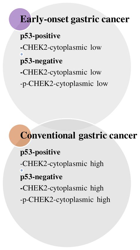
2 MACHLOWSKA et al: STATUS OF CHEK2 AND p53 IN PATIENTS WITH GASTRIC CANCER
DNA damage response genes are involved in the preserva‑ CHEK2 and p53 proteins in both conventional (C)GC and
tion of a healthy genome. Defects in cell cycle checkpoints and early onset (EO)GC, to assess the effects of CHEK2 and p53
DNA repair genes, such as ATM, CHEK2, BRCA1, BRCA2 and in the development of the heterogeneous nature of GC.
TP53, particularly mutations and/or aberrant downregulation,
may be associated with the development of gastric cancer (6). Materials and methods
The cell cycle checkpoint kinase 2 (CHEK2) gene serves
a significant role in the DNA damage response signaling Patients. The tissue samples used in the present study
network, particularly in cancer development (7). Germline included 89 cases of CGC, diagnosed between 1993‑2003
mutations in the gene have been reported in families of patients and 107 samples of EOGC, obtained from the Academic
with cancer; particularly for breast cancer (8‑10) and prostate Medical Centre in Amsterdam as well as collected from
cancer (11,12), but also in other types of cancer (7,13,14), 24 institutions in the Netherlands, Finland and Poland. Tumor
including gastric cancer (15). The most prevalent mutant samples were classified according using the Laurén clas‑
alleles of CHEK2 are three mutant alleles resulting in protein sification system (23) as either intestinal or diffuse gastric
truncation (1100delC, IVS2G>A, del5395), one which is a adenocarcinomas (Table I). The tissues were previously used
missense variant (I157T) (16). for a previous analysis (24), where immunohistochemical
In humans, the CHEK2 protein is a 65 kDa polypeptide labelling was performed to determine the expression patterns
that consists of 543 amino acids and three different functional of several gastric cancer markers amongst EOGC and CGC.
domains. The N terminal SQ/TQ cluster domain contains a For sequencing analysis, 53 GC tissues were selected, 35 with
region rich in serine‑glutamine and threonine‑glutamine EOGC and 18 with CGC, taking into consideration the DNA
motifs, which are potential sites of phosphorylation by ATR quality status of the samples. The present study was approved
and ATM kinases (part of the PI3K family) (17). Under physi‑ by the Medical University of Lublin Bioethical Committee
ological conditions, CHEK2 in retained in the nucleus in an and was performed in accordance with the Declaration of
inactive, monomeric form. DNA damage initiates phosphoryla‑ Helsinki.
tion at Thr68 and other sites of the SQ/TQ cluster domain. The
phosphorylated site binds to the forkhead‑associated domain DNA extraction. Tissues were obtained as previously
and forms a dimer that is further activated by autophosphory‑ described by Sitarz et al (25). Genomic DNA extraction
lation at the C‑terminal serine/threonine kinase domain (18). from formalin‑fixed paraffin‑embedded (FFPE) tissues was
As a result, the dimer dissociates into fully active monomers. performed using the QIAamp DNA Mini kit (Qiagen GmbH)
The Thr68 phosphorylated form of CHEK2 creates distinct or a Puregene DNA Isolation kit (Gentra, Inc.) in accordance
nuclear foci as a reaction to ionizing radiation (19). Only the with the manufacturers' protocols. DNA was quantified using
activated form of CHEK2 is present at sites of DNA double a Quantus™ Fluorometer with the QuantiFluor ® dsDNA
stranded breaks. These observations suggest that CHEK2 is system according to the manufacturer's protocol (Promega
regulated at the site of the DNA double stranded break, and Corporation). DNA quality was assessed using an Agilent
that phosphorylated CHEK2 has an important role in the 2200 TapeStation and Genomic DNA ScreenTape (Agilent
DNA damage response. Our current study is focused on both Technologies, Inc.). Further evaluation of the suitability of
forms of CHEK2 to examine their role in the development of the samples for sequencing was performed using an Infinium
age‑dependent subtypes of gastric carcinoma. HD FFPE QC assay protocol (Illumina, Inc.). The Real‑Time
The TP53 gene is located on the short arm of chromosome PCR using a CFX96 Touch™ Real‑Time PCR Detection
17 (17p13.1) (18). Multiple studies have demonstrated the system (Bio‑Rad Laboratories, Inc.) was used to assess the
genetic association between TP53 alterations and cancer amplification status of each sample.
susceptibility. The TP53 gene is the most frequently mutated
gene (>50%) in human cancer, suggesting that the TP53 gene Library preparation using a target enrichment workflow for
serves a pivotal role in preventing the formation of cancer (19). next generation sequencing. Libraries for the sequencing
In gastric carcinoma the mutations are located broadly across a experiments were prepared using a Trusight Rapid Capture
gene, from exons 4‑11 with hot spots of mutations at codons 175, Preparation kit (Illumina, Inc.) according to the manufac‑
248, 273, 282, 245 and 213. Additionally, G:C>A:T transitions turer's protocol. DNA samples were standardized to 50 ng and
at CpG sites are the most popular types of mutation (20). underwent a tagmentation step. The produced libraries from
The p53 protein serves an important role in regulation or input of genomic DNA were amplified and adapter tagged
cancer progression through the regulation of apoptosis, the multiple short library fragments of 220‑350 base pairs were
cell cycle and genomic stability (21). p53 can stimulate DNA prepared. Libraries for each sample were combined to ensure
repair proteins when DNA damage is persistent or serious. proper complexity for a single run. Biotin labelled probes
It can prevent proliferation by arresting the cell cycle at the were used to target regions of interest. Further hybridiza‑
G1/S regulation point following recognition of DNA damage. tion steps were performed using custom synthesized oligos
When a cell is arrested at this checkpoint, the DNA repair that captured genes with an assigned predisposition towards
proteins attempt a repair. However, if the cells are arrested at multiple cancer types. The sequencing panel, TruSight Cancer
this checkpoint for a sufficient amount of time, the cell may Panel (Illumina, Inc.), targeted 94 genes and 284 SNPs linked
instead undergo apoptosis (22). to common and rare cancers. The complete list of target genes
The present study focused on the frequency of genetic and SNPs is available from illumina.com/techniques/popular‑
alterations in the CHEK2 and TP53 genes, and the expression applications/genotyping.html. Standard pools were enriched
patterns of the phosphorylated and non‑phosphorylated with regions of interest through streptavidin coated beads thatONCOLOGY LETTERS 21: 348, 2021 3
Table I. Histological types of GC samples analyzed in the Inc.; cat. no. 2197; 1:50; ARS pH 9; 72 h incubation at 4˚C).
study (n=196). The sections (4 µm) were deparaffinized and endogenous
peroxidase activity was blocked by immersion in 0.3% H2O2
Early‑onset GC Conventional GC and in methanol for 20 min. Antigen retrieval was performed
Characteristic (n=107) (n=89) in sodium citrate/ARS buffer (0.01 M/pH 6.0) for 10 min
at 120˚C or in Tris/EDTA buffer (10 mM/1 mM; pH 9.0)
Age (range), years ≤45 (21‑45) >45 (47‑86) for 10 min at 120˚C. Subsequently, once the samples had
Histology, n (%) cooled for 10 min, they were washed in PBS and blocked
Intestinal 21 (20) 42 (47) using PBS with 5% normal goat serum. Then the material
Diffuse 78 (73) 37 (42) was incubated with the antibodies. Antibody binding was
Mixed 8 (7) 10 (11) visualized using a Powervision + poly‑HRP detection system
(Leica Microsystems; cat. no. PV6107) with Bright DAB
GC, gastric cancer. (Immunologic; cat. no. BS04‑110) as the chromogen for
CHEK2, and PowerDAB (Immunologic; cat. no. BS03‑25)
used as the chromogen for p‑CHEK2. Sections were
counterstained using hematoxylin.
bound to the biotinylated probes. Then, DNA fragments were TP53 staining; sections (4‑µm thick) were deparaffinized
eluted from the beads and another round of hybridization and and antigen retrieval was performed by boiling in 10 mM
enrichment was performed. The final amplified post‑capture Tris/1 mM EDTA (pH 9.0) for 10 min. Subsequently, slides
library concentrations were assessed using a Quantus™ were immersed in 0.3% hydrogen peroxide in methanol for
Fluorometer according to the manufacturer's protocol. 30 min and nonspecific binding was blocked with 5% normal
Libraries were quantified by qPCR using a KAPA Library goat serum for 1 h at room temperature. The sections were
Quantification kit (KAPA Biosystems, Inc.). The size of the incubated for 1 h at room temperature with an anti‑p53
obtained libraries fragments was evaluated using an Agilent antibody (monoclonal antibody combination of DO‑7 and
2200 TapeStation and High Sensitivity D1000 ScreenTape BP53‑12; Neomarkers, Inc.; 1:2,000). The Ultravision
system (Agilent Technologies, Inc.). Post capture enriched anti‑polyvalent HRP detection system (Lab Vision Corp.) was
libraries were sequenced on a MiSeq Sequencing Platform used to visualize antibody binding with 3,30‑diaminobenzi‑
(Illumina, Inc.) using the manufacturer's workflow. The dine used as the chromogen. Sections were counterstained
concentration of loaded libraries amounted to 10 pM. The using hematoxylin.
sequencing experiments were performed using a MiSeq
Reagent kit version 2 (300 cycles). Scoring. Scoring of immunohistochemistry was performed for
analysis of CHEK2 and p53 expression. Scoring was performed
Data processing. Results from each sample were mapped to independently by two researchers, any disagreements in
the human reference genome GRCh37, also known as hg19 score were reanalyzed by the expert using a multiheaded
(Human Genome version 19) using Burrows‑Wheeler Aligner microscopes in accordance with his judgement. Cases where
(BWA‑mem, version 0.7.5). The reads with low mapping the staining was not clearly abnormal were categorized into
quality scores, unmapped reads and duplicates were filtered the normal group. No slides exhibited increased negative
out using Samtools version 0.1.19 (26). Local realignment of staining around the edges, and the age of the block had no
reads around indels (insertion or deletion) and detection of impact on immunohistochemical staining. Multiple scoring
systematic errors in base quality scores were performed using systems in the literature were considered to ensure a system
Genome Analysis Toolkit (27). The reads mapped outside the that adequately reflected the results generated was used, and
target region were removed. Variant calling for germline SNPs all cases were re‑examined based on the agreed upon scoring
and indels was performed using the GATK HaplotypeCaller parameters.
tool (28). The call sets of SNPs and small insertions and Immunohistochemical labelling revealed the patterns of
deletions were separated for further filtering. Hard filters CHEK2 and p‑CHEK2 expression in GC (Fig. 1). The scoring
applied to variant call sets for SNPs were, QualByDepth criteria for the quantity of the tumor cells were: 1, No staining;
(QD)13.0, QRankSum4 MACHLOWSKA et al: STATUS OF CHEK2 AND p53 IN PATIENTS WITH GASTRIC CANCER
Figure 1. Immunohistochemical staining of CHEK2 and p‑CHEK2 in gastric cancer tissues (magnification, x100). The following scoring criteria of the tumor
cells were used: 0+, no staining; 1+, weak diffuse cytoplasmic staining (may contain stronger intensity in 50% of the tumor cells stained with strong intensity. p‑CHEK2, phosphorylated checkpoint kinase 2.
protein expression levels) were performed using the χ2 test. TP53 genetic alterations amongst EOGC and CGC. Results
For this purpose we used GraphPad programme and applied from next‑generation sequencing for TP53 are shown in
the 2x2 contingency table. We chose chi‑square test with the Table III. A total of 9 variant alterations in TP53 were detected
Yates' continuity correction, which is designed to make the in 53 patients: 5 missense variants, 2 synonymous vari‑
chi‑square approximation better. We calculated two‑tailed ants, 1 frameshift variant and 1 premature stop codon. The
(also called two‑sided) P‑values. PC missense variant was found in one case (EOGC,
indicate a statistically significant difference. 1 HET‑heterozygote) and was described as a medium impact
mutation, according to Gemini (Table SII), and present in
Results dbSNP and ClinVar where it was described to be associated
with Li‑Fraumeni syndrome, but not present in the COSMIC
CHEK2 genetic alterations amongst EOGC and CGC. database. The c.1014C>T synonymous variant was present in
Next‑generation sequencing results were generated for 1 EOGC case as heterozygous, was considered a low impact
53 patients with GC, 35 patients with EOGC and 18 patients alteration, described in clinical repositories as related to
with CGC. DNA from all samples was successfully amplified, hereditary cancer predisposing syndrome and Li‑Fraumeni
libraries were constructed and a minimum coverage amongst syndrome, and in COSMIC it was described as being related
regions of interest was obtained in all tested samples. Results to skin and upper aerodigestive tract. A deletion (frameshift
from next‑generation targeted sequencing for CHEK2 are variant): c.851_852delCA was observed in one CGC case,
shown in Table II. A total of 5 different variants in CHEK2 described as high impact, and was considered as rare as it was
were found amongst 53 cases: 2 missense variants, 2 synony‑ not described in any of the databases. The missense alteration
mous variants and 1 intron alteration. The c.1741G>T missense c.844C>G was reported in 1 EOGC case, as heterozygous,
alteration in CHEK2 was observed only in one case and was was considered medium impact and was associated with
described as a medium impact mutation, according to Gemini. hereditary cancer predisposing syndrome, and in COSMIC, it
This variant seemed to be very rare, as it had not been previ‑ was described as being associated with lung and breast cancer,
ously described in CHEK2 and has never been reported in as well as hematopoietic and lymphoid, upper aerodigestive
NCBI ClinVar or dbSNP (Table SI). Both missense variants: tract and pancreatic neoplastic disease. This alteration was
c.1246A>G (medium) and the synonymous variant: c.1245C>T also found in stomach cancer by Rugge et al (30) and by
(low) were detected in 5 out of 53 cases. The CHEK2 c.252A>G Xu et al (31).
synonymous variant, which according to NCBI ClinVar Missense variant c.818G>A was found in one patient
database, is associated with a hereditary predisposition to with CGC, as heterozygous, medium impact, related to
colorectal cancer and familial breast cancer, was detected in Li‑Fraumeni syndrome, thyroid cancer and hereditary cancer
5 cases. Interestingly the intron variant c.319+379A>G was predisposing syndrome according to the ClinVar database;
detected in 47 cases, but according to Gemini, its impact was and according to COSMIC, it was found in multiple studies
low. The detected mutations are fairly uncommon for gastric in stomach cancer, but predominantly in large intestine,
carcinoma; they were not found in Catalogue of Somatic breast, lung, central nervous system and ovary cancer.
Mutations in Cancer (COSMIC). Variant c.734G>A was a missense alteration, found in oneONCOLOGY LETTERS 21: 348, 2021 5
Table II. Presentation of variants identified in CHEK2 gene in EOGC (n=35) and CGC (n=18) cases.
Variant type Protein change Molecular consequence Exon CGC mutations EOGC mutations
c.1741G>T p. Val581Leu Missense variant 16 ‑ 1 HET
c.1246A>G p. Lys416Glu Missense variant 12 3 HET 2 HET
c.1245C>T p. Ser415Ser Synonymous variant 12 3 HET 2 HET
c.252A>G p. Glu84Glu Synonymous variant 2 3 HET 2 HET
c.319+379A>G ‑ Intron variant 2 7 HET + 10 ALT 12 HET + 18 ALT
HET, heterozygous; ALT, alternative homozygous (identical mutation of both paternal and maternal alleles); CHEK2, checkpoint kinase 2;
GC, gastric cancer; EOGC, early‑onset GC; CGC, conventional GC.
Table III. Presentation of variants detected in TP53 gene in EOGC (n=35) and CGC (n=18) cases.
Variant type Protein change Molecular consequence Exon CGC mutations EOGC mutations
c.1129A>C p. Thr377Pro Missense variant 11 ‑ 1 HET
c.1014C>T p. Phe338Phe Synonymous variant 10 ‑ 1 HET
c.851_852delCA p. Thr284fs Frameshift variant 8 1 DEL ‑
c.844C>G p. Arg282Gly Missense variant 8 ‑ 1 HET
c.818G>A p. Arg273His Missense variant 8 1 HET ‑
c.734G>A p. Gly245Asp Missense variant 7 1 HET ‑
c.639A>G p. Arg213Arg Synonymous variant 6 3 HET 1 HET
c.586C>T p. Arg196* Nonsense variant 6 1 HET ‑
c.215C>G p. Pro72Arg Missense variant 4 11 HET + 6 ALT 12 HET + 17 ALT
HET, heterozygous; ALT, alternative homozygous (identical mutation of both paternal and maternal alleles); DEL, deletion; GC, gastric cancer;
EOGC, early‑onset GC; CGC, conventional GC.
case (CGC, 1 HET) and was described as a medium impact intestine and soft tissue cancers, but also in the stomach
mutation, described in dbSNP and ClinVar as associated with according to the International Cancer Genome Consortium.
Li‑Fraumeni syndrome and hereditary cancer predisposing
syndrome. In the COSMIC database, it was shown to be found Immunohistochemistry staining of CHEK2 and p‑CHEK2
in large intestine, esophagus, hematopoietic and lymphoid, proteins. The analyzed group encompassed 196 patients, 89
lung and breast neoplasms. In cancers of the stomach, the with CGC and 107 with EOGC (Table IV). The first approach
variant was previously reported by Gleeson et al (32) and was to compare the results from immunohistochemical
Kim et al (33). staining in EOGCs and CGC cases. Two categories, high and
Interestingly, variant c.639A>G (synonymous) was found low expression, were distinguished. Differential expression in
in 3 CGC cases and 1 EOGC case as heterozygous. This the cytoplasmic CHEK2 and nuclear‑phosphorylated CHEK2
was a low impact variant, associated with hereditary cancer (PT in EOGC, it was observed in only 34% of samples. The
was considered high impact and was detected in one CGC differences in the nuclear CHEK2 (CHEK2‑nucleus) and
case (HET), associated with hereditary cancer predisposing cytoplasmic phosphorylated CHEK2 (phospho‑CHEK2‑
syndrome and Li‑Fraumeni syndrome; and in the COSMIC cytoplasmic) expression patterns between CGC and EOGC
database was described to be associated with large intestine, were not significant (P=0.178 and P=0.133 respectively).
breast, esophagus, skin and stomach cancer. The most frequent Based on the result that nuclear CHEK2 expression was
variant was the missense c.215C>G variant, found in 17 cases upregulated in both subtypes of GC, this parameter was used
of CGC (6 ALT and 11 HET) and 29 EOGC cases (17 ALT to distinguish a subgroup of samples with high nuclear CHEK2
and 12 HET), and was also present in patients with hereditary expression, the ‘CHEK2‑nucleus high subgroup’, and compared
cancer predisposing syndrome and Li‑Fraumeni syndrome. with the other parameters (Table V). In the CHEK2‑nucleus
Based on the COSMIC database, it was found in the large high subgroup, there was a statistically significant difference6 MACHLOWSKA et al: STATUS OF CHEK2 AND p53 IN PATIENTS WITH GASTRIC CANCER Table IV. Comparison of nuclear and cytoplasmic CHEK2 and p‑CHEK2 expression in CGC (n=89) and EOGC (n=107) samples. Expression EOGC, n (%) CGC, n (%) P‑value CHEK2‑nucleus high 93 (87) 70 (79) 0.178 CHEK2‑nucleus low 14 (13) 19 (21) CHEK2‑cytoplasmic high 39 (36) 56 (63)
ONCOLOGY LETTERS 21: 348, 2021                                                  7

Table VII. Comparison of high and low CHEK2 and p‑CHEK2 expression in the nucleus and cytoplasm among EOGC
p53‑positive (n=29) and CGC p53‑positive (n=44) subgroups.

Expression                               EOGC p53‑positive, n (%)                  CGC p53‑positive, n (%)               P‑value

CHEK2‑nucleus high                                 27 (93)                                  38 (86)                       0.604
CHEK2‑nucleus low                                   2 (7)                                    6 (14)
CHEK2‑cytoplasmic high                              8 (28)                                  30 (68)                       0.002a
CHEK2‑cytoplasmic low                              21 (72)                                  14 (32)
p‑CHEK2‑nucleus high                               10 (34)                                  24 (55)                       0.149
p‑CHEK2‑nucleus low                                19 (66)                                  20 (45)
p‑CHEK2‑cytoplasmic high                            5 (17)                                  14 (32)                       0.264
p‑CHEK2‑cytoplasmic low                            24 (83)                                  30 (68)

PG group (82% of cases). Between CGC and
CHEK2‑cytoplasmic expression was low in EOGC p53‑nega‑            EOGC with two SNPs in TP53 gene: c.215C>G, c.639A>G and
tive patients (66%), whereas high expression was found in 57%     p53‑positive and negative staining, there was no statistically
of CGC p53‑negative patients. Phospho‑CHEK2‑cytoplasmic           significant correlations.
low expression was predominant in the EOGC p53‑negative
group (76%), and high expression was observed in the CGC          Discussion
p53‑negative group (P=0.033). The other groups in Table VIII
were analyzed; however, none of the assessed parameters were      The primary function of CHEK2 is suppressing tumor
statistically significant.                                        growth, and exhibits proapoptotic and mitotic functions.
                                                                  The association between changes in CHEK2 gene expres‑
Correlation between the genetic alterations of CHEK2              sion and cancer risk development have been demonstrated
and TP53 genes and their protein expression levels. Next,         in numerous case‑controlled studies (34‑36). The CHEK2
the EOGC and CGC cases were compared for the different            gene encodes a serine/threonine kinase (CHEK2), which is
mutation status for SNPs most frequently occurring among          activated by ATM, following mobilization of the cascade
the analyzed groups in both genes: CHEK2 and TP53, and            of DNA damage response molecules to double‑stranded
their protein expression levels (Table SIII). Correlations        breaks. Additionally, CHEK2 is widely phosphorylated at
between mutations in CHEK2: c.319+379A>G, c.252A>G,               Thr68, resulting in its activation, particularly during the
c.1246A>G, c.1245C>T and CHEK2 protein expression                 development of precancerous lesions and in the progression
in the nucleus and cytoplasm both in the phosphorylated           of cancer (37).
8 MACHLOWSKA et al: STATUS OF CHEK2 AND p53 IN PATIENTS WITH GASTRIC CANCER
In the Polish population, three founder alleles were found:
A missense substitution of an isoleucine for a threonine in
exon 3: I157T and two alterations that truncate the CHEK2
protein: IVS2+1G>A in exon 3 and 1100delC in exon 10, all of
which have been previously investigated with regard to their
association with predisposition to GC (16).
Li and Stern (38) found that prior to DNA damage, CHEK2
is associated with chromatin, and irradiation or topoisomerase
inhibitors decrease this association. They observed that
phospho‑CHEK2 was released from chromatin following
DNA damage, and accumulated in the soluble cytoplasmic
and soluble nuclear fraction. CHEK2 in human cells exposed
to DNA‑damaging agents resulted in immediate redistribu‑
tion of the activated CHEK2 throughout the nucleus, rapidly
spreading the checkpoint arrest signal from localized sites of
DNA damage to the soluble mobile proteins, such as Cdc25
or p53 (34). Ćmielová et al (39) showed increased levels of
phospho‑CHEK2 in the cytoplasmic fraction after 24 and 48 h
of mitoxantrone treatment. In the present study, only a weak,
insignificant signal was observed in the nuclear fraction at the
same time points.
The reports of the subcellular distribution of CHEK2
are inconsistent and the redistribution of CHEK2 after DNA
damage is very poorly described. An improved understanding
of the DNA damage response and protein distribution in
the cells may assist in improving the efficiency of treatment
of several types of cancer and may highlight specific novel
therapeutic targets.
Sequencing results revealed 5 different SNPs within the
studied group of patients. Variants c.1246A>G (missense
variant), c.1245C>T (synonymous variant) and c.252A>G
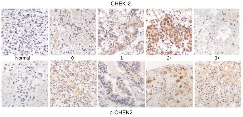
(synonymous variant) were similarly distributed between the Figure 2. Differences between early‑onset gastric cancer and conven‑
subgroups. Amongst the SNPs detected, 3 were HET in CGC tional gastric cancer with respect to CHEK2 and p53 staining. p‑CHEK2,
phosphorylated checkpoint kinase 2.
and 2 were HET in EOGC. Interestingly, the intron variant
c.319+379A>G was present in a high number of cases; 94%
of CGCs and 86% EOGCs cases. Only variant c.252A>G has
been previously shown to be associated with several types of and hereditary cancer predisposing syndrome. According to
cancer, when searching NCBI ClinVar database. However, COSMIC, five of the detected variants were previously shown
in reference to GC, all the listed SNPs seem to be very rare to be associated with stomach cancer (c.844C>G, c.818G>A,
and thus far, have not been broadly described. Interestingly, c.734G>A, c.586C>T and c.215C>G). In the present study, it
the intron variant c.319+379A>G, detected in the present was demonstrated that the TP53 mutations may accumulate
study, was previously shown to be associated with esophageal over a very long period of time (lifetime) (44). With aging,
squamous cell carcinoma risk, as reported by Li et al (40). genetic damage may increase, increasing the importance of
The tumor suppressor gene TP53 is one of the most regu‑ the function of TP53, and therefore making an individual more
larly mutated genes in human cancers, and TP53 alterations are vulnerable to loss of function mutations.
present in ~77% of stomach cancers (41). Mutations of TP53 Immunohistochemistry analysis may have additional value
have been detected in the early stages of gastric carcinoma, for the assessment of the ATM>CHEK2>p53 pathway. When
and their frequency increases as the tumor progressed (42). CHEK2 is phosphorylated at Thr68, it is activated. Therefore,
TP53 has been termed ‘the guardian of the genome’ (41). expression of the phosphorylated protein may be used as a
Genomic instability causes genetic variability and accelerates potential marker of active CHEK2 status (45). The constitutive
the rate at which a cell acquires and accumulates mutations, activation of the DNA damage checkpoint pathway may be
which may underlie the initiation of tumorigenesis (43). In the associated with an increased level of p53 alterations in cancer,
present study, 9 different genetic alterations of the TP53 gene taking into consideration that p53 is a downstream target
were detected. Most of these occurred sporadically; however, of ATM and CHEK2. The immunohistochemical analyses
variant c.215C>G was present in 17 cases of CGC (6 ALT and performed in the present study included analysis of cytoplasmic
11 HET) and 29 cases of EOGC cases (17 ALT and 12 HET), and nuclear expression of CHEK2 and phospho‑CHEK2.
whereas variant c.639A>G was present in 3 CGC cases and The levels and distribution of expression of these proteins
1 EOGC cases (all HET). The clinical significance of the appeared to vary between CGC and EOGC. Interestingly,
detected variants was assessed using dbSNP and ClinVar. the levels of p‑CHEK2 increased with age, and a statistical
Most of these were associated with the Li‑Fraumeni syndrome difference between the two subtypes with regard to nuclearONCOLOGY LETTERS 21: 348, 2021 9
p‑CHEK2 expression was observed. Additionally, high nuclear Acknowledgements
CHEK2 expression was also more prevalent in CGC, following
stratification based on levels of nuclear CHEK2 expression. Not applicable.
Cytoplasmic phospho‑CHEK2 expression was increased.
The phosphorylated forms of both CHEK2 types may be an Funding
indicator of GC development. The levels of CHEK2 protein
in the nuclei was elevated in both subtypes of GC, and this The present study was partly supported by a grant from the
outcome supports further study into the potential therapeutic Polish Ministry of Science and Higher Education (grant
value of CHEK2. no. NN402423838).
p53 staining was performed on 181 tumor samples, in both
age dependent subtypes of GC; expression of the p53 protein Availability of data and materials
increased with age. Immunohistochemistry analysis of p53
may explain the biology of the tumor. Multiple studies have The NGS datasets generated and/or analyzed during the current
used immunohistochemistry analysis to assess p53 expression. study are available in the Open Science Framework Repository.
In lung cancer, a meta‑analysis showed that positive immuno‑ Outcome for the 94 genes and 284 SNPs presented in the NGS
histochemistry staining of p53 may be used as a prognostic panel are available at https://osf.io/9483c/(snps.variants‑all.csv)
biomarker (46). The results of the present study showed that and https://osf.io/zxwg9/(genes.variants‑all.csv).
p53 expression could be used to distinguish age dependent
onset of GC. Authors' contributions
The EOGC p53‑positive and CGC p53‑positive groups
were compared based on phosphorylation of CHEK2. PW and RS conceptualized the study. JM, FM, GJAO and
There was a statistically significant difference between RS developed the methodology. PK and PW performed the
the expression of CHEK2‑cytoplasmic high and low, and data processing. JM, PK, AB and RS analyzed the results.
the EOGC p53‑positive and CGC p53‑positive patients. JM, PK, MS, AB, FM, PW, RM, GJAO and RS performed the
The expression of CHEK2 in the cytoplasm was increased experiments. PW and RS confirmed the authenticity of the
(68%) in the CGC p53‑positive group. Statistically important data. JM, MS, AB and RS wrote the original draft. JM, PK,
differences between CHEK2‑cytoplasmic high and low, MS, AB, FM, PW, RM, GJAO and RS reviewed and edited
and the EOGC and CGC p53‑negative patients were also the manuscript. PW, GJAO and RS supervised the study. RM
observed. CHEK2‑cytoplasmic expression was increased and RS acquired funding. All authors read and approved the
in CGC p53‑negative patients (57%), and its expression final manuscript.
was higher in the CGC p53‑positive subgroup (68%).
Phospho‑CHEK2‑cytoplasmic was also higher amongst the Ethics approval and consent to participate
CGC TP53‑negative group. Together, these results suggests a
link between the expression of p53 and CHEK2 cytoplasmic All procedures involving human participants were in accor‑
proteins in GC development based on age. Fig. 2 summa‑ dance with the ethical standards of the Medical University of
rizes the primary differences between EOGC and CGC with Lublin Bioethical Committee (Lublin, Poland; approval no.
respect to CHEK2 and p53 staining. KE‑0254/322/2019) and with the 1964 Declaration of Helsinki
Non‑coding regions possess regulatory functions for the and its later amendments or comparable ethical standards. Due
coding regions. Thus coding regions are the primary actors, to the retrospective nature of the study, patient consent was
but their function is regulated by non‑coding regions. Thus waived by the ethics committee.
any modifications to the non‑coding may have an impact on
the expression and regulation of the expressed genes. There Patient consent for publication
was a statistically significant correlation between occurrence
of the c.319+379A>G intronic alteration in both CGC and Not applicable.
EOGC subtypes and phospho‑CHEK2‑nuclear expression,
which increased with age. Competing interests
The present study has some limitations. First, the number
of biopsies from patients was a limiting factor. The selected The authors declare that they have no competing interests.
subgroups were small, and thus it is difficult to observe the
differences in SNP frequencies in the CHEK2 gene. Use of References
a benign gastric disease control group may better allow for
determination of whether the protein expression of these 1. Zou Z and Jemal A: Cancer statistics, 2014. CA Cancer J Clin 64:
9‑29, 2014.
two genes may be used as biomarkers of early gastric cancer 2. Howlader N, Noone AM, Krapcho M, Neyman N, Aminou R,
development. In vitro studies are required to determine the Waldron W, Altekruse SF, Kosary CL, Ruhl J, Tatalovich Z, et al:
functional effects of the detected SNPs in more detail. It is also SEER Cancer Statistics Review, 1975‑2008, National Cancer
Institute. Bethesda, MD, pp19, 2011.
probable that different allele frequency amongst individuals 3. Milne AN, Sitarz R, Carvalho R, Carneiro F and Offerhaus GJ:
of different ethnicities may have an impact on the obtained Early onset gastric cancer: On the road to unraveling gastric
data. Thus, a more diverse cohort may allow for identification carcinogenesis. Curr Mol Med 7: 15‑28, 2007.
4. Milne AN, Carneiro F, O'Morain C and Offerhaus GJ: Nature
of differences in frequencies of SNPs in different populations meets nurture: Molecular genetics of gastric cancer. Hum
around the world. Genet 126: 615‑628, 2009.10 MACHLOWSKA et al: STATUS OF CHEK2 AND p53 IN PATIENTS WITH GASTRIC CANCER
5. Milne AN, Offerhaus GJ and Carneiro F: Histopathology of 27. McKenna A, Hanna M, Banks E, Sivachenko A, Cibulskis K,
familial and early‑onset gastric cancer. Diagnostic Histopathol 17: Kernytsky A, Garimella K, Altshuler D, Gabriel S, Daly M
62‑68, 2011. and DePristo MA: The genome analysis toolkit: A MapReduce
6. Slavin T, Neuhausen SL, Rybak C, Solomon I, Nehoray B, framework for analyzing next‑generation DNA sequencing data.
Blazer K, Niell‑Swiller M, Adamson AW, Yuan YC, Genome Res 20: 1297‑1303, 2010.
Yang K, et al: Genetic gastric cancer susceptibility in the inter‑ 28. Narzisi G, O'Rawe JA, Iossifov I, Fang H, Lee YH, Wang Z,
national clinical cancer genomics community research network. Wu Y, Lyon GJ, Wigler M and Schatz MC: Accurate de novo
Cancer Genet 216‑217: 111‑119, 2017. and transmitted indel detection in exome‑capture data using
7. Cybulski C, Górski B, Huzarski T, Masojć B, Mierzejewski M, microassembly. Nat Methods 11: 1033‑1036, 2014.
Debniak T, Teodorczyk U, Byrski T, Gronwald J, Matyjasik J, et al: 29. Cingolani P, Platts A, Wang le L, Coon M, Nguyen T, Wang L,
CHEK2 is a multiorgan cancer susceptibility gene. Am J Hum Land SJ, Lu X and Ruden DM: A program for annotating and
Genet 75: 1131‑1135, 2004. predicting the effects of single nucleotide polymorphisms,
8. Al‑Rakan MA, Hendrayani SF and Aboussekhra A: CHEK2 SnpEff: SNPs in the genome of Drosophila melanogaster strain
represses breast stromal fibroblasts and their paracrine w1118; iso‑2; iso‑3. Fly (Austin) 6: 80‑92, 2012.
tumor‑promoting effects through suppressing SDF‑1 and IL‑6. 30. Rugge M, Shiao YH, Busatto G, Cassaro M, Strobbe C,
BMC Cancer 16: 575, 2016. Russo VM, Leo G, Parenti AR, Scapinello A, Arslan P and
9. Desrichard A, Bidet Y, Uhrhammer N and Bignon YJ: CHEK2 Egarter‑Vigl E: The p53 gene in patients under the age of 40 with
contribution to hereditary breast cancer in non‑BRCA families. gastric cancer: Mutation rates are low but are associated with a
Breast Cancer Res 13: R119, 2011. cardiac location. Mol Pathol 53: 207‑210, 2000.
10. Wang N, Ding H, Liu C, Li X, Wei L, Yu J, Liu M, Ying M, 31. Xu JH, Chen LR, Wang HJ and Yao LF: Application of
Gao W, Jiang H and Wang Y: A novel recurrent CHEK2 Y390C immunohistochemistry‑Laser microdissection‑PCR technique
mutation identified in high‑risk Chinese breast cancer patients in detecting p53 gene mutation in paraffin sections of
impairs its activity and is associated with increased breast cancer gastric cancer. Zhonghua Bing Li Xue Za Zhi 33: 342‑345, 2004
risk. Oncogene 34: 5198‑5205, 2015. (In Chinese).
11. Dong X, Wang L, Taniguchi K, Wang X, Cunningham JM, 32. Gleeson CM, Sloan JM, McManus DT, Maxwell P, Arthur K,
McDonnell SK, Qian C, Marks AF, Slager SL, Peterson BJ, et al: McGuigan JA, Ritchie AJ and Russell SE: Comparison of p53
Mutations in CHEK2 associated with prostate cancer risk. Am and DNA content abnormalities in adenocarcinoma of the
J Hum Genet 72: 270‑280, 2003. oesophagus and gastric cardia. Br J Cancer 77: 277‑286, 1998.
12. Hale V, Weischer M and Park JY: CHEK2 (*) 1100delC mutation 33. Kim SS, Bhang CS, Min KO, Chae HS, Choi SW, Lee CD,
and risk of prostate cancer. Prostate Cancer 2014: 294575, 2014. Lim KW, Chung IS and Park DH: p53 mutations and microsat‑
13. Cybulski C, Masojc B, Oszutowska D, Jaworowska E, Grodzki T, ellite instabilities in the subtype of intestinal metaplasia of the
Waloszczyk P, Serwatowski P, Pankowski J, Huzarski T, stomach. J Korean Med Sci 17: 490‑496, 2002.
Byrski T, et al: Constitutional CHEK2 mutations are associ‑ 34. Bartek J and Lukas J: Chk1 and Chk2 kinases in checkpoint
ated with a decreased risk of lung and laryngeal cancers. control and cancer. Cancer Cell 3: 421‑429, 2003.
Carcinogenesis 29: 762‑765, 2008. 35. Cybulski C, Wokołorczyk D, Jakubowska A, Huzarski T, Byrski T,
14. Kilpivaara O, Alhopuro P, Vahteristo P, Aaltonen LA and Gronwald J, Masojć B, Deebniak T, Górski B, Blecharz P, et al:
Nevanlinna H: CHEK2 I157T associates with familial and Risk of breast cancer in women with a CHEK2 mutation with
sporadic colorectal cancer. J Med Genet 43: e34, 2006. and without a family history of breast cancer J Clin Oncol 29:
15. Skierucha M, Milne AN, Offerhaus GJA, Polkowski WP, 3747‑3752, 2011.
Maciejewski R and Sitarz R: Molecular alterations in gastric 36. Siołek M, Cybulski C, Gąsior‑Perczak D, Kowalik A,
cancer with special reference to the early‑onset subtype. World Kozak‑Klonowska B, Kowalska A, Chłopek M, Kluźniak W,
J Gastroenterol 22: 2460‑2474, 2016. Wokołorczyk D, Pałyga I, et al: CHEK2 mutations and the risk
16. Teodorczyk U, Cybulski C, Wokołorczyk D, Jakubowska A, of papillary thyroid cancer. Int J Cancer 137: 548‑552, 2015.
Starzyńska T, Lawniczak M, Domagała P, Ferenc K, Marlicz K, 37. Zoppoli G, Solier S, Reinhold WC, Liu H, Connelly JW Jr,
Banaszkiewicz Z, et al: The risk of gastric cancer in carriers of Monks A, Shoemaker RH, Abaan OD, Davis SR, Meltzer PS, et al:
CHEK2 mutations. Fam Cancer 12: 473‑478, 2013. CHEK2 genomic and proteomic analyses reveal genetic inactiva‑
17. Buscemi G, Carlessi L, Zannini L, Lisanti S, Fontanella E, tion or endogenous activation across the 60 cell lines of the US
Canevari S and Delia D: DNA damage‑induced cell cycle National Cancer Institute. Oncogene 31: 403‑418, 2012.
regulation and function of novel Chk2 phosphoresidues. Mol Cell 38. Li J and Stern DF: DNA damage regulates Chk2 association with
Biol 26: 7832‑7845, 20060. chromatin. J Biol Chem 280: 37948‑37956, 2005.
18. McBride OW, Merry D and Givol D: The gene for human p53 39. Ćmielová J, Lesná M and Řezáčová M: Subcellular localization
cellular tumor antigen is located on chromosome 17 short arm of proteins responding to mitoxantrone‑induced DNA damage in
(17p13). Proc Natl Acad Sci USA 83: 130‑134, 1986. leukaemic cells. Folia Biol (Praha) 61: 60‑65, 2015.
19. Barnoud T, Parris JLD and Murphy ME: Common genetic 40. Li WQ, Hu N, Hyland PL, Gao Y, Wang ZM, Yu K, Su H,
variants in the TP53 pathway and their impact on cancer. J Mol Wang CY, Wang LM, Chanock SJ, et al: Genetic variants in
Cell Biol 11: 578‑585, 2019. DNA repair pathway genes and risk of esophageal squamous cell
20. Fenoglio‑Preiser CM, Wang J, Stemmermann GN and carcinoma and gastric adenocarcinoma in a Chinese population.
Noffsinger A: TP53 and gastric carcinoma: A review. Hum Carcinogenesis 34: 1536‑1542, 2013.
Mutat 21: 258‑270, 2003. 41. Lane DP: Cancer. p53, guardian of the genome. Nature 358:
21. Blondal JA and Benchimol S: The role of p53 in tumor progres‑ 15‑16, 1992.
sion. Semin Cancer Biol 5: 177‑186, 1994. 42. Vousden KH and Prives C: P53 and prognosis: New insights and
22. Lakin ND and Jackson SP: Regulation of p53 in response to further complexity. Cell 120: 7‑10, 2005.
DNA damage. Oncogene 18: 7644‑7655, 1999. 43. Hanahan D and Weinberg RA: Hallmarks of cancer: The next
23. Laurén P: The two histological main types of gastric carcinoma: generation. Cell 144: 646‑674, 2011.
Diffuse and so‑called intestinal‑type carcinoma. An attempt at 44. Feng Z, Hu W, Teresky AK, Hernando E, Cordon‑Cardo C
a histo‑clinical classification. Acta Pathol Microbiol Scand 64: and Levine AJ: Declining p53 function in the aging process: A
31‑49, 1965. possible mechanism for the increased tumor incidence in older
24. Milne AN, Carvalho R, Morsink FM, Musler AR, de Leng WW, populations. Proc Natl Acad Sci USA 104: 16633‑16638, 2007.
Ristimäki A and Offerhaus GJ: Early‑onset gastric cancers have 45. DiTullio RA Jr, Mochan TA, Venere M, Bartkova J,
a different molecular expression profile than conventional gastric Sehested M, Bartek J and Halazonetis TD: 53BP1 functions in
cancers. Mod Pathol 19: 564‑572, 2006. an ATM‑dependent checkpoint pathway that is constitutively
25. Sitarz R, Leguit RJ, de Leng WW, Polak M, Morsink FM, activated in human cancer. Nat Cell Biol 4: 998‑1002, 2002.
Bakker O, Maciejewski R, Offerhaus GJ and Milne AN: The 46. Steels E, Paesmans M, Berghmans T, Branle F, Lemaitre F,
COX‑2 promoter polymorphism‑765 G>C is associated with Mascaux C, Meert AP, Vallot F, Lafitte JJ and Sculier JP: Role of
early‑onset, conventional and stump gastric cancers. Mod p53 as a prognostic factor for survival in lung cancer: A system‑
Pathol 21: 685‑690, 2008. atic review of the literature with a meta‑analysis. Eur Respir J 18:
26. Li H, Handsaker B, Wysoker A, Fennell T, Ruan J, Homer N, 705‑719, 2001.
Marth G, Abecasis G and Durbin R; 1000 Genome Project Data This work is licensed under a Creative Commons
Processing Subgroup: The sequence Alignment/Map format and Attribution-NonCommercial-NoDerivatives 4.0
SAMtools. Bioinformatics 25: 2078‑2079, 2009. International (CC BY-NC-ND 4.0) License.You can also read