Metformin attenuates blood-brain barrier disruption in mice following middle cerebral artery occlusion
←
→
Page content transcription
If your browser does not render page correctly, please read the page content below
Liu et al. Journal of Neuroinflammation 2014, 11:177                                                                                JOURNAL OF
http://www.jneuroinflammation.com/content/11/1/177
                                                                                                                                    NEUROINFLAMMATION

 RESEARCH                                                                                                                                      Open Access

Metformin attenuates blood-brain barrier
disruption in mice following middle
cerebral artery occlusion
Yanqun Liu1, Guanghui Tang2, Yaning Li2, Yang Wang1, Xiaoyan Chen2, Xiang Gu2, Zhijun Zhang2,
Yongting Wang2,3* and Guo-Yuan Yang1,2,3*

  Abstract
  Background: Metformin, a widely used hypoglycemic drug, reduces stroke incidence and alleviates chronic
  inflammation in clinical trials. However, the effect of metformin in ischemic stroke is unclear. Here, we investigated
  the effect of metformin on ischemic stroke in mice and further explored the possible underlying mechanisms.
  Methods: Ninety-eight adult male CD-1 mice underwent 90-minute transient middle cerebral artery occlusion
  (tMCAO). Metformin (200 mg/kg) was administrated for up to 14 days. Neurobehavioral outcomes, brain infarct
  volume, inflammatory factors, blood-brain barrier (BBB) permeability and AMPK signaling pathways were evaluated
  following tMCAO. Oxygen glucose deprivation was performed on bEND.3 cells to explore the mechanisms of
  metformin in inhibiting inflammatory signaling pathways.
  Results: Infarct volume was reduced in metformin-treated mice compared to the control group following tMCAO
  (P < 0.05). Neurobehavioral outcomes were greatly improved in metformin-treated mice (P < 0.05). MPO+ cells, Gr1+
  cells, MPO activity and BBB permeability were decreased after metformin administration (P < 0.05). In addition,
  metformin activated AMPK phosphorylation, inhibited NF-κB activation, down-regulated cytokine (IL-1β, IL-6, TNF-α)
  and ICAM-1 expression following tMCAO (P < 0.05). Furthermore, metformin activated AMPK signaling pathway and
  alleviated oxygen-glucose deprivation-induced ICAM-1 expression in bEND.3 cells (P < 0.05). Compound C, a selective
  AMPK inhibitor, eliminated this promotional effect.
  Conclusions: Metformin down-regulated ICAM-1 in an AMPK-dependent manner, which could effectively prevent
  ischemia-induced brain injury by alleviating neutrophil infiltration, suggesting that metformin is a promising therapeutic
  agent in stroke therapy.
  Keywords: Blood-brain barrier, ICAM-1, Inflammation, Ischemic stroke, Metformin

Background                                                                           onset. However, due to its narrow therapeutic window,
Ischemic stroke is the second leading cause of death                                 less than 5% of patients benefit from it [2]. Therefore,
worldwide [1]. Due to its high disability, it is also a big                          developing effective drugs to treat ischemic stroke is an
burden on our society. So far, the only Food and Drugs                               important task.
Administration (FDA) approved drug for the treatment                                   Metformin is a drug widely prescribed for the treat-
of ischemic stroke is rtPA, which improves clinical out-                             ment of type 2 diabetes and other metabolic syndromes
comes if administrated within 4.5 hours after the stroke                             since 1960s [3]. Through activating AMP-activated kin-
                                                                                     ase (AMPK), metformin inhibits hepatic glucose produc-
* Correspondence: yongting.wang@gmail.com; gyyang0626@gmail.com                      tion and increases peripheral glucose utilization, which
2
 Neuroscience and Neuroengineering Research Center, Med-X Research Institute         effectively controls blood glucose level [4]. However, its
and School of Biomedical Engineering, Shanghai Jiao Tong University, Shanghai
200030, China
                                                                                     ability is not limited to lowering glucose. The benefits of
1
 Department of Neurology, Ruijin Hospital, School of Medicine, Shanghai Jiao         metformin have been demonstrated in clinical trials.
Tong University, Shanghai 200025, China                                              Metformin reduces stroke incidence and diabetes related
Full list of author information is available at the end of the article

                                        © 2014 Liu et al.; licensee BioMed Central Ltd. This is an Open Access article distributed under the terms of the Creative
                                        Commons Attribution License (http://creativecommons.org/licenses/by/4.0), which permits unrestricted use, distribution, and
                                        reproduction in any medium, provided the original work is properly credited. The Creative Commons Public Domain
                                        Dedication waiver (http://creativecommons.org/publicdomain/zero/1.0/) applies to the data made available in this article,
                                        unless otherwise stated.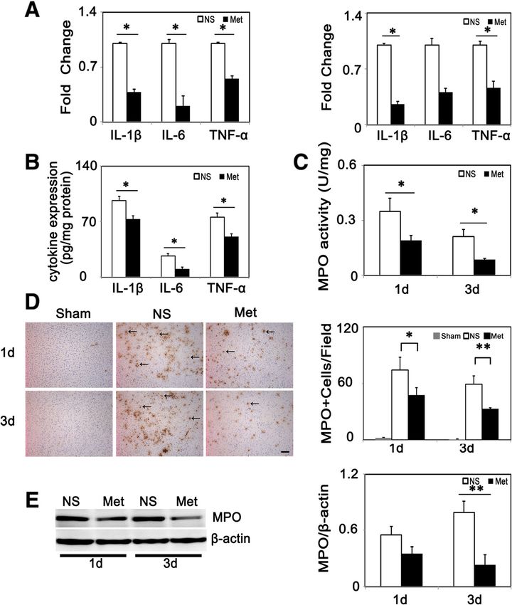
Liu et al. Journal of Neuroinflammation 2014, 11:177                                                                       Page 2 of 12
http://www.jneuroinflammation.com/content/11/1/177

death [5]. Metformin also reduces intercellular adhesion         divided into two groups that either underwent metformin
molecule-1 (ICAM-1) and vascular cell adhesion molecule-         or saline treatment. At 1 and 3 days after tMCAO, mice
1 (VCAM-1) levels in plasma and alleviates chronic in-           were sacrificed and samples were collected for further
flammation in patients [6]. Nevertheless, these effects were     study. Metformin (Sigma, St. Louis, MO, USA) was dis-
independent of its glycemic management properties, sug-          solved in sterile saline at a concentration of 30 mg/ml and
gesting that metformin may have other functions through          200 mg/kg was administered intra-peritoneally immediately
mechanisms other than glucose reduction. AMPK is a tri-          after reperfusion and then administered daily until the ani-
metric enzyme comprising a catalytic α-subunit and regu-         mals were sacrificed. An equal volume of saline was used
latory β- and γ-subunits [7]. An alteration in AMP/ATP           for the control group. The dose was chosen according to a
ratio activates AMPK and promotes AMPK phosphoryl-               previous study [20]. The whole experimental design and
ation at a threonine residue (Thr-172) [3]. A series of          the number of animal used are displayed in Figure 1.
pathological conditions such as glucose deprivation, ische-
mia, starvation and oxidative stress increase AMPK activ-        Transient middle cerebral artery occlusion (tMCAO)
ity [8]. Agents such as resveratrol and adiponectin can          in mice
also activate AMPK. AMPK activation is a protective reac-        tMCAO was carried out as previously described [21].
tion that occurs after injury. AMPK activated by metfor-         Adult CD1 mice weighing 30 ± 5 grams were anesthetized
min showed to reduce endothelial cell apoptosis and              with ketamine/xylazine (100 mg/10 mg/kg; Sigma, St.
diminish cardiomyocyte death [9,10]. It has been demon-          Louis, MO, USA) through intra-peritoneal injection. After
strated that increasing AMPK activity in neurons protects        isolation of the left common carotid artery, the external
neurons from various injuries [11]. In addition, metformin       carotid artery (ECA) and the internal carotid artery (ICA),
reduced TNF-α-induced inflammation via activation of             a silicone-coated 6-0 suture (Covidien, Mansfield, MA,
AMPK in vascular endothelial cells (ECs) [12]. It has been       USA) was gently inserted into the ICA and stopped at the
indicated that AMPK activation in cells in the immune            opening of the middle cerebral artery (MCA). Successful
system promotes the switch from a pro-inflammatory to            occlusion was ascertained by a decrease of surface cerebral
an anti-inflammatory phenotype [13]. Thus, metformin             blood flow to 10% of baseline using a laser Doppler flow-
could be a promising anti-inflammatory agent.                    metry (Moor Instruments, Devon, UK). Reperfusion was
   ICAM-1 is expressed on many cell types including ECs          performed 90 minutes after tMCAO with suture with-
and lymphocytes [14]. ICAM-1 is expressed constitutively         drawal. Sham-operated mice underwent the same proced-
on ECs at low level and its expression is significantly in-      ure except for the insertion of the suture into the ICA.
creased in hypoxic condition [15]. ICAM-1 expressed on           The mortality in our study was less than 5%.
ECs facilitates neutrophil adhesion and tissue infiltration,
which play critical roles in the progress of ischemic stroke     Infarct volume measurement
[16,17]. Infiltrated leukocytes induce a secondary injury        Infarct volume was measured using cresyl violet (Sigma,
after reperfusion by producing detrimental substances that       St. Louis, MO, USA) staining as previously described [22].
damage brain cells and disrupt the blood-brain barrier           The ischemic area of each section was depicted by image
(BBB) [18,19]. BBB disruption after ischemia increases
brain edema and exacerbates ischemic injury [14]. Since
ICAM-1 plays a vital role in neutrophil infiltration and
cerebral injury after reperfusion, it is a promising target in
the treatment of ischemic stroke.
   Studies have illustrated that metformin provides cardi-
oprotection against myocardial infarction [4]. However,
the function of metformin in inflammation after ische-
mic stroke is unknown. In our research, we explored
whether metformin could reduce ischemic brain injury
using a mouse transient middle cerebral artery occlusion
(tMCAO) model, and attempted to define the underlying
mechanism of metformin.

Methods
Experimental design                                               Figure 1 Experimental design. Graph illustrating the experimental
Animal protocol was approved by the Institutional Animal          design including transient middle cerebral artery occlusion (tMCAO),
                                                                  metformin administration, infarct volume, protein expression and
Care and Use Committee of Shanghai Jiao Tong University,
                                                                  neurobehavioral assessments.
Shanghai, China. Ninety-eight adult male CD1 mice were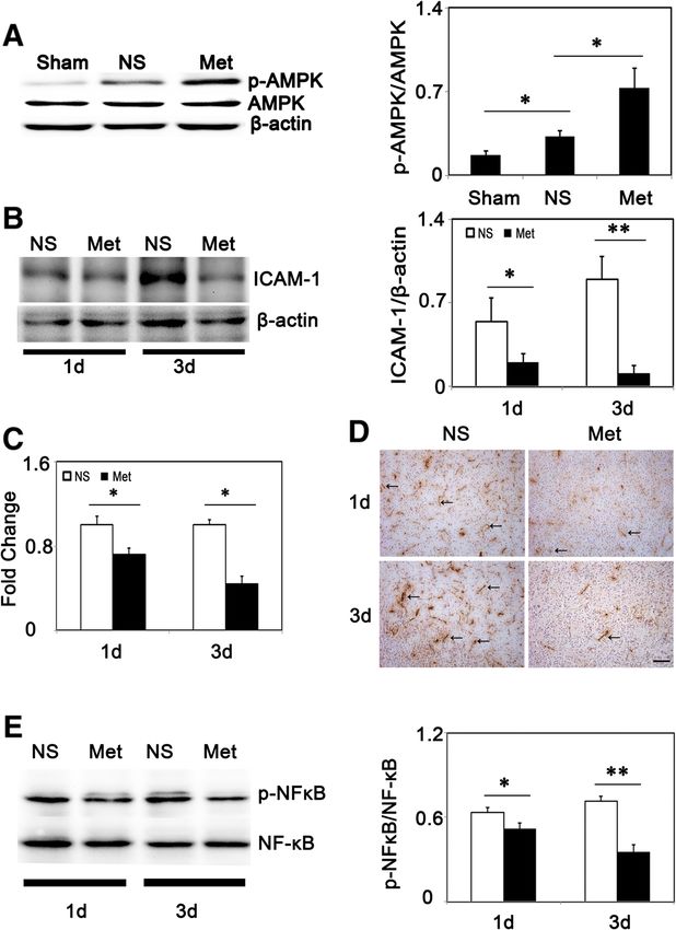
Liu et al. Journal of Neuroinflammation 2014, 11:177                                                            Page 3 of 12
http://www.jneuroinflammation.com/content/11/1/177

analysis software (Image J, NIH, MD, USA). Infarct             the treatment group. IgG leakage was examined as pre-
volume was calculated as described in our previous             viously reported and the procedure was similar to MPO
study [23].                                                    staining except for the primary antibody incubating
                                                               process [23]. Four areas of ischemic penumbra from
Neurobehavioral assessments                                    each slide were photographed. And mean optical dens-
Neurobehavioral assessments were conducted by an ex-           ity was measured using Image-Pro Plus software (Media
perimenter who was blind to the treatment conditions.          Cybernetics, Bethesda, MD, USA).
The rotarod test was used to evaluate the motor and bal-
ance functions of the mice. The mice were trained to           Western blot analysis
stay on an accelerating rotating cylinder for 3 days be-       Samples were lysed in radioimmunoprecipitation assay
fore tMCAO, and time remained on the rotating rod              (RIPA) (Millipore, Bedford, MA, USA) supplemented with
was recorded before surgery and at 1, 3, 7 and 14 days         1 mmol/L PMSF (Thermo, Waltham, MA, USA), cocktail
after surgery. The velocity was increased slowly from 4 to     (Thermo, Waltham, MA, USA) and phosphatase inhibitor
40 rpm within 2 minutes. For each test, every animal was       (Thermo, Waltham, MA, USA). For Western blot ana-
tested three times, and the average time maintained on         lyses, samples containing the same amount of proteins
the rod was recorded. For neurological function assess-        were loaded onto the resolving gel (Promoton, Shanghai,
ment, a modified Neurological Severity Scores (mNSS)           China) for electrophoresis after denaturing. Proteins were
ranging from 0 to 14 score was adopted, which included         transferred onto a nitrocellulose membrane (Whatman,
raising the mouse by the tail (0 to 3), walking on the floor   Piscataway, NJ, USA). After being blocked with 5% non-fat
(0 to 3), beam balance tests (0 to 6), and the relaxes ab-     milk, the membrane was incubated with primary antibodies
sence (0 to 2) [22].                                           at the following dilution MPO (1:500), ICAM-1 (1:2,000),
                                                               ZO-1 (1:500), occludin (1:500), claudin-5 (1:500), p-AMPK
Immunostaining                                                 (1:1,000, Cell Signaling Technology, Beverly, MA, USA),
Double staining: ZO-1/CD31, occludin/CD31 or claudin-          AMPK (1:1,000, Cell Signaling Technology, Beverly,
5/CD31 double staining was conducted as previously de-         MA, USA), p-NF-κB (1:1,000, Cell Signaling Technol-
scribed [23]. Briefly, brain sections were blocked with 10%    ogy, Beverly, MA, USA), NF-κB (1:1,000, Cell Signaling
FBS for 1 hour and then incubated with ZO-1 (1:100 dilu-       Technology, Beverly, MA, USA), β-actin (1:1,000, Santa
tion, Invitrogen, Carlsbad, CA, USA) and CD31 (1:200 di-       Cruz Technology, Santa Cruz, CA, USA) at 4°C overnight,
lution, R&D Systems, Minneapolis, MN, USA); occludin           respectively. After washing, the membrane was incubated
(1:100 dilution, Invitrogen, Carlsbad, CA, USA) and            with the appropriate horseradish peroxidase (HRP)-conju-
CD31; claudin-5 (1:100 dilution, Invitrogen, Carlsbad, CA,     gated secondary antibody for 1 hour and then reacted with
USA) and CD31 at 4°C overnight. After washing, brain           enhanced chemiluminescence substrate (Pierce, Rockford,
sections were incubated with the appropriate second anti-      IL, USA). The results were recorded by Quantity One
bodies for 1 hour. Brain sections were examined using a        image software (Bio-Rad, Hercules, CA, USA) and relative
confocal microscope (Leica, Solms, Germany) and photo-         intensity was calculated using Gel-Pro Analyzer software
graphs were taken for further analysis.                        (Media Cybernetics, Bethesda, MD, USA).
   DAB staining: for myeloperoxidase (MPO), ICAM-1 and
Gr1 (Ly 6G) immunostaining, brain sections were incu-          MPO activity assay
bated in 0.3% H2O2 in methanol for 30 minutes. After           MPO activity assay was performed as described previously
blocking with FBS, the primary anti-body MPO (1:300 dilu-      [23]. In brief, brain protein (10 μL) from ipsilateral hemi-
tion, R&D Systems, Minneapolis, MN, USA) and ICAM-1            sphere was added to 180 μL of work solution, which con-
(1:200 dilution, R&D Systems, Minneapolis, MN, USA),           tained 2 mmol/L O-dianisidin-dihydrochloride (Sigma)
Gr1 (1:100 dilution, Millipore, Darmstadt, Germany) were       dissolved in 180 μl of 50 mmol/L potassium phosphate buf-
incubated overnight at 4°C. Sections were incubated with       fer (pH =6). Before measurement, 10 μL of 100 mmol/L
biotinylated-conjugated secondary antibody (Vector La-         H2O2 was added. Changes in absorbance at 460 nm over
boratories, Burlingame, CA, USA) and then incubated with       10 minutes were measured. MPO activity was expressed
Vectastain ABC Reagent. The reaction product was vi-           as U/mg tissue, and 1U of MPO activity represents the
sualized using a DAB substrate (Vector Laboratories,           amount of enzyme degrading 1 μmol H2O2 per minute
Burlingame, CA, USA). Eight interested fields in each          at 25°C.
ipsilateral hemisphere, including the perifocal region in
both cortex and striatum, were photographed in each            Real-Time PCR
section and five consecutive sections spaced at 200 μm         Total RNA from the ischemic hemisphere was extracted
were counted in each mouse. MPO+ and Gr1+ (Ly 6G)              using TRIzol reagent (Invitrogen, Carlsbad, CA, USA)
cells were counted in each field by a person blinded to        and dissolved in 60 μL RNA free water according to the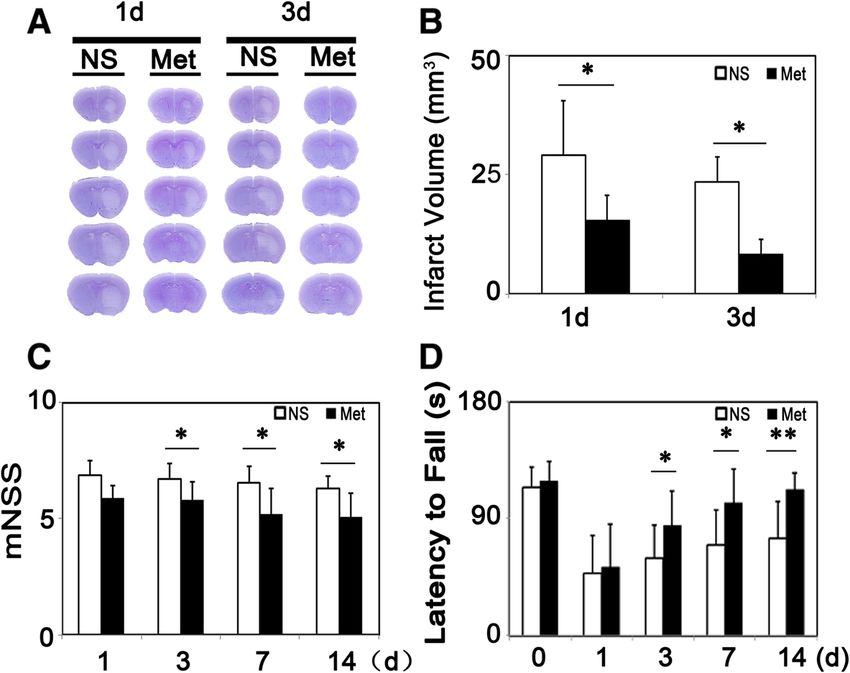
Liu et al. Journal of Neuroinflammation 2014, 11:177                                                            Page 4 of 12
http://www.jneuroinflammation.com/content/11/1/177

manufacturer’s instructions. A universal 2-step RT-PCR          Statistical analysis
cycling condition was used: 95°C for 30 seconds followed        Results were presented as mean ± SD. Statistical analysis
by 40 cycles of 95°C for 5 seconds and 60°C for 30 sec-         was evaluated by Prism 4 software (GraphPad Software,
onds. mRNA levels were normalized to the endogenous             San Diego, CA, USA). For comparison between the two
control, GAPDH expression, and were calculated using            groups, statistical significance was determined through a
fold change relative to the saline control group [23].          Student's t test. For comparison among multiple groups,
                                                                statistical significance was evaluated using one-way ANOVA
Enzyme Linked Immunosorbent Assay (ELISA) analysis              followed by a Student-Newman-Keuls test. A probability
Protein levels of IL-1β, IL-6, and TNF-α were quantified        value of P < 0.05 was considered statistically significant.
using an ELISA kit (R&D systems, Minneapolis, MN,
USA) according to the manufacturer’s instruction. Ab-           Results
sorbance at 450 nm was recorded and the concentration           Metformin reduced infarct volume and improved
of the target protein was read according to the standard        neurobehavioral outcomes
curve. Result was expressed as pg/mg protein.                   We found that after metformin treatment, infarct volume
                                                                was reduced at 1 and 3 days after tMCAO (Figure 2A-B).
Evans blue extravasation                                        To further explore the function of metformin, we used
Evans blue extravasation was measured as previously de-         mNSS to examine the motor, balance and reflex functions
scribed. In brief, 3 days after tMCAO, 4 ml/kg of 2% Evans      of mice after tMCAO. We showed that mice had
blue (Sigma, St. Louis, MO, USA) in saline was adminis-         significantly lower scores after 3 days in the metformin-
tered intraperitoneally. After 2 hours circulation, mice were   treated group after tMCAO for at least 14 days
anesthetized and perfused with saline through the left ven-     (Figure 2C). A similar result was obtained from the
tricle until colorless fluid outflowed from the right atrium.   rotarod test (Figure 2D). To investigate whether met-
Then, ipsilateral and contralateral hemispheres were col-       formin influenced glucose levels after tMCAO, fasting
lected after decapitated. Each hemisphere was weighed           blood-glucose (FBG) was tested before, 3 days and
rapidly, homogenized in 1 ml of 50% trichloroacetic acid        14 days after tMCAO, indicating metformin did not
(wt/vol). After centrifugation (12,000 × g, 20 minutes),        influence FBG level, blood gas or body weight after
supernatant was collected and mixed with ethanol (1:3).         both 3 (Table 1) days and 14 days treatment (Data
The concentration of Evans blue was determined by meas-         were shown in Additional file 1: Table S1)
uring the 610 nm absorbance and tissue content of Evans
blue was quantified from a linear standard curve and            Metformin alleviated neutrophil infiltration and IL-1β,
expressed in terms of Evans blue (μg)/tissue (g).               IL-6, TNF-α expression
                                                                To investigate the effect of metformin on neutrophil in-
Oxygen glucose deprivation                                      filtration in the acute phase of cerebral ischemia, we
bEND.3 was purchased from American Type Culture                 performed 3,3’-diaminobenzidine (DAB) staining to de-
Collection (ATCC) and cultured in DMEM (Gibco La-               tect MPO+ cells. Results showed MPO+ cells were al-
boratories, Grand Island, NY, USA) supplemented with            most undetectable in the sham group, and there was a
10% FBS. Ischemia-like conditions in vitro were induced         decrease in MPO+ cells at 1 and 3 days after tMCAO in
by oxygen glucose deprivation and reperfusion-like con-         the metformin-treated group compared to the control
ditions in vitro were induced by reoxygenation. After           group (Figure 3D). Western blot analysis indicated that
cells reached a 90% confluence, the medium was replaced         MPO were reduced in metformin-treated mice (P < 0.01,
with DMEM without glucose. Then, cultures were trans-           Figure 3E). MPO activity is an indicator of inflammation
ferred to an anaerobic chamber infused with a gas mixture       and could be used to evaluate neutrophil accumulation
containing 5% CO2, 95% N2. After incubating for 6 hours,        [23]. MPO activity was attenuated in the metformin-
cells were further cultured in DMEM supplemented with           treated group compared to the control group (P < 0.05,
10% FBS under normal conditions for another 24 hours            Figure 3C). In addition, we used another neutrophil
with or without metformin. p-AMPK analysis in bEND.3            marker Gr1 (Ly 6G) to evaluate neutrophil infiltration
cells was determined 1 hour, 4 hours, and 24 hours after        after cerebral ischemia. Results showed that metformin re-
6 hours oxygen glucose deprivation (OGD). The dose used         duced Gr1 positive cells effectively at 1 day and 3 days
was 10 mM, which was chosen according to previous re-           after tMCAO (Additional file 2: Figure S1, P < 0.01). We
ports [20]. Compound C (Sigma, St. Louis, MO, USA), a           further used RT-PCR and ELISA to evaluate changes in
selective AMPK inhibitor was added to the medium at a           inflammation-related cytokine expression in mRNA and
final concentration of 10 μmol/L before OGD treatment           protein levels. Metformin reduced IL-β, IL-6, TNF-α
and maintained throughout the whole experiment [24].            mRNA at 1 day after tMCAO. Although there was a
An equal volume of PBS was used in the control group.           downward tendency in IL-6 mRNA, only changes in IL-β,
Liu et al. Journal of Neuroinflammation 2014, 11:177 Page 5 of 12
http://www.jneuroinflammation.com/content/11/1/177
Figure 2 Metformin reduced infarct volume and improved neurobehavioral outcomes following transient middle cerebral artery (tMCAO)
in mice. (A) Photographs show a series of coronal sections with cresyl violet staining following tMCAO in metformin- and saline-treated mice. (B) Bar
graph shows a quantification of the infarct volume from (A). n =6 per group. The changes in neurological scores (C) and the rotarod test (D) at 1, 3, 7
and 14 days following tMCAO in metformin- and saline-treated mice. n =9 per group, data are mean ± SD, *P < 0.05, metformin versus control group,
**P < 0.01, metformin versus control group.
and TNF-α were statistically significant in the metformin Metformin reduced BBB disruption
group compared to the control group at 3 days after To evaluate endothelial cell permeability after metformin
tMCAO (Figure 3A). There was a decrease of IL-β, IL-6, treatment, we conducted occludin/CD31, ZO-1/CD31 and
TNF-α expression at 3 days after tMCAO in protein level claudin-5/CD31 double staining to observe tight junction
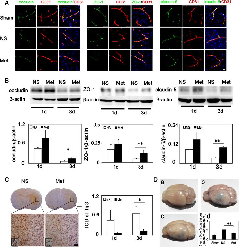
(Figure 3B, P < 0.01). distribution in situ at 3 days after tMCAO. Result indi-
cated that occludin and ZO-1 were continuously located
Table 1 Metformin did not influence blood gas, glucose on the margin of ECs in sham group, claudin-5 was con-
level and body weight in transient middle cerebral artery tinuously located along ECs, and fewer gaps were formed
occlusion (tMCAO) mice in the metformin-treated group (Figure 4A). Gap forma-
Before tMCAO After tMCAO tion and rearrangement were used to evaluate tight junc-
NS Met NS Met tion disruption after injury. To evaluate tight junction
pH 7.35 ± 0.05 7.34 ± 0.03 7.34 ± 0.07 7.35 ± 0.03
rearrangement, Western blot was adopted and we found
that metformin-treated mice demonstrated occludin, ZO-
PCO2 (mmHg) 38 ± 5 33 ± 3 30 ± 7 30 ± 5
1 and claudin-5 hyper-expression (Figure 4B). In addition,
PO2 (mmHg) 62 ± 4 60 ± 7 64 ± 2 66 ± 12 we performed IgG immunostaining and Evans blue ex-
SO2 (%) 91 ± 8% 87 ± 10% 91 ± 2% 91 ± 5% travasation to evaluate endothelial permeability and found
Na(mmol/L) 155 ± 2 153 ± 2 154 ± 3 152 ± 2 that there was significantly reduced IgG and Evans blue
K (mmol/L) 3.2 ± 0.1 3.1 ± 0.4 3.5 ± 0.3 3.9 ± 0.2 leakage at 3 days after tMCAO in metformin-treated mice
iCa (mmol/L) 1.37 ± 0.02 1.34 ± 0.06 1.22 ± 0.04 1.26 ± 0.04
(Figure 4C-D).
Glu (mg/dL) 132 ± 20 127 ± 33 123 ± 16 129 ± 24
Metformin down-regulated ICAM-1 expression via AMPK
Hct (%PCV) 37 ± 1% 35 ± 1% 34 ± 6% 35 ± 2% signaling pathway
Hb (g/dL) 12.4 ± 0.4 11.9 ± 0.3 11.4 ± 2.2 11.8 ± 0.7 To assess the phosphorylation status of AMPK at threonine
Body weight (g) 33 ± 2 32 ± 1 25 ± 6 28 ± 4 residue, Western blot was used. We demonstrated that
Table showed vein blood gas analysis results, glucose levels and body weight ischemia-reperfusion increased AMPK phosphorylation
at 1 day before tMCAO and at 3 days after tMCAO in metformin-(Met) and and this induction was increased after metformin treatment
saline(NS)-treated mice (n =4 per group). Data were mean ± SD.
Legend: Glu, glucose; Hb, hemoglobin; Hct, hematocrit; PCO2, partial pressure of
(Figure 5A). To further explore mechanisms of metformin
CO2; PO2, partial pressure of O2; PCV, packed cell volume; SO2, oxygen saturation. in neuroprotection, we analyzed ICAM-1 expression afterLiu et al. Journal of Neuroinflammation 2014, 11:177 Page 6 of 12 http://www.jneuroinflammation.com/content/11/1/177 Figure 3 Metformin alleviated neutrophil infiltration and inflammatory cytokine expression in mice. (A) Relative fold changes of inflammatory cytokines (IL-1β, IL-6, TNF-α in metformin and control mice at 1 (left) and 3 days (right) following tMCAO (n =3 per group). (B) Protein level of inflammatory cytokines IL-1β, IL-6, TNF-α expression in metformin and control mice at 3 days following tMCAO (n =3 per group). (C) Bar graph shows MPO activity in metformin and control mice (n =3 per group). (D) MPO+ cells (arrows) and their quantification in sham group, control and metformin-treated mice at 1 and 3 days following tMCAO. Scale bar =100 μm. (E) Western blot of MPO expression in metformin and control mice. Bar graph shows a quantification of MPO. Data are mean ± SD, *P < 0.05, **P < 0.01, metformin versus control group. tMCAO. ICAM-1 was expressed constituently at low level metformin after 6 hours of OGD, there was also an in- in ECs, and after cerebral ischemia its expression was crease in p-AMPK expression 1 hour after reoxygenation elevated hugely (Figure 5D). Furthermore, Western blot and p-AMPK levels were unregulated for at least 24 hours and RT-PCR results indicated that ICAM-1 was reduced after reoxygenation (Figure 6B). Second, we examined after metformin-treatment compared to the control group ICAM-1 expression after 6 hours of OGD and reoxygena- (Figure 5B-C). We also found that metformin inhibited tion with or without metformin. After OGD/reoxygena- NF-κB phosphorylation at 1 day and 3 days after tMCAO tion treatment, ICAM-1 expression was up-regulated in (Figure 5E). To determine whether metformin-induced mRNA level and metformin inhibited this up-regulation: down-regulation of ICAM-1 was AMPK-dependent or the inhibitive effects began at 4 hours after reoxygenation not, we analyzed the effect of metformin at a cellular level, and were sustained for at least 24 hours after reoxygena- using OGD models to mimic in vivo ischemia/reperfusion tion (Figure 6C). To test whether AMPK signaling was in- injury. First, we treated bEND.3 cells with metformin to volved, a selective AMPK inhibitor, compound C, was used determine whether metformin could also increase p- to block the AMPK phosphorylation. First, we found that AMPK in vitro. We found that there was an increase in p- compound C reduced metformin-induced AMPK phos- AMPK expression at 60 and 120 minutes after metformin phorylation, then, we used RT-PCR and Western blot to as- treatment: the maximum effect was at 60 minutes in nor- sess ICAM-1 expression after treatment. Results indicated mal conditions (Figure 6A) and while treatment with that metformin reduced ICAM-1 expression in both
Liu et al. Journal of Neuroinflammation 2014, 11:177                                                                                     Page 7 of 12
http://www.jneuroinflammation.com/content/11/1/177

 Figure 4 Metformin promoted ZO-1, occludin and claudin-5 rearrangement and lessened IgG and Evans blue extravasation. (A) Occludin,
 ZO-1 and claudin-5 expression in sham group, NS and metformin-treated mice at 3 days following tMCAO. Scale bar =10 μm. (B) Representative result of
 occludin, ZO-1 and claudin-5 expression at 1 and 3 days after tMCAO. Bar graphs show a quantification of occludin, ZO-1 and claudin-5 expression.
 (C) IgG leakage at 3 days following tMCAO in saline and metformin-treated group. Higher magnifications are shown below. Boxes displayed
 representative IgG staining. Scale bar =1 mm (upper) and 100 μm (lower). Bar graph shows a semi-quantification of integrated optical density
 (IOD) of IgG at 1 and 3 days after tMCAO (n =3 per group). (D) Images show Evans blue extravasation in sham (a), saline (b) and metformin (c)
 group at 3 days after tMCAO. Blue area indicates extravasation of Evans blue. Bar graph shows a quantification analysis of Evans blue contents
 in brain tissue (n =3 per group). Data were mean ± SD, *P < 0.05, metformin versus saline group, **P < 0.01, metformin versus saline group.

mRNA and protein levels under OGD/reoxygenation con-                         phosphorylation and this function was abolished by com-
ditions (Figure 6D-E). Furthermore, we used Western blot                     pound C (Figure 6G). Thus, we concluded that metformin
to evaluate the effects of metformin on NF-κB activation;                    diminished ICAM-1 via an AMPK mediated signaling
the result indicated that metformin inhibited NF-κB                          pathway and AMPK-NF-κB might be involved.
Liu et al. Journal of Neuroinflammation 2014, 11:177                                                                                    Page 8 of 12
http://www.jneuroinflammation.com/content/11/1/177

 Figure 5 Metformin promoted phosphorylation of AMPK and reduced ICAM-1 expression in transient middle cerebralartery occlusion
 (tMCAO) mice. (A) p-AMPK and AMPK expression in sham, saline- and metformin-treated groups at 1 day after tMCAO. Bar graph showed a
 quantification of p-AMPK/AMPK ratio (n =3/group). (B) ICAM-1 expression and quantification at 1 and 3 days after tMCAO in metformin and
 control mice (n =3 per group). (C) Fold change of ICAM-1 expression in mRNA level in metformin and control mice at 1 day and 3 days after tMCAO
 (n =3 per group). (D) ICAM-1 expression in control and metformin-treated mice at 1 and 3 days after tMCAO. Arrows indicated representative ICAM-1
 expression. Scale bar =100 μm. (E) p-NF-κB and NF-κB expression in saline- and metformin-treated groups at 1 day and 3 days after tMCAO. Bar graph
 show a quantification of p-NF-κB/NF-κB ratio (n =3 per group). Data are mean ± SD, *P < 0.05, metformin versus control, sham versus control group,
 **P < 0.01, metformin versus control group.

Discussion                                                                  exerts its protective effect after cerebral ischemia partly
In the present study, we demonstrated that metformin pro-                   through diminished ICAM-1 expression.
tected the brain from ischemic injury through alleviating                      Metformin is a glucose-lowering agent, and is one of the
inflammatory responses in tMCAO mice, thus improving                        first-line drugs recommended to treat type II diabetes mel-
long-term recovery. Metformin diminished neutrophil infil-                  litus [25]. The UK Prospective Diabetes Study (UKPDS)
tration, thereby alleviating endothelial injury and lowering                has revealed that metformin reduced the risk of all-cause
BBB permeability. These effects were potentially mediated                   mortality and stroke clinically; however, these benefits
via an AMPK-dependent ICAM-1 down-regulation. We                            were independent of its anti-hyperglycemic effects, since
also found that inhibiting AMPK activation by compound                      metformin reduced glycated hemoglobin (HbA1c) to the
C could reverse metformin-induced down-regulation of                        same extent as sulphonylurea and insulin [5]. Metformin
ICAM-1 in vitro. Thus, we concluded that metformin                          decreased myocardial injury in non-diabetic and diabetic
Liu et al. Journal of Neuroinflammation 2014, 11:177   Page 9 of 12
http://www.jneuroinflammation.com/content/11/1/177

 Figure 6 (See legend on next page.)
Liu et al. Journal of Neuroinflammation 2014, 11:177                                                                              Page 10 of 12
http://www.jneuroinflammation.com/content/11/1/177

 (See figure on previous page.)
 Figure 6 Metformin promoted phosphorylation of AMPK and reduced ICAM-1 expression in vitro in an AMPK-dependent manner. (A) p-AMPK
 and AMPK expression and quantification at 30, 60 and 120 minutes after metformin treatment in vitro in normal conditions. (B) p-AMPK and
 AMPK expression and quantification in control, 1, 4 and 24 hours in reoxygenation group in the presence of metformin in vitro. (C) Fold change
 of ICAM-1 expression in mRNA level 1, 4 and 24 hours after reoxygenation in saline- and metformin-treated group. (D) ICAM-1 expression in
 mRNA after metformin and AMPK inhibitor and compound C treatment in the oxygen glucose deprivation (OGD) model. (E) ICAM-1 expression and
 quantification after metformin, AMPK inhibitor and compound C treatment in OGD model. (F) p-AMPK and AMPK expression and quantification after
 metformin and compound C treatment in OGD model. (G) p-NF-κB and NF-κB expression and quantification after metformin and compound C
 treatment in OGD model. Data were mean ± SD, *P < 0.05, **P < 0.01. Representative results from three independent experiments are shown. Com.
 C = compound C.

mice [4] and prevented the progression of heart failure in                the mTOR signaling pathway, AMPK regulates cell
dogs [10]. However, reports regarding neuroprotection of                  growth and autophagy [7]. Via activation of the Nrf2/
metformin in cerebral ischemia were controversial. Using                  SKN-1 signaling pathway, AMPK increases antioxidant
a 90-minute tMCAO model, McCullough demonstrated                          gene expression [32]. By promotion of the eNOS pathway,
that chronic treatment, both pre- and post- (3 weeks),                    AMPK reduces endothelial cell apoptosis and improves
with metformin reduced infarct volume effectively; how-                   endothelial functions [9]. Furthermore, emerging evidence
ever, pre-treatment (3 days) enhanced injury in ischemic                  shows that AMPK is beneficial to neurons suffering from
stroke [26]. Harada et al. showed that using 3-day metfor-                injuries such as ischemia, starvation, and oxidative damage
min treatment after 2 hours of tMCAO, metformin effect-                   [11]. Collectively, these results suggest that metformin-
ively reduced infarct volume [27], which was consistent                   AMPK signaling pathways exert protective effects in stress
with our result. In addition, Li has suggested that chronic               conditions and protect the brain from ischemic stroke.
treatment (14 days in drinking water, 300 mg/kg) with                        The mechanism by which metformin reduces inflamma-
metformin in diabetic rats was protective, but acute treat-               tion after cerebral ischemia is poorly understood. ICAM-1
ment (1 day in drinking water, 300 mg/kg) exerted differ-                 expression can be regulated by the nuclear transcriptional
ent effects [28]. However, we must note that different                    factor NF-κB [33]. Recently, the potency of metformin
models were used. For acute treatment, 3-hour occlusion                   blocking NF-κB signaling has been illustrated [34]. In
and 21 hours reperfusion model were used; however, for the                addition, metformin decreased TNF-α-induced ICAM-1
chronic treatment, a 90-minute occlusion and 14 days re-                  by inhibiting NF-κB activation in ECs [12]. Therefore, we
perfusion model was adopted, and glucose level was normal                 suppose that metformin reduced ICAM-1 expression via
in chronic treatment rats. Thus, different animal strains and             the AMPK-NF-κB pathway. We also detected an inhib-
different models may explain the different effects observed               ition of NF-κB activation after metformin treatment both
from different studies [26]. In our study, using a 90-minute              in vivo and in vitro. Previous study showed that there was
tMCAO model in mice and treatment at the time of reper-                   a huge increase of ICAM-1 expression at the time of re-
fusion, we demonstrated that metformin could alleviate                    perfusion; in our study, metformin was administrated at
ischemic injury and improve neurobehavioral outcomes.                     the time of reperfusion. Increased expression of adhesion
   We demonstrated the protection by metformin on is-                     cytokine is detrimental during ischemic injury since it in-
chemic stroke involved, at least in part, AMPK activity                   creases neutrophil adhesion to ECs and thus promotes
[3]. However, McCullough’s group found that metformin                     their infiltration [16]. Anti-ICAM-1 treatment significantly
enhanced ischemic injury in tMCAO animals in a 3-day                      reduced infarct volume [15]. Coincidently, we demon-
precondition study through activating AMPK [26]. We                       strated that metformin decreased ICAM-1 both in vivo
believe this result can be due to the period of treatment.                and in vitro, and this reduction in ICAM-1 in vivo was ac-
Metformin could increase AMPK phosphorylation both                        companied by alleviated neutrophil infiltration and re-
in vitro and in vivo [4,9]. We detected an induction of                   duced infarct size in tMCAO mice. Furthermore, our
AMPK phosphorylation in both bEND.3 cells and the                         study indicated that this effect was possibly mediated by
mouse brain after tMCAO. Notably, conditions that                         AMPK in a dependent manner. We concluded that met-
could activate AMPK have been proven to be beneficial                     formin conferred resistance to ischemic stroke through
in stress, particularly in ischemia. Ischemic precondition,               decreasing ICAM-1 via the AMPK signaling pathway.
which can activate AMPK due to an increase of the                            Besides increasing inflammation, neutrophil infiltration
AMP/ATP ratio, is supposed to be protective in ischemic                   also induces EC injury and increases BBB permeability
stroke [29]. Adiponectin reduces infarct size in cerebral is-             [35]. BBB disruption exacerbates brain injury after ische-
chemia and myocardial injury has been shown to be partly                  mia. Injuries such as ischemia and trauma lead to a dis-
through promoting AMPK phosphorylation [30,31]. In                        ruption and reconstruction of ZO-1 and occludin, and an
addition, AMPK involves pleiotropic pathways that play                    increase in BBB permeability [23]. Reduction of BBB per-
critical roles in cerebral ischemia. Through suppression of               meability alleviates cerebral ischemia injury in both
Liu et al. Journal of Neuroinflammation 2014, 11:177                                                                                                   Page 11 of 12
http://www.jneuroinflammation.com/content/11/1/177

transient and permanent cerebral ischemia [19,22,23]. Re-                          Author details
                                                                                   1
cently, it has been reported that metformin-induced im-                             Department of Neurology, Ruijin Hospital, School of Medicine, Shanghai Jiao
                                                                                   Tong University, Shanghai 200025, China. 2Neuroscience and Neuroengineering
provement of BBB functions in ECs in vitro is due to                               Research Center, Med-X Research Institute and School of Biomedical Engineering,
activating AMPK activity [24]. In the present study, we                            Shanghai Jiao Tong University, Shanghai 200030, China. 3Department of
demonstrated that after metformin treatment, IgG and Ev-                           Neurology, Med-X Research Institute and School of BME, Ruijin Hospital, School of
                                                                                   Medicine, Shanghai Jiao Tong University, 1954 Hua-shan Road, Shanghai 200030,
ans blue leakage was significantly reduced and tight junc-                         China.
tion protein profoundly increased, leading to better
outcomes in tMCAO mice.                                                            Received: 18 April 2014 Accepted: 30 September 2014

Conclusions
                                                                                   References
We demonstrated that metformin is beneficial to the                                1. Mathers CD, Boerma T, Ma Fat D: Global and regional causes of death.
treatment of ischemic stroke, which is possible through                                Br Med Bull 2009, 92:7–32.
inhibiting inflammation via AMPK signaling pathways.                               2. Kleindorfer D, Lindsell CJ, Brass L, Koroshetz W, Broderick JP: National US
                                                                                       estimates of recombinant tissue plasminogen activator use: ICD-9 codes
Since metformin is a widely used drug with few adverse                                 substantially underestimate. Stroke 2008, 39:924–928.
effects, using this long-established drug for a new use                            3. Martin-Montalvo A, Mercken EM, Mitchell SJ, Palacios HH, Mote PL,
may be a promising way to develop an effective therapy                                 Scheibye-Knudsen M, Gomes AP, Ward TM, Minor RK, Blouin MJ, Schwab M,
                                                                                       Pollak M, Zhang Y, Yu Y, Becker KG, Bohr VA, Ingram DK, Sinclair DA, Wolf
for ischemic stroke [36]. Metformin has the potential to                               NS, Spindler SR, Bernier M, de Cabo R: Metformin improves healthspan
be useful in the clinical treatment of ischemic stroke.                                and lifespan in mice. Nat Commun 2013, 4:2192.
                                                                                   4. Calvert JW, Gundewar S, Jha S, Greer JJ, Bestermann WH, Tian R, Lefer DJ:
                                                                                       Acute metformin therapy confers cardioprotection against myocardial
                                                                                       infarction via AMPK-eNOS-mediated signaling. Diabetes 2008, 57:696–705.
Additional files                                                                   5. Group. UPDSU: Effect of intensive blood-glucose control with metformin on
                                                                                       complications in overweight patients with type 2 diabetes (UKPDS 34). UK
 Additional file 1: Table S1. Metformin did not influence blood gas,                   Prospective Diabetes Study (UKPDS) Group. Lancet 1998, 352:854–865.
 glucose level and body weight 14 days after transient middle cerebral             6. Diamanti-Kandarakis E, Paterakis T, Alexandraki K, Piperi C, Aessopos A,
 artery occlusion (tMCAO) in mice.                                                     Katsikis I, Katsilambros N, Kreatsas G, Panidis D: Indices of low-grade
 Additional file 2: Figure S1. Metformin reduced neutrophil infiltration               chronic inflammation in polycystic ovary syndrome and the beneficial
 in transient middle cerebral artery occlusion (tMCAO) mice. Gr1 (Ly 6G)+              effect of metformin. Hum Reprod 2006, 21:1426–1431.
 cells (arrows) and their quantification in control and metformin treated          7. Mihaylova MM, Shaw RJ: The AMPK signalling pathway coordinates cell
 mice at 1 and 3 days following tMCAO (n = 3/group). Scale bar = 100 μm.               growth, autophagy and metabolism. Nat Cell Biol 2011, 13:1016–1023.
 Data are mean ± SD, **P
Liu et al. Journal of Neuroinflammation 2014, 11:177 Page 12 of 12
http://www.jneuroinflammation.com/content/11/1/177
20. Wang J, Gallagher D, DeVito LM, Cancino GI, Tsui D, He L, Keller GM,
Frankland PW, Kaplan DR, Miller FD: Metformin activates an atypical
PKC-CBP pathway to promote neurogenesis and enhance spatial
memory formation. Cell Stem Cell 2012, 11:23–35.
21. He X, Li Y, Lu H, Zhang Z, Wang Y, Yang GY: Netrin-1 overexpression
promotes white matter repairing and remodeling after focal cerebral
ischemia in mice. J Cereb Blood Flow Metab 2013, 33:1921–1927.
22. Liu Y, Tang GH, Sun YH, Lin XJ, Wei C, Yang GY, Liu JR: The protective role
of Tongxinluo on blood-brain barrier after ischemia-reperfusion brain
injury. J Ethnopharmacol 2013, 148:632–639.
23. Huang J, Li Y, Tang Y, Tang G, Yang GY, Wang Y: CXCR4 antagonist AMD3100
protects blood-brain barrier integrity and reduces inflammatory response
after focal ischemia in mice. Stroke 2013, 44:190–197.
24. Takata F, Dohgu S, Matsumoto J, Machida T, Kaneshima S, Matsuo M,
Sakaguchi S, Takeshige Y, Yamauchi A, Kataoka Y: Metformin induces
up-regulation of blood-brain barrier functions by activating AMP-activated
protein kinase in rat brain microvascular endothelial cells. Biochem Biophys
Res Commun 2013, 433:586–590.
25. Force ICGT: Global Guideline for Type 2 Diabetes: recommendations for
standard, comprehensive, and minimal care. Diabet Med 2006, 23:579–593.
26. Li J, Benashski SE, Venna VR, McCullough LD: Effects of metformin in
experimental stroke. Stroke 2010, 41:2645–2652.
27. Harada S, Fujita-Hamabe W, Tokuyama S: The importance of regulation of
blood glucose levels through activation of peripheral 5'-AMP-activated
protein kinase on ischemic neuronal damage. Brain Res 2010, 1351:254–263.
28. Li W, Qu Z, Prakash R, Chung C, Ma H, Hoda MN, Fagan SC, Ergul A:
Comparative analysis of the neurovascular injury and functional
outcomes in experimental stroke models in diabetic Goto-Kakizaki rats.
Brain Res 2013, 1541:106–114.
29. Gidday JM: Cerebral preconditioning and ischaemic tolerance. Nat Rev
Neurosci 2006, 7:437–448.
30. Shibata R, Sato K, Pimentel DR, Takemura Y, Kihara S, Ohashi K, Funahashi T,
Ouchi N, Walsh K: Adiponectin protects against myocardial ischemia-
reperfusion injury through AMPK- and COX-2-dependent mechanisms.
Nat Med 2005, 11:1096–1103.
31. Nishimura M, Izumiya Y, Higuchi A, Shibata R, Qiu J, Kudo C, Shin HK,
Moskowitz MA, Ouchi N: Adiponectin prevents cerebral ischemic injury
through endothelial nitric oxide synthase dependent mechanisms.
Circulation 2008, 117:216–223.
32. Salminen A, Kaarniranta K: AMP-activated protein kinase (AMPK) controls
the aging process via an integrated signaling network. Ageing Res Rev
2012, 11:230–241.
33. Tak PP, Firestein GS: NF-kappaB: a key role in inflammatory diseases. J Clin
Invest 2001, 107:7–11.
34. Isoda K, Young JL, Zirlik A, MacFarlane LA, Tsuboi N, Gerdes N, Schonbeck U,
Libby P: Metformin inhibits proinflammatory responses and nuclear
factor-kappaB in human vascular wall cells. Arterioscler Thromb Vasc Biol
2006, 26:611–617.
35. Chopp M, Zhang ZG, Jiang Q: Neurogenesis, angiogenesis, and MRI
indices of functional recovery from stroke. Stroke 2007, 38:827–831.
36. Potts MB, Lim DA: An old drug for new ideas: metformin promotes adult
neurogenesis and spatial memory formation. Cell Stem Cell 2012, 11:5–6.
doi:10.1186/s12974-014-0177-4
Cite this article as: Liu et al.: Metformin attenuates blood-brain barrier
disruption in mice following middle cerebral artery occlusion. Journal
of Neuroinflammation 2014 11:177.
Submit your next manuscript to BioMed Central
and take full advantage of:
• Convenient online submission
• Thorough peer review
• No space constraints or color figure charges
• Immediate publication on acceptance
• Inclusion in PubMed, CAS, Scopus and Google Scholar
• Research which is freely available for redistribution
Submit your manuscript at
www.biomedcentral.com/submitYou can also read