Nicotinamide mononucleotide attenuates glucocorticoid induced osteogenic inhibition by regulating the SIRT1/PGC 1α signaling pathway
←
→
Page content transcription
If your browser does not render page correctly, please read the page content below
Molecular Medicine REPORTS 22: 145-154, 2020
Nicotinamide mononucleotide attenuates
glucocorticoid‑induced osteogenic inhibition by
regulating the SIRT1/PGC‑1α signaling pathway
RUI‑XIONG HUANG1,2 and JUN TAO1
1
Department of Orthopedics, The Second Affiliated Hospital of Nanchang University, Nanchang,
Jiangxi 330000; 2Department of Orthopedics, Gao'an People's Hospital, Gao'an, Jiangxi 330800, P.R. China
Received August 19, 2019; Accepted March 3, 2020
DOI: 10.3892/mmr.2020.11116
Abstract. Long‑term and high‑dose glucocorticoid treatment spine (anteroposterior), femoral neck and total hip, and/or 33%
is recognized as an important influencing factor for osteo- (one‑third) radius, even in the absence of a prevalent fracture.
porosis and osteonecrosis. Nicotinamide mononucleotide Osteoporosis may also be diagnosed in patients with osteopenia
(NMN) is an intermediate of NAD + biosynthesis, and is and increased fracture risk, using FRAX® country‑specific
widely used to replenish the levels of NAD +. However, the thresholds. Osteopenia is defined as T‑score between ‑1.0 and
potential role of NMN in glucocorticoid‑induced osteo- ‑2.5, based on bone mineral density testing (3). There are two
genic inhibition remains to be demonstrated. In the present major categories of osteoporosis: Primary and secondary.
study, the protective effects of NMN on dexamethasone Sex and age are the primary influencing factors for primary
(Dex)‑induced osteogenic inhibition, and its underlying osteoporosis, whereas secondary osteoporosis is associated
mechanisms, were investigated. Bone mesenchymal stem with long‑term and high‑dose glucocorticoid treatment (4).
cells were treated with Dex, which decreased the levels of the Glucocorticoids such as dexamethasone (Dex) and hydrocorti-
osteogenic markers alkaline phosphatase, Runt‑related tran- sone are widely used to treat inflammation and immunological
scription factor 2 and osteocalcin. NMN treatment attenuated rejection, as well as autoimmune diseases (5). These diseases
Dex‑induced osteogenic inhibition and promoted the expres- not only include rheumatoid arthritis and systemic lupus
sion of sirtuin 1 (SIRT1) and peroxisome proliferator‑activated erythematosus, but also asthma, chronic obstructive pulmo-
receptor gamma coactivator (PGC)‑1α. SIRT1 knockdown nary disease, Crohn's disease and ulcerative colitis (6‑12).
reversed the protective effects of NMN and reduced the Dex‑induced osteogenic inhibition has been considered as the
expression levels of PGC‑1α. Collectively, the results of the most severe side‑effect of this particular treatment type (13),
present study reveal that NMN may be a potential therapeutic and long‑term administration of glucocorticoids can result in
target for glucocorticoid‑induced osteoporosis. osteoporosis or osteonecrosis (14). However, the molecular
mechanism underlying glucocorticoid‑induced osteogenic
Introduction inhibition is not clear.
Nicotinamide mononucleotide (NMN) is an important
Osteoporosis is a chronic disease with a heavy global NAD+ intermediate whose levels decrease with age. As such,
socioeconomic burden. It is defined as a skeletal disorder, NMN administration is an effective treatment for age‑related
characterized by decreased bone strength, which in turn predis- diseases and bone metabolism (Table I), and can be used to
poses affected individuals to fractures (1) and damage to the alleviate age‑related type 2 diabetes, ischemia‑reperfusion
bone microarchitecture (2). Osteoporosis is diagnosed based injury and Alzheimer's disease (15). NMN has previously
on the presence of fragility fractures in the absence of other been reported to improve osteogenesis by regulating sirtuin 1
metabolic bone disorders, or a T‑score of ≤2.5 in the lumbar (SIRT1) (16). The present study aimed to investigate the role
of NMN against the glucocorticoid‑induced loss of bone cell
viability via mesenchymal stromal cell (MSC) regulation.
MSCs are non‑hematopoietic pluripotent stem cells with
regenerative capacity, and with age, a reduction in the number
Correspondence to: Professor Jun Tao, Department of Orthopedics,
The Second Affiliated Hospital of Nanchang University, 1 Minde or function of MSCs severely limits tissue regeneration (17).
Road, Donghu, Nanchang, Jiangxi 330000, P.R. China However, the mechanism of action of NMN in glucocorticoid‑
E‑mail: 2431835455@qq.com induced loss of bone cell viability remains unclear.
SIRT1, also known as silent mating type information
Key words: nicotinamide mononucleotide, osteoporosis, sirtuin 1, regulation 2 homolog, was discovered in humans in 1999 (18),
peroxisome proliferator‑activated receptor gamma coactivator and is an NAD‑dependent class III protein deacetylase (19).
As a deacetylase, SIRT1 is closely associated with various
other proteins such as p53, Ku70, forkhead box protein O1,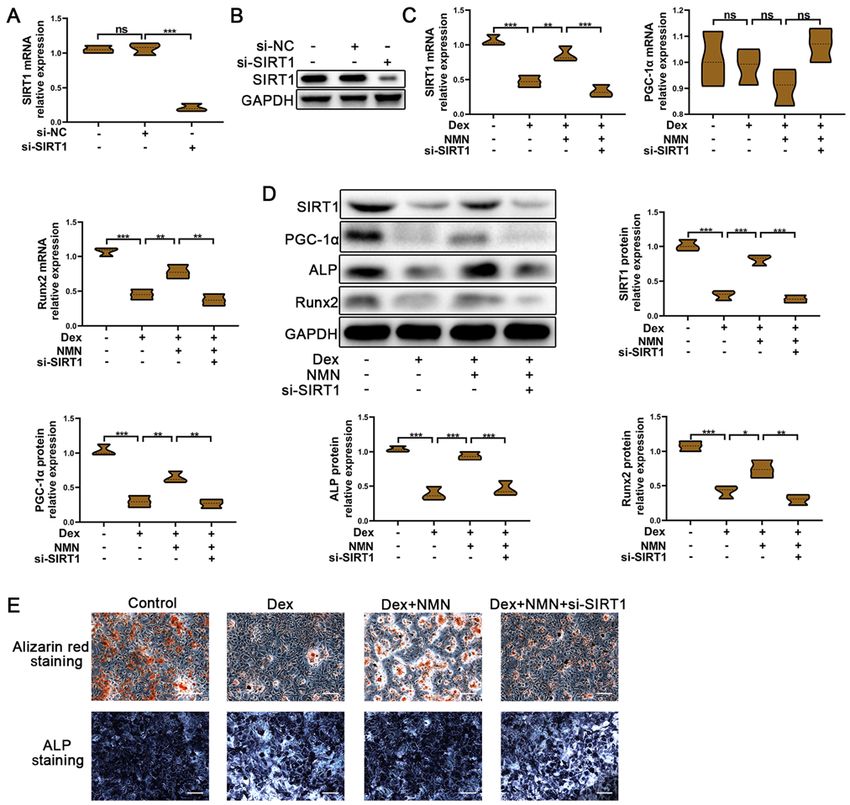
146 HUANG and TAO: NICOTINAMIDE MONONUCLEOTIDE ALLEVIATES GLUCOCORTICOID‑INDUCED OSTEOPOROSIS
NF‑κ B, peroxisome proliferator‑activated receptor‑ γ and RT‑qPCR analysis. Total RNA was isolated from cells using
p300, and is involved in the regulation of cell senescence and TRIzol® reagent (Invitrogen; Thermo Fisher Scientific, Inc.)
apoptotic death under stress conditions, thereby enhancing and quantified using an absorbance measurement at a wave-
cellular activity, self‑healing and survival capacity (20‑23). length of 260 nm. The RNA was reverse‑transcribed into
Numerous studies have reported the involvement of SIRT1 cDNA using a SYBR® PrimeScript™ RT‑PCR kit (Takara
in bone metabolism, and as a mediator of bone mass regula- Bio, Inc.) The specific primers for mouse ALP, Runt‑related
tion (24‑26). Conditional knockout of SIRT1 leads to low transcription factor 2 (Runx2), osteocalcin (OCN), SIRT1 and
bone density and mass, and a significant increase in body peroxisome proliferator‑activated receptor gamma coactivator
weight, skeletal size, bone volume, osteoblast numbers, alka- (PGC)‑1α are listed in Table II. qPCR was performed using
line phosphatase (ALP)‑ and type I collagen‑positive areas in SYBR® Premix Ex Taq (Takara Bio, Inc.) with the ABI 7500
SIRT1 transgenic mice (27). system (Applied Biosystems; Thermo Fisher Scientific, Inc.).
In the present study, the effects of NMN on glucocorti- The thermocycling conditions used for qPCR were as follows:
coid‑induced loss of bone cell viability, and its underlying Initial denaturation at 95˚C for 5 min; followed by 40 cycles
mechanisms, underwent preliminary investigation. It was of denaturation at 95˚C for 10 sec, annealing at 60˚C for
hypothesized that NMN plays a protective role in glucocor- 20 sec and a final extension at 72˚C for 20 sec, as previously
ticoid‑induced osteoporosis, and the present study provides described (28). Melting curve analysis was used to analyze
a potential therapeutic method for glucocorticoid‑induced the specificity of transcript amplification, and target gene
osteoporosis. expression was quantified using the 2‑ΔΔCq method and normal-
ized to the internal reference gene GAPDH (29).
Materials and methods
Protein extraction and western blotting. For nuclear and
Cell culture and osteogenic induction. Bone mesenchymal cytoplasmic proteins, the Nuclear and Cytoplasmic Protein
stem cells (BMSCs) at passage 6 were obtained from Cyagen Extraction kit (Beyotime Institute of Biotechnology) with
Biosciences, Inc. The cells were cultured in C57BL/6 1% Triton X‑100 (Beyotime Institute of Biotechnology) was
Mouse Mesenchymal Stem Cell growth medium (Cyagen added to cells in order to solubilize plasma membrane and
Biosciences, Inc.) containing 10% FBS, 1% glutamine and 1% keep the nuclear membrane intact. The supernatant was
penicillin‑streptomycin, and incubated at 37˚C in a humidi- incubated at 4˚C for 20 min, and then 500 µl nuclear isola-
fied atmosphere (5% CO2). For osteogenic induction, BMSCs tion buffer was added. Next, homogenates were centrifuged
were seeded into 6‑well plates at a density of 2x10⁵ cells per at 600 x g (10 min, 4˚C) for separation into the supernatant
well. The culture medium was substituted for C57BL/6 Mouse cytosolic fraction and pellet nuclear fraction. Proteins were
Mesenchymal Stem Cell osteogenic differentiation medium then lysed using RIPA lysis buffer (Beyotime Institute of
(Cyagen Biosciences, Inc.; 10% FBS, 1% penicillin‑strep- Biotechnology) (30). Total protein was extracted from cells
tomycin, 0.2% ascorbate, 1% glutamine, 10 ‑10 M Dex and using RIPA lysis buffer containing 10% phenylmethylsulfonyl
10 mM ß‑glycerophosphate), and the cells were incubated until fluoride (Beyotime Institute of Biotechnology) according to
reaching 80% confluence. The negative control group was the manufacturer's instructions. Protein concentration was
incubated in osteogenic differentiation medium supplemented assessed using a bicinchoninic acid kit (Beyotime Institute
with 10‑10 M Dex. For subsequent experiments, BMSCs were of Biotechnology) according to the manufacturer's protocol.
used between passages 7 and 10. The osteogenic induction Protein samples (30 µg) were separated by 8‑12% SDS‑PAGE
medium was replaced every 3 days. After incubation for and electro‑transferred onto PVDF membranes. Membranes
7 days, subsequent experiments were performed. were blocked with 5% non‑fat milk for 2 h at room tempera-
ture, and then incubated with primary antibodies against ALP
Nicotinamide mononucleotide (NMN) treatment. BMSCs (cat. no. ab229126, 1:1,000), Runx2 (Abcam; cat. no. ab192256,
were treated with 1, 5 or 10 mM NMN (cat. No. 1094‑61‑7) 1:1,000), SIRT1 (Abcam; cat. no. ab110304, 1:1,000), prolif-
for 7 days. The vehicle group was cultured with osteogenic erating cell nuclear antigen (PCNA; Abcam; cat. no. ab29,
differentiation medium for 7 days. The Dex groups were 1:1,000), proliferator‑activated receptor gamma coactivator
cultured with osteogenic differentiation medium containing (PGC)‑1α (Abcam; cat. no. ab54481, 1:1,000) and GAPDH
10‑6 M Dex for 7 days. The NMN groups were cultured with (Beyotime Institute of Biotechnology; cat. no. AF5009,
osteogenic differentiation medium containing 10‑6 M Dex and 1:1,000) at 4˚C overnight. The membranes were washed three
1, 5 or 10 mM NMN for 7 days. times with TBS‑Tween 20 (1% Tween) and incubated with a
horseradish peroxidase (HRP)‑labeled goat anti‑rabbit IgG
RNAi and transfection. SIRT1‑small interfering (si)RNA (H+L) (Beyotime Institute of Biotechnology; cat. no. A0208;
and their negative control (NC) siRNA were purchased from 1:2,000) or HRP‑labeled goat anti‑mouse IgG (H+L)
Shanghai GenePharma Co., Ltd. BMSCs were transfected (Beyotime Institute of Biotechnology; cat. no. A0216; 1:2,000)
with 5 µl SIRT1 siRNA (si‑SIRT1: 5'‑CCACCUGAGUUG for 1 h at room temperature. The bands were visualized using
GAUGAUA‑3'; 20 µM) or NC siRNA (5'‑ACGUGACACGUU the Western Chemiluminescent HRP Substrate kit (EMD
CGGAGAATT‑3'; 20 µM) were transfected into BMSCs using Millipore), and ImageJ software (version 1.8.0; National
Lipofectamine® RNAi Max reagent (Thermo Fisher Scientific, Institutes of Health) was used for densitometric quantification.
Inc.), according to the manufacturer's protocol. After 48 h, the
effect of knockdown was confirmed by reverse transcription‑ Alizarin red and ALP staining. Following osteogenic induction,
quantitative PCR (RT‑qPCR) and western blotting. BMSCs were washed three times with PBS and fixed with 4%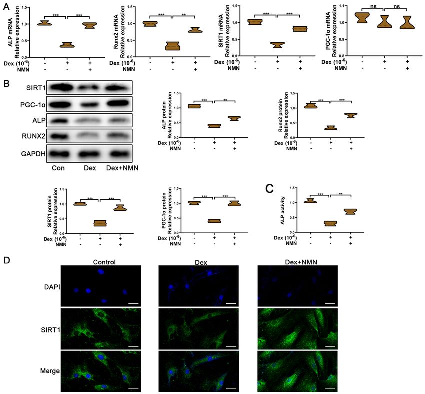
Molecular Medicine REPORTS 22: 145-154, 2020                                                                                 147

Table I. Study characteristics.

Study                                       Type                                                       Primary findings                                                           (Refs.)

Song et al, 2019 	Research	NMN promotes osteogenesis via SIRT1                                                                                                                      (16)
Zainabadi, 2019 	Review	NMN improve osteogenesis                                                                                                                                    (45)
Liang et al, 2019 	Research	NMN alleviates aluminum‑induced bone loss by inhibiting the                                                                                             (42)
		                           thioredoxin‑interacting protein‑NLRP3 inflammasome
Hassan et al, 2018  Research Nicotinamide phosphoribosyltransferase expression in osteoblasts                                                                                       (46)
		                           controls osteoclast recruitment in alveolar bone remodeling
Mills et al, 2016   Research Long‑term NMN administration significantly improves bone density                                                                                       (47)
Baek et al, 2017    Research Nicotinamide phosphoribosyltransferase inhibits receptor activator                                                                                     (48)
		                           of nuclear factor‑κB ligand‑induced osteoclast differentiation in vitro
Abed et al, 2014	Research	Low SIRT1 levels in human osteoarthritis subchondral osteoblasts lead                                                                                    (49)
		                           to abnormal sclerostin expression which decreases Wnt/β‑catenin activity

NMN, nicotinamide mononucleotide; SIRT1, sirtuin 1; NLRP3, nucleotide‑binding oligomerization domain, leucine rich repeat and pyrin
domain containing 3.

Table II. Primers used for qPCR.

                                                                                                  Primer sequence (5'→3')
	‑‑‑‑‑‑‑‑‑‑‑‑‑‑‑‑‑‑‑‑‑‑‑‑‑‑‑‑‑‑‑‑‑‑‑‑‑‑‑‑‑‑‑‑‑‑‑‑‑‑‑‑‑‑‑‑‑‑‑‑‑‑‑‑‑‑‑‑‑‑‑‑‑‑‑‑‑‑‑‑‑‑‑‑‑‑‑‑‑‑‑‑‑‑‑‑‑‑‑‑‑‑‑‑‑‑‑‑‑‑‑‑‑‑‑‑‑‑‑‑‑‑‑‑‑‑‑‑‑‑‑‑‑‑‑‑‑‑‑‑‑‑‑‑‑‑‑‑‑‑‑‑‑‑‑‑‑‑‑‑‑‑‑‑‑‑‑‑‑‑‑‑‑‑‑‑‑‑‑‑‑‑‑‑‑‑‑
Gene                                                              Forward                                                                                 Reverse

Runx2    GACGAGGCAAGAGTTTCACC   GGACCGTCCACTGTCACTTT
ALP      TCGGGACTGGTACTCGGATAAC	GTTCAGTGCGGTTCCAGACATAG
OCN	CAAGCAGGGAGGCAATAAGG	CGTCACAAGCAGGGTTAAGCQ
SIRT1	CACATGCCAGAGTCCAAGTT	AAATCCAGA TCCTCCAGCAC
PGC‑1α   AACCACACCCACAGGATCAGA	TCTTCGCTTTATTGCTCCATGA
GAPDH	AGGAGCGAGACCCCACTAACA	AGGGGGGCTAAGCAGTTGGT

Runx2, Runt‑related transcription factor 2; ALP, alkaline phosphatase; OCN, osteocalcin; SIRT1, sirtuin 1; PGC‑1α, peroxisome
proliferator‑activated receptor gamma coactivator‑1α.

paraformaldehyde for 10 min at room temperature. Alizarin                                       density of each well at a wavelength of 450 nm was measured
working solution (1%; 1 g Alizarin diluted in aqueous solu-                                     using a microplate reader and analyzed using GraphPad Prism
tion; Cyagen Biosciences, Inc.) was used to perform Alizarin                                    software (version 8.0; GraphPad Software, Inc.).
staining for 3‑5 min, and a 5‑bromo, 4‑chloro, 3‑indolylphos-
phate/Nitro‑Blue Tetrazolium ALP Color Development                                              ALP activity assay. After osteogenic induction, BMSCs were
kit (Beijing Leagene Biotechnology, Co., Ltd.) was used to                                      harvested and lysed with RIPA lysis buffer (Beyotime Institute
perform ALP staining as previously described (31). After                                        of Biotechnology). Following centrifugation at 18407 x g at
washing three times, images of the stained cells were imme-                                     4˚C for 10 min, the lysate supernatants were collected and
diately captured. Alizarin red staining was used to determine                                   added to 96‑well plates. ALP activity was detected with
the effectiveness of osteogenic differentiation by the number                                   the ALP Assay kit (Beyotime Institute of Biotechnology)
and size of red calcium nodules. ALP staining was used to                                       using the p‑nitrophenylphosphate method, according to the
determine the ALP of BMSCs according to color shade.                                            manufacturer's instructions. The cells were incubated at 37˚C
Stained cells were observed using a BX51 light microscope                                       for 30 min, and absorbance was measured with a microplate
(Olympus Corporation; magnification, x100).                                                     reader (Omega Bio‑Tek, Inc.) at 405 nm. The ALP level was
                                                                                                normalized to the total protein content, and ALP activity was
Cell viability assay. Cell viability was assessed using a                                       demonstrated as a fold change over the corresponding control
Cell Counting Kit‑8 (CCK‑8) assay (Beyotime Institute of                                        group.
Biotechnology), according to the manufacturer's protocol.
BMSCs were cultured and induced in 96‑well plates at density                                    Immunofluorescence assay. After osteogenic induction and
of 5x10³ cells/well for 7 days. Subsequently, 10 µl CCK‑8 solu-                                 treatment, BMSCs were washed with PBS and then fixed in 4%
tion was added to each well and cultured for 1 h. The optical                                   paraformaldehyde for 15 min at room temperature. The cells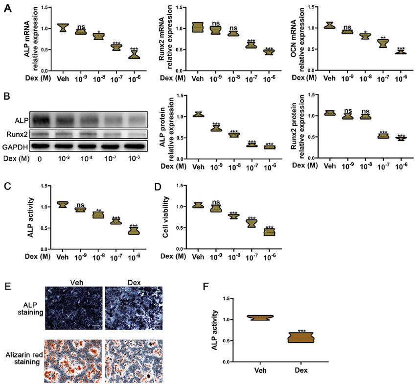
148          HUANG and TAO: NICOTINAMIDE MONONUCLEOTIDE ALLEVIATES GLUCOCORTICOID‑INDUCED OSTEOPOROSIS

Figure 1. Dex‑induced osteogenic inhibition in BMSCs. BMSCs were exposed to Dex (range, 10 ‑9‑10 ‑6 M) for 7 days. (A) Reverse transcription‑quantitative
PCR was used to detect Runx2, ALP and OCN mRNA expression in BMSCs. (B) Western blotting was used to determine the Runx2 and ALP protein expres-
sion levels in BMSCs. (C) Relative ALP activity. (D) Cell Counting Kit‑8 was used to assess the viability of BMSCs. (E) Alizarin red and ALP staining assays
were used to measure the osteogenic function of BMSCs treated with 10 ‑6 M Dex for 7 days. Scale bar, 20 µm. (F) Relative ALP activity of BMSCs treated
with 10 ‑6 M Dex for 7 days. All experiments were performed ≥3 times. ***P
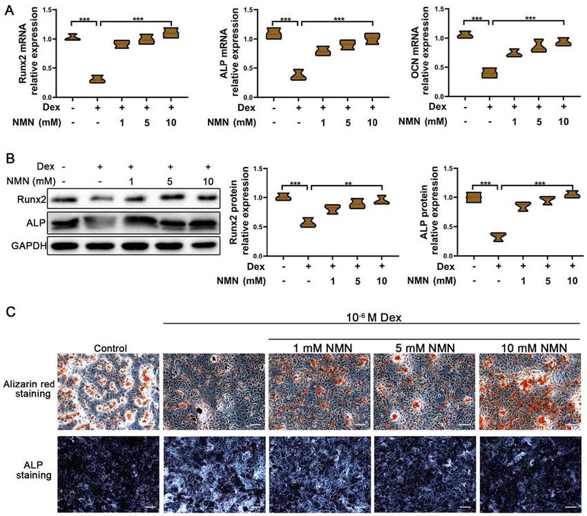
Molecular Medicine REPORTS 22: 145-154, 2020                                                           149

Figure 2. Role of NMN in Dex‑induced osteogenic inhibition of BMSCs. BMSCs were exposed to 10 ‑5 M Dex and NMN (1, 5 and 10 mM). (A) Reverse
transcription‑quantitative PCR was used to detect Runx2, ALP and OCN mRNA expression in BMSCs. (B) Western blotting was used to determine the Runx2
and ALP protein expression levels in BMSCs. (C) Alizarin red and ALP staining assays were used to assess the osteogenic function of BMSCs treated with
10 ‑5 M Dex for 7 days. Scale bar=20 µm. All experiments were performed ≥3 times. P
150         HUANG and TAO: NICOTINAMIDE MONONUCLEOTIDE ALLEVIATES GLUCOCORTICOID‑INDUCED OSTEOPOROSIS

Figure 3. SIRT1/PGC‑1α signaling is involved in the protective effects of NMN on Dex‑induced osteogenic inhibition. BMSCs were treated with control,
Dex and Dex + NMN for 7 days. (A) Reverse transcription‑quantitative PCR was used to detect the SIRT1, PGC‑1α, Runx2 and ALP mRNA expression
levels in BMSCs. (B) Western blotting was used to determine the SIRT1, PGC‑1α, Runx2 and ALP protein expression levels in BMSCs. (C) Relative ALP
activity. (D) Immunofluorescence was used to detect SIRT1 protein expression in BMSCs following 2 days of treatment. Scale bar, 5 µm. All experiments
were performed ≥3 times. ***P
Molecular Medicine REPORTS 22: 145-154, 2020                                                           151

Figure 4. SIRT1 knockdown reduces the protective effects of NMN on Dex‑induced osteogenic inhibition. Si‑NC or si‑SIRT1 were transfected into BMSCs
before Dex and NMN treatment. SIRT1‑knockdown was confirmed by (A) RT‑qPCR and (B) western blotting. (C) RT‑qPCR was used to detect the SIRT1,
PGC‑1α and Runx2 mRNA expression levels in BMSCs. (D) Western blotting assays were used to determine the SIRT1, PGC‑1α, ALP and Runx2 protein
expression levels in BMSCs. (E) Alizarin red and ALP staining assays were used to assess the osteogenic ability of BMSCs. Scale bar, 20 µm. All experiments
were performed ≥3 times. ***P
152 HUANG and TAO: NICOTINAMIDE MONONUCLEOTIDE ALLEVIATES GLUCOCORTICOID‑INDUCED OSTEOPOROSIS
deacetylase, which regulates metabolism in a variety of
cell types (21,22). Qu et al (43) revealed that SIRT1 was
involved in osteogenic proliferation and differentiation
by regulating miR‑132‑3p. Furthermore, Wang et al (44)
demonstrated that SIRT1 promotes osteogenic differentia-
tion and increases alveolar bone mass via B cell‑specific
Moloney murine leukemia virus integration site 1. In the
present study, the expression of SIRT1 and its downstream
target PGC‑1α was discovered to be decreased in osteo-
blasts exposed to Dex. Therefore, it was speculated that the
SIRT1/PGC‑1α signaling pathway played an important role
in the protective effects of NMN in Dex‑treated BMSCs.
In the present study, NMN treatment was found to increase
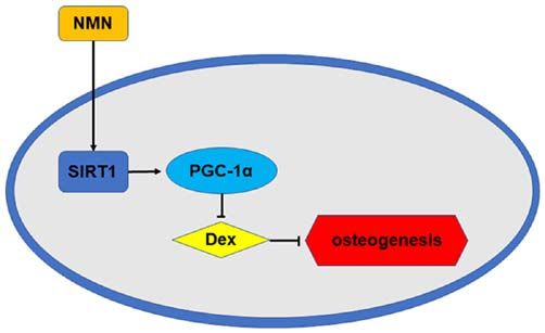
Figure 5. NMN alleviates Dex‑induced osteogenesis by regulating the the mRNA and protein expression levels of SIRT1. The
SIRT1/PGC‑1α signaling pathway. NMN, nicotinamide mononucleotide;
Dex, dexamethasone; SIRT1, sirtuin 1; PGC, peroxisome proliferator‑acti-
protein expression of PGC‑1α was also enhanced by NMN
vated receptor gamma coactivator. treatment, whereas the mRNA levels remained unchanged.
Thus, it was hypothesized that SIRT1 regulates the expres-
sion of PGC‑1α protein by deacetylation, while leaving
mRNA expression unaltered. Meanwhile, the results of
Inhibiting osteogenesis increases the risk of osteoporosis and immunofluorescence also indicated that Dex treatment
osteonecrosis, resulting in bone fracture (39). Due to glucocor- decreased protein expression of SIRT1 in the cytoplasm,
ticoid‑induced osteogenic inhibition of BMSCs, long‑term and while NMN promoted the SIRT1 expression in BMSCs
high‑dose administration of glucocorticoids can lead to serious treated with Dex.
side effects, such as osteoporosis (40). In the present study, To further confirm the role of SIRT1 in this process,
a potential therapeutic method for glucocorticoid‑induced si‑SIRT1 was used to knock down SIRT1, which inhibited the
osteogenic inhibition was investigated. protective effect of NMN in glucocorticoid‑induced osteo-
The present study suggested NMN as a potential thera- genic inhibition. Knockdown of SIRT1 was found to reduce
peutic target for Dex‑induced inhibition of osteogenesis, and the expression of the osteogenic markers that were increased
that SIRT1 was an important downstream target of NMN. with NMN treatment. Alizarin red and ALP staining also
The expression of BMSC osteogenic markers was decreased confirmed the importance of SIRT1 in the protective effect of
following exposure to Dex (range, 10‑9‑10‑6 M); these included NMN in Dex‑treated BMSCs. Importantly, SIRT1 knockdown
ALP, Runx2 and OCN. ALP staining and alizarin red staining was able to reduce the protein expression of PGC‑1α improved
also confirmed above results. These results suggest that by NMN treatment in BMSCs exposed to Dex. Together, these
Dex, as a glucocorticoid, can inhibit the differentiation and results suggest that NMN attenuates Dex‑induced osteogenic
osteogenesis of BMSCs. inhibition by regulating the SIRT1/PGC‑1α signaling pathway,
Various studies have demonstrated that the administra- and that SIRT1 regulates this process through improving the
tion of NMN significantly increases the intracellular levels of protein expression of PGC‑1α rather than PGC‑1α mRNA
NAD+. Much evidence has also confirmed that intracellular (Fig. 5).
NAD+ is closely associated with bone diseases. Li et al (41) Song et al (16) reported that NMN could promote osteo-
demonstrated that intracellular NAD+ levels were enhanced genesis in aged bone marrow. However, the effect of NMN
during osteogenic differentiation. NAD+ is also involved in the in glucocorticoid‑induced osteoporosis was unknown. Our
maintenance of osteoblast differentiation, and an increase in work confirmed the effect of NMN in glucocorticoid‑induced
the intracellular levels of NAD+ is a necessary event for the osteogenic inhibition. Together, these two studies suggest a
development of senile osteoporosis. Liang et al (42) reported therapeutic role of NMN in osteoporosis caused by age and
that NMN attenuates aluminum‑induced bone loss. However, glucocorticoids. However, as the present study was performed
the protective effects of NAD + depletion on glucocorti- in vitro, future in vivo studies will be required to validate the
coid‑induced osteogenic inhibition have yet to be elucidated. findings.
In the present study, NMN was found to alleviate Dex‑induced In conclusion, the results of the present study show that
osteogenic inhibition. NMN treatment was able to promote Dex is capable of inhibiting the differentiation and mineraliza-
osteogenic marker expression in BMSCs pre‑treated with Dex. tion of BMSCs. Moreover, NMN can alleviate Dex‑induced
Furthermore, the mineralization ability and ALP activity of osteogenic inhibition by regulating SIRT1/PGC‑1α expres-
Dex‑treated BMSCs was enhanced by NMN. These results sion. These findings provide a novel mechanism to improve
suggest that NMN protects against Dex‑induced osteogenic the understanding of glucocorticoid‑induced osteogenic inhi-
impairment, although the exact mechanism requires further bition, and indicate that NMN may be a potential therapeutic
investigation. target.
A previous study found that in aged bone marrow,
NMN improved osteogenesis and reduced adipogenesis Acknowledgements
by regulating MSCs via the SIRT1 pathway (17). NMN, a
key NAD + intermediate, can stimulate BMSC differentia- The authors would like to thank Professor Yan Jiang (The
tion and osteogenesis (17). SIRT1 is an NAD + ‑dependent Second Affiliated Hospital of Nanchang University) forMolecular Medicine REPORTS 22: 145-154, 2020 153
provided suggestions regarding experimental design and 10. Caramori G, Ruggeri P, Arpinelli F, Salvi L and Girbino G:
Long‑term use of inhaled glucocorticoids in patients with stable
article writing. chronic obstructive pulmonary disease and risk of bone fractures:
A narrative review of the literature. Int J Chron Obstruct Pulmon
Funding Dis 14: 1085‑1097, 2019.
11. Krela‑Kaźmierczak I, Szymczak A, Łykowska‑Szuber L, Eder P
and Linke K: Osteoporosis in gastrointestinal diseases. Adv Clin
No funding was received. Exp Med 25: 185‑190, 2016.
12. Kitazaki S, Mitsuyama K, Masuda J, Harada K, Yamasaki H,
Kuwaki K, Takedatsu H, Sugiyama G, Tsuruta O and Sata M:
Availability of data and materials Clinical trial: Comparison of alendronate and alfacalcidol in
glucocorticoid‑associated osteoporosis in patients with ulcerative
All data generated or analyzed during the present study are colitis. Aliment Pharmacol Ther 29: 424‑430, 2009.
13. Li J, Zhang N, Huang X, Xu J, Fernandes JC, Dai K and Zhang X:
included in this published article. Dexamethasone shifts bone marrow stromal cells from osteo-
blasts to adipocytes by C/EBPalpha promoter methylation. Cell
Authors' contributions Death Dis 4: e832, 2013.
14. Sosa M and Gomez de Tejada MJ: Glucocorticoid‑Induced
osteoporosis. N Engl J Med 380: 1378‑1379, 2019.
RH performed the experiments and collected the data. 15. Wang X, Hu X, Yang Y, Takata T and Sakurai T: Nicotinamide
JT designed the study, analyzed the data and drafted the mononucleotide protects against β ‑amyloid oligomer‑induced
manuscript. All authors read and approved the final manuscript. cognitive impairment and neuronal death. Brain Res 1643: 1‑9,
2016.
16. Song J, Li J, Yang F, Ning G, Zhen L, Wu L, Zheng Y, Zhang Q,
Ethics approval and consent to participate Lin D, Xie C and Peng L: Nicotinamide mononucleotide
promotes osteogenesis and reduces adipogenesis by regulating
mesenchymal stromal cells via the SIRT1 pathway in aged bone
Not applicable. marrow. Cell Death Dis 10: 336, 2019.
17. Signer RA and Morrison SJ: Mechanisms that regulate stem cell
Patient consent for publication aging and life span. Cell Stem Cell 12: 152‑165, 2013.
18. Frye RA: Characterization of five human cDNAs with homology
to the yeast SIR2 gene: Sir2‑like proteins (sirtuins) metabolize
Not applicable. NAD and may have protein ADP‑ribosyltransferase activity.
Biochem Biophys Res Commun 260: 273‑279, 1999.
19. Sherman JM, Stone EM, Freeman‑Cook LL, Brachmann CB,
Competing interests Boeke JD and Pillus L: The conserved core of a human SIR2 homo-
logue functions in yeast silencing. Mol Biol Cell 10: 3045‑3059, 1999.
The authors declare that they have no competing interests. 20. Liang F, Kume S and Koya D: SIRT1 and insulin resistance. Nat
Rev Endocrinol 5: 367‑373, 2009.
21. Chua KF, Mostoslavsky R, Lombard DB, Pang WW, Saito S,
References Franco S, Kaushal D, Cheng HL, Fischer MR, Stokes N, et al:
Mammalian SIRT1 limits replicative life span in response to
chronic genotoxic stress. Cell Metab 2: 67‑76, 2005.
1. Baccaro LF, Conde DM, Costa‑Paiva L and Pinto‑Neto AM: 22. Haigis MC and Guarente LP: Mammalian sirtuins‑emerging
The epidemiology and management of postmenopausal osteopo- roles in physiology, aging, and calorie restriction. Genes Dev 20:
rosis: A viewpoint from Brazil. Clin Interv Aging 10: 583‑591, 2913‑2921, 2006.
2015. 23. Leibiger IB and Berggren PO: Sirt1: A metabolic master switch
2. Chen Q, Shou P, Zhang L, Xu C, Zheng C, Han Y, Li W, Huang Y, that modulates lifespan. Nat Med 12: 34‑36; discussion 36, 2006.
Zhang X, Shao C, et al: An osteopontin‑integrin interaction 24. Lee HW, Suh JH, Kim AY, Lee YS, Park SY and Kim JB: Histone
plays a critical role in directing adipogenesis and osteogenesis by deacetylase 1‑mediated histone modification regulates osteoblast
mesenchymal stem cells. Stem Cells 32: 327‑337, 2014. differentiation. Mol Endocrinol 20: 2432‑2443, 2006.
3. Camacho PM, Petak SM, Binkley N, Clarke BL, Harris ST, 25. Cohen‑Kfir E, Artsi H, Levin A, Abramowitz E, Bajayo A,
Hurley DL, Kleerekoper M, Lewiecki EM, Miller PD, Gurt I, Zhong L, D'Urso A, Toiber D, Mostoslavsky R and
Narula HS, et al: American association of clinical endocrinolo- Dresner‑Pollak R: Sirt1 is a regulator of bone mass and a
gists and American college of endocrinology clinical practice repressor of Sost encoding for sclerostin, a bone formation
guidelines for the diagnosis and treatment of postmenopausal inhibitor. Endocrinology 152: 4514‑4524, 2011.
osteoporosis‑2016‑executive summary. Endocr Pract 22: 26. Iyer S, Han L, Bartell SM, Kim HN, Gubrij I, de Cabo R,
1111‑1118, 2016. O'Brien CA, Manolagas SC and Almeida M: Sirtuin1 (Sirt1)
4. Xu D, Gao Y, Hu N, Wu L and Chen Q: miR‑365 ameliorates promotes cortical bone formation by preventing beta‑catenin
dexamethasone‑induced suppression of osteogenesis in MC3T3‑E1 sequestration by FoxO transcription factors in osteoblast
cells by targeting HDAC4. Int J Mol Sci 18: E977, 2017. progenitors. J Biol Chem 289: 24069‑24078, 2014.
5. Yuasa M, Yamada T, Taniyama T, Masaoka T, Xuetao W, 27. Sun W, Qiao W, Zhou B, Hu Z, Yan Q, Wu J, Wang R, Zhang Q
Yoshii T, Horie M, Yasuda H, Uemura T, Okawa A and Sotome S: and Miao D: Overexpression of Sirt1 in mesenchymal stem cells
Dexamethasone enhances osteogenic differentiation of bone protects against bone loss in mice by FOXO3a deacetylation and
marrow‑ and muscle‑derived stromal cells and augments ectopic oxidative stress inhibition. Metabolism 88: 61‑71, 2018.
bone formation induced by bone morphogenetic protein‑2. PLoS 28. Yu W, Zhu C, Xu W, Jiang L and Jiang S: Neuropeptide
One 10: e0116462, 2015. Y1 receptor regulates glucocorticoid‑induced inhibition of
6. Fardet L, Petersen I and Nazareth I: Prevalence of long‑term oral osteoblast differentiation in murine MC3T3‑E1 cells via ERK
glucocorticoid prescriptions in the UK over the past 20 years. signaling. Int J Mol Sci 17: E2150, 2016.
Rheumatology (Oxford) 50: 1982‑1990, 2011. 29. Livak KJ and Schmittgen TD: Analysis of relative gene expression
7. Hoes JN, Bultink IE and Lems WF: Management of osteoporosis data using real‑time quantitative PCR and the 2(‑Delta Delta
in rheumatoid arthritis patients. Expert Opin Pharmacother 16: C(T)) method. Methods 25: 402‑408, 2001.
559‑571, 2015. 30. Tang Z, Hu B, Zang F, Wang J, Zhang X and Chen H: Nrf2 drives
8. Edens C and Robinson AB: Systemic lupus erythematosus, oxidative stress‑induced autophagy in nucleus pulposus cells via
bone health, and osteoporosis. Curr Opin Endocrinol Diabetes a Keap1/Nrf2/p62 feedback loop to protect intervertebral disc
Obes 22: 422‑431, 2015. from degeneration. Cell Death Dis 10: 510, 2019.
9. Monadi M, Javadian Y, Cheraghi M, Heidari B and Amiri M: 31. Shi GX, Zheng XF, Zhu C, Li B, Wang YR, Jiang SD and
Impact of treatment with inhaled corticosteroids on bone mineral Jiang LS: Evidence of the role of R‑Spondin 1 and its receptor
density of patients with asthma: Related with age. Osteoporos Lgr4 in the transmission of mechanical stimuli to biological
Int 26: 2013‑2018, 2015. signals for bone formation. Int J Mol Sci 18: E564, 2017.154 HUANG and TAO: NICOTINAMIDE MONONUCLEOTIDE ALLEVIATES GLUCOCORTICOID‑INDUCED OSTEOPOROSIS
32. Lane NE: Glucocorticoid‑Induced osteoporosis: New insights 43. Qu H, Li T, Jin H, Zhang S and He B: Silent mating type
into the pathophysiology and treatments. Curr Osteoporos information regulation 2 homolog (SIRT1) influences osteogenic
Rep 17: 1‑7, 2019. proliferation and differentiation of MC3T3‑E1 cells via regulation
33. Ohnaka K, Tanabe M, Kawate H, Nawata H and Takayanagi R: of miR‑132‑3p. Med Sci Monit 25: 2289‑2295, 2019.
Glucocorticoid suppresses the canonical Wnt signal in cultured 44. Wang H, Hu Z, Wu J, Mei Y, Zhang Q, Zhang H, Miao D and
human osteoblasts. Biochem Biophys Res Commun 329: 177‑181, Sun W: Sirt1 promotes osteogenic differentiation and increases
2005. alveolar bone mass via Bmi1 activation in mice. J Bone Miner
34. Briot K and Roux C: Glucocorticoid‑induced osteoporosis. RMD Res 34: 1169‑1181, 2019.
Open 1: e000014, 2015. 45. Zainabadi K: Drugs targeting SIRT1, a new generation of
35. Nogueiras R, Habegger KM, Chaudhary N, Finan B, Banks AS, therapeutics for osteoporosis and other bone related disorders?
Dietrich MO, Horvath TL, Sinclair DA, Pfluger PT and Pharmacol Res 143: 97‑105, 2019.
Tschöp MH: Sirtuin 1 and sirtuin 3: Physiological modulators of 46. Hassan B, Baroukh B, Llorens A, Lesieur J, Ribbes S,
metabolism. Physiol Rev 92: 1479‑1514, 2012. Chaussain C, Saffar JL and Gosset M: NAMPT expression in
36. Jung HY, Lee D, Ryu HG, Choi BH, Go Y, Lee N, Lee D, Son HG, osteoblasts controls osteoclast recruitment in alveolar bone
Jeon J, Kim SH, et al: Myricetin improves endurance capacity remodeling. J Cell Physiol 233: 7402‑7414, 2018.
and mitochondrial density by activating SIRT1 and PGC‑1α. Sci 47. Mills KF, Yoshida S, Stein LR, Grozio A, Kubota S, Sasaki Y,
Rep 7: 6237, 2017. Redpath P, Migaud ME, Apte RS, Uchida K, et al: Long‑term
37. Henneicke H, Gasparini SJ, Brennan‑Speranza TC, Zhou H and administration of nicotinamide mononucleotide mitigates age‑asso-
Seibel MJ: Glucocorticoids and bone: Local effects and systemic ciated physiological decline in mice. Cell Metab 24: 795‑806, 2016.
implications. Trends Endocrinol Metab 25: 197‑211, 2014. 48. Baek JM, Ahn SJ, Cheon YH, Lee MS, Oh J and Kim JY:
38. Canalis E, Mazziotti G, Giustina A and Bilezikian JP: Nicotinamide phosphoribosyltransferase inhibits receptor
Glucocorticoid‑induced osteoporosis: Pathophysiology and activator of nuclear factor‑kB ligand‑induced osteoclast
therapy. Osteoporos Int 18: 1319‑1328, 2007. differentiation in vitro. Mol Med Rep 15: 784‑792, 2017.
39. Infante A and Rodriguez CI: Osteogenesis and aging: Lessons 49. Abed É, Couchourel D, Delalandre A, Duval N, Pelletier JP,
from mesenchymal stem cells. Stem Cell Res Ther 9: 244, 2018. Martel‑Pelletier J and Lajeunesse D: Low sirtuin 1 levels in
40. Chen TL: Inhibition of growth and differentiation of osteopro- human osteoarthritis subchondral osteoblasts lead to abnormal
genitors in mouse bone marrow stromal cell cultures by increased sclerostin expression which decreases Wnt/β ‑catenin activity.
donor age and glucocorticoid treatment. Bone 35: 83‑95, 2004. Bone 59: 28‑36, 2014.
41. Li Y, He J, He X, Li Y and Lindgren U: Nampt expression
increases during osteogenic differentiation of multi‑ and omnip- This work is licensed under a Creative Commons
otent progenitors. Biochem Biophys Res Commun 434: 117‑123, Attribution-NonCommercial-NoDerivatives 4.0
2013. International (CC BY-NC-ND 4.0) License.
42. Liang H, Gao J, Zhang C, Li C, Wang Q, Fan J, Wu Z and Wang Q:
Nicotinamide mononucleotide alleviates Aluminum induced
bone loss by inhibiting the TXNIP‑NLRP3 inflammasome.
Toxicol Appl Pharmacol 362: 20‑27, 2019.You can also read