Mercury in the human adrenal medulla could contribute to increased plasma noradrenaline in aging
←
→
Page content transcription
If your browser does not render page correctly, please read the page content below
www.nature.com/scientificreports
OPEN Mercury in the human adrenal
medulla could contribute
to increased plasma noradrenaline
in aging
Roger Pamphlett1,2*, Stephen Kum Jew1, Philip A. Doble3 & David P. Bishop3
Plasma noradrenaline levels increase with aging, and this could contribute to the sympathetic
overactivity that is associated with essential hypertension and the metabolic syndrome. The
underlying cause of this rise in noradrenaline is unknown, but a clue may be that mercury increases
noradrenaline output from the adrenal medulla of experimental animals. We therefore determined the
proportion of people from 2 to 104 years of age who had mercury in their adrenal medulla. Mercury
was detected in paraffin sections of autopsied adrenal glands using two methods of elemental
bioimaging, autometallography and laser ablation-inductively coupled plasma-mass spectrometry.
Mercury first appeared in cells of the adrenal medulla in the 21–40 years group, where it was present
in 52% of samples, and increased progressively in frequency in older age groups, until it was detected
in 90% of samples from people aged over 80 years. In conclusion, the proportion of people having
mercury in their adrenal medulla increases with aging. Mercury could alter the metabolism of
catecholamines in the adrenal medulla that leads to the raised levels of plasma noradrenaline in aging.
This retrospective autopsy study was not able to provide a definitive link between adrenal mercury,
noradrenaline levels and hypertension, but future functional human and experimental studies could
provide further evidence for these associations.
Plasma noradrenaline increases with a ge1, and increased noradrenaline activity is thought to play a role in
the pathogenesis of essential h ypertension2–4 and the metabolic syndrome5. One proposed mechanism for the
increased noradrenaline levels during aging is overactivity of central nervous system sympathetic neurons, fol-
lowed by a spillover of noradrenaline into the plasma from postganglionic neurons4,6, but the cause of this central
neuronal overactivity is obscure. In contrast to elevated noradrenaline, adrenaline secretion from the adrenal
medulla decreases with a ge6. The cause of this decreased adrenaline output remains unknown.
Another possible origin for the elevated noradrenaline in aging is the uptake of toxic metals by the adrenal
medulla. Exposures to toxic metals, such as mercury and cadmium, release noradrenaline from the adrenal
medulla of experimental a nimals7,8. Several case studies, mostly in children, have reported acute hypertension
after accidental exposure to m ercury9–16. Mercury in the adrenal medulla has been proposed to deactivate the
cofactor S-adenosylmethionine (SAM), due to its high number of mercury-binding sulfhydryl groups12. This
would decrease the activity of two SAM-dependent enzymes, catechol-O-methyltransferase that deactivates
noradrenaline, and phenylethanolamine N-methyltransferase that converts noradrenaline into adrenaline (Fig. 1).
The result would be an increased output of noradrenaline and a decreased secretion of adrenaline, the situation
found in human aging. This mechanism provides a potential link between the findings, in both essential hyper-
tension and the metabolic syndrome, of increased sympathetic a ctivity4,5 and previous exposure to m ercury17,18.
It may also underlie the acutely-raised noradrenaline levels and hypertension that follow accidental exposure
to mercury14.
Selective uptake of mercury by the adrenal medulla has been demonstrated in r odents19,20 and p rimates21,22,
but it is not known if mercury is taken up by the human adrenal medulla. To see if mercury in the adrenal medulla
could contribute to the changes in catecholamine levels found in aging, we looked for the presence of mercury
and other toxic metals in the adrenal glands of people aged between 2 and 104 years.
1
Discipline of Pathology, Sydney Medical School, Brain and Mind Centre, The University of Sydney, Sydney, NSW,
Australia. 2Department of Neuropathology, Royal Prince Alfred Hospital, Sydney, NSW, Australia. 3Elemental
Bio‑Imaging Facility, School of Mathematical and Physical Sciences, University of Technology Sydney, Sydney,
NSW, Australia. *email: roger.pamphlett@sydney.edu.au
Scientific Reports | (2021) 11:2961 | https://doi.org/10.1038/s41598-021-82483-y 1
Vol.:(0123456789)
www.nature.com/scientificreports/
Figure 1. Pathways for mercury-induced increased noradrenaline and decreased adrenaline output. Mercury,
by binding to and inhibiting the cofactor S-adenosyl-l-methionine (SAM) in the adrenal medulla, reduces the
ability of catechol-O-methyltransferase (COMT) to deactivate noradrenaline, resulting in an increased output
of noradrenaline. Mercury binding to SAM also reduces the ability of phenylethanolamine N-methyltransferase
(PNMT) to convert noradrenaline into adrenaline, resulting in a decreased output of adrenaline. Overactivity
of central nervous system sympathetic neurons (of unknown cause) may contribute to increased sympathetic
activity via elevated noradrenaline output from postganglionic neurons.
Methods
Sample collection. Paraffin-embedded samples of adrenal glands were examined from 89 individuals who
had autopsies performed in the New South Wales Department of Forensic Medicine, Sydney (Supplementary
Table S1). Ages ranged between 2 and 104 years, and comprised 56 males (mean age 52 years, SD 26 years, range
2–98 years) and 33 females (mean age 59 years, SD 29 years, range 2–104 years). Clinical histories were: no major
known disorder (N = 36), neurodegenerative disorder (N = 26), psychosis (N = 22), two with epilepsy, and one
each of post-traumatic stress disorder, Down syndrome, and anorexia nervosa. Causes of death were: suicide
(N = 23), trauma (N = 15), cardiovascular (N = 14), drug overdose (N = 10), drowning (N = 8), choking (N = 4),
infection (N = 4), undetermined (N = 4), cerebrovascular (N = 2), respiratory (N = 2), and one each of cancer,
cirrhosis, and undernutrition.
Autometallography. Seven-micron paraffin sections of the adrenal medulla were stained for inorganic
mercury bound to sulphide or selenide using silver nitrate autometallography (AMG), which represents the
presence of mercury as black grains23. Briefly, sections were placed in physical developer containing 50% gum
arabic, citrate buffer, hydroquinone and silver nitrate at 26 °C for 80 min in the dark then washed in 5% sodium
thiosulphate to remove unbound silver. Sections were counterstained with mercury-free hematoxylin and
viewed with bright-field microscopy using an Olympus BX50 microscope. Each staining run included a control
section of mouse spinal cord where motor neuron cell bodies contained mercury following an intraperitoneal
injection of mercuric c hloride24. The proportion of adrenal chromaffin cells containing AMG was categorised
as ‘low’ if AMG was present in up to 25% of cells, and ‘high’ if AMG was present in more than 25% of cells in at
least four 200× microscopic fields.
Laser ablation‑inductively coupled plasma‑mass spectrometry (LA‑ICP‑MS). In addition to
mercury, AMG detects the presence of inorganic silver and bismuth25,26. To confirm that AMG in the adrenal
medulla detected mercury, and to look for the presence of other toxic metals, seven-micron paraffin sections
Scientific Reports | (2021) 11:2961 | https://doi.org/10.1038/s41598-021-82483-y 2
Vol:.(1234567890)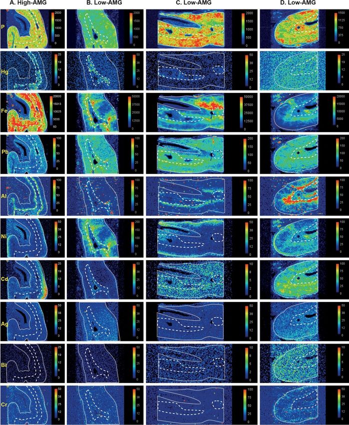
www.nature.com/scientificreports/
of 12 adrenal glands (8 AMG-positive, 4 AMG-negative) were subjected to LA-ICP-MS for mercury, silver,
bismuth, phosphorus, aluminium, cadmium, gold, lead, chromium, nickel and iron. Analyses were carried out
on an NWR-193 excimer laser hyphenated to an Agilent Technologies 7700 ICP-MS, with argon used as the car-
rier gas. LA-ICP-MS conditions were optimised on NIST 612 Trace Element in Glass CRM and the sample was
ablated with a 50 µm spot size and a scan speed of 100 µm/s at a frequency of 20 Hz. The data were collated into
a single image file using in-house developed software and visualised using FIJI.
Ethics statement. This study (X14-029) was approved by the Human Research Committee, Sydney Local
Health District (Royal Prince Alfred Hospital Zone), in accordance with the Declaration of Helsinki as revised
in 2000. The institutional review board (the Human Research Committee, Sydney Local Health District) waived
the need for written informed consent from relatives of individuals studied since this was a de-identified retro-
spective study of autopsy tissue.
Results
Autometallography. Hematoxylin and eosin stained sections showed no histological abnormalities of the
adrenal cortex or medulla in any samples. Hematoxylin-only stained sections showed no black grains in any
adrenal cells, in contrast to the black-stained grains in the medulla of AMG-positive samples (Fig. 2). The AMG
staining patterns in chromaffin cells were either: (1) single or multiple dense black granules of varying size, either
adjacent to the nucleus or within the cytoplasm (the most common pattern), or (2) small black grains dispersed
throughout the cytoplasm, either alone or together with dense black granules (Fig. 3). The density of AMG-con-
taining chromaffin cells often varied across the medulla. AMG was not seen in the adrenal cortex of any samples.
AMG-detected mercury deposits in the adrenal medulla were present in none of the 2–20 years group, in
52% of the 21–40 years group (12% high-AMG), in 80% of the 41–60 years group (40% high-AMG), in 87% of
the 61–80 years group (40% high-AMG), and in 90% of the 81–104 years group (40% high-AMG) (chi-square
trend for aging p < 0.0001) (Fig. 4, Supplementary Table S1).
The 33 females in the study had a slightly higher proportion (73%) of mercury in the adrenal medulla than
the 56 males (65%), probably because females were older on average than males. The proportion of people with
high-AMG adrenal medulla mercury varied between the major categories of clinical diagnoses (14% for psy-
chosis, 28% for no major clinical condition, 42% for neurodegeneration), most likely due to the different average
ages in these groups (30, 52, and 74 years respectively).
LA‑ICP‑MS. An example of the co-localisation of mercury in the adrenal medulla in both AMG and LA-
ICP-MS samples is shown in Fig. 5 (from sample A in Fig. 6). The results of the LA-ICP-MS multi-elemental
analyses of the adrenal glands, together with AMG staining and ages of the individuals sampled, are summarised
in Table 1, and can be cross-matched with the LA-ICP-MS images in Figs. 6, 7 and 8.
In high-AMG adrenal samples, the anatomical regions where mercury was detected by AMG (corresponding
to the medulla) matched those of mercury detected by LA-ICP-MS (Figs. 6, 7). In low-AMG samples, LA-ICP-
MS could not detect mercury when only a few percent of chromaffin cells stained with AMG. In samples with
no AMG staining, no mercury was detected on LA-ICP-MS (Fig. 8). Other toxic metals present in adrenal sam-
ples were located either in both the medulla and cortex, or in the medulla or cortex alone (Figs. 6, 7, 8). Metals
detected in both the medulla and cortex were iron (N = 8), lead (N = 8), nickel (N = 4), aluminium (N = 2), and
silver (N = 1). In the medulla alone, metals detected were cadmium (N = 2), aluminium (N = 2), bismuth (N = 1)
and chromium (N = 1). Metals present in the cortex alone were aluminium (N = 5), iron (N = 3), and silver (N = 2).
Gold was not seen in any samples.
Discussion
The key finding of this study is that the proportion of people with mercury in their adrenal medulla increases
steadily after the age of 20 years, until mercury is present in the medulla of 90% or more of people over the age of
80 years. This raises the possibility that mercury induces changes to catecholamine metabolism within the adrenal
gland that contribute to the raised plasma noradrenaline and decreased adrenaline secretion found in aging.
Humans are exposed to mercury mostly from consuming mercury-containing larger fi sh27. The amount of
mercury in the atmosphere is rising, due mostly to the burning of c oal28. Atmospheric mercury enters the global
atmosphere-water-soil cycle and has been found in increasing amounts in predatory fish29. Others sources of
mercury exposure include those from occupations, mercury-containing dental amalgam restorations27 and from
food such as rice in some c ountries30. Mercury can be detected in the chromaffin cells of primates after they have
been fitted with as few as four mercury amalgam dental fi llings22.
Our findings are paralleled by studies of the effect methylmercury has on hypertension in local communities
living around Minamata Bay in Japan. Hypertension was found to be more common in the Minamata area (high
methylmercury exposure) than in an area of low methylmercury exposure, and dose response trends with hair
mercury levels were observed for hypertension, supporting a causal relationship between methylmercury expo-
sure and hypertension31. Later work from the same group showed that mortality from hypertension was greater
in Minamata city than in the whole prefecture in which Minamata is s ited32, with further analyses supporting a
link between mercury exposure and h ypertension33.
The reason mercury preferentially localises to adrenal medulla cells is not known, but one possibility is that
noradrenaline-containing cells are predisposed to take up circulating toxic metals, since noradrenergic locus
ceruleus neurons in the brain stem also accumulate mercury on a ging34. Another possibility is that chromaffin
cells contain a high level of one or more of a range of metal transporters, for example, human organic anion
Scientific Reports | (2021) 11:2961 | https://doi.org/10.1038/s41598-021-82483-y 3
Vol.:(0123456789)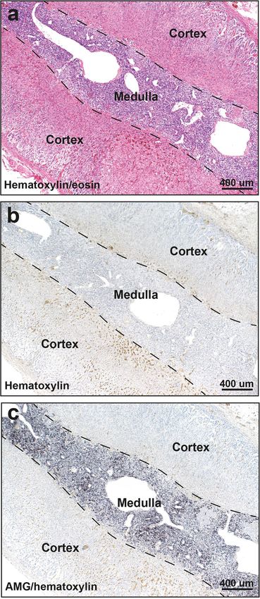
www.nature.com/scientificreports/
Figure 2. Routine and AMG staining of an AMG-positive adrenal medulla. All adjacent sections are from
the same sample (from a 39 year-old male). (a) No histological abnormalities are seen in the adrenal cortex or
medulla on hematoxylin and eosin staining. The medullary chromaffin cells have a bluish tinge. The large spaces
in the medulla are empty veins. (b) No black grains are present in the adrenal cortex or medulla in a section
stained with hematoxylin only. The yellow in the cortex shows erythrocytes. (c) Black AMG staining is seen in
the adrenal medulla, but not in the cortex, in a section stained with AMG and hematoxylin.
transporter 1 or breast cancer resistance protein, that are found in other cells in the body that take up mercury,
such as those of renal proximal tubules35,36.
Scientific Reports | (2021) 11:2961 | https://doi.org/10.1038/s41598-021-82483-y 4
Vol:.(1234567890)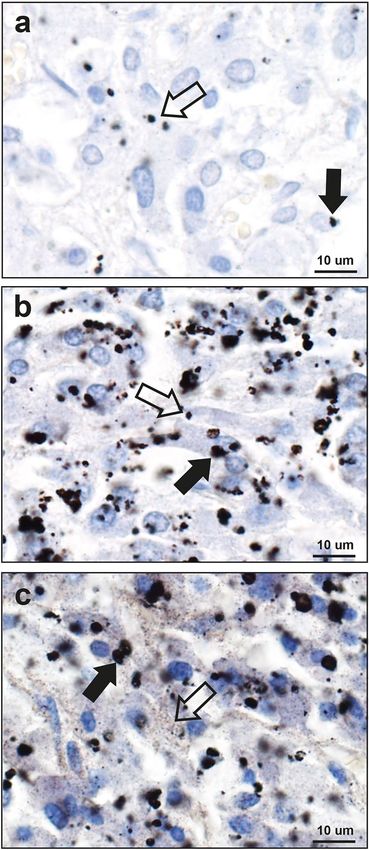
www.nature.com/scientificreports/
Figure 3. Patterns of AMG staining in the adrenal medulla. (a) Small dense black AMG granules, either
adjacent to nuclei (filled arrow) or within the cytoplasm (open arrow), are present in fewer than 25% of these
chromaffin cells (69 year-old male). (b) Black AMG granules, either large (filled arrow) or small (open arrow),
often multiple, are present in the cytoplasm of more than 25% of chromaffin cells (32 year-old male). (c) Black
AMG staining is present in most chromaffin cells, either as small grains dispersed in the cytoplasm (open
arrow), or as these small grains accompanied by dense granules (filled arrow) (39 year-old male). AMG and
hematoxylin.
Our samples were not suitable for ultrastructural studies, so we were not able to establish the subcellular loca-
tion of the mercury. However, animal studies indicate that mercury in chromaffin cells is situated mostly within
lysosomes, and to a lesser extent in secretory granules22,37. In most of our samples, AMG-stained mercury was
present within compact granules in the cells, suggesting past mercury exposure with lysosomal accumulation.
In a few samples, fine AMG grains were present throughout the cytoplasm, implying widespread cytoplasmic
mercury from recent or ongoing exposure. This implies catecholamine metabolism could be disturbed either
early after mercury exposure when cytoplasmic mercury can bind to sulfhydryl-containing proteins, or later due
to disruption of lysosomal metabolic p rocesses38.
Scientific Reports | (2021) 11:2961 | https://doi.org/10.1038/s41598-021-82483-y 5
Vol.:(0123456789)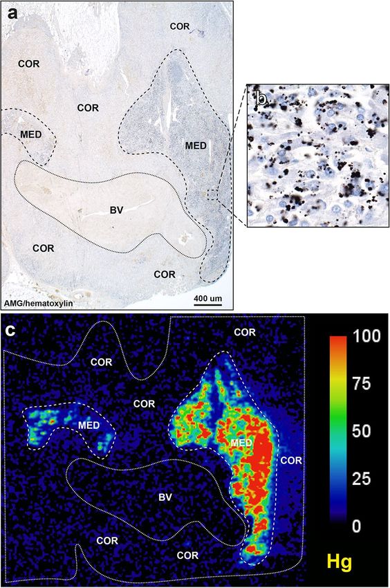
www.nature.com/scientificreports/
Figure 4. Mercury in the adrenal medulla in increasing ages. Mercury starts appearing in the adrenal medulla
in the 21–40 years group, where it is present in 52% of people. The proportion of people with mercury in the
medulla increases to 80% in the 41–60 years group, and reaches a maximum of 90% in the final 81–104 years
group. Numbers of people in groups are above the bars.
Our findings, suggesting links between mercury uptake in the adrenal medulla, age-related increases in
circulating noradrenaline, and hypertension, give a potential pathophysiological explanation for a number of
clinical and epidemiological findings: (1) the prevalence of hypertension rises with aging39,40, at about the same
age-related increase in the proportion of individuals in our study having mercury in their adrenal medulla. (2)
Indirect evidence of sympathetic overactivity in hypertension comes from the frequent finding of t achycardia41
and tremor42 in people with raised blood pressure. (3) Hypertension has been associated with mercury exposure
in people living in the vicinity of v olcanoes43,44 (with eruptions being sources of m ercury45), as well as in people
46,47 28
exposed to atmospheric p ollution that often has a mercury c omponent . (4) Hypertension and abdominal
obesity are features of the metabolic syndrome, where sympathetic overdrive is i mplicated5, but why these two
conditions are linked is u nclear48–50. Several epidemiological studies report associations of circulating levels of
mercury with h ypertension17,51, obesity52,53, and with the metabolic s yndrome18,53,54. The reason for this link
could be accumulation of mercury in both the adrenal medulla (with increased noradrenaline output causing the
hypertension) together with mercury that accumulates on aging in pituitary somatotrophs55, since a decrease in
growth hormone levels promotes abdominal obesity56. (5) The onset of hypertension has been related to psycho-
logical stress57. Raised noradrenaline levels from mercury in chromaffin cells could cause symptoms associated
with stress58, rather than stress being a trigger for hypertension. (6) Mercury has been suggested to underlie the
hypertension seen in both mercury poisoning and in Kawasaki d isease59.
Noradrenaline-containing cells, such as adrenal chromaffin cells and locus ceruleus neurons, take up mercury
selectively, so it is possible that noradrenaline-containing sympathetic ganglion neurons also have a predilection
to take up mercury, which could be a factor in the increased sympathetic nerve activity in hypertension described
in humans4. Finding toxic metals in sympathetic ganglion neurons would imply that both nervous system and
adrenal medulla-derived noradrenaline play roles in essential hypertension (Fig. 1). The paravertebral sympa-
thetic chain is seldom removed routinely at autopsy, so a prospective autopsy study of people with and without
hypertension would be needed to undertake an elemental analysis study of this type.
Metals other than mercury detected by LA-ICP-MS in the adrenal medulla included cadmium and chromium,
both of which can release catecholamines from the adrenal m edulla8,60. Lead was found in most adrenal glands,
probably because of exposure to lead in petrol and domestic paints before these sources of lead were banned in
Australia61. Iron and nickel were found mostly in the adrenal cortex, possibly because erythrocytes were more
numerous in the cortex, suggesting that these were circulating metals. Silver has been found to enter the chromaf-
fin cells of rats, where it is localised within lysosomes62, and silver is commonly present in noradrenergic cells of
the human locus c eruleus34. Bismuth uptake into cells closely mirrors that of m ercury63, but bismuth is thought to
Scientific Reports | (2021) 11:2961 | https://doi.org/10.1038/s41598-021-82483-y 6
Vol:.(1234567890)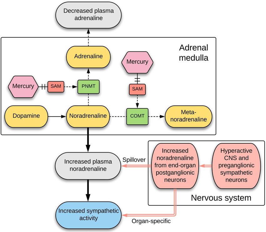
www.nature.com/scientificreports/
Figure 5. Co-localisation of mercury by AMG and LA-ICP-MS. (a) Black AMG staining is present in two
regions of the adrenal medulla (MED), but not in the adjacent adrenal cortex (COR), or in a large blood vessel
(BV). AMG and hematoxylin. (b) An enlargement of the area within the box in (a) shows numerous chromaffin
cells containing black AMG grains. (c) LA-ICP-MS of an adjacent section shows mercury in the two adrenal
medulla regions, but not in the cortex or blood vessel. All images from a 98 year-old male. Scale = counts per
second (proportional to abundance).
be non-toxic for humans in low doses. Bismuth use is uncommon in Australia, unlike countries such as the USA
where it is often taken for gastrointestinal symptoms. The finding of multiple metals within individual adrenal
medulla samples may have implications for catecholamine metabolism since harmful synergistic interactions
between toxic metals, in particular mercury and cadmium, have been d escribed64.
An unexpected finding was the frequent detection of aluminium in the zona reticularis of the cortex, adjacent
to the medulla. Apart from its known function as a source of adrenal androgens such as dehydroepiandrosterone,
important for the adrenarche, other functions of the human reticularis remain unclear, though it may be involved
in xenobiotic metabolism and maintaining circadian r hythms65. High levels of aluminium in the reticularis
could compromise these important functions. Zona reticularis cells often contain lipofuscin66, a wear-and-tear
pigment, which is consistent with these cells having to deal with damaging xenobiotics. Exposure to aluminium
phosphide, a commonly used fumigant for stored grains, can cause adrenocortical n ecrosis67, which may be
because the aluminium moiety targets reticularis cells before toxic phosphine is released.
Scientific Reports | (2021) 11:2961 | https://doi.org/10.1038/s41598-021-82483-y 7
Vol.:(0123456789)www.nature.com/scientificreports/
Figure 6. LA-ICP-MS I. The dotted lines outline the adrenal gland in the section, the medulla is outlined by
the internal dashed lines, and the cortex occupies the space between the dashed and dotted lines. Phosphorus
LA-ICP-MS indicates the degree of cellularity. Mercury is seen selectively in the medulla of all four samples
(A–D) that had high-AMG. Metals present in both cortex and medulla are iron (A–D), lead (A–D), cadmium
(A,C,D), bismuth (B,C), nickel (B,C), silver (B) and aluminium (in the cortical zona reticularis) in (D). Bismuth
is present in the medulla in (A) and silver in the cortex in (C). Scale = counts per second (proportional to
abundance).
Scientific Reports | (2021) 11:2961 | https://doi.org/10.1038/s41598-021-82483-y 8
Vol:.(1234567890)www.nature.com/scientificreports/
Age y Site AMG Hg Fe Pb Al Ni Cd Ag Bi Cr Au Fig
Medulla ++ + + + − − + − + − −
98 6A
Cortex 0 − + + − − + − − − −
Medulla ++ + + + − + − + + − −
39 6B
Cortex 0 − + + − + − + + − −
Medulla ++ + + + − − + − + − −
59 6C
Cortex 0 − + + − − + + + − −
Medulla ++ + + + + − + − − − −
46 6D
Cortex 0 − + + + − + − − − −
Medulla ++ + + − − − + − − − −
42 7A
Cortex 0 − + − + − − − − − −
Medulla + + − + − + + − − − −
49 7B
Cortex 0 − + + − + − − − − −
Medulla + + − + − + − − − − −
32 7C
Cortex 0 − + + + + − − − − −
Medulla + − − + − + + − − − −
41 7D
Cortex 0 − + + + + + + + − −
Medulla 0 − + − − − − − − − −
44 8A
Cortex 0 − + - + − − − − − −
Medulla 0 − + + + − + − − + −
40 8B
Cortex 0 − + + − − + − − − −
Medulla 0 − + − + − − − − − −
69 8C
Cortex 0 − + − − − + − − − −
Medulla 0 − − − − − − − − − −
32 8D
Cortex 0 − + − + − − − − − −
Table 1. Potentially toxic metals found by LA-ICP-MS and AMG in 12 human adrenal glands. AMG: 0
none,+low, ++ high. LA-ICP-MS metals: − not detected,+detected. Fig: figure. Gold (Au) LA-ICP-MS results
(all negative) are not shown in Figs. 6, 7 and 8.
This study has several limitations. (1) This was a retrospective autopsy study, so we do not know the noradren-
aline and adrenaline status of the people whose adrenal tissue we studied. We did not have access to fresh blood
samples to measure catecholamine levels, or unfixed adrenal tissue to measure intra-adrenal catecholamine levels.
At present, there is no imaging method of detecting toxic metals in the adrenal gland in vivo, so undertaking
clinical catecholamine-adrenal mercury studies on humans is currently not possible. Substantiating links between
adrenal toxic metals, catecholamine levels and hypertension would require future laboratory investigations. (2)
Autometallography demonstrates inorganic mercury, but not organic mercury. However, inorganic mercury is
considered to be the proximate toxic form of the metal in t issues27 and is therefore the most important type to
measure. (3) Coronial autopsy populations, aimed largely at investigating unnatural deaths, cannot precisely rep-
licate conditions in general populations. We tried to minimise these differences by studying people with a range
of disorders, as well as those without known medical conditions who had died suddenly and unexpectedly. (4)
Assessing the proportion of AMG-stained chromaffin cells was difficult, since the distribution of mercury within
the medulla was often variable, chromaffin cell borders are not clearly d elineated66, and the adrenal glands had
not been dissected in a uniform way. The method used to categorise the proportion of cells containing mercury
into low and high is therefore qualitative in nature. (5) We did not confirm the presence of chromaffin using his-
tochemistry (which would have obscured AMG staining), but virtually all the cells in the human adrenal medulla
are chromaffin cells, with only a few interspersed ganglion cells that are readily identified m icroscopically66. (6)
In one sample, LA-ICP-MS did not detect mercury when AMG was seen in only a few chromaffin cells. This is
to be expected, since autometallography, being an amplification technique, can detect nanogram amounts of
inorganic mercury bound to selenide or sulphide in single cells23, whereas the detection limit for tissue mercury
by LA-ICP-MS is 0.08 μg/g68.
In conclusion, mercury is found often within the adult adrenal medulla, and the proportion of people having
mercury in their adrenal medulla increases on aging. The finding of mercury in the adrenal medulla provides
a potential link between mercury exposure, increased sympathetic activity, and the pathogenesis of essential
hypertension and the metabolic syndrome. Because of the severe health consequences of chronically raised
sympathetic tone, precautionary approaches would be to limit consumption of seafood containing high mercury
levels, to consider alternatives to mercury-containing amalgam fillings, to ensure that the diet contains enough
selenium to help counteract the toxic effects of m ercury69, and to take continued steps to reduce workplace
exposure to mercury.
Scientific Reports | (2021) 11:2961 | https://doi.org/10.1038/s41598-021-82483-y 9
Vol.:(0123456789)www.nature.com/scientificreports/
Figure 7. LA-ICP-MS II. The dotted lines outline the adrenal gland in the section, the medulla is outlined by
the internal dashed lines, and the cortex occupies the space between the dashed and dotted lines. Phosphorus
LA-ICP-MS indicates the degree of cellularity. Mercury is present selectively in the medulla of one high-AMG
sample (A). In the three low-AMG samples, mercury is present in the medulla in two (B,C), but not detected in
one (D). Metals present in both cortex and medulla are lead (B–D), nickel (B,D), cadmium (C,D), mercury (D),
iron (A), and bismuth (D). Cadmium is present selectively in one medulla (B). Metals mostly in the cortex are
iron (B–D), aluminium (in the zona reticularis, A,D), cadmium (A) and silver (D). Scale = counts per second
(proportional to abundance).
Scientific Reports | (2021) 11:2961 | https://doi.org/10.1038/s41598-021-82483-y 10
Vol:.(1234567890)www.nature.com/scientificreports/
Figure 8. LA-ICP-MS III. The dotted lines outline the adrenal gland in the section, the medulla is outlined by the internal dashed
lines, and the cortex occupies the space between the dashed and dotted lines. Phosphorus LA-ICP-MS indicates the degree of
cellularity. No mercury is seen in adrenal glands that did not stain with AMG (A–D). Metals in both cortex and medulla are iron
(A–C), cadmium (B), lead (B), and aluminium (in the zona reticularis, C). Metals present selectively in the medulla are aluminium (B)
and chromium (B). Aluminium is seen mostly in the zona reticularis of the cortex in (A,D). Cadmium is present mostly in the cortex
in (C), and iron in the inner cortex in (D). In (D), thyroid tissue (containing cadmium) is present at the upper right and lymph node
tissue (containing iron) at the lower left. Scale = counts per second (proportional to abundance).
Scientific Reports | (2021) 11:2961 | https://doi.org/10.1038/s41598-021-82483-y 11
Vol.:(0123456789)www.nature.com/scientificreports/
Data availability
All data generated or analysed during this study are included in this published article and its Supplementary
Information files.
Received: 13 July 2020; Accepted: 18 January 2021
References
1. Ziegler, M. G., Lake, C. R. & Kopin, I. J. Plasma noradrenaline increases with age. Nature 261, 333–335 (1976).
2. Louis, W. J., Doyle, A. E. & Anavekar, S. Plasma norepinephrine levels in essential hypertension. N. Engl. J. Med. 288, 599–601
(1973).
3. Goldstein, D. S. Plasma catecholamines and essential hypertension. An analytical review. Hypertension 5, 86–99 (1983).
4. Guyenet, P. G. The sympathetic control of blood pressure. Nat. Rev. Neurosci. 7, 335–346 (2006).
5. Carnagarin, R. et al. Effects of sympathetic modulation in metabolic disease. Ann. N. Y. Acad Sci 7, 20 (2019).
6. Seals, D. R. & Esler, M. D. Human ageing and the sympathoadrenal system. J. Physiol. 528, 407–417 (2000).
7. Hart, D. T. & Borowitz, J. L. Adrenal catecholamine release by divalent mercury and cadmium. Arch. Int. Pharmacodyn. Ther. 209,
94–99 (1974).
8. Shanbaky, I. O., Borowitz, J. L. & Kessler, W. V. Mechanisms of cadmium- and barium-induced adrenal catecholamine release.
Toxicol. Appl. Pharmacol. 44, 99–105 (1978).
9. Henningsson, C., Hoffmann, S., McGonigle, L. & Winter, J. S. Acute mercury poisoning (acrodynia) mimicking pheochromocytoma
in an adolescent. J. Pediatr. 122, 252–253 (1993).
10. Velzeboer, S. C., Frenkel, J. & de Wolff, F. A. A hypertensive toddler. Lancet 349, 1810 (1997).
11. Wossmann, W., Kohl, M., Gruning, G. & Bucsky, P. Mercury intoxication presenting with hypertension and tachycardia. Arch. Dis.
Child. 80, 556–557 (1999).
12. Torres, A. D., Rai, A. N. & Hardiek, M. L. Mercury intoxication and arterial hypertension: Report of two patients and review of
the literature. Pediatrics 105, E34 (2000).
13. Beck, C., Krafchik, B., Traubici, J. & Jacobson, S. Mercury intoxication: It still exists. Pediatr. Dermatol. 21, 254–259 (2004).
14. Brannan, E. H., Su, S. & Alverson, B. K. Elemental mercury poisoning presenting as hypertension in a young child. Pediatr. Emerg.
Care 28, 812–814 (2012).
15. Evans, S., Smith, J. & Caron, E. A case of mercury toxicity complicated by acute inflammatory demyelinating polyneuropathy. J.
Child. Neurol. 33, 817–819 (2018).
16. Yan, J., Pan, Y., Tang, Z. & Song, Y. Mercury poisoning presenting with hypertension: Report of 2 cases. Am. J. Med. Genet. B
Neuropsychiatr. Genet. 132, 1475–1477 (2019).
17. Hu, X. F., Singh, K. & Chan, H. M. Mercury exposure, blood pressure, and hypertension: A systematic review and dose-response
meta-analysis. Environ. Health Perspect. 126, 076002 (2018).
18. Roy, C., Tremblay, P. Y. & Ayotte, P. Is mercury exposure causing diabetes, metabolic syndrome and insulin resistance? A systematic
review of the literature. Environ. Res. 156, 747–760 (2017).
19. Berlin, M. & Ullberg, S. Accumulation and retention of mercury in the mouse. II. An autoradiographic comparison of phenylmer-
curic acetate with inorganic mercury. Arch. Environ. Health 6, 602–609 (1963).
20. Kozma, L., Papp, L., Varga, E. E. & Gomba, S. Accumulation of Hg(II) Ions in Mouse Adrenal Gland. Pathol. Oncol. Res. 2, 52
(1996).
21. Khayat, A. & Dencker, L. Organ and cellular distribution of inhaled metallic mercury in the rat and Marmoset monkey (Callithrix
jacchus): Influence of ethyl alcohol pretreatment. Acta. Pharmacol. Toxicol. (Copenh.) 55, 145–152 (1984).
22. Danscher, G., Horsted-Bindslev, P. & Rungby, J. Traces of mercury in organs from primates with amalgam fillings. Exp. Mol. Pathol.
52, 291–299 (1990).
23. Danscher, G. & Moller-Madsen, B. Silver amplification of mercury sulfide and selenide: A histochemical method for light and
electron microscopic localization of mercury in tissue. J. Histochem. Cytochem. 33, 219–228 (1985).
24. Pamphlett, R. & Png, F. Y. Shrinkage of motor axons following systemic exposure to inorganic mercury. J. Neuropathol. Exp. Neurol.
57, 360–366 (1998).
25. Danscher, G., Stoltenberg, M. & Juhl, S. How to detect gold, silver and mercury in human brain and other tissues by autometal-
lographic silver amplification. Neuropathol. Appl. Neurobiol. 20, 454–467 (1994).
26. Danscher, G., Stoltenberg, M., Kemp, K. & Pamphlett, R. Bismuth autometallography: Protocol, specificity, and differentiation. J.
Histochem. Cytochem. 48, 1503–1510 (2000).
27. Clarkson, T. W. The toxicology of mercury. Crit. Rev. Clin. Lab. Sci. 34, 369–403 (1997).
28. Streets, D. G. et al. All-time releases of mercury to the atmosphere from human activities. Environ. Sci. Technol. 45, 10485–10491
(2011).
29. Schartup, A. T. et al. Climate change and overfishing increase neurotoxicant in marine predators. Nature 572, 648–650 (2019).
30. Zhao, H. et al. Mercury contents in rice and potential health risks across China. Environ. Int. 126, 406–412 (2019).
31. Yorifuji, T., Tsuda, T., Kashima, S., Takao, S. & Harada, M. Long-term exposure to methylmercury and its effects on hypertension
in Minamata. Environ. Res. 110, 40–46 (2010).
32. Inoue, S., Yorifuji, T., Tsuda, T. & Doi, H. Short-term effect of severe exposure to methylmercury on atherosclerotic heart disease
and hypertension mortality in Minamata. Sci. Total Environ. 417–418, 291–293 (2012).
33. Yorifuji, T. & Tsuda, T. Epidemiological studies of neurological signs and symptoms and blood pressure in populations near the
industrial methylmercury contamination at Minamata, Japan. Arch. Environ. Occup. Health 71, 231–236 (2016).
34. Pamphlett, R., Bishop, D. P., Kum Jew, S. & Doble, P. A. Age-related accumulation of toxic metals in the human locus ceruleus.
PLoS One 13, e0203627 (2018).
35. Zalups, R. K., Aslamkhan, A. G. & Ahmad, S. Human organic anion transporter 1 mediates cellular uptake of cysteine-S conjugates
of inorganic mercury. Kidney Int. 66, 251–261 (2004).
36. Bridges, C. C., Zalups, R. K. & Joshee, L. Toxicological significance of renal Bcrp: Another potential transporter in the elimination
of mercuric ions from proximal tubular cells. Toxicol. Appl. Pharmacol. 285, 110–117 (2015).
37. Rasmussen, B. L. & Thorlacius-Ussing, O. Ultrastructural localization of mercury in adrenals from rats exposed to methyl mercury.
Virchows Arch. B Cell. Pathol. Incl. Mol. Pathol. 52, 529–538 (1987).
38. Lawrence, R. E. & Zoncu, R. The lysosome as a cellular centre for signalling, metabolism and quality control. Nat. Cell Biol. 21,
133–142 (2019).
39. Benjamin, E. J. et al. Heart disease and stroke statistics-2019 update: A report from the American Heart Association. Circulation
139, e56–e528 (2019).
40. Kit, B. K. et al. Prevalence of and trends in dyslipidemia and blood pressure among US children and adolescents, 1999–2012. JAMA
Pediatr. 169, 272–279 (2015).
Scientific Reports | (2021) 11:2961 | https://doi.org/10.1038/s41598-021-82483-y 12
Vol:.(1234567890)www.nature.com/scientificreports/
41. Palatini, P. et al. Relationship of tachycardia with high blood pressure and metabolic abnormalities: A study with mixture analysis
in three populations. Hypertension 30, 1267–1273 (1997).
42. Hu, T. et al. Postural hand tremor and incident hypertension in young to middle-aged adults: The Bogalusa heart study. J. Hypertens.
34, 1273–1278 (2016).
43. Longo, B. M. Adverse health effects associated with increased activity at Kilauea Volcano: A repeated population-based survey.
ISNR Public Health 2013, 20 (2013).
44. Brook, R. D., Brook, J. R. & Tam, E. K. Volcanic smog and cardiometabolic health: Hawaiian hypertension?. J. Clin. Hypertens.
(Greenwich) 21, 533–535 (2019).
45. Varekamp, J. C. & Buseck, P. R. Global mercury flux from volcanic and geothermal sources. J. Appl. Geochem. 1, 65–73 (1986).
46. Brook, R. D. et al. Extreme air pollution conditions adversely affect blood pressure and insulin resistance: The air pollution and
cardiometabolic disease study. Hypertension 67, 77–85 (2016).
47. Dong, G. H. et al. Association between long-term air pollution and increased blood pressure and hypertension in China. Hyperten-
sion 61, 578–584 (2013).
48. Yanai, H. et al. The underlying mechanisms for development of hypertension in the metabolic syndrome. Nutr. J. 7, 10 (2008).
49. Mendizabal, Y., Llorens, S. & Nava, E. Hypertension in metabolic syndrome: Vascular pathophysiology. Int. J. Hypertens. 2013,
230868 (2013).
50. Mule, G., Calcaterra, I., Nardi, E., Cerasola, G. & Cottone, S. Metabolic syndrome in hypertensive patients: An unholy alliance.
World J. Cardiol. 6, 890–907 (2014).
51. da CunhaMartins Jr, A., Carneiro, M. F. H., Grotto, D., Adeyemi, J. A. & Barbosa, F. Jr. Arsenic, cadmium, and mercury-induced
hypertension: Mechanisms and epidemiological findings. J. Toxicol. Environ. Health B Crit. Rev. 20, 1–22 (2018).
52. Park, J. S., Ha, K. H., He, K. & Kim, D. J. Association between blood mercury level and visceral adiposity in adults. Diabetes Metab.
J. 41, 113–120 (2017).
53. Lee, K. Blood mercury concentration in relation to metabolic and weight phenotypes using the KNHANES 2011–2013 data. Int.
Arch. Occup. Environ. Health 91, 185–193 (2018).
54. Planchart, A., Green, A., Hoyo, C. & Mattingly, C. J. Heavy metal exposure and metabolic syndrome: Evidence from human and
model system studies. Curr. Environ. Health Rep. 20, 20 (2018).
55. Pamphlett, R., Kum Jew, S., Doble, P. A. & Bishop, D. P. Elemental analysis of aging human pituitary glands implicates mercury as
a contributor to the somatopause. Front. Endocrinol. (Lausanne) 10, 419 (2019).
56. Rudman, D. et al. Effects of human growth hormone on body composition in elderly men. Horm. Res. 36(Suppl 1), 73–81 (1991).
57. Liu, M. Y., Li, N., Li, W. A. & Khan, H. Association between psychosocial stress and hypertension: A systematic review and meta-
analysis. Neurol. Res. 39, 573–580 (2017).
58. Kantorovich, V., Eisenhofer, G. & Pacak, K. Pheochromocytoma: An endocrine stress mimicking disorder. Ann. N. Y. Acad. Sci.
1148, 462–468 (2008).
59. Mutter, J. & Yeter, D. Kawasaki’s disease, acrodynia, and mercury. Curr. Med. Chem. 15, 3000–3010 (2008).
60. Liu, P. S. & Lin, M. K. Biphasic effects of chromium compounds on catecholamine secretion from bovine adrenal medullary cells.
Toxicology 117, 45–53 (1997).
61. Daley, G. M., Pretorius, C. J. & Ungerer, J. P. Lead toxicity: An Australian PERSPECTIVE. Clin. Biochem. Rev. 39, 61–98 (2018).
62. Rungby, J. Exogenous silver in dorsal root ganglia, peripheral nerve, enteric ganglia, and adrenal medulla. Acta Neuropathol. 69,
45–53 (1986).
63. Ross, J. F., Switzer, R. C., Poston, M. R. & Lawhorn, G. T. Distribution of bismuth in the brain after intraperitoneal dosing of
bismuth subnitrate in mice: Implications for routes of entry of xenobiotic metals into the brain. Brain Res. 725, 137–154 (1996).
64. Cobbina, S. J. et al. Interaction of four low dose toxic metals with essential metals in brain, liver and kidneys of mice on sub-chronic
exposure. Environ. Toxicol. Pharmacol. 39, 280–291 (2015).
65. Vinson, G. P. Functional zonation of the adult mammalian adrenal cortex. Front. Neurosci. 10, 238 (2016).
66. Carney, J. A. In Histology for Pathologists, Vol 46 (ed. Mills, S. E.) 1231–1254 (Wolters Kluwer Health, Philadelphia, 2012).
67. Chugh, S. N. et al. Adrenocortical involvement in aluminium phosphide poisoning. Indian J. Med. Res. 90, 289–294 (1989).
68. Niehoff, A. C. et al. Quantitative bioimaging to investigate the uptake of mercury species in Drosophila melanogaster. Anal. Chem.
87, 10392–10396 (2015).
69. Ralston, N. V. & Raymond, L. J. Dietary selenium’s protective effects against methylmercury toxicity. Toxicology 278, 112–123
(2010).
Acknowledgements
RP is supported by the Aimee Stacy Memorial and Ignacy Burnett bequests. PAD is supported by Australian
Research Council Discovery Project Grants DP170100036 and DP190102361. DPB is supported by an Australian
Research Council Discovery Early Career Researcher Award DE180100194.
Author contributions
R.P. conceived the study and drafted the manuscript. R.P. and D.P.B. planned the experiments. D.P.B. performed
the LA-ICP-MS, and D.P.B. and P.A.D. analysed the results. S.K.J. performed the autometallography. All authors
reviewed the submitted manuscript.
Competing interests
The authors declare no competing interests.
Additional information
Supplementary Information The online version contains supplementary material available at https://doi.
org/10.1038/s41598-021-82483-y.
Correspondence and requests for materials should be addressed to R.P.
Reprints and permissions information is available at www.nature.com/reprints.
Publisher’s note Springer Nature remains neutral with regard to jurisdictional claims in published maps and
institutional affiliations.
Scientific Reports | (2021) 11:2961 | https://doi.org/10.1038/s41598-021-82483-y 13
Vol.:(0123456789)www.nature.com/scientificreports/
Open Access This article is licensed under a Creative Commons Attribution 4.0 International
License, which permits use, sharing, adaptation, distribution and reproduction in any medium or
format, as long as you give appropriate credit to the original author(s) and the source, provide a link to the
Creative Commons licence, and indicate if changes were made. The images or other third party material in this
article are included in the article’s Creative Commons licence, unless indicated otherwise in a credit line to the
material. If material is not included in the article’s Creative Commons licence and your intended use is not
permitted by statutory regulation or exceeds the permitted use, you will need to obtain permission directly from
the copyright holder. To view a copy of this licence, visit http://creativecommons.org/licenses/by/4.0/.
© The Author(s) 2021
Scientific Reports | (2021) 11:2961 | https://doi.org/10.1038/s41598-021-82483-y 14
Vol:.(1234567890)You can also read