Improved resolution in single-molecule localization microscopy using QD-PAINT - Nature
←
→
Page content transcription
If your browser does not render page correctly, please read the page content below
Chang et al. Experimental & Molecular Medicine (2021) 53:384–392
                                    https://doi.org/10.1038/s12276-021-00572-4                                                                            Experimental & Molecular Medicine

                                     ARTICLE                                                                                                                                                    Open Access

                                    Improved resolution in single-molecule localization
                                    microscopy using QD-PAINT
                                    Yeonho Chang1, Do-Hyeon Kim1, Kai Zhou1, Min Gyu Jeong2, Soyeon Park1, Yonghoon Kwon1, Triet Minh Hong1,
                                    Jungeun Noh1 and Sung Ho Ryu1

                                      Abstract
                                      Single-molecule localization microscopy (SMLM) has allowed the observation of various molecular structures in cells
                                      beyond the diffraction limit using organic dyes. In principle, the SMLM resolution depends on the precision of
                                      photoswitching fluorophore localization, which is inversely correlated with the square root of the number of photons
                                      released from the individual fluorophores. Thus, increasing the photon number by using highly bright fluorophores,
                                      such as quantum dots (QDs), can theoretically fundamentally overcome the current resolution limit of SMLM.
                                      However, the use of QDs in SMLM has been challenging because QDs have no photoswitching property, which is
                                      essential for SMLM, and they exhibit nonspecificity and multivalency, which complicate their use in fluorescence
                                      imaging. Here, we present a method to utilize QDs in SMLM to surpass the resolution limit of the current SMLM
                                      utilizing organic dyes. We confer monovalency, specificity, and photoswitchability on QDs by steric exclusion via
                                      passivation and ligand exchange with ptDNA, PEG, and casein as well as by DNA point accumulation for imaging in
                                      nanoscale topography (DNA-PAINT) via automatic thermally driven hybridization between target-bound docking and
                                      dye-bound complementary imager strands. QDs are made monovalent and photoswitchable to enable SMLM and
                  1234567890():,;
1234567890():,;
                  1234567890():,;
1234567890():,;

                                      show substantially better photophysical properties than Cy3, with higher fluorescence intensity and an improved
                                      resolution factor. QD-PAINT displays improved spatial resolution with a narrower full width at half maximum (FWHM)
                                      than DNA-PAINT with Cy3. In summary, QD-PAINT shows great promise as a next-generation SMLM method for
                                      overcoming the limited resolution of the current SMLM.

                                    Introduction                                                                                        emission between a fluorescent state (“on”) and a non-
                                      Single-molecule localization microscopy (SMLM) has                                                fluorescent state (“off”), allowing sequential detection and
                                    become a popular technique to investigate the molecular                                             localization of individual fluorophores5,6. In DNA point
                                    structures, spatial distribution, clustering, and diffusion of                                      accumulation for imaging in nanoscale topography
                                    receptors below the diffraction limit of light, allowing the                                        (DNA-PAINT), the fluorescence transition is achieved by
                                    observation of many biological phenomena never seen                                                 automatic thermally driven hybridization between the
                                    prior to the realization of these breakthrough techni-                                              target molecule-attached ‘docking’ strands and the
                                    ques1–4. SMLM techniques share the common principle                                                 fluorophore-attached complementary ‘imager’ strands7.
                                    that a small subset of fluorophores labeling target proteins                                           In principle, the spatial resolution of SMLM depends on
                                    are spatiotemporally separated by switching their                                                   the uncertainty of photoswitching fluorophore localiza-
                                                                                                                                        tion, which is inversely proportional to the square root of
                                                                                                                                        the number of detected photons8–11. The current SMLM
                                    Correspondence: Do-Hyeon Kim (genesis@postech.ac.kr) or Sung
                                    Ho Ryu (sungho@postech.ac.kr)                                                                       techniques frequently utilize photoswitchable organic
                                    1
                                     Department of Life Sciences, Pohang University of Science and Technology,                          dyes that allow achievable resolutions of 20–40 nm at
                                    Pohang 37673, Republic of Korea
                                    2                                                                                                   best12. The resolution limit is attributed to the funda-
                                     Integrative Biosciences and Biotechnology, Pohang University of Science and
                                    Technology, Pohang 37673, Republic of Korea                                                         mental photophysical characteristics of organic dyes with
                                    These authors contributed equally: Yeonho Chang, Do-Hyeon Kim

                                    © The Author(s) 2021
                                                        Open Access This article is licensed under a Creative Commons Attribution 4.0 International License, which permits use, sharing, adaptation, distribution and reproduction
                                                        in any medium or format, as long as you give appropriate credit to the original author(s) and the source, provide a link to the Creative Commons license, and indicate if
                                    changes were made. The images or other third party material in this article are included in the article’s Creative Commons license, unless indicated otherwise in a credit line to the material. If
                                    material is not included in the article’s Creative Commons license and your intended use is not permitted by statutory regulation or exceeds the permitted use, you will need to obtain
                                    permission directly from the copyright holder. To view a copy of this license, visit http://creativecommons.org/licenses/by/4.0/.

                                    Official journal of the Korean Society for Biochemistry and Molecular Biology
Chang et al. Experimental & Molecular Medicine (2021) 53:384–392                                                                      385

a limited number of photons released during the fluor-                          employing the highly bright property of QDs in DNA-
escence transition between fluorescent and non-                                 PAINT, named QD-PAINT. We conferred monovalency,
fluorescent states8,13. In other words, a fluorescent label                      specificity, and photoswitchability on QDs via steric
with a higher photon output can further fundamentally                          exclusion and DNA-PAINT. We analyzed the fluores-
increase the spatial resolution14.                                             cence intensity of ptDNA-PEG-casein-passivated mQDs,
  Quantum dots (QDs), as one of the brightest fluor-                            which was superior to that of Cy3. We reconstructed
ophores, are renowned for their potential as next-                             images of single molecules in QD-PAINT that showed a
generation fluorophores in a broad spectrum of biologi-                         narrower full width at half maximum than that in DNA-
cal imaging applications15–18. QDs are known to be 10–20                       PAINT with Cy3.
times brighter than organic dyes19,20. This brightness of
QDs arises because their extinction coefficients are 10–50                      Materials and methods
times larger than those of organic dyes, allowing the                          Reagents
absorption of 10–50 times more photons than organic                              Organic QD585 (#Q21711MP) and Lipofectamine LTX
dyes at the same excitation photon flux21. Furthermore,                         (#15338100) were purchased from Invitrogen. Glass cov-
QDs are hundreds to several thousands of times more                            erslips were purchased from Marienfeld Laboratory
photostable than organic dyes, indicating that QDs can                         Glassware (25 mm, #0111580). Tetrabutylammonium
tolerate much higher excitation photon flux and resist                          bromide (TBAB, #426288), chloroform (#C2432), fibro-
photobleaching19,21. In theory, QDs with 10–20 times                           nectin (#F2006), hydrofluoric acid (#695068), and casein
higher fluorescence intensity than organic dyes can                             (#C6554) were purchased from Sigma-Aldrich.
achieve an ~3–5-fold improvement in resolution. Despite                        2,5,8,11,14,17,20-Heptaoxadocosane-22-thiol     (mPEG
these excellent photophysical properties, an SMLM                              thiol) was purchased from Polypure (#11156–0695).
method that can distinguish many different molecules                           Carboxy PEG6 alkane thiol, or HS-(CH2)11-(OCH2CH2)6-
within the diffraction-limited region while taking advan-                      OCH2CO2H (HSC11EG6CO2H), was purchased from
tage of the full potential of the substantially high photon                    ProChimia (#TH003-m11.n6-0.1). Sodium hydroxide was
yield of QDs to improve the resolution beyond the current                      purchased from ACROS (#S/4845). Sephadex NAP5
limit of SMLM has not been reported22–24. QDB3 utilizes                        (#17085301) and NAP10 (#17085401) columns were
the ‘uncontrolled’ intrinsic blinking of QDs for very short                    purchased from GE Healthcare. A 30 kDa Centricon spin
periods, limiting the number of QDs that can be dis-                           column was purchased from Amicon (#Z717185). A
tinguished within the diffraction-limited region, thereby                      Fixation/Permeabilization Solution Kit was purchased
making this technique difficult to extend to super-                             from BD Biosciences (#554714). Acetone was purchased
resolution imaging. QSTORM induces the stochastic                              from Samchun Chemical (#A0097). Absolute ethanol was
blueing of QDs upon laser illumination at high power in a                      purchased from Merck (#1.00983.1011). Bovine serum
‘controlled’ manner by altering the oxygen concentration                       albumin was purchased from Affymetrix (#9048-46-8).
with 10 or 20% glycerol. In QSTORM, the blueing process                        Modified DNA oligonucleotides for docking and imager
decreases the fluorescence intensity of QDs, leading to a                       strands were purchased from Integrated DNA Technol-
24 nm resolution for individual QDs (FWHM) being                               ogies (Supplementary Table 1). The docking strands
obtained on microtubules. Furthermore, QD-utilizing                            (2 mM) with NH2 in HEPES buffer (200 mM, pH 8.5)
SMLM on proteins in cells has been challenging. First                          were reacted with BG-GLA-NHS (20 mM, #S9151S, New
and most importantly, QDs are not photoswitchable,                             England Biolabs) in anhydrous dimethyl sulfoxide
which is essential for SMLM techniques to achieve the                          (DMSO, #D8418, Sigma-Aldrich) at RT for 30 min
necessary stochastic switching on and off of subsets of                        according to the manufacturer’s instructions (New Eng-
QDs for sequential detection and localization of each QD                       land Biolabs).
within the diffraction limit23. Second, QDs are nonspecific
and multivalent, which complicates the use of QDs by                           Phase transfer of organic QDs into the aqueous phase
potentially inducing undesired on- or off-target effects                         Organic QD585 (600 µl in chloroform) was mixed with
such as oligomerization, activation, internalization, or                       TBAB (400 µl, 0.3 M in chloroform) in a 5 ml glass vial,
redistribution of molecules25. If QDs with a substantially                     and then, mPEG thiol (CH3O(CH2CH2O)6C2H5SH)
high photon output could be properly utilized for SMLM                         (36 µl, neat) was slowly added dropwise. After shaking O/
by conferring photoswitchability, specificity, and mono-                        N, aqueous NaOH (800 µl, 0.2 M) was added to the mix-
valency on them, then the current resolution limit in                          ture and shaken for 30 min. The successfully phase-
photoswitchable organic dye-utilizing SMLM would be                            transferred orange-colored aqueous particles were posi-
overcome.                                                                      tioned on top of the denser and clear organic phase. The
  Here, we developed a method to increase the resolution                       orange-colored aqueous-phase particles were collected
of conventional SMLM utilizing organic dyes by                                 and pre-equilibrated via a Sephadex NAP10 desalting

Official journal of the Korean Society for Biochemistry and Molecular Biology
Chang et al. Experimental & Molecular Medicine (2021) 53:384–392                                                                     386

column with elution buffer (10 mM Tris, 30 mM NaCl, pH                         37 °C, and washed once with washing buffer. Then, the
8.0). The buffer-exchanged particles were concentrated                         cells were incubated with washing buffer containing 0.1%
with a Centricon spin column (30 kDa molecular weight                          NaBH4 for 30 min and washed once with washing buffer
cutoff). The QD concentration was measured in a Nano-                          and three times with PBS.
Drop 2000 based on the absorbance at 350 nm (the
extinction coefficient of QD585 is 3,500,000 M−1 cm−1).                         Microscope setup
                                                                                 Fluorescence imaging was carried out on a homemade
Preparation of mQDs                                                            objective-type total internal reflection fluorescence (TIRF)
   For the wrapping of QDs with ptDNA at a 1:1 stoi-                           microscope built on an inverted microscope (IX-81,
chiometry, 0 (50 µl, 10 mM Tris 30 mM NaCl buffer), 0.5                        Olympus) equipped with an XYZ automated stage (MS-
(50 µl, 100 nM), and 1 (50 µl, 200 nM) equivalents of                          2000, Applied Scientific Instrumentation). A 561-nm laser
ptDNA were slowly added dropwise to the phase-                                 (YLK 6150T, Lasos) was aligned with an oil-immersion
transferred QDs (100 µl, 100 nM) under vigorous stir-                          TIRF objective lens (APON 100XOTIRF/1.49, Olympus).
ring (for the sequence, see Supplementary Table 1). After                      The fluorescence from QDs and Cy3 was collected by an
shaking O/N, 10 µl of the ptDNA-wrapped QDs was                                electron multiplying charge-coupled device (EM-CCD)
removed and run on an analytical agarose gel (0.8% in                          camera (iXon Ultra 897, Andor Technology) in an adaptor
sodium borate buffer) at 100 V for 10 min. After the                           (TuCam, Andor Technology). A 1.6X amplifier and a
complete conjugation of all QDs with ptDNA, the surface                        1.43X tube lens were used to increase the magnification.
ligands were exchanged with carboxy PEG6 alkane thiol                          All instrument operations and data acquisitions were
((CO2H)CH2O(CH2CH2O)6C11H23SH) in 10 mM Tris                                   controlled by MetaMorph (Molecular Devices) and cus-
30 mM NaCl buffer (pH 8.0) for 10 min. Then, 0.5 ml of                         tom plug-ins written in MATLAB (MathWorks).
the PEG6-ptDNA-QD solution was pre-equilibrated with
elution buffer (10 mM Tris, 30 mM NaCl, pH 8.0) by                             Imaging of SNAP-EGFR by using mQDs and Cy3 in DNA-
using a Sephadex NAP5 column to remove excess alkane                           PAINT
PEG6 thiol. The QDs were concentrated and collected                               Glass coverslips (25 mm) were cleaned by sonication in
with a Centricon spin column (30 kDa molecular weight                          a water bath (1510R-DTH, Branson) with deionized water
cutoff) for storage at 4 °C. Prior to use for imaging, the                     for 5 min, acetone for 30 min, and 1% hydrofluoric acid
QDs were incubated with 0.5% casein to further reduce                          for 15 min. Then, the coverslips were thoroughly rinsed
nonspecific binding to cells.                                                   more than 20 times with deionized water to remove all
                                                                               traces of hydrofluoric acid. Next, the coverslips were
Plasmid DNA                                                                    sterilized in ethanol under UV light for 30 min and
  SNAP-EGFR was prepared as previously described26.                            washed three times with PBS. The coverslips were coated
                                                                               with fibronectin (100 µg/ml) dissolved in PBS for 1 h prior
Sample preparation                                                             to seeding COS7 cells expressing SNAP-EGFR labeled
  COS7 cells (American Type Culture Collection, ATCC)                          with BG-docking strands (for the sequence, see Supple-
were cultured in Dulbecco’s modified Eagle’s medium                             mentary Table 1). Prior to treatment with 0.5% casein-
(DMEM, 12-604F, Lonza) supplemented with 10% (v/v)                             passivated mQDs (200 pM–2 nM), the fixed, fixed and
FBS (Gibco) at 37 °C, 5% CO2, and 95% humidity in a 6-                         permeabilized, or live cells were washed once with 1%
well plate. Transient expression of SNAP-EGFR was                              BSA in PBS with 300 mM NaCl (pH 8.0) and incubated
achieved by plasmid transfection using Lipofectamine                           with 3% BSA in PBS or DMEM with 300 mM NaCl (pH
LTX according to the manufacturer’s protocol. After 36 h,                      8.0) for 20 min to reduce the nonspecific binding of
the cells were treated with 1 μM BG-docking strands for                        mQDs to cells and glass. The cells were incubated with
30 min and then washed three times with PBS. The cells                         mQDs or Cy3-bearing imager strands for 20 min and TIR
were detached with 1 mM EDTA and then seeded onto a                            illuminated using a 561-nm laser with an excitation
25 mm glass coverslip in phenol red-free DMEM with                             intensity of 150 W/cm2 (for mQDs) or 10–20 W/cm2 (for
10% FBS. For cell fixation, the phenol red-free DMEM was                        Cy3) for an exposure time of 500 ms (for Cy3) or 5000 ms
removed, and the coverslips were rinsed with PBS. Then,                        (for mQDs).
the cells were fixed with 4% paraformaldehyde and 0.1%
glutaraldehyde in PBS for 15 min at 37 °C and washed                           On-time of mQDs and Cy3 in SMLM
three times with PBS. For cell fixation and permeabiliza-                         The number of frames for which single molecules of
tion, the coverslips were rinsed with PBS after removal of                     mQDs or Cy3 were fluorescent, or switched ‘on’, was
the phenol red-free DMEM. The cells were fixed and                              measured. The on-frame number was multiplied by the
permeabilized with fixation/permeabilization buffer con-                        exposure time to calculate the on-time of mQDs and Cy3.
taining 0.1% glutaraldehyde, incubated for 30 min at                           The on-times of individual fluorescent molecules were

Official journal of the Korean Society for Biochemistry and Molecular Biology
Chang et al. Experimental & Molecular Medicine (2021) 53:384–392 387
fitted by exponential decay curves to obtain the mean on- wrapped QDs at a 0.5:1 ratio of ptDNA:QDs, the ptDNA-
time. wrapped mQDs at a 1:1 stoichiometric ratio moved
toward the positively charged pole in a single band in gel
Fluorescence intensity profiles of mQDs and Cy3 electrophoresis, indicating the complete conjugation of all
The single-molecule intensities of mQDs and Cy3 were QDs with negatively charged ptDNA and the production
measured by using MATLAB. The statistical distribution of a single species (Fig. 1b). The ptDNA-wrapped mQDs
of fluorescence intensity per fluorescence population were passivated with commercially available polyethylene
density during SMLM imaging in QD-PAINT and DNA- glycol (carboxy PEG6 alkane thiol) ligands by ligand
PAINT with Cy3 was plotted. exchange and then with 0.5% casein immediately before
imaging (Fig. 1a). The hydrodynamic diameter of the
Reconstruction and measurement of the FWHM mQDs (QD585) including the imager strand was esti-
mQD- and Cy3-utilizing single-molecule localization mated to increase from ~5 to 15 nm, as measured and
microscopy was performed as previously described2. estimated by using dynamic light scattering as in previous
Subsequently, the FWHM of the point spread function of reports27,28. We next applied imager strand-bearing
each reconstructed mQD or Cy3 signal was calculated in ptDNA-PEG6-casein-passivated mQDs to imaging of
MATLAB. SNAP-EGFR covalently attached to O6-benzylguanine
(BG)-conjugated 20-nucleotide complementary docking
Results strands at a 1:1 stoichiometry. The small-sized SNAP-tag
Specific and monovalent quantum dots enable (~5 nm) allowed a decrease in the linkage error compared
fluorescence imaging in cells via DNA hybridization to traditional primary and DNA-conjugated secondary
To produce specific and monovalent QDs (mQDs) by antibody labeling (~20 nm, as the size of a typical IgG
steric exclusion27, CdSe:ZnS QDs that emit at 585 nm antibody is ~10 nm). Determination of the spatial orga-
(QD585) were phase transferred from the organic to nization of EGFR on the plasma membrane is essential for
aqueous phase and wrapped with a polymer, a 50- understanding its physiological and pathological roles29
adenosine phosphorothioate DNA (ptDNA, As50, where and has been investigated by using SMLM techniques
‘S’ refers to sulfur modification on the DNA backbone) such as dSTORM30 and Exchange-PAINT31. We also
domain with a 20-nucleotide linker strand and a 20- chose EGFR as a target molecule to apply QD-PAINT and
nucleotide imager strand (Fig. 1a). In comparison with the test its applicability in cell imaging with improved spatial
unwrapped bare QDs (0:1 ptDNA:QDs) and partially resolution compared with the current SMLM resolution.
Fig. 1 Formation of monovalent QDs for fluorescence imaging in cells. a Experimental scheme of the formation of mQDs for imaging in cells.
b Agarose gel electrophoresis of ptDNA-wrapped QDs yielding monovalent products. Representative TIRF images of COS7 cells expressing SNAP-
EGFR with (c) and without (d) BG-docking strands attached obtained by using mQDs bearing complementary imager strands via 20-nucleotide
hybridization. Scale bars, 5 μm.
Official journal of the Korean Society for Biochemistry and Molecular BiologyChang et al. Experimental & Molecular Medicine (2021) 53:384–392 388
The large size of the mQDs with long ptDNA-linker dissociation constant for a 1-nucleotide-longer hybridi-
imager strands hindered their binding to the central zone zation sequence32,33. Interestingly, the mQDs exhibited a
of fixed cells (Supplementary Fig. 1). An organic dye, Cy3, multimodal distribution of the fluorescence intensity
bearing a 20-nucleotide linker and a 20-nucleotide imager profiles, with higher fluorescence intensity by ~10- to 80-
strand, also showed reduced binding to the central zone of fold than that in DNA-PAINT with Cy3, which had a
cells at low concentrations (Supplementary Fig. 2). unimodal distribution (Fig. 2c). The mQDs appeared to
Although Cy3 bearing long imager strands could bind contain four different species that showed clear differ-
throughout the cells at highly increased concentrations, ential fluorescence intensities of up to ~9-fold between
unfortunately, the mQDs had the fundamental issue that the mQD species with the highest and lowest fluorescence
their concentration could not be increased above a certain intensities. Considering the 8.9-fold higher on-time of
level due to their high brightness, which would increase QD-PAINT than that of DNA-PAINT with Cy3, the
the overall background signal and thus affect the imaging overall fluorescence intensity difference was ~8–9-fold,
quality. Therefore, while keeping the mQD concentration under the assumption that their on-times were similar,
low, we fixed and permeabilized cells, which allowed both leading to a theoretical improvement in resolution by a
mQDs and Cy3 with long imager strands to show factor of ~3.
improved binding throughout the cells (Fig. 1c and Sup-
plementary Fig. 3). Next, we wanted to ensure that the QD-PAINT improves the spatial resolution over that in
mQD binding is specific to SNAP-EGFR, not to other DNA-PAINT with Cy3
proteins or to the glass surface. Because mQDs are We examined whether QD-PAINT can surpass the
immobile upon binding to SNAP-EGFR, other molecules, spatial resolution limit of DNA-PAINT with Cy3. We
or the glass surface for fixed-permeabilized cells, we reconstructed EGFR images in QD-PAINT and DNA-
applied mQDs to live SNAP-EGFR-expressing COS7 PAINT with Cy3 (Supplementary Figs. 4, 5) and analyzed
cells. In live cells, the mQDs were primarily mobile the single-molecule resolution of mQD- and Cy3-labeled
(Supplementary Video 1), ruling out the possibility of EGFR (Fig. 3a–d). The full width at half maximum
mQD nonspecific binding to glass surfaces. In addition, in (FWHM) of the single molecules of mQDs and Cy3 in the
the absence of the BG-docking strands, the mQDs showed TIRF images were similar, with values of 283.9 ± 11.9 nm
low nonspecific binding, suggesting that the mQD bind- and 280.4 ± 8.3 nm, respectively. After image reconstruc-
ing to both mobile and immobile proteins on the plasma tion, the FWHM of the single molecules of mQDs and
membrane is specific to SNAP-EGFR via hybridization Cy3 in SMLM narrowed to 7.7 ± 0.1 nm and 22.4 ±
between the docking and imager strands (Fig. 1d). 0.8 nm, respectively, showing an improvement in spatial
resolution in QD-PAINT of 2.9-fold. One proper way to
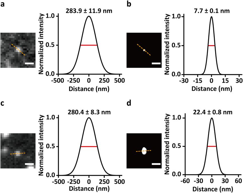
Photoswitchable mQDs in QD-PAINT are superior to Cy3 in estimate the imaging localization precision is to distin-
DNA-PAINT guish two closely localized imaging points34. We esti-
Next, we investigated mQD photoswitchability for QD- mated the imaging localization precision of QD-PAINT
PAINT in cells (Fig. 2a). As transient hybridization and after reconstruction by measuring the distance between
specificity can be tuned by changing the length and the maximum of each PSF of two closely localized SNAP-
sequence of the DNA hybridization pair between the EGFR pairs. QD-PAINT was capable of resolving two
docking and imager strands, we reduced the length of the SNAP-EGFR pairs separated by ~8 nm (Supplementary
docking strands from 20 nucleotides to 8 nucleotides Fig. 6).
(Supplementary Table 1). The mQDs that were hardly
dissociable in the 20-nucleotide hybridization between the Discussion
docking and imager strands (Supplementary Video 2) In this study, we introduce a promising next-generation
became photoswitchable by transient hybridization with SMLM method named QD-PAINT that increases the
an on-time of ~17.8 ± 0.7 s (Fig. 2b and Supplementary spatial resolution of conventional SMLM by utilizing the
Video 3). We compared the photoswitching and intensity high brightness of QDs. Until now, sub-20 nm spatial
profiles of QD-PAINT with those of DNA-PAINT with resolution has been feasible in a study using DNA-PAINT
Cy3 (Supplementary Video 4). While the exposure time in in vitro by trace averaging, templated, and geometry-
QD-PAINT was 10-fold higher than that in DNA-PAINT templated drift correction with origami as fiducial mar-
with Cy3, the on-time was 8.9-fold higher in QD-PAINT kers, with a photon count cutoff, achieving a resolution of
than in DNA-PAINT with Cy3. This 8.9-fold difference in ~5 nm35. QD-PAINT shows great promise for achieving
the on-time occurred because the hybridization sequence sub-20 nm spatial resolution in vivo by fundamentally
length in QD-PAINT was 1 nucleotide longer than that in increasing the photon number that critically determines
DNA-PAINT with Cy3, and this result was consistent the spatial resolution. Our methodology overcame the
with previous studies that reported an 8- to 10-fold higher major limitations of QDs in the utilization of SMLM due
Official journal of the Korean Society for Biochemistry and Molecular BiologyChang et al. Experimental & Molecular Medicine (2021) 53:384–392 389 Fig. 2 QD-PAINT and DNA-PAINT with Cy3. a Schematic of conducting DNA-PAINT with Cy3 (left) and QD-PAINT (right) on cells. b Plots of single- molecule measurements of the on-time of mQDs (left) and Cy3 (right). The error bars represent the accuracy of the fitting at the 95% confidence interval. c Statistical distribution of fluorescence intensity per fluorescence population density during SMLM imaging in QD-PAINT (red) and DNA- PAINT with Cy3 (black). The exposure times were 500 and 5000 ms for Cy3 and mQDs, respectively. to their high nonspecificity, multivalency, and, most ligands used in the preparation of monovalent QDs, importantly, nonphotoswitchability by combining steric including mPEG thiol, ptDNA, and carboxy PEG6 alkane exclusion and DNA-PAINT methods7,36. After passiva- thiol. However, as shown in Fig. 2c, the fluorescence tion with ptDNA-PEG6-casein and tuning of hybridiza- intensity profile of mQDs showed a multimodal dis- tion sequences, monovalent and specific QDs became tribution, suggesting the inclusion of multiple mQD photoswitchable via QD-PAINT and maintained super- species with differential fluorescence intensities ranging iority to Cy3 in fluorescence intensity, showing a theo- from 10- to 100-fold higher than that of Cy3. The retical improvement in resolution by a factor of ~3. ‘reduced’ fluorescence intensity could be recovered by QD-PAINT achieved improved spatial resolution with a uncaging mQDs passivated with surface-bound thiol narrower FWHM than that of Cy3 by 2.9-fold. molecules upon laser excitation (Supplementary Video 5), Although bare QDs are generally known to be sub- as also observed in a previous report39. Therefore, the stantially brighter than organic dyes, the improvement in mQDs with a fluorescence intensity of 104 in Fig. 3 may be the localization accuracy of QD-PAINT was limited to an responsible for the 9-fold higher intensity of mQDs than ~3-fold increase derived from the ~9-fold higher fluor- Cy3, and the mQDs with a fluorescence intensity of 105, escence intensity than that of Cy3 (Figs. 2c and 3). As thus 100-fold higher intensity than Cy3, may lead to a 10- previously reported regarding the diminishing effect of fold improvement in resolution. To utilize the brightest thiol ligands on the fluorescence intensity of QDs37,38, this mQDs with further increased photon output for SMLM, unexpectedly ‘reduced’ fluorescence intensity of mQDs is sufficient pre-excitation with laser illumination could be also probably due to the caging effect induced by the thiol employed. Although the proof-of-concept of QD-PAINT Official journal of the Korean Society for Biochemistry and Molecular Biology
Chang et al. Experimental & Molecular Medicine (2021) 53:384–392                                                                                      390

  Fig. 3 Improvement in spatial resolution with QD-PAINT over that in DNA-PAINT with Cy3. Representative single-molecule images of EGFR
  obtained using QD-PAINT before (a left) and after (b left) reconstruction and using DNA-PAINT with Cy3 before (c left) and after (d left)
  reconstruction. Scale bars, 500 nm (a, c) and 100 nm (b, d). Cross-sectional histograms of the single molecules of EGFR for measuring the full width at
  half maximum (FWHM) over the dashed lines using QD-PAINT before (a right) and after (b right) reconstruction and using DNA-PAINT with Cy3
  before (c right) and after (d right) reconstruction.

led to a limited resolution improvement of a 3-fold                            such as PALM and DNA-PAINT also took several hours
increase, the current QD-PAINT in its primary stage of                         up to days9, we focused on presenting a general method to
technique development has not reached its full potential,                      generate the blinking of bright monovalent QDs required
and further optimization to maximize the photon output                         for SMLM through DNA-PAINT and steric exclusion
will lead to further improved spatial resolution. In addi-                     with ptDNA and demonstrated the proof-of-concept of
tion, given that the DNA-PAINT system allows for con-                          our method. For optimization of QD-PAINT, the balance
stant renewal of QDs on the sample by exchange with                            between the on-time and off-time, typically quantified as a
reservoir QDs from solution, QDs can be detected mul-                          duty cycle in SMLM, critically contributes to the spatio-
tiple times at each spot, enabling accumulation of photons                     temporal resolution of superresolution images. The on-
for much higher localization precision and further                             time is crucially proportional to the length of DNA linkers
improvement of the resolution.                                                 for hybridization between targets and mQDs. However,
  The major limitation of the current QD-PAINT is the                          the off-time is not significantly affected by the length of
long image acquisition time required for the whole                             DNA linkers but mainly depends on the concentrations of
structure of the imaging sample compared to conven-                            docking strand-bearing target proteins and imager strand-
tional SMLM. In the current QD-PAINT, each QD is                               bearing mQDs because the hybridization between short
detected only once, but due to the high quantum yield of                       DNA oligomers is primarily diffusion-limited. As the
QDs, substantially more photons can still be collected,                        concentration of docking strand-bearing target proteins
resulting in improved resolution. Conceptually, as QD-                         varies in different samples, the concentration of imager
PAINT with an on-time of ~17.8 s detects ~100 molecules                        strand-bearing mQDs should be adjusted sample-by-
in each frame every 5 s, the total acquisition time for                        sample to optimize the off-time. For practical use of
imaging a total of 1 × 10⁶ molecules on the plasma                             QD-PAINT, extensive optimizations, including the on-
membrane is ~49 h. Although a long image acquisition                           time and off-time, will be required in a further study.
time is required, as the initial forms of SMLM techniques                      Upon optimization, the image acquisition time of QD-

Official journal of the Korean Society for Biochemistry and Molecular Biology
Chang et al. Experimental & Molecular Medicine (2021) 53:384–392                                                                                                                 391

PAINT may be reduced down to a similar extent as in the                                       Conflict of interest
speed-optimized DNA-PAINT methods using various                                               The authors declare no competing interests.

approaches40–42, in which resolving 20-nm distances with
                                                                                              Publisher’s note
sufficient sampling in 5 min is possible. The optimized                                        Springer Nature remains neutral with regard to jurisdictional claims in
imaging speed will allow the extremely photostable QDs                                        published maps and institutional affiliations.
to be detected multiple times at each spot, leading to the
                                                                                              Supplementary information The online version contains supplementary
accumulation of photons for higher localization precision                                     material available at https://doi.org/10.1038/s12276-021-00572-4.
and further improvement of the resolution within an
overall reasonably short image acquisition time.                                              Received: 5 October 2020 Revised: 7 December 2020 Accepted: 29
  The large hydrodynamic size of mQDs is not expected                                         December 2020.
                                                                                              Published online: 2 March 2021
to affect their labeling efficiency in terms of density or
fidelity because mQDs detach from the binding site,
allowing binding of another mQD to the same spot. The                                         References
                                                                                               1. Chojnacki, J. et al. Maturation-dependent HIV-1 surface protein redistribution
large hydrodynamic size of the mQDs can instead be                                                 revealed by fluorescence nanoscopy. Science 338, 524–528 (2012).
advantageous by sterically hindering the binding of sec-                                       2. Kanchanawong, P. et al. Nanoscale architecture of integrin-based cell adhe-
ond molecules at close positions during the time they                                              sions. Nature 468, 580–584 (2010).
                                                                                               3. Xu, K., Zhong, G. & Zhuang, X. Actin, spectrin, and associated proteins form a
bind to the target molecules. This is preferable in SMLM                                           periodic cytoskeletal structure in axons. Science 339, 452–456 (2013).
because it reduces the chance of simultaneous binding of                                       4. Sahl, S. J., Hell, S. W. & Jakobs, S. Fluorescence nanoscopy in cell biology. Nat.
two or more QDs within the same diffraction-limited                                                Rev. Mol. cell Biol. 18, 685 (2017).
                                                                                               5. Hell, S. W. et al. The 2015 super-resolution microscopy roadmap. J. Phys. D:
region.                                                                                            Appl. Phys. 48, 443001 (2015).
  Another potential application of QD-PAINT is the                                             6. Thompson, M. A., Lew, M. D. & Moerner, W. Extending microscopic resolution
imaging of live specimens with the resolution of dynamic                                           with single-molecule imaging and active control. Annu. Rev. biophysics 41,
                                                                                                   321–342 (2012).
target molecules at a higher resolution than in sptPALM,                                       7. Jungmann, R. et al. Multiplexed 3D cellular super-resolution imaging with
which typically utilizes photoactivatable or photoswitch-                                          DNA-PAINT and Exchange-PAINT. Nat. methods 11, 313–318 (2014).
able fluorescent proteins or organic dyes43. As QDs                                             8. Calarese, D. A. et al. Antibody domain exchange is an immunological solution
                                                                                                   to carbohydrate cluster recognition. Science 300, 2065–2071 (2003).
absorb many more photons than organic dyes at the same                                         9. Betzig, E. et al. Imaging intracellular fluorescent proteins at nanometer reso-
excitation photon flux and subsequently release a higher                                            lution. Science 313, 1642–1645 (2006).
number of photons, a low laser power can be sufficient for                                     10. Pinaud, F., Clarke, S., Sittner, A. & Dahan, M. Probing cellular events, one
                                                                                                  quantum dot at a time. Nat. Methods 7, 275–285 (2010).
detecting QD-labeled molecules, avoiding the phototoxic                                       11. Huang, F. et al. Ultra-high resolution 3D imaging of whole cells. Cell 166,
effects on live cells of high-power laser illumination44.                                         1028–1040 (2016).
  Ultraresolution QD-PAINT will enable the fluores-                                            12. Dempsey, G. T., Vaughan, J. C., Chen, K. H., Bates, M. & Zhuang, X. Evaluation of
                                                                                                  fluorophores for optimal performance in localization-based super-resolution
cence imaging of organelles and molecular complexes at                                            imaging. Nat. Methods 8, 1027 (2011).
the nanoscale, a level that has not been properly                                             13. Zhao, Q., Young, I. T. & De Jong, J. G. S. Photon budget analysis for fluores-
observed due to the limited resolution of the current                                             cence lifetime imaging microscopy. J. Biomed. Opt. 16, 086007 (2011).
                                                                                              14. Thompson, R. E., Larson, D. R. & Webb, W. W. Precise nanometer localization
SMLM that functions by utilizing photoactivatable or                                              analysis for individual fluorescent probes. Biophys. J. 82, 2775–2783 (2002).
photoswitchable fluorescent proteins or organic dyes45.                                        15. Saxton, M. J. Lateral diffusion in an archipelago. Single-particle diffusion. Bio-
From the perspective of basic biology, the composition                                            phys. J. 64, 1766–1780 (1993).
                                                                                              16. Michalet, X. et al. Quantum dots for live cells, in vivo imaging, and diagnostics.
and molecular architecture of protein complexes or                                                Science 307, 538–544 (2005).
dense protein networks will be revealed, while altera-                                        17. Groc, L. et al. Surface trafficking of neurotransmitter receptor: comparison
tions may become critical markers of disease conditions                                           between single-molecule/quantum dot strategies. J. Neurosci. 27,
                                                                                                  12433–12437 (2007).
in the health industry. By realizing the unseen, QD-                                          18. Byers, R. J. & Hitchman, E. R. Quantum dots brighten biological imaging. Prog.
PAINT may become a cornerstone, if not the vanguard,                                              Histochem. Cytochem. 45, 201–237 (2011).
of ultraresolution SMLM.                                                                      19. Chan, W. C. & Nie, S. Quantum dot bioconjugates for ultrasensitive nonisotopic
                                                                                                  detection. Science 281, 2016–2018 (1998).
                                                                                              20. Gao, X., Cui, Y., Levenson, R. M., Chung, L. W. & Nie, S. In vivo cancer targeting
Acknowledgements                                                                                  and imaging with semiconductor quantum dots. Nat. Biotechnol. 22, 969–976
This work was supported by the Global Research Laboratory (GRL) Program                           (2004).
through the National Research Foundation of Korea (NRF) funded by the                         21. Xing, Y. & Rao, J. Quantum dot bioconjugates for in vitro diagnostics & in vivo
Ministry of Science and ICT (No. NRF-2016K1A1A2912722) and a National                             imaging. Cancer Biomark. 4, 307–319 (2008).
Research Foundation of Korea (NRF) grant funded by the Ministry of Education                  22. Wang, Y., Fruhwirth, G., Cai, E., Ng, T. & Selvin, P. R. 3D super-resolution imaging
Science and Technology of Korea (MEST) (No. NRF-2019R1A2C2002152).                                with blinking quantum dots. Nano Lett. 13, 5233–5241 (2013).
                                                                                              23. Xu, J., Tehrani, K. F. & Kner, P. Multicolor 3D super-resolution imaging by
                                                                                                  quantum dot stochastic optical reconstruction microscopy. Acs Nano 9,
Author contributions                                                                              2917–2925 (2015).
Conceptualization: D.-H.K. and Y.C.; methodology: D.-H.K. and Y. C.;                          24. Jung, S. et al. Light-induced fluorescence modulation of quantum dot-crystal
investigation: D.-H.K., Y.C., K.Z., Y.K., S.P., M.J., J.N., and T.H.; analysis: D.-H.K. and       violet conjugates: stochastic off–on–off cycles for multicolor patterning and
Y.C.; manuscript preparation: D.-H.K. and Y.C.; funding acquisition: S.R. and D.-H.               super-resolution. J. Am. Chem. Soc. 139, 7603–7615 (2017).
K.; supervision: S.R. and D.-H.K.

Official journal of the Korean Society for Biochemistry and Molecular Biology
Chang et al. Experimental & Molecular Medicine (2021) 53:384–392 392
25. Howarth, M. et al. Monovalent, reduced-size quantum dots for imaging 35. Dai, M., Jungmann, R. & Yin, P. Optical imaging of individual biomolecules in
receptors on living cells. Nat. Methods 5, 397–399 (2008). densely packed clusters. Nat. Nanotechnol. 11, 798–807 (2016).
26. Kim, D.-H. et al. Direct visualization of single-molecule membrane protein 36. Schnitzbauer, J., Strauss, M. T., Schlichthaerle, T., Schueder, F. & Jungmann,
interactions in living cells. PLoS Biol. 16, e2006660 (2018). R. Super-resolution microscopy with DNA-PAINT. Nat. Protoc. 12, 1198 (2017).
27. Farlow, J. et al. Formation of targeted monovalent quantum dots by steric 37. Gao, X., Chan, W. C. & Nie, S. Quantum-dot nanocrystals for ultrasensitive
exclusion. Nat. Methods 10, 1203–1205 (2013). biological labeling and multicolor optical encoding. J. Biomed. Opt. 7, 532–537
28. Seo, D., Farlow, J., Southard, K., Jun, Y.-W. & Gartner, Z. J. Production and (2002).
targeting of monovalent quantum dots. JoVE, e52198, https://doi.org/10.3791/ 38. Uyeda, H. T., Medintz, I. L., Jaiswal, J. K., Simon, S. M. & Mattoussi, H. Synthesis of
52198 (2014). compact multidentate ligands to prepare stable hydrophilic quantum dot
29. Valley, C. C. et al. Enhanced dimerization drives ligand-independent activity of fluorophores. J. Am. Chem. Soc. 127, 3870–3878 (2005).
mutant epidermal growth factor receptor in lung cancer. Mol. Biol. Cell 26, 39. Han, G., Mokari, T., Ajo-Franklin, C. & Cohen, B. E. Caged quantum dots. J. Am.
4087–4099 (2015). Chem. Soc. 130, 15811–15813 (2008).
30. Gao, J. et al. Mechanistic insights into EGFR membrane clustering revealed by 40. Schickinger, M., Zacharias, M. & Dietz, H. Tethered multifluorophore motion
super-resolution imaging. Nanoscale 7, 2511–2519 (2015). reveals equilibrium transition kinetics of single DNA double helices. Proc. Natl
31. Werbin, J. L. et al. Multiplexed Exchange-PAINT imaging reveals ligand- Acad. Sci. USA 115, E7512–e7521 (2018).
dependent EGFR and Met interactions in the plasma membrane. Sci. Rep. 7, 41. Schueder, F. et al. An order of magnitude faster DNA-PAINT imaging by
12150 (2017). optimized sequence design and buffer conditions. Nat. Methods 16,
32. Jungmann, R. et al. Single-molecule kinetics and super-resolution microscopy 1101–1104 (2019).
by fluorescence imaging of transient binding on DNA origami. Nano Lett. 10, 42. Strauss, S. & Jungmann, R. Up to 100-fold speed-up and multiplexing in
4756–4761 (2010). optimized DNA-PAINT. Nat. Methods 17, 789–791 (2020).
33. Auer, A., Strauss, M. T., Schlichthaerle, T. & Jungmann, R. Fast, background-free 43. Li, H. & Vaughan, J. C. Switchable fluorophores for single-molecule localization
DNA-PAINT imaging using FRET-based probes. Nano Lett. 17, 6428–6434 microscopy. Chem. Rev. 118, 9412–9454 (2018).
(2017). 44. Wäldchen, S., Lehmann, J., Klein, T., Van De Linde, S. & Sauer, M. Light-induced
34. Endesfelder, U., Malkusch, S., Fricke, F. & Heilemann, M. A simple method cell damage in live-cell super-resolution microscopy. Sci. Rep. 5, 15348 (2015).
to estimate the average localization precision of a single-molecule 45. Sochacki, K. A., Dickey, A. M., Strub, M.-P. & Taraska, J. W. Endocytic proteins are
localization microscopy experiment. Histochem. Cell Biol. 141, 629–638 partitioned at the edge of the clathrin lattice in mammalian cells. Nat. Cell Biol.
(2014). 19, 352–361 (2017).
Official journal of the Korean Society for Biochemistry and Molecular BiologyYou can also read