2021 Advocacy Statements for the Role of 99mTc-Pyrophosphate Scintigraphy in the Diagnosis of Transthyretin Cardiac Amyloidosis: A Report of the ...
←
→
Page content transcription
If your browser does not render page correctly, please read the page content below
Acta Cardiol Sin 2021;37:221-231
Consensus                                                                               doi: 10.6515/ACS.202105_37(3).20210420A

            2021 Advocacy Statements for the Role of
             99m
                 Tc-Pyrophosphate Scintigraphy in the
         Diagnosis of Transthyretin Cardiac Amyloidosis:
          A Report of the Taiwan Society of Cardiology
           and the Society of Nuclear Medicine of the
                         Republic of China
                  Yih-Hwen Huang,1 Yen-Hung Lin,2,3 Ruoh-Fang Yen,1,2 Charles Jia-Yin Hou,4,5 Shan-Ying Wang,6,7
                    Shih-Chuan Tsai,8,9 Kung-Chu Ho,10 Ming-Hsien Lin,11,12 Chin-Ho Tsao,13 Chih-Yung Chang,7,14
                               Jin-Long Huang,15,16 Mei-Fang Cheng1,2* and Yen-Wen Wu1,2,3,6,16,17*

     Transthyretin cardiac amyloidosis (ATTR-CM) is an increasingly recognized cause of heart failure with preserved
     ejection fraction. Favorable prognosis depends on early diagnosis and correct treatment strategy. Among patients
     for whom there is a high clinical suspicion of cardiac amyloidosis, 99mTc-labeled bone avid scintigraphy including
     99m
         Tc-pyrophosphate (PYP) scintigraphy may be of diagnostic and prognostic importance. Various international
     guidelines support the non-biopsy diagnosis of ATTR-CM using 99mTc-PYP scintigraphy, yet emphasize the gap in
     standardization of acquisition and imaging analysis protocols, as well as the appropriateness of its clinical use.
     Therefore, a joint expert consensus has been reached by the Taiwan Society of Cardiology and the Society of
     Nuclear Medicine of the Republic of China, to advocate for the application of 99mTc-PYP scintigraphy in the diagnosis
     of ATTR-CM. This article aims to highlight the recommendations on image acquisition, qualitative and quantitative
     assessments of cardiac 99mTc-PYP uptake, and diagnostic algorithms. We hope the implementation of these
     recommendations in Taiwan will facilitate the process and enhance the diagnostic rate of ATTR-CM.

     Key Words:         Cardiac amyloidosis · Cardiomyopathy · Heart failure · Scintigraphy ·              99m
                                                                                                                 Tc-pyrophosphate
                        (PYP) · Transthyretin

Received: April 12, 2021          Accepted: April 20, 2021
1
  Department of Nuclear Medicine, National Taiwan University Hospital; 2National Taiwan University College of Medicine; 3Department of
Internal Medicine, National Taiwan University Hospital, Taipei; 4Mackay Medical College, New Taipei City; 5Cardiovascular Center, MacKay
Memorial Hospital, Taipei; 6Department of Nuclear Medicine, Far Eastern Memorial Hospital, New Taipei City; 7Department of Biomedical
Imaging and Radiological Sciences, National Yang Ming Chiao Tung University, Taipei; 8Department of Nuclear Medicine, Taichung Veterans
General Hospital; 9Department of Medical Imaging and Radiological Sciences, Central Taiwan University of Science and Technology, Taichung;
10
   Department of Nuclear Medicine, Chang Gung Memorial Hospital, Taoyuan; 11Division of Nuclear Medicine, Department of Nuclear
Medicine, Cheng Hsin General Hospital, Taipei; 12Division of Nuclear Medicine, Camillian Saint Mary’s Hospital, Luodong; 13Department of
Nuclear Medicine, Mackay Memorial Hospital; 14Department of Nuclear Medicine, Taipei Veterans General Hospital, Taipei; 15Heart Failure
Division, Cardiovascular Center, Education Department, Taichung Veterans General Hospital, Taichung; 16National Yang Ming Chiao Tung
University, School of Medicine, Faculty of Medicine, Taipei; 17Division of Cardiology, Cardiovascular Medical Center, Far Eastern Memorial
Hospital, New Taipei City, Taiwan.
Corresponding author: Dr. Yen-Wen Wu, Cardiology Division of Cardiovascular Medical Center, Far Eastern Memorial Hospital, No. 21, Sec. 2,
Nanya S. Rd., Banciao Dist., New Taipei City, Taiwan. Tel: 886-2-8966-7000 ext. 1019; Fax: 886-2-7728-2378; E-mail: wuyw0502@gmail.com
* These authors contributed equally to the study.

                                                                   221                                   Acta Cardiol Sin 2021;37:221-231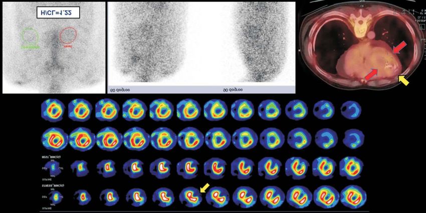
Yih-Hwen Huang et al.

INTRODUCTION                                                        mately 80% of them have LVH.14 Therefore, raising early
                                                                    awareness that ATTR-CM in Taiwan can manifest as a
     Heart failure (HF) is the leading cause of cumulative          dominant cardiac phenotype mimicking hypertrophic
hospitalization days for cardiovascular disease in Taiwan,          cardiomyopathy (HCM) at presentation will facilitate the
and rates of re-hospitalization and mortality are high.1,2          clinical recognition of this under-diagnosed disease.
Due to different etiologies and disease severity, there is               ATTR is debilitating and associated with poor life ex-
a large gap in treatment response and prognosis.3-5                 pectancy, especially in those with cardiac dysfunction.
     The incidence and prevalence of HF with preserved              Delayed or lack of treatment after diagnosis will result in
ejection fraction (HFpEF) have increased in recent years.6          poor prognosis, with a median survival period of only 2
Among the different causes of HFpEF, cardiac amylo-                 to 3 years. Recently, a variety of new and promising
idosis (CA), where amyloid deposition occurs in the                 treatment options have become available,7 including
heart, still poses challenges to physicians in early diag-          pharmacological therapy that slows or halts ATTR-CM
nosis and treatment.7,8 The majority (> 95%) of patients            progression and favorably affects clinical outcomes. The-
with cardiac amyloidosis are affected either by a mis-              refore, early recognition is essential to afford the best
folded immunoglobulin light chain (AL), or by trans-                treatment efficacy.
thyretin (TTR) deposition in the heart.8 Cardiomyopathy                  Several clinical signs can raise suspicion for CA. These
is a clinical feature of transthyretin amyloidosis (ATTR),          red flags include abnormalities on electrocardiography
which is an under-recognized systemic disease whereby               (ECG), echocardiography, cardiac magnetic resonance
the transthyretin protein misfolds to form fibrils that de-         imaging (CMR), or cardiac biomarkers.15-18 Noninvasive
posit in various tissues and organs. Transthyretin cardiac          methods including ECG, echocardiography, CMR, and
amyloidosis (ATTR-CM) can be divided into two major                 nuclear scintigraphy can assist in diagnosis.19 LVH is a
forms — wild-type ATTR (ATTRwt) and ATTR from a ge-                 common indicator used to detect CA. Red flags of CA on
netic mutation (ATTRm).7                                            ECG include a low QRS voltage in combination with in-
     The primary manifestations of ATTRwt are cardiac               creased left ventricle thickness, a pseudo-infarction pat-
amyloidosis and carpal tunnel syndrome. The preva-                  tern, conduction abnormality, and arrhythmia.19-21 Red
lence of ATTRwt in HFpEF patients with left ventricular             flags on echocardiography indicating CA include thicken-
hypertrophy (LVH) (wall thickness > 12 mm) is about                 ing of the atrioventricular valves and interatrial septum,
13%, and the prevalence of ATTRwt in patients with aor-             a speckled and granular appearance of the myocardium,
tic stenosis after aortic valve replacement is 6-16%.9-11           and a base-to-apex strain gradient with relative apical
The age of onset of ATTRm is variable, ranging from 30              sparing of longitudinal strain (the so-called “cherry-on-
to 80 years old. The symptoms of ATTRm correspond to                top pattern”).19,22,23 CMR might show global diffuse myo-
different gene mutations. This hereditary form of ATTR              cardial late gadolinium enhancement in the subendo-
manifests predominantly as polyneuropathy, cardiomyo-               cardial layers, elevated native T1 and extracellular vol-
pathy, or mixed phenotype.12 The Transthyretin Amyloi-              ume fraction in T1 mapping.18,19,24 Endocardial biopsy is
dosis Outcomes Survey (THAOS) study found that among                considered as the gold standard for the diagnosis of
symptomatic patients with TTR mutations, approxi-                   ATTR.19 Once CA is suspected, further confirmatory diag-
mately half have neurological manifestations as their               nosis could be reached through either non-invasive me-
primary symptom, followed by cardiac abnormalities                  thods such as nuclear scintigraphy and the free light
and mixed phenotype, each constituting approximately                chain (FLC) test,25-27 or invasive methods such as biopsy
a quarter, respectively. 12 ATTR genotypes and pheno-               and amyloid typing.
types are highly heterogeneous. Because there are no                     Bone-avid radiotracers including technetium-99m
large-scale screening and registration studies, the actual          (99mTc)-pyrophosphate (PYP) have been reported to be
prevalence of ATTR-CM in Taiwan remains unclear. The                useful in detecting CA.8,15-19,22-27 The exact mechanism is
Ala97Ser mutation is the most common cause of ATTRm                 not fully understood. It may be related to amyloid micro-
in Taiwan.13,14 It is considered both a form of cardiomyo-          calcification. Historically, 99mTc-PYP has been well-known
pathy and a polyneuropathic syndrome, and approxi-                  for its role in diagnosing acute/recent myocardial infarc-

Acta Cardiol Sin 2021;37:221-231                              222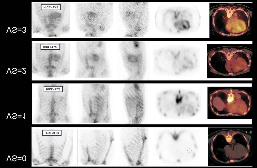
TSOC/SNMROC Statements for PYP Scan in ATTR-CM

tion or rhabdomyolysis, thus also referred to as infarct               scan will make diagnosis of ATTR-CM more applicable
or muscle scintigraphy. Semi-quantitative analyses based               for both clinicians and patients.27
on the radiotracer uptake in the heart and surrounding                      Last but not least, with respect to promising thera-
ribs, expressed on a 0-3 point grading scale, help to dis-             peutic outcome from recent clinical trials, imaging tech-
tinguish different types of CA.25-27 Most patients with AL             niques and reporting format standardization are of in-
amyloidosis are of lower grades (grades 0-2), while pa-                creasing importance. The Taiwan Society of Cardiology
tients with ATTR-CM are of higher grades (grades 2-                    (TSOC) and the Society of Nuclear Medicine of the Re-
3).25-27 In a study which enrolled 1,217 patients after ex-            public of China (SNMROC) apprehend the acute neces-
cluding AL with suspected CA and using PYP scan grades                 sity of establishing advocacy statements on when and
2-3 as criteria, the specificity and positive predictive               how to use nuclear imaging procedures, adjust relevant
values were both as high as 100%.28 PYP scan has thus                  acquisition parameters, standardize reporting format,
been considered a suitable alternative to EMB in some                  and facilitate earlier therapeutic intervention and clini-
patients suspect of ATTR-CM. 29,30 In contrast to echo-                cal research.
cardiography, EMB can truly demonstrate amyloid fibrils
in clinically suspected patients mimicking hypertensive
                                                                       99m
heart disease or HCM. It strengthens the importance of                    Tc-PYROPHOSPHATE SCINTIGRAPHY
pathological confirmation of CA from other HCM, albeit
diagnostic yield of extracardiac biopsies with the pres-               Image acquisition procedures
ence of amyloid protein in high-risk patients with con-                     Recommendations for 99mTc-PYP scintigraphy acqui-
duction disturbances and advanced age could be rela-                   sition parameters are listed in Table 1. Standard planar
tively low. A feasible and less invasive tool such as PYP              images for anterior, left anterior oblique and lateral
                               99m
Table 1. Recommendations for         Tc-pyrophosphate scintigraphy acquisition parameters
                                                                                         Parameters
Imaging procedure
  Specific preparation                                         None; fasting is not required
  Scan                                                         Rest scan
           99m
  Dose of Tc-PYP                                               · Recommended: 20 mCi for 3-hour acquisition
                                                               · Adjust according to different models of instrument
  Time interval between injection and acquisition              · Recommended: 3-hour planar and SPECT
                                                               · Optional: 1-hour planar
Imaging parameters
  Field of view                                                Cardiac or chest
  Image type                                                   Cardiac or chest planar and SPECT (or SPECT/CT)
  Position                                                     Supine
  Energy window                                                140 keV, 15-20%
  Collimators                                                  Low energy, high resolution
  Matrix                                                       128 ´ 128 recommended (64 ´ 64 minimum required) for SPECT,
                                                               256 ´ 256 for planar
  Pixel size                                                   2.3-6.5 mm (may change with different matrix)
Planar imaging specific parameters
  Number of views                                              Anterior, lateral, and left anterior oblique (LAO)
  Detector configuration                                       180 degrees
  Image duration                                               750,000 counts
  Magnification                                                1.45-1.5
SPECT imaging specific parameters
  Angular range                                                360 degrees
  Detector configuration                                       180 degrees
  ECG gating                                                   Off; non-gated imaging
  Number of views/detectors                                    32-64
  Time per stop                                                20 seconds
ECG, electrocardiography; keV, kilo-electronvolts; mCi, millicurie; PYP, pyrophosphate; SPECT, single-photon emission computed
tomography; SPECT/CT, SPECT/computed tomography.

                                                                 223                                    Acta Cardiol Sin 2021;37:221-231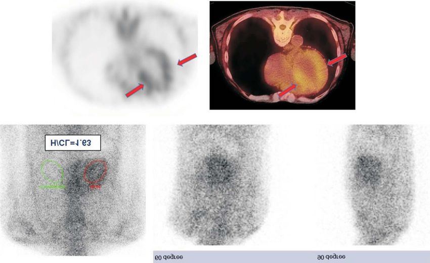
Yih-Hwen Huang et al.

views are shown in Figure 1. Bilateral shoulders should                          (SPECT/CT) is preferred for attenuation correction and
be visible in the image to evaluate for musculoskeletal                          anatomical localization. It accentuates differences in
manifestations of amyloid deposition, such as the shoul-                         myocardial uptake patterns, allowing for more accu-
der pad sign or joint swelling.31 Symmetric renal uptake                         rate classifications — absent, focal, diffuse, or focal on
should be minimized in planar images to avoid interfer-                          diffuse. Therefore, an injection dose of 20 millicurie
ence with the myocardial counts.                                                 (mCi) of 99mTc PYP is recommended for 3-hour planar
     One-hour imaging results have higher detection                              and SPECT imaging. Although delayed-time-point imag-
sensitivity, while 3-hour imaging results offer higher                           ing is expected to improve qualitative and semi-quanti-
detection specificity. This is because delayed clearance                         tative results, SPECT or SPECT/CT at 1 hour with low
of blood pool activity after tracer injection is not un-                         blood activity is also acceptable. If SPECT or SPECT/CT
common in patients with poor renal function or low car-                          is performed, planar images of left anterior oblique
diac output. In our recommendation, 3-hour planar im-                            and lateral views are optional to shorten the image ac-
ages are recommended for visual scoring and semi-                                quisition time.
quantitation and 1-hour images are optional. Single-                                  The differences in 99m Tc-PYP scintigraphy imaging
photon emission computed tomography (SPECT) is ne-                               acquisition parameters between our recommendations
cessary to distinguish blood pool activity from myocar-                          and those of the 2019 American Society of Nuclear Car-
dial activity. In addition, SPECT/computed tomography                            diology (ASNC) practice points26 are listed in Table 2.

          A                                                 B                                             C
Figure 1. Standard planar images for (A) ANT (anterior), (B) LAO (left anterior oblique) and (C) LAT (lateral) views, acquired at 1 hour (hr) and/or 3
hrs, respectively. Optimal field of view (FOV) should cover the whole chest region, including bilateral shoulders while avoiding highly radioactive sites,
such as injection sites or areas with marked renal activity.

Table 2. Differences in 99mTc-pyrophosphate scintigraphy acquisition parameters between ASNC* and TSOC/SNMROC consensus
         recommendations
                                                                        TSOC and SNMROC                            2019 ASNC practice points
                                                                        recommendations                               recommendations*
Imaging procedure
          99m
  Dose of Tc-PYP                                  · Recommended: 20 mCi for 3-hour                            · 10-20 mCi
                                                    acquisition
                                                  · Adjust according to different models
                                                    of instrument
  Time interval between injection and acquisition · Recommended: 3-hour planar and                            · Recommended: 1-hour planar and
                                                    SPECT                                                       SPECT
                                                  · Optional: 1-hour planar                                   · Optional: 3-hour SPECT or planar
Imaging parameters
  Matrix                                          · Recommended: 128 ´ 128 for SPECT                          64 ´ 64 minimum
                                                  · Required: 64 ´ 64 minimum for SPECT
                                                  · 256 ´ 256 for planar
Planar imaging specific parameters
  Detector configuration                          180 degrees                                                 90 degrees
  Magnification                                   1.45-1.5                                                    1.46
SPECT imaging specific parameters
  Number of views/detector                        32-64                                                       40
ASNC, American Society of Nuclear Cardiology; ECG, electrocardiography; mCi, millicurie; PYP, pyrophosphate; SNMROC, Society of
Nuclear Medicine of the Republic of China; SPECT, single-photon emission computed tomography; TSOC, Taiwan Society of
Cardiology.
                                                       26
* Source: Dorbala S, et al. 2019 ASNC Practice Points.

Acta Cardiol Sin 2021;37:221-231                                           224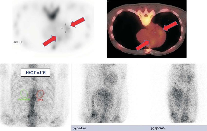
TSOC/SNMROC Statements for PYP Scan in ATTR-CM

SEMI-QUANTITATION                                                              ing careful to exclude any right heart or mediastinal ac-
                                                                               tivity and avoiding the subdiaphragmatic organs. Total
     A visual grading scale is used to interpret the imag-                     and absolute mean counts are measured in each ROI. A
ing results (Table 3). A visual grade of 0 is not suggestive                   H/CL ratio is calculated as the fraction of heart ROI mean
of ATTR-CM. Grade 1 is equivocal. If clinically highly sus-                    counts to contralateral chest ROI mean counts. Alterna-
picious for CA, a specialist should perform further as-                        tively, geometric means of the average counts of ROIs
sessment. Grade 2 or 3 is diagnostic of ATTR-CM.                               calculated from both anterior and posterior planar views
     A heart to contralateral lung ratio (H/CL) on the pla-                    can be used. Although different H/CL ratios and cut-off
nar anterior view is used for semi-quantitation as shown                       values for one hour or three hours have been repor-
in Figure 2. A circular or elliptical region of interest (ROI)                 ted,25-27,32 our consensus uphold the criteria of the 2019
is drawn to include the left ventricle maximally while                         ASNC recommendations25,26 and the 2020 JCS guideline.27
cautiously avoiding the sternal region and the adjacent                        For one-hour images, an H/CL ratio > 1.5 is used to iden-
lung. The ROI is mirrored to the contralateral chest, be-                      tify ATTR-CM. For 3-hour images, an H/CL ratio ³ 1.3 is

                                                                       99m
Table 3. Recommendations for standardized interpretation of                  Tc-pyrophosphate scintigraphy
Visual interpretation
· Evaluate for cardiac radiotracer uptake on planar and myocardial uptake on SPECT or SPECT/CT images.
· If SPECT images show radiotracer uptake in the myocardium, proceed with visual grading. In the absence of any myocardial tracer
  uptake, a visual grade of 0 is given.
Visual grading
The relative tracer uptake in the myocardium to ribs seen on planar and SPECT images is graded on the following scale:
  · Grade 0                   No myocardial uptake and normal bone uptake
  · Grade 1                   Myocardial uptake less than rib uptake
  · Grade 2                   Myocardial uptake equal to rib uptake
  · Grade 3                   Myocardial uptake greater than rib uptake with mild/absent rib uptake
H/CL ratio interpretation
If tracer uptake is not evident on SPECT images, obtaining the H/CL ratio is not recommended. H/CL ratios can help identify ATTR-
CM if systemic AL amyloidosis is excluded by the following values:
   · 1-hour images            H/CL ratio > 1.5
   · 3-hour images            H/CL ratio ³ 1.3
AL, immunoglobulin light chain amyloidosis; ATTR-CM, transthyretin cardiac amyloidosis; H/CL, heart to contralateral lung; SPECT,
single-photon emission computed tomography; SPECT/CT, SPECT/computed tomography.
Adapted and modified from references 25, 26.

               A                                                              B
Figure 2. Quantitation of cardiac Tc-PYP uptake using the H/CL ratio. (A) Planar anterior view, (B) Region of interest (ROI) demonstration. Circu-
                                    99m

lar target ROI (red circles), which includes the heart, is drawn over the planar anterior image, and is mirrored over the contralateral lung. Total and
mean counts are calculated, and the H/CL ratio is derived from mean counts of ROIs. BKG, background; H/CL, heart to contralateral lung; PYP,
pyrophosphate; stddev, standard deviation.

                                                                         225                                       Acta Cardiol Sin 2021;37:221-231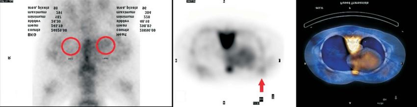
Yih-Hwen Huang et al.

used to identify ATTR-CM.                                                      Interpretation of 99mTc-PYP scintigraphy should in-
      99m
          Tc-PYP quantitative SPECT is a feasible and objec-              clude a visual evaluation of planar and SPECT images for
tive tool for assessing the burden of CA in the diagnosis                 any radiotracer uptake, visual grading, and the semi-
of ATTR-CM. 33,34 Dual-isotope SPECT using 99m Tc-PYP/                    quantitative H/CL ratio. In the absence of any tracer up-
201
    Tl might augment visual differentiation compared to                   take in the myocardium on SPECT imaging, a visual
single-isotope SPECT. 35 However, the methodology is                      grade of 0 is given. Visual scores ³ 2 on planar imaging
not standardized. As such, further investigation is war-                  are regarded as ATTR-positive. An H/CL ratio > 1.5 on
ranted.                                                                   one-hour imaging or ³ 1.3 on three-hour imaging is in-
      The recently introduced cadmium zinc telluride SPECT                dicative of ATTR-CM if plasma cell dyscrasia has been ex-
cameras have the potential to reduce radiation exposure                   cluded. If myocardial uptake patterns on SPECT are focal
to patients and shorten imaging time. The value of 99mTc                  or focal on diffuse, use the maximal uptake for visual
PYP imaging utilizing the newer “cardiac only” SPECT                      grading is recommended.
cameras also needs further validation.36-40                                    After excluding AL, 99mTc-labeled bone tracer scinti-
                                                                          graphy is used to diagnose ATTR-CM with high sensitiv-
                                                                          ity and specificity.37 False positives on 99mTc-PYP scans
INTERPRETATION AND REPORTING                                              may arise from AL, blood pool uptake, rib fractures,
                                                                          myocardial infarction, quinine drug toxicity, or other
    Recommendations for image interpretation and re-                      rare forms of CA. False negatives may be due to minimal
porting of 99mTc-PYP scintigraphy are listed in Tables 3 and 4.           myocardial infiltration (early-stage disease) or scar tis-

                                                           99m
Table 4. Recommendations for standardized reporting of           Tc-pyrophosphate scintigraphy
Parameters                                                                 Components
Demographics          Patient name, age, sex, study date and reason, previous imaging procedures, and results of biopsy if available.
                      Heart failure severity (NYHA classification, LVEF %), recent myocardial infarction history (within 3 months), and
                      renal function (eGFR or cCr within 3-6 months, or presence of ESRD) should be provided as supportive
                      information (recommended)
Methods               Image acquisition parameters and technique, dose and mode of radiotracer administration, time interval
                      between injection and scan, and scan (planar and SPECT) performed (required)
Findings              Quality of the image
                      Visual grading interpretation in relation to rib uptake (required)
                      Semi-quantitative H/CL ratio (required)
Ancillary findings    Consider attenuation correction in CT interpretation if SPECT/CT scanners are used (recommended)
Conclusions           1. Findings can be interpreted and categorized as follows:
                                                        Visual Score                H/CL ratio at 1-hour      H/CL ratio at 3-hour
                      Not suggestive                         0                               1.5                ³ 1.3 (positive)
                      Equivocal                              1                             1-1.5
                      2. Interpret the results in the context of prior cardiac evaluation, assessment for systemic AL amyloidosis using
                         serum and urine immunofixation, and serum free light chain assay.
                      3. If echo/CMR are strongly positive and 99mTc-PYP scan is negative, consider further evaluation including
                         endomyocardial biopsy.
                                                     99m
Notes                 A negative or mildly positive Tc-PYP does NOT exclude AL
                      Equivocal results could represent either AL or early ATTR
AL, immunoglobulin light chain amyloidosis; ATTR, transthyretin amyloidosis; cCr, creatinine clearance; CMR, cardiac magnetic
resonance; eGFR, estimated glomerular filtration rate; ESRD, end stage renal disease; H/CL ratio, heart to contralateral lung ratio;
LVEF, left ventricular ejection fraction; NYHA, New York Heart Association; PYP, pyrophosphate; SPECT, single-photon emission
computed tomography;SPECT/CT, SPECT/computed tomography. Adapted and modified from reference 25, 26.

Acta Cardiol Sin 2021;37:221-231                                    226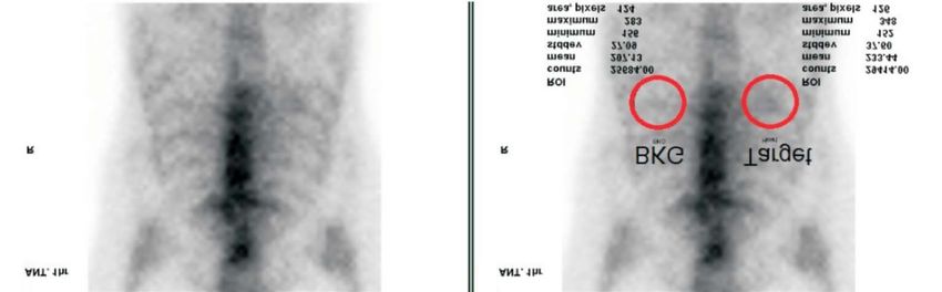
TSOC/SNMROC Statements for PYP Scan in ATTR-CM
sue formation after myocardial infarction. Therefore, and 99mTc-PYP scintigraphy can reveal four possible out-
the severity of heart failure [i.e., New York Heart Associ- comes. Firstly, if both tests are negative, it is unlikely to
ation (NYHA) functional classification or left ventricular be AL or ATTR-CM. Secondly, if the FLC test is negative
ejection fraction (LVEF) percentage], a recent history of and 99m Tc-PYP scintigraphy is positive with grade 2-3
myocardial infarction (within 3 months), and current re- radiotracer uptake, ATTR-CM is implicated. Thirdly, if the
nal function [estimated glomerular filtration rate (eGFR) FLC test is positive and Tc-99mTc-PYP scintigraphy is nega-
or creatinine clearance (cCr) within 3-6 months, or pres- tive, it points to AL, and histological confirmation is re-
ence of end-stage renal disease] are recommended sup- quired. Lastly, if FLC test is positive and 99mTc-PYP scin-
portive information to consider to avoid false positives. tigraphy shows visual grade of 1 to 3, further histolo-
Visual grading and semi-quantitative H/CL ratios are re- gical confirmation is necessary to differentiate between
quired. SPECT/CT with attenuation correction is also re- AL and ATTR-CM. To diagnosis ATTR-CM noninvasively
commended to distinguish blood pool or rib activity. If (i.e. not requiring EMB), the patient should demonstrate
background or blood pool activity is still high, further both a positive 99mTC-PYP scan (visual grade 2 or greater)
delayed imaging may be considered. An overall interpre- and a negative FLC test. Otherwise, a single negative or
tation of the findings separates patients into three cate- mildly positive 99mTc-PYP scan does not exclude AL. Equi-
gories — not suggestive, strongly suggestive, or equi- vocal results (visual grade 1) on 99mTc-PYP scintigraphy
vocal — for ATTR-CM. Examples of 99m Tc-PYP scinti- only could represent either AL or early ATTR-CM. The
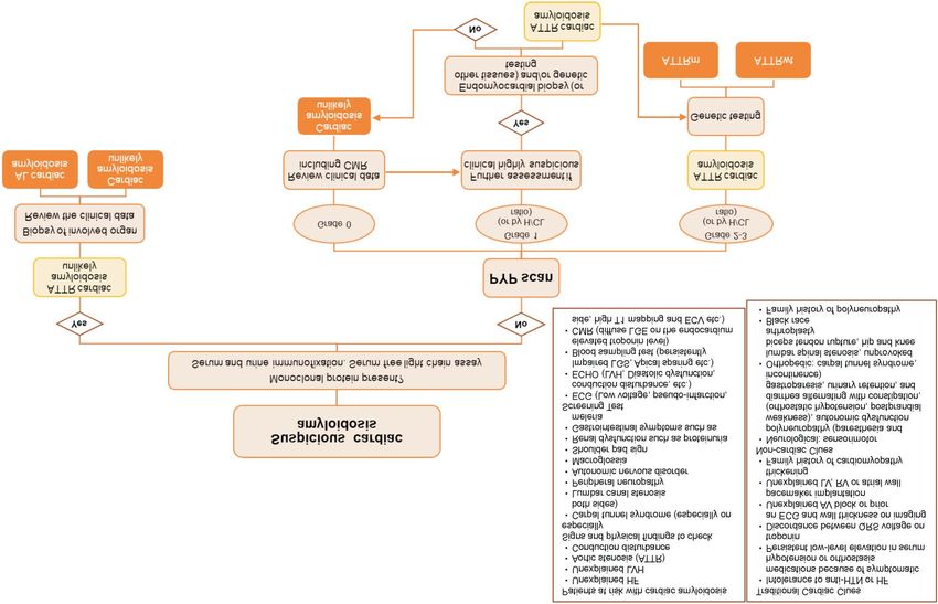
graphy are shown in Figures 3 to 7. clinical information should be clearly reviewed and an
There is an increasing trend towards the use of non- EMB may be needed in the case of equivocal imaging
invasive testing with 99m Tc-PYP for the evaluation of findings or discordant data. Herein, we proposed a sim-
ATTR-CM. In addition, monoclonal protein testing and plified clinical diagnostic algorithm of CA where the
SPECT imaging are critical components for ruling out AL presence of monoclonal proteins in serum and urine
and distinguishing myocardial retention from blood should be analyzed before performing the 99m Tc-PYP
pooling.36,41 The diagnostic combination of the FLC test scintigraphy (Figure 8).
Figure 3. Visual scores. Examples of visual scores from 0 to 3 based on 99mTc-PYP planar (anterior, left anterior oblique, and lateral views), axial
SPECT, and axial fused SPECT/CT images at 3 hours post-injection of radiotracer. H/CL ratio, heart to contralateral lung ratio; PYP, pyrophosphate;
SPECT/CT, single-photon emission computed tomography/computed tomography.
227 Acta Cardiol Sin 2021;37:221-231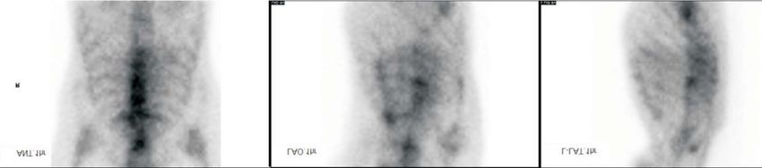
Yih-Hwen Huang et al.

                  A

                                B
Figure 4. ATTR amyloidosis, hereditary subtype. A 62-year-old man presented with a two-year history of progressive numbness and tingling in all
four limbs. In the last six months, he developed exertional dyspnea. Coronary catheterization revealed patent coronary arteries and endomyocardial
biopsy showed amyloidosis cardiomyopathy. 99mTc-PYP planar (A) and SPECT/CT (B) imaging demonstrated diffuse intense uptake in the LV
myocardium at 3 hours post-injection, significantly greater than rib activity with mildly decreased rib uptake (red arrows, visual score of 3). The H/CL
ratio at 3 hours post-injection was 1.63. Nerve conduction velocity exam revealed sensorimotor polyneuropathy with axonal degeneration, manifest-
ing as bilateral median entrapment neuropathy. Urine, blood, and cerebral spinal fluid immunofixation electrophoresis showed no evidence of
monoclonal gammopathy. Sural nerve biopsy also revealed amyloid neuropathy. Familial amyloid polyneuropathy stemming from a transthyretin
(TTR) Ala97Ser mutation was further confirmed. ATTR, transthyretin amyloidosis; H/CL, heart to contralateral lung; LV, left ventricular; PYP,
pyrophosphate; SPECT/CT, single-photon emission computed tomography/computed tomography.

                         A

                   B                                                                                C
Figure 5. AL amyloidosis. A 60-year-old man who presented with paroxysmal nocturnal dyspnea and orthopnea was diagnosed with heart failure
with preserved ejection fraction of 58.2% (NYHA Functional Classification II). Standardized dipyridamole stress myocardial perfusion imaging showed
scarring at the apical inferolateral wall (A, yellow arrow), but cardiac angiography revealed patent coronary arteries (figure not shown). Further
99m
   Tc-PYP planar and SPECT/CT images demonstrated diffuse, mildly elevated tracer activity in the LV myocardium at 3 hours post-injection (B, red
circle; and C, red arrows), equal to rib activity (visual score of 2). The H/CL ratio at 3 hours post-injection was 1.22. SPECT/CT images revealed the in-
creased tracer uptake unseen at the LV apex, corresponding to an area of scarred myocardium (C, calcification seen at yellow arrow). Endomyocardial
biopsy confirmed the patient has AL amyloidosis. Interstitial plasmacytosis (10-20%) was seen in the bone marrow biopsy, and a diagnosis of plasma
cell myeloma was given. AL, immunoglobulin light chain amyloidosis; H/CL, heart to contralateral lung; LV, left ventricular; NYHA, New York Heart As-
sociation; PYP, pyrophosphate; SPECT/CT, single-photon emission computed tomography/computed tomography.

Acta Cardiol Sin 2021;37:221-231                                           228
TSOC/SNMROC Statements for PYP Scan in ATTR-CM
A
B
Figure 6. ATTR amyloidosis, hereditary subtype. A 59-year-old man presented with a one-year history of progressive bilateral ascending numbness,
as well as orthostatic hypotension, frequent diarrhea, and frequent episodes of palpitation. Many of his family members were diagnosed with
familial amyloidotic polyneuropathy. A transthyretin gene study was performed with positive confirmation of the Ala97Ser mutation. 99mTc-PYP
planar (A) and SPECT/CT (B) images demonstrated a visual score of 2, with radiotracer uptake localized to the LV myocardium (red arrows). The H/CL
ratio at 3 hours post-injection was 1.6. ATTR, transthyretin amyloidosis; H/CL, heart to contralateral lung; LV, left ventricular; PYP, pyrophosphate;
SPECT/CT, single-photon emission computed tomography/computed tomography.
A B C
Figure 7. Breast uptake overlapping the heart in planar and SPECT images. (A) Although the ROI (red circles) was well-delineated in planar view,
marked breast physiological uptake was also noted. (B) An axial SPECT slice reveals marked breast uptake (red arrow) and blood pool activity. (C)
SPECT/CT fusion images reveal potential causes for false positives of cardiac 99mTc-PYP uptake including blood pool activity, soft tissue uptake such as
in the breast, osseous uptake, lung parenchymal lesions, overlying devices, or myocardial infarction. SPECT or SPECT/CT clearly demonstrates uptake
in the myocardium or other areas. BKG, background; PYP, pyrophosphate; ROI, region of interest; SPECT/CT, single-photon emission computed tomog-
raphy/computed tomography; stddev, standard deviation.
CONCLUSIONS ther improved and therapies applied earlier through the
implementation of these recommendations in Taiwan.
Transthyretin cardiac amyloidosis is an increasingly
recognized cause of HFpEF. Favorable prognosis depends
on early diagnosis and correct treatment strategy. This ar- ACKNOWLEDGMENTS
ticle highlights the recommendations of 99mTc-PYP imag-
ing among patients with clinical suspicion of cardiac amy- This advocacy is a joint collaboration between the
loidosis. The diagnostic accuracy of ATTR-CM can be fur- Taiwan Society of Cardiology and the Society of Nuclear
229 Acta Cardiol Sin 2021;37:221-231Yih-Hwen Huang et al.
Figure 8. The proposed diagnostic algorithm of suspected cardiac amyloidosis in Taiwan. AL, immunoglobulin light chain amyloidosis; ATTR,
transthyretin amyloidosis; AV, atrioventricular; CMR, cardiac magnetic resonance; ECG, electrocardiography; ECV, extracellular volume fraction; Gr.,
grade; H/CL ratio, heart to contralateral lung ratio; HF, heart failure; HTN, hypertension; LGE, late gadolinium enhancement; LGS, longitudinal global
strain; LV, left ventricle; LVH, left ventricular hypertrophy; PYP, pyrophosphate; RV, right ventricle. (Adapted and modified from references 25, 41).
Medicine of the Republic of China, and is partly sup- of hospitalized patients with decompensated systolic heart fail-
ported by the Ministry of Science and Technology of Tai- ure: description of population and management. Acta Cardiol Sin
wan (MOST 107-2314-B-418-006-MY3). We thank the 2016;32:400-11.
3. Chang HY, Wang CC, Wei J, et al. Gap between guidelines and clini-
Rare Disease medical affairs team of Pfizer Taiwan Inc.
cal practice in heart failure with reduced ejection fraction: results
for providing editorial support. Pfizer Taiwan has no con-
from TSOC-HFrEF registry. J Chin Med Assoc 2017;80:750-7.
flict of interests throughout the preparation or publica- 4. Chang HY, Wang CC, Wu YW, et al. One-year outcomes of acute
tion of this manuscript. decompensated systolic heart failure in Taiwan: lessons from
TSOC-HFrEF registry. Acta Cardiol Sin 2017;33:127-38.
5. Wang CC, Wu CK, Tsai ML, et al. 2019 focused update of the
CONFLICT OF INTEREST guidelines of the Taiwan Society of Cardiology for the diagnosis
and treatment of heart failure. Acta Cardiol Sin 2019;35:244-83.
6. Redfield MM. Heart failure with preserved ejection fraction. N
All the authors declare no conflict of interest.
Engl J Med 2016;375:1868-77.
7. Griffin JM, Maurer MS. Transthyretin cardiac amyloidosis: a
treatable form of heart failure with a preserved ejection fraction.
REFERENCES Trends Cardiovasc Med 2021;31:59-66.
8. Maurer MS, Elliott P, Comenzo R, et al. Addressing common
1. Lin GM, Li YH, Yin WH, et al. The obesity-mortality paradox in questions encountered in the diagnosis and management of car-
patients with heart failure in Taiwan and a collaborative meta- diac amyloidosis. Circulation 2017;135:1357-77.
analysis for east Asian patients. Am J Cardiol 2016;118:1011-8. 9. Castano A, Narotsky DL, Hamid N, et al. Unveiling transthyretin
2. Wang CC, Chang HY, Yin WH, et al. TSOC-HFrEF registry: a registry cardiac amyloidosis and its predictors among elderly patients
Acta Cardiol Sin 2021;37:221-231 230TSOC/SNMROC Statements for PYP Scan in ATTR-CM
with severe aortic stenosis undergoing transcatheter aortic valve Pyrophosphate.2019.pdf>. Accessed on March 11, 2021.
replacement. Eur Heart J 2017;38:2879-87. 27. Kitaoka H, Izumi C, Izumiya Y, et al. JCS 2020 guideline on diagnosis
10. Nitsche C, Scully PR, Patel KP, et al. Prevalence and outcomes of and treatment of cardiac amyloidosis. Circ J 2020;84:1610-71.
concomitant aortic stenosis and cardiac amyloidosis. J Am Coll 28. Perugini E, Guidalotti PL, Salvi F, et al. Noninvasive etiologic diag-
Cardiol 2021;77:128-39. nosis of cardiac amyloidosis using 99mTc-3,3-diphosphono-1,2-
11. Gonzalez-Lopez E, Gallego-Delgado M, Guzzo-Merello G, et al. propanodicarboxylic acid scintigraphy. J Am Coll Cardiol 2005;
Wild-type transthyretin amyloidosis as a cause of heart failure 46:1076-84.
with preserved ejection fraction. Eur Heart J 2015;36:2585-94. 29. Bokhari S, Castano A, Pozniakoff T, et al. (99m)Tc-pyrophosphate
12. Coelho T, Maurer MS, Suhr OB. THAOS - The Transthyretin Amy- scintigraphy for differentiating light-chain cardiac amyloidosis
loidosis Outcomes Survey: initial report on clinical manifesta- from the transthyretin-related familial and senile cardiac amylo-
tions in patients with hereditary and wild-type transthyretin idoses. Circ Cardiovasc Imaging 2013;6:195-201.
amyloidosis. Curr Med Res Opin 2013;29:63-76. 30. Hanna M, Ruberg FL, Maurer MS, et al. Cardiac scintigraphy with
13. Chao HC, Liao YC, Liu YT, et al. Clinical and genetic profiles of technetium-99m-labeled bone-seeking tracers for suspected
hereditary transthyretin amyloidosis in Taiwan. Ann Clin Transl amyloidosis: JACC review topic of the week. J Am Coll Cardiol
Neurol 2019;6:913-22. 2020;75:2851-62.
14. Lai HJ, Huang KC, Liang YC, et al. Cardiac manifestations and prog- 31. Hoffman JE, Dempsey NG, Sanchorawala V. Systemic amyloidosis
nostic implications of hereditary transthyretin amyloidosis as- caused by monoclonal immunoglobulins: soft tissue and vascular
sociated with transthyretin Ala97Ser. J Formos Med Assoc 2020; involvement. Hematol Oncol Clin North Am 2020;34:1099-113.
119:693-700. 32. Castano A, Haq M, Narotsky DL, et al. Multicenter study of planar
15. Donnelly JP, Hanna M. Cardiac amyloidosis: an update on diag- technetium 99m pyrophosphate cardiac imaging: predicting sur-
nosis and treatment. Cleve Clin J Med 2017;84:12-26. vival for patients with ATTR cardiac amyloidosis. JAMA Cardiol
16. Gonzalez-Lopez E, Lopez-Sainz A, Garcia-Pavia P. Diagnosis and 2016;1:880-9.
treatment of transthyretin cardiac amyloidosis. Progress and 33. Dorbala S, Kijewski MF, Park MA. Quantitative bone-avid tracer
hope. Rev Esp Cardiol (Engl Ed) 2017;70:991-1004. SPECT/CT for cardiac amyloidosis: a crucial step forward. JACC
17. Martinez-Naharro A, Baksi AJ, Hawkins PN, Fontana M. Diagnostic Cardiovasc Imaging 2020;13:1364-7.
imaging of cardiac amyloidosis. Nat Rev Cardiol 2020;17:413-26. 34. Ren C, Ren J, Tian Z, et al. Assessment of cardiac amyloidosis with
18. Siddiqi OK, Ruberg FL. Cardiac amyloidosis: an update on patho- (99m)Tc-pyrophosphate (PYP) quantitative SPECT. EJNMMI Phys
physiology, diagnosis, and treatment. Trends Cardiovasc Med 2021;8:3.
2018;28:10-21. 35. Tamarappoo B, Otaki Y, Manabe O, et al. Simultaneous Tc-99m
19. Ruberg FL, Berk JL. Transthyretin (TTR) cardiac amyloidosis. Cir- PYP/Tl-201 dual-isotope SPECT myocardial imaging in patients
culation 2012;126:1286-300. with suspected cardiac amyloidosis. J Nucl Cardiol 2020;27:28-37.
20. Dungu JN, Anderson LJ, Whelan CJ, Hawkins PN. Cardiac trans- 36. Poterucha TJ, Elias P, Bokhari S, et al. Diagnosing transthyretin car-
thyretin amyloidosis. Heart 2012;98:1546-54. diac amyloidosis by technetium 99m pyrophosphate: a test in evo-
21. Rapezzi C, Merlini G, Quarta CC, et al. Systemic cardiac amylo- lution. JACC Cardiovasc Imaging 2020;S1936-878X(20)30816-0.
idoses: disease profiles and clinical courses of the 3 main types. 37. Treglia G, Glaudemans A, Bertagna F, et al. Diagnostic accuracy of
Circulation 2009;120:1203-12. bone scintigraphy in the assessment of cardiac transthyretin-
22. Dubrey SW, Hawkins PN, Falk RH. Amyloid diseases of the heart: related amyloidosis: a bivariate meta-analysis. Eur J Nucl Med
assessment, diagnosis, and referral. Heart 2011;97:75-84. Mol Imaging 2018;45:1945-55.
23. Fontana M, Corovic A, Scully P, Moon JC. Myocardial amyloidosis: 38. Flaherty KR, Morgenstern R, Pozniakoff T, et al. (99m)Technetium
the exemplar interstitial disease. JACC Cardiovasc Imaging 2019; pyrophosphate scintigraphy with cadmium zinc telluride cameras
12:2345-56. is a highly sensitive and specific imaging modality to diagnose
24. Arbustini E, Merlini G. Early identification of transthyretin-re- transthyretin cardiac amyloidosis. J Nucl Cardiol 2020;27:371-80.
lated hereditary cardiac amyloidosis. JACC Cardiovasc Imaging 39. Manrique A, Dudoignon D, Brun S, et al. Quantification of myo-
2014;7:511-4. cardial (99m)Tc-labeled bisphosphonate uptake with cadmium
25. Dorbala S, Ando Y, Bokhari S, et al. ASNC/AHA/ASE/EANM/HFSA/ zinc telluride camera in patients with transthyretin-related car-
ISA/SCMR/SNMMI expert consensus recommendations for multi- diac amyloidosis. EJNMMI Res 2019;9:117.
modality imaging in cardiac amyloidosis: part 1 of 2-evidence 40. Dorbala S, Park MA, Cuddy S, et al. Absolute quantitation of car-
base and standardized methods of imaging. J Nucl Cardiol 2019; diac (99m)Tc-pyrophosphate using cadmium zinc telluride-based
26:2065-123. SPECT/CT. J Nucl Med 2020 Sep 4;jnumed.120.247312.
26. Dorbala S BS, Miller E, Bullock-Palmer R, et al. 99mTechnetium- 41. Kittleson MM, Maurer MS, Ambardekar AV, et al. Cardiac amy-
pyrophosphate imaging for transthyretin cardiac amyloidosis. loidosis: evolving diagnosis and management: a scientific state-
2019 ASNC Practice Points. Available atYou can also read