Cinematic Rendering in Mixed-Reality Holograms: A New 3D Preoperative Planning Tool in Pediatric Heart Surgery - opus4 ...
←
→
Page content transcription
If your browser does not render page correctly, please read the page content below
ORIGINAL RESEARCH
                                                                                                                                            published: 09 February 2021
                                                                                                                                        doi: 10.3389/fcvm.2021.633611

                                            Cinematic Rendering in
                                            Mixed-Reality Holograms: A New 3D
                                            Preoperative Planning Tool in
                                            Pediatric Heart Surgery
                                            Pia Gehrsitz 1 , Oliver Rompel 2 , Martin Schöber 1 , Robert Cesnjevar 3 , Ariawan Purbojo 3 ,
                                            Michael Uder 2 , Sven Dittrich 1 and Muhannad Alkassar 1*
                                            1
                                              Department of Pediatric Cardiology, University Hospital Erlangen, Friedrich-Alexander University Erlangen-Nürnberg (FAU),
                                            Erlangen, Germany, 2 Institute of Radiology, University Hospital Erlangen, Friedrich-Alexander University Erlangen-Nürnberg
                                            (FAU), Erlangen, Germany, 3 Department of Pediatric Cardiac Surgery, University Hospital Erlangen, Friedrich-Alexander
                                            University Erlangen-Nürnberg (FAU), Erlangen, Germany

                                            Cinematic rendering (CR) is based on a new algorithm that creates a photo-realistic
                                            three-dimensional (3D) picture from cross-sectional images. Previous studies have
                                            shown its positive impact on preoperative planning. To date, CR presentation has
                                            only been possible on 2D screens which limited natural 3D perception. To depict
                          Edited by:
                     Sebastian Kelle,       CR-hearts spatially, we used mixed-reality technology and mapped corresponding hearts
              Deutsches Herzzentrum         as holograms in 3D space. Our aim was to assess the benefits of CR-holograms in the
                     Berlin, Germany
                                            preoperative planning of cardiac surgery. Including 3D prints allowed a direct comparison
                      Reviewed by:
                       Dirk Loßnitzer,
                                            of two spatially resolved display methods. Twenty-six patients were recruited between
      Universität Mannheim, Germany         February and September 2019. CT or MRI was used to visualize the patient’s heart
                       Radu Tanacli,        preoperatively. The surgeon was shown the anatomy in cross-sections on a 2D screen,
                 German Heart Center
                     Berlin, Germany        followed by spatial representations as a 3D print and as a high-resolution hologram.
                *Correspondence:            The holographic representation was carried out using mixed-reality glasses (HoloLens® ).
                Muhannad Alkassar           To create the 3D prints, corresponding structures were segmented to create STL files
  muhannad.alkassar@uk-erlangen.de
                                            which were printed out of resin. In 22 questions, divided in 5 categories (3D-imaging
                    Specialty section:
                                            effect, representation of pathology, structure resolution, cost/benefit ratio, influence on
          This article was submitted to     surgery), the surgeons compared each spatial representation with the 2D method, using
               Cardiovascular Imaging,
                                            a five-level Likert scale. The surgical preparation time was assessed by comparing
                a section of the journal
  Frontiers in Cardiovascular Medicine      retrospectively matched patient pairs, using a paired t-test. CR-holograms surpassed
       Received: 25 November 2020           2D-monitor imaging in all categories. CR-holograms were superior to 3D prints in all
        Accepted: 13 January 2021           categories (mean Likert scale 4.4 ± 1.0 vs. 3.7 ± 1.3, P < 0.05). Compared to 3D prints it
       Published: 09 February 2021
                                            especially improved the depth perception (4.7 ± 0.7 vs. 3.7 ± 1.2) and the representation
                             Citation:
    Gehrsitz P, Rompel O, Schöber M,
                                            of the pathology (4.4 ± 0.9 vs. 3.6 ± 1.2). 3D imaging reduced the intraoperative
     Cesnjevar R, Purbojo A, Uder M,        preparation time (n = 24, 59 ± 23 min vs. 73 ± 43 min, P < 0.05). In conclusion, the
      Dittrich S and Alkassar M (2021)
                                            combination of an extremely photo-realistic presentation via cinematic rendering and
 Cinematic Rendering in Mixed-Reality
  Holograms: A New 3D Preoperative          the spatial presentation in 3D space via mixed-reality technology allows a previously
       Planning Tool in Pediatric Heart     unattained level of comprehension of anatomy and pathology in preoperative planning.
                              Surgery.
   Front. Cardiovasc. Med. 8:633611.        Keywords: mixed-reality, cinematic rendering, 3D printing, preoperative planning, pediatric heart surgery,
     doi: 10.3389/fcvm.2021.633611          congenital heart disease

Frontiers in Cardiovascular Medicine | www.frontiersin.org                         1                                          February 2021 | Volume 8 | Article 633611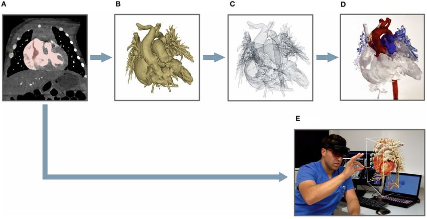
Gehrsitz et al.                                                                                  Cinematically Rendered Holograms in Cardio-Surgery

INTRODUCTION                                                              for 3D imaging (2–4, 6, 7, 12–15), cross-sectional images were
                                                                          recorded using MRI or CT depending on the clinical question. If
Due to the complex and highly individual anatomy of patients              morphology was the only question, CT was used. If there were
with congenital heart disease (CHD), it is essential to have              additional functional questions, MRI was carried out.
precise preoperative planning and good morphologic imaging
for surgical success. Currently, three-dimensional (3D) imaging           Data Acquisition
offers the most realistic representation of cardiac structures, and       The CT scans were performed during the post venous phase, after
has therefore gained importance in recent years (1–5).                    injecting contrast medium peripheral. The images were acquired
    3D images are generated from two-dimensional (2D) cross-              in 0.6 mm slices, using either a second-generation 128-slice
sectional images produced using computed tomography (CT)                  dual-source CT scanner (SOMATOM Definition Flash; Siemens
and magnetic resonance imaging (MRI). There are two methods               Healthcare GmbH, Erlangen, Germany) or a third-generation
for generating 3D images from 2D datasets: (1) creating 3D                192-slice dual-source CT scanner (SOMATOM Definition Force;
segmentation by manually selecting interesting structures, and            Siemens Healthcare, Erlangen, Germany). Modern low-dose
(2) calculating a 3D image automatically, based on rendering              (0.2–0.5 mSv) protocols were used. The MRI datasets were
algorithms. Siemens Healthineers has developed a new volume               collected in diastolic heart phase, in a whole-heart sequence,
rendering technique called cinematic rendering (CR). CR                   in 0.8 mm slices, with a 1.5-Tesla MRI-scanner (MAGNETOM
generates a more photo-realistic 3D depiction than previously             Aera; Siemens Healthcare GmbH, Erlangen, Germany).
used rendering algorithms, by imitating natural light interactions
(3, 6). Multiple previous studies have confirmed that CR provides         Spatial View
a more photo-realistic view and improvements in shape and                 To set up the spatial representation, datasets were exported from
depth perception compared with cross-sectional imaging or                 the advanced visualization imaging software, syngo.via (Version
volume rendering (7–10).                                                  VB30A; Siemens Healthcare GmbH, Erlangen, Germany), and
    Until recently, 3D-rendered images can be presented only on           saved in the standard “Digital Imaging and Communications in
a 2D screen. Currently, it is possible to present these 3D images         Medicine” (DICOM)-format.
in 3D space with either physical or virtual 3D imaging. Physical              DICOM-data were used to visualize the heart directly in the
3D imaging is generated by producing a 3D-printed model from              cinematically rendered view with the newly developed prototype
a manually-generated 3D image. Virtual 3D imaging is generated            mixed-reality syngo.via application “Cinematic Reality.” The
by creating a hologram by using mixed-reality technology. The             new syngo.via application generated a cinematically rendered
latest development is an application that integrates CR and               hologram (CR-hologram), which could be viewed with the
mixed-reality techniques for use with the HoloLens R (Microsoft,          HoloLens R . The hologram was projected in a firmly fixed
Redmond, USA) (11). The current gold standard for spatial                 position in the room, so the observer could walk around the heart
imaging in preoperative planning is 3D printing. However, it has          and examine it from every side. Figure 1 shows a surgeon looking
mostly been described in case reports and systematic reviews of           at a CR-hologram through the HoloLens R .
its advantages are still rare. Furthermore, a significant benefit             To create a realistic 3D-printed model from cross-sectional
compared to 2D imaging regarding the overall surgery time could           images, various pre-processing steps were necessary. First,
not be shown yet for 3D printing. Therefore, we compared both             DICOM-data were exported into the open-source software, 3D
3D imaging methods additional to the standard preoperative                Slicer (Version 4.11; http://www.slicer.org). In this software, the
imaging on 2D screen.                                                     image was segmented based on an adjustable threshold chosen
    The aim of this study was to determine the benefits of                so that only the voxels of interest were marked. The marking
spatial representation of CR-reconstructed heart structures in the        depended on the master volume intensity range of the individual
preoperative planning of pediatric heart surgery. By including            voxels. In addition to the whole heart, neighboring vessels were
3D prints, a direct comparison of two spatially resolving display         marked that were relevant to later surgery.
methods was possible.                                                         The created model was saved in standard tessellation language
                                                                          (STL) file format, which was compatible with the 3D printer.
MATERIALS AND METHODS                                                     Next, the model was produced by 3D printer (Form 2, Formlabs,
                                                                          Sommerville, USA) out of resin at a resolution of 0.1 mm layers
This prospective study was performed in accordance with the               using standardized printing plans. As a post-processing step,
Guidelines for Good Clinical Practice. All CT and MRI datasets            important structures such as coronary arteries were marked in
were accessed with permission through informed consent from               different colors to facilitate orientation. An overview of the main
both parents; in no case were the images taken exclusively for            steps for generating a 3D-printed model in comparison to a CR-
this study.                                                               hologram is given in Figure 2. The imaging processing time for
   The patients were recruited between February and September             each technique was recorded.
2019. All patients who underwent cross-sectional imaging in
preparation for surgery were included in the study. The decision          Study Design
to perform cross-sectional imaging was based on comprehensive             First, the patient’s cross-sectional images were presented on a
echocardiography performed previously. Because high-quality               2D screen, which was previously the common technique for
cross-sectional images from MRI and CT are equally suitable               presenting preoperative imaging material. The CR-hologram,

Frontiers in Cardiovascular Medicine | www.frontiersin.org            2                                    February 2021 | Volume 8 | Article 633611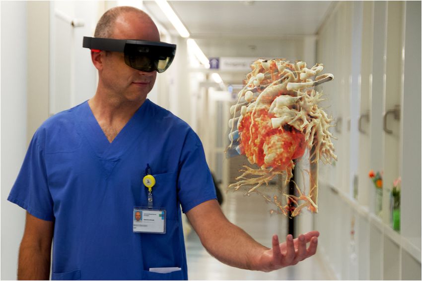
Gehrsitz et al.                                                                                                     Cinematically Rendered Holograms in Cardio-Surgery

 FIGURE 1 | Surgeon looking at a CR-hologram of a patient’s heart with dextro-transposition of the great arteries using HoloLens® . The figure illustrates an image for
 which the view of the surgeon through the HoloLens® was combined with a photo in which he is working with the HoloLens® .

and subsequently the 3D-printed model, were then presented to                          time was defined as the time from the initial cut until the
the surgeon in an upright position. To provide a comparison                            first vessel was clamped. The intraoperative preparation times
of the newly introduced spatial 3D-imaging techniques and the                          for study participants were compared with intraoperative
representation on the 2D screen, the surgeons were asked to                            preparation times for patients with matching characteristics
complete a questionnaire for each spatial 3D-imaging technique.                        for whom preoperative imaging in 3D space was not used.
The two 3D techniques were also directly compared with                                 The patients were matched for age, weight, operative
each other.                                                                            procedure, previous operations, and the general state of
                                                                                       health preoperatively (Table 1). As intraoperative preparation
Questionnaire                                                                          time was analyzed, only patients who underwent the same
In total, the questionnaire comprised 22 items, each rated on                          surgical procedures with the same complexity were matched.
a 5-point Likert scale. The five response options ranged from                          Additionally, the intraoperative preparation times of the matched
“clearly superior” (5 points) to “clearly inferior” (1 point).                         patient pairs were analyzed after being divided into groups in
The questionnaire items were structured in five subgroups to                           which the “facilitation of preparation” was, respectively, rated
provide a better overview: 3D-imaging effect, representation                           of excellent benefit (5 points on the Likert scale) or not (≤ 4
of the pathology, anatomical structure resolution, cost/benefit                        points). A Wilcoxon test was performed to compare the two
ratio, and influence on the surgery (Table 2). The reliability                         groups because the F-test showed no equality of variance for the
of the different items of each dimension was checked using                             “advantageous” group.
Cronbach’s alpha.

Analysis of Surgical Preparation Time                                                  Statistical Evaluation
In order to investigate the influence of preoperative spatial                          Statistical analyses were performed using IBM SPSS statistics
representation on the surgical procedure, we compared                                  (Version 21; IBM, Armonk, USA). Results of the questionnaires
intraoperative preparation times. Intraoperative preparation                           were expressed as mean values with standard deviation

Frontiers in Cardiovascular Medicine | www.frontiersin.org                         3                                           February 2021 | Volume 8 | Article 633611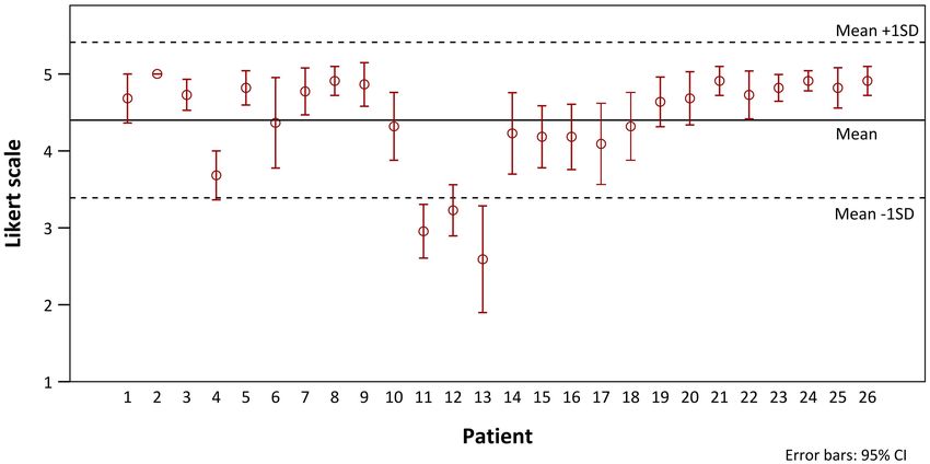
Gehrsitz et al.                                                                                              Cinematically Rendered Holograms in Cardio-Surgery

 FIGURE 2 | Processing steps for generating a 3D-printed model (A–D) vs. a CR-hologram (E): (A) DICOM-data viewed in the 3D Slicer application, marked based on
 an appropriate threshold; (B) a segmented 3D model; (C) a 3D model as an STL file; (D) a finished printed 3D model; (E) cross-sectional images converted to a
 CR-hologram.

TABLE 1A | Demographic information about the analyzed patient population.              The contrast-to-noise ratio (CNR) and signal-to-noise ratio
                                                                                    (SNR) were determined independently for CT and MRI via
Patient characteristics                          Cases       Matched controls
                                                                                    regions of interest (ROIs) using the software syngo.via. The SNR
Number                                           n = 26           n = 24            was calculated by dividing the mean signal intensity of the aorta
Gender                 Male                      61.5%            58.3%             in cross-section (ROI = 1.2 ± 0.1 mm2 ) by the SD of the extra
                       Female                    38.5%            41.7%             thoracic background noise (ROI = 2.2 ± 0.1 cm2 ) measured from
Age (years)                                     2.0 ± 3.9        2.0 ± 4.7
                                                                                    the air surrounding the patient. For the CNR the mean signal
Weight (kg)                                    10.5 ± 12.9      10.7 ± 15
                                                                                    intensity of the left ventricular muscle (ROI = 1.2 ± 0.1 mm2 )
Heart lung machine     Yes                       92.3%            95.8%
                                                                                    was subtracted from the mean signal intensity of the aorta and
                       No                         7.7%             4.2%
                                                                                    afterwards divided by the SD of the background noise.
Access                 Median sternotomy         84.6%            87.5%
                       Posterolateral            15.4%            12.5%
Previous operations    Yes                       34.6%            29.2%
                       No                        65.4%            70.8%
                                                                                    RESULTS
                                                                                    We recruited 26 patients with an average age of 2.0 ± 3.9 years.
                                                                                    Of the surgeries, 77% concerned mainly outer cardiovascular
                                                                                    structures (e.g., great vessels) and 23% inner cardiovascular
                                                                                    structures (e.g., valves). Twenty-four patients underwent CT as
(SD). Each questionnaire item was analyzed separately and                           preoperative imaging modality and 2 patients MRI. CT and MRI
summarized in the five subgroups.                                                   datasets were of comparable quality (CNR: MRI: 16.4 ± 1.4, CT:
   A paired t-test was performed to compare the questionnaire                       13.7 ± 6.4; SNR: MRI: 19.8 ± 2.1, CT: 20.28 ± 8.5). Cross-
results (Likert scale data) of CR-holograms with those of 3D-                       sectional images for all 26 patients were of high quality without
printed models. As Jeffrey and Norman have shown, parametric                        artifacts and could be used successfully for both rendering and 3D
tests are superior to non-parametric tests when analyzing Likert                    printing. Further demographic information about the patients
scale data (16, 17). To compare the intraoperative preparation                      and the conducted surgeries is shown in Table 1. The cross-
times between the patients and matched controls a paired t-test                     sectional imaging was taken on average 13 days before the surgery
was used. The statistical significance level was defined as P < 0.05                took place. The patients’ images were of comparable quality;
for all analyses.                                                                   in particular, no difference between the cases and the matched

Frontiers in Cardiovascular Medicine | www.frontiersin.org                      4                                       February 2021 | Volume 8 | Article 633611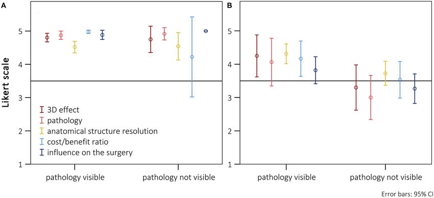
Gehrsitz et al.                                                                                                                       Cinematically Rendered Holograms in Cardio-Surgery

TABLE 1B | Demographic information about the analyzed patient population.

Case        Imaging                                             Cases                                                                           Matched controls

                            Main diagnosis and kind of                    Age (years)         Weight (kg)          Main diagnosis and kind of                    Age (years)         Weight (kg)
                            operation                                                                              operation

1           CT              Single ventricle (1)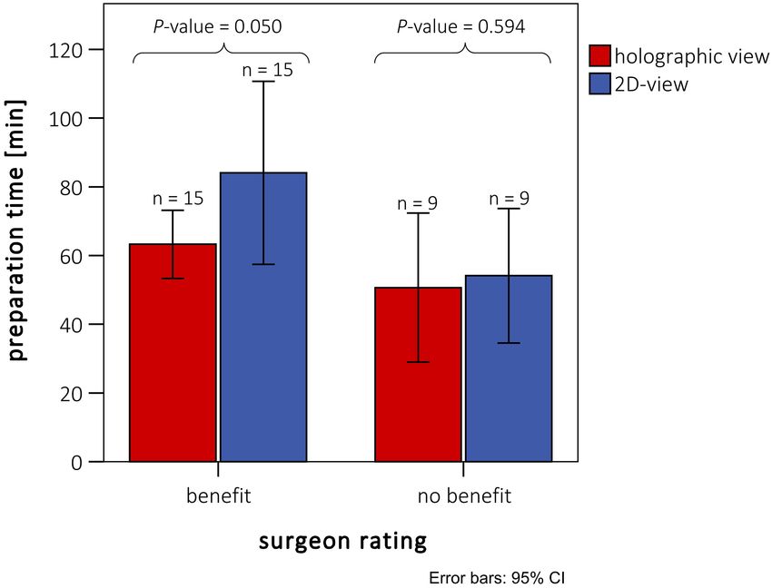
Gehrsitz et al.                                                                                                         Cinematically Rendered Holograms in Cardio-Surgery

TABLE 2 | Evaluation of CR-holograms and 3D-printed models divided in                             5, n = 17). No disadvantage in the overall rating was seen, even
subgroups.                                                                                        if the main pathology could not be presented holographically.
Items                                         CR-      3D printing vs.
                                                                                                  For patients for whom the preparation was rated worse, a
                                         holograms vs.  2D imaging                                significant deterioration of the ratings in all subgroups could be
                                          2D imaging                                              shown (Figure 4B, n = 9). A particularly poor evaluation could
                                                                                                  be shown for patients in whom, additionally, the holographic
                                           mean (SD)           mean (SD)        P-value#
                                                                                                  presentation of the pathology was not possible.
3D-imaging effect                            4.4 (0.8)          3.4 (1.1)*       0.000§
Comparison with CR on 2D screen              4.1 (1.3)          2.7 (1.5)*       0.000§           Superiority of the HoloLens® Over
Sufficient visualization options             4.5 (1.0)           3.7 (1.4)       0.001§           Previously Used 3D-Imaging Techniques
Sufficient quality                           4.4 (1.1)           3.5 (1.4)       0.000§           The CR-holograms were rated significantly higher than the 3D-
Improved depth perception                    4.7 (0.7)           3.7 (1.2)       0.000§           printed models in all categories (Table 2). Nevertheless, clear
Representation of the pathology              4.4 (0.9)           3.6 (1.2)       0.001§           differences in the ratings of the individual subgroups could be
All necessary areas presented                4.3 (1.1)           3.5 (1.4)       0.001§           identified. Concerning cost/benefit ratio, only a small benefit
                                                                                         ‡
Important details not hidden                 3.9 (1.5)          3.2 (1.6)*       0.010            could be shown for the holographic presentation compared
Improved comprehensibility                   4.6 (0.9)           3.9 (1.3)       0.008§           to 3D-printed models (CR-holograms: 4.5 ± 0.7; 3D print:
Adequate pathology assessment                4.7 (0.7)           4.0 (1.2)       0.003§           4.0 ± 0.9; P < 0.05), while no significant difference between
Anatomical structure resolution              4.3 (0.5)           3.8 (0.7)       0.000§           the 3D-imaging methods was observed for use in education.
                                                                                         ‡
Confluence of vessels                        4.8 (0.5)           4.4 (0.9)       0.022            The surgeons rated the time expenditure for 3D printing
Out-flowing vessels                          4.7 (0.6)           4.2 (1.0)       0.005§           only a bit higher. In the overall evaluation of the anatomical
                                                                                         †
Aortic arch                                  4.9 (0.3)           4.9 (0.3)       1.000            structure resolution, CR-holograms showed significantly better
Coronaries                                   3.4 (1.4)*         2.5 (1.3)*       0.003§           results (CR-holograms: 4.3 ± 0.5; 3D print: 3.8 0.7; P
                                                                                         ‡
Pulmonary veins                              4.7 (0.5)           4.1 (1.2)       0.023            < 0.05). Nevertheless, both evaluated 3D-imaging methods
                                                                                         ‡
Atrial appendages                            4.8 (0.4)           4.4 (0.7)       0.010            showed no benefit compared with 2D imaging regarding the
Structures of the inner heart                3.3 (1.1)*         3.0 (1.3)*       0.397
                                                                                         †
                                                                                                  representation of the coronaries as well as the intracardial
Neighboring structures                       4.3 (1.0)          3.1 (1.4)*       0.000§           structures. However, CR-holograms were rated significantly
Cost/benefit ratio                           4.5 (0.7)           4.0 (0.9)       0.012
                                                                                         ‡        higher than 3D-printed models for the representation of the
Appropriate expenditure of time              4.5 (1.0)           3.9 (1.1)        0.03
                                                                                         ‡        coronaries. Concerning the remaining anatomical structures,
Educational potential                        4.7 (0.6)           4.5 (0.8)       0.056
                                                                                         †        only a small significant difference could be observed between
Adequate costs and benefits                  4.2 (1.1)           3.7 (1.3)       0.020
                                                                                         ‡        the two spatial representation methods, especially considering
Influence on the surgery                     4.4 (0.8)           3.8 (1.0)       0.004§           large vessel structures. While the benefit of CR-holograms in
Concordant intraoperative view               4.7 (0.7)           4.1 (1.0)       0.006§           representing the pathology was clearly higher compared with 2D
Facilitation of preparation                  4.5 (0.8)           3.9 (1.0)       0.003§           imaging, for 3D printing the benefit was only marginal (CR-
Positive impact on surgery time              4.2 (1.0)           3.5 (1.2)       0.016
                                                                                         ‡        holograms: 4.4 ± 0.9; 3D print: 3.6 ± 1.2; P < 0.05). In the
                                                                                                  assessment of the 3D-imaging effect, the HoloLens R was clearly
                          †                           ‡
# pairedt-test for analysis, No significant difference, p < 0.05, § p < 0.01. *presentation       superior to 3D printing (CR-holograms: 4.4 ± 0.8; 3D print: 3.4
on 2D screen superior to presentation in 3D space. The bold values correspond to the              ± 1.1; P < 0.05).
five subgroups that were evaluated. The corresponding items from which a subgroup was
composed are listed below the respective bold subgroup.
                                                                                                  Significant Shortening of the Intraoperative
                                                                                                  Preparation Time
order are shown in Figure 3. There was no indication of a                                         We evaluated the measurable influence of spatial 3D methods
chronological dependency of the ratings. In 3 of 26 patients, no                                  in preoperative planning on the course of surgery by analyzing
benefit for the average values could be identified in comparison                                  the intraoperative preparation times. Two cases could not
with 2D imaging (patients 11, 12, and 13). On closer examination                                  be included, because no suitable case could be found for
it became apparent that these patients underwent a repair of inner                                comparison, due to the complexity of the respective surgery.
cardiovascular structures that cannot be adequately presented via                                 In five cases, the main diagnosis of cases and matched controls
CR-holograms. Additionally, for these cases the usefulness of the                                 differed, but the operative procedure and other conditions were
hologram for the preparation received no excellent ratings (3.6 ±                                 the same in these patients.
0.5). To assess whether these two aspects (neighboring structures                                    The mean intraoperative preparation time was 58.5 ±
and holographic visibility) influenced the overall evaluation,                                    22.6 min (n = 24, minimum: 23 min, maximum: 104 min),
the patients were evaluated after separation into groups that,                                    when spatial 3D models were used for preoperative planning.
respectively, did and did not benefit from the representation of                                  In contrast, the preoperative planning for the relevant control
the pathology. There was only a benefit for the representation of                                 group was carried out completely on a 2D monitor, and no
intracardial pathologies when additional vascular surgery aspects                                 representation in 3D space was used. The control intraoperative
were present. In Figure 4A, only patients with an excellent rating                                preparation time was 72.8 ± 43.1 min (n = 24, minimum: 24 min,
of the usefulness for preparation were included (Likert scale =                                   maximum: 186 min). The intraoperative preparation times of

Frontiers in Cardiovascular Medicine | www.frontiersin.org                                    6                                   February 2021 | Volume 8 | Article 633611
Gehrsitz et al. Cinematically Rendered Holograms in Cardio-Surgery FIGURE 3 | Distribution (means with 95% confidence intervals) of the Likert scale ratings of CR-holograms over the progress of the study. FIGURE 4 | Presentation of the scores (means with 95% confidence intervals) separately listed for the five subgroups, divided between surgeries where the main pathology was visible or not using the HoloLens® . (A) Cases with an excellent rating for preparation-time benefit (Likert scale = 5; n = 17). (B) Cases without an excellent rating for preparation-time benefit (Likert scale
Gehrsitz et al.                                                                                                     Cinematically Rendered Holograms in Cardio-Surgery

 FIGURE 5 | Intraoperative preparation times of patients with preoperative holographic representation (red) and matched patient pairs (blue) without spatial
 representation. Ratings are divided between cases rated with and without benefit for preparation by the surgeons. Patients rated with “no benefit” did not differ from
 the rest either in diagnosis or in kind of surgery (see also Table 1B).

patients rated “non-advantageous” by the surgeon, there was no                          subgroups were confirmed using Cronbach’s alpha, validating
significant benefit measured for the preparation time.                                  the selection criteria. MRI was performed in two cases because
                                                                                        additional functional parameters were needed. The quality of
                                                                                        these images did not differ from that of CT. Furthermore, the
DISCUSSION                                                                              possibility that discrepancies in the results arose from differences
                                                                                        between the cases and the controls in the quality of the underlying
Recent developments made the use of CR in 3D space possible                             images could be rejected.
for the first time. This study is the first to compare CR-
holograms with previously used imaging techniques in which the
images were projected onto a 2D screen (CT or MRI). Precise                             CR-Holograms as the Most Realistic
morphological imaging is essential for surgical success, especially                     Representation Method
in complex cases.                                                                       In fields like engineering or design review, the fundamental
   A direct comparison of CR-holograms to 2D imaging and                                advantage of 3D imaging in 3D space is already proven. Spatial
3D printing quickly reveals the superiority of the photo-realistic                      3D representation has been established for many years in those
3D holographic representation (Figure 6). The holographic                               areas of expertise. It has already been shown that the spatial
representation by the HoloLens R surpassed the standard                                 3D presentation improves shape and depth perception, reduces
representation on a 2D screen in all five analyzed parameters                           mental workload, and makes it possible to complete tasks faster
(Table 2). Furthermore, the assessment of an already established                        and with higher quality results (22, 23). In accordance with these
method of 3D imaging, 3D printing, was used as a spatial                                observations, our results showed the advantage of using spatial
comparison method (2, 12–15, 19–21). The questions used to                              3D imaging as, the most significant improvement in preoperative
create the questionnaire were carefully selected. The absence of                        planning by enhancing depth perception and the representation
unnecessary redundancy and good inter-item correlation in the                           of the pathology (Table 2). These aspects were rated better for

Frontiers in Cardiovascular Medicine | www.frontiersin.org                          8                                          February 2021 | Volume 8 | Article 633611
Gehrsitz et al. Cinematically Rendered Holograms in Cardio-Surgery FIGURE 6 | Preoperative imaging of a patient’s heart with truncus arteriosus communis from two different perspectives. (A1–A3) A direct comparison of CT on a 2D screen (2D imaging), (B1–B3) cinematically rendered hologram, and (C1–C3) 3D printing. In (A3), the coronaries (arrows) show only a weak contrast, but the 3D view can be demonstrated as a cinematically rendered hologram (B3). A reconstruction of the coronary structure for 3D printing was not possible in this case (C3). CR-holograms than for 3D-printed models, even though the 3D the realistic spatial 3D view with an extremely realistic rendering prints correspond in dimensions and anatomical presentation algorithm clearly improved depth perception and provided a exactly to the patient’s heart and have a haptic advantage. The better delineation of complex anatomical structures. main difference between the 3D print and a real heart is the It is known that repeatedly visually presented objects can representation of surface features like color, texture, and lighting be processed much faster than unknown structures (24). The characteristics. The absence of these realistic features makes exact preoperative visualization of the operative field allows the additional mental transformational work necessary since an surgeon to plan the operation’s steps directly. Therefore, the intuitive recognition intraoperatively is not possible. In contrast, aim of preoperative imaging is to provide the surgeon a virtual CR makes it possible to create a 3D image which strongly operative field in advance that is as close to reality as possible. Our corresponds to the familiar intraoperative tissue texture. This study showed that the intraoperative findings corresponded to is achieved by using an algorithm that takes the interaction the preoperative images significantly better when the latter were between light photons and human tissue into account (3, 6–10). presented as CR-holograms than when visualized on a 2D screen This could already prove to be a significant advantage, especially or by 3D printing (Table 2). We assume that this is the reason for in shape and depth perception, over previously used rendering the facilitated visual comprehension of the pathology. algorithms (e.g., volume rendering) when presented on 2D screen The only barriers to easier comprehension of pathologies (7–10). However, our results showed that CR on a 2D screen using CR-holograms were structures which were either not improved the 3D perception so that it could show results even imaged in the primary dataset or could not be presented equal to those obtained using 3D printing (Table 2). This explains holographically (intracardial structures). The lack of information the consistently excellent review of the 3D perception of the in the primary data set is most evident in the representation holographic representation of CR-images. The combination of of the coronary arteries. The representation is dependent on Frontiers in Cardiovascular Medicine | www.frontiersin.org 9 February 2021 | Volume 8 | Article 633611
Gehrsitz et al.                                                                                     Cinematically Rendered Holograms in Cardio-Surgery

the perfusion and the phase in which the single shot was                         Nevertheless, the holographic representation was superior,
taken. Since the images were not taken especially to display the             considering the time expense. In the preparation process the
coronary arteries, contrast was lacking in some patients and                 cost/benefit of 3D printing is clearly inferior to CR-holograms.
this limited the coronary arteries’ visibility. Nevertheless, CR-            While the acquisition costs are similar for the 3D printer
holograms exhibited an advantage over 3D printing in cases                   and the HoloLens R , the processing costs are much higher for
with limited coronary representation in raw 2D images. In these              3D printing. Each print adds personnel and material costs.
cases, 3D printing is inferior to 2D representation, because a               Furthermore, the intraoperative preparation time for a 3D
weak coronary contrast makes 3D reconstruction impossible.                   print is much longer, which is why short-term production—
On the other hand, CR-holograms enable weakened coronary                     for example, from an emergency CT for subsequent surgery—
visibility, which allows equivalent visibility of coronaries as in 2D        is not possible. In contrast, a CR-hologram can be prepared in
images (Figure 6). Apart from this, it is currently not possible to          little time.
view intracardial structures using the HoloLens R . However, even
in cases in which intracardial pathology is the main diagnosis,
surgical preparation could be facilitated by the presentation of             CR-Holograms Shorten the Intraoperative
outer cardiovascular and neighboring structures.                             Preparation Time
   Comparing the distribution of all ratings for the individual              To assess whether the subjectively identified benefit influenced
patients, in three cases the holographic presentation provided               the clinical outcome, we performed an objective evaluation
no benefit for preoperative planning compared with imaging                   of the spatial 3D-presentation methods by comparing the
on a 2D screen (Figure 3). In these cases, the rating of the                 intraoperative preparation times between retrospectively
facilitation of the preparation seemed to be most important for              matched pairs of patients. We found that the group that
the overall evaluation. If this advantage was missing, the total             received preoperative planning based on 3D-printed models
benefit was clearly reduced (Figure 4). If there was no benefit              and CR-holograms showed significant reductions in the average
for the representation of the pathology (e.g., intracardial) and             intraoperative preparation time compared with the control group
the preparation (e.g., very superficial pathology), the holographic          (cases: 58.5 ± 22.6 min vs. matched controls: 72.8 ± 43 min).
representation was overall inferior to the representation on a               Both spatial imaging techniques thus proved to be superior to
2D screen.                                                                   standard imaging on 2D screen. We analyzed the intraoperative
   Considering the responses of the surgeons to the patients                 preparation time because our 3D-imaging methods are especially
in chronological order, no chronological dependency could be                 suitable for the representation of outer cardiovascular structures.
identified (Figure 3). The partly observed fluctuations in the               Furthermore, as illustrated in Figure 4, the usefulness of the
graphics can be explained by patient-dependent weaknesses of                 preoperative representation in reducing the intraoperative
the holographic representation. This rules out the possibility that          preparation time was decisive for the total benefit. We assumed
the benefit of the method was seriously influenced by its first-time         that the 3D tissue imaging allows faster recognition of the outer
use as a new preoperative planning tool or by a habituation effect.          structures, which makes a more precise and rapid preparation
                                                                             of the operative field possible. An assessment of the overall
                                                                             operative time would be influenced by a wide range of additional
                                                                             parameters, making the comparison much more difficult.
CR-Holograms Led in Cost/Benefit                                                In nine patients the 3D imaging provided, according to the
Analysis                                                                     surgeon’s opinion, no benefit to preparation time, independent
Looking at the costs and benefits of the evaluated methods,                  of the reason for surgery. When examining the intraoperative
two different aspects must be considered: the cost (e.g., time               preparation times of these patients, we identified no benefit
expenditure)/benefit (e.g., reduction surgery time) balance for              relative to the matched patients (P = 0.59). Therefore, the
the surgeon in preoperative usage and the cost/benefit balance               subjective assessment by the surgeons corresponded to our
from a financial perspective of synthesizing the different types of          objective findings regarding the preparation time. On the other
3D representations.                                                          hand, a significant reduction in the intraoperative preparation
   For the surgeon, CR-holograms as well as 3D-printed models                time could be measured in the “as advantageous” rated patients (P
provided a clear benefit in comparison with monitor-imaging                  = 0.05). Though only a small patient cohort was analyzed, a clear
(Table 2). The time required for both 3D imaging modalities was              difference was identified with slight significance. A few studies
rated better than the time needed for 2D imaging, whereas CR-                have suggested that using spatial 3D imaging preoperatively
holograms were superior to 3D printing. The actual planning                  can improve surgical outcomes and reduce operative times
time was not measured due to the retrospective study design, but             in patients with CHD (15, 21, 27). However, a systematically
the surgeons’ assessments revealed a clear cost-benefit advantage            measured significant shortening of the operative time has not
when 3D spatial representation was used for preoperative                     before been determined. This is probably due to the fact
planning. Regarding their educational potential, both 3D-                    that the overall operative time or the aortic cross-clamp time
imaging methods were rated better than imaging on a 2D                       were analyzed, and no test of correlation with the surgeon’s
screen and thus equally useful. This finding was consistent with             assessment was performed. For the first time, a significant
results from previous studies regarding the valuable educational             positive influence of 3D spatial imaging in preoperative planning
potential of 3D printing (13, 14, 25, 26).                                   on the operative time in patients with CHD could be proven.

Frontiers in Cardiovascular Medicine | www.frontiersin.org              10                                    February 2021 | Volume 8 | Article 633611
Gehrsitz et al. Cinematically Rendered Holograms in Cardio-Surgery
Shortening of the overall operative time has already been shown Prospects
to greatly influence postoperative outcome in cardiovascular There are clear limits to the representation of intracardial
surgery (28, 29). Nevertheless, the intraoperative preparation and intravascular structures since the current version of the
time is only one factor; many other factors, such as cross-clamp HoloLens R only allows viewing of the outer surface without
time and preoperative complications play an important role interactive cutting through the patient’s heart. In the next
influencing overall operation time and outcome. To demonstrate version of the HoloLens R , an interactive cutting through the
this correlation, a prospective study with larger patient cohorts hologram will be possible. A further potential expansion of the
(e.g., multicenter study) is needed. This has the potential to presentation of CR-holograms would be a multi-user system
improve the total outcome (e.g., by reducing postoperative that would enable joint discussion in 3D space. Furthermore,
complications and improving long-term survival). Since CR- the visualization of hemodynamic information will likely be
holograms surpassed 3D printing in all analyzed subgroups, possible in future versions. The presentation of functional
we assume that the benefit can be attributed mainly to the examinations (e.g., heart beating, 4D phase contrast, 4D speckle
CR-holograms. It can therefore be concluded that detailed tracking) in 3D space is conceivable and would facilitate locating
preoperative planning has a significant influence on the operation anatomical structures.
procedure, depending on the realism of the representation.
Limitations DATA AVAILABILITY STATEMENT
Due to the study design, the surgeon assessed the imaging
The raw data supporting the conclusions of this article will be
material of the patient more intensively. A bias regarding the
made available by the authors, without undue reservation.
reduction of the intraoperative preparation time through this
cannot be excluded. Additionally, the surgeon could not be
blinded to the used imaging technique. Furthermore, since the ETHICS STATEMENT
patient pairs were matched retrospectively, it could not be
granted that both patients were respectively treated by the same Written informed consent was obtained from the individual(s)
surgeon. A small patient cohort was used for analyses. The for the publication of any potentially identifiable images or data
intraoperative benefit identified here was significant but should included in this article.
be validated in a larger study.
AUTHOR CONTRIBUTIONS
Summary
In conclusion, this study demonstrated that spatial imaging PG, RC, SD, and MA contributed to conception and design of the
provides a clear benefit in preoperative planning of pediatric study. PG organized the database and performed together with
heart surgery compared with the previously used representation MA the statistical analysis. PG, MA, RC, and AP were involved
on a 2D screen. The combination of an extremely photo- in the implementation of the study. SD and MA were responsible
realistic surface representation by cinematic rendering and for the supervision of the study. PG and MA wrote the first
the presentation of the cardiovascular structures in 3D space draft of the manuscript. MS and SD reviewed and edited all
improves the 3D perception enormously. This provides a better sections of the manuscript. MU and OR provided cross-sectional
subjective assessment as well as a measurable shortening of imaging resources for the study. All authors read and approved
the intraoperative preparation time. The cinematically rendered the submitted manuscript version.
holographic presentation using mixed-reality glasses surpassed
the previously used spatial 3D presentation method (3D printing) ACKNOWLEDGMENTS
in all analyzed aspects. Therefore, it is reasonable to assume
that the future of preoperative imaging lies in 3D-spatial The present work was performed in fulfillment of the
representations, and particularly in CR-holograms. requirements for obtaining the degree Dr. med.
REFERENCES 4. Goo HW, Park SJ, Yoo SJ. Advanced medical use of three-dimensional
imaging in congenital heart disease: augmented reality, mixed reality, virtual
1. Brun H, Bugge RA, Suther LK, Birkeland S, Kumar R, Pelanis E, et al. reality, and three-dimensional printing. Korean J Radiol. (2020) 21:133–45.
Mixed reality holograms for heart surgery planning: first user experience in doi: 10.3348/kjr.2019.0625
congenital heart disease. Eur Heart J Cardiovasc Imaging. (2018) 20:883–8. 5. Sørensen TS, Beerbaum P, Mosegaard J, Greil GF. Developing
doi: 10.1093/ehjci/jey184 and evaluating virtual cardiotomy for preoperative planning in
2. Sun Z, Lau I, Wong YH, Yeong CH. Personalized three-dimensional congenital heart disease. Stud Health Technol Inform. (2009) 142:340–5.
printed models in congenital heart disease. J Clin Med. (2019) 8:522. doi: 10.3233/978-1-58603-964-6-340
doi: 10.3390/jcm8040522 6. Fellner FA. Introducing cinematic rendering: a novel technique
3. Comaniciu D, Engel K, Georgescu B, Mansi T. Shaping the future for post-processing medical imaging data. JBiSE. (2016) 9:170–5.
through innovations: from medical imaging to precision medicine. doi: 10.4236/jbise.2016.93013
Med Image Anal. (2016) 33:19–26. doi: 10.1016/j.media.2016. 7. Dappa E, Higashigaito K, Fornaro J, Leschka S, Wildermuth S, Alkadhi
06.016 H. Cinematic rendering—an alternative to volume rendering for 3D
Frontiers in Cardiovascular Medicine | www.frontiersin.org 11 February 2021 | Volume 8 | Article 633611Gehrsitz et al. Cinematically Rendered Holograms in Cardio-Surgery
computed tomography imaging. Insights Imaging. (2016) 7:849–56. for complex congenital heart disease. World J Pediatr. (2019) 15:246–54.
doi: 10.1007/s13244-016-0518-1 doi: 10.1007/s12519-019-00228-4
8. Röschl F, Purbojo A, Rüffer A, Cesnjevar R, Dittrich S, Glöckler M. Initial 21. Zhao L, Zhou S, Fan T, Li B, Liang W, Dong H. Three-dimensional printing
experience with cinematic rendering for the visualization of extracardiac enhances preparation for repair of double outlet right ventricular surgery. J
anatomy in complex congenital heart defects. Interact Cardiovasc Thorac Surg. Card Surg. (2018) 33:24–7. doi: 10.1111/jocs.13523
(2019) 28:916–21. doi: 10.1093/icvts/ivy348 22. Wang X, Dunston PS. User perspectives on mixed reality tabletop
9. Ebert LC, Schweitzer W, Gascho D, Ruder TD, Flach PM, Thali MJ, visualization for face-to-face collaborative design review. Autom Constr.
et al. Forensic 3D visualization of CT data using cinematic volume (2008) 17:399–412. doi: 10.1016/j.autcon.2007.07.002
rendering: a preliminary study. AJR Am J Roentgenol. (2017) 208:233–40. 23. Gabriel B Dadi, Paul M Goodrum, Timothy RB Taylor, William
doi: 10.2214/AJR.16.16499 F Maloney. Effectiveness of communication of spatial engineering
10. Elshafei M, Binder J, Baecker J, Brunner M, Uder M, Weber GF, et al. information through 3D CAD and 3D printed models. Vis Eng. (2014)
Comparison of cinematic rendering and computed tomography for speed 2:9. doi: 10.1186/s40327-014-0009-8
and comprehension of surgical anatomy. JAMA Surg. (2019) 154:738–44. 24. Grill-Spector K, Henson R, Martin A. Repetition and the brain: neural
doi: 10.1001/jamasurg.2019.1168 models of stimulus-specific effects. Trends Cogn Sci. (2006) 10:14–23.
11. Tepper OM, Rudy HL, Lefkowitz A, Weimer KA, Marks SM, Stern CS, doi: 10.1016/j.tics.2005.11.006
et al. Mixed reality with HoloLens: where virtual reality meets augmented 25. Su W, Xiao Y, He S, Huang P, Deng X. Three-dimensional printing
reality in the operating room. Plast Reconstr Surg. (2017) 140:1066–70. models in congenital heart disease education for medical students:
doi: 10.1097/PRS.0000000000003802 a controlled comparative study. BMC Med Educ. (2018) 18:178.
12. Batteux C, Haidar MA, Bonnet D. 3D-Printed models for surgical planning in doi: 10.1186/s12909-018-1293-0
complex congenital heart diseases: a systematic review. Front Pediatr. (2019) 26. White SC, Sedler J, Jones TW, Seckeler M. Utility of three-dimensional models
7:23. doi: 10.3389/fped.2019.00023 in resident education on simple and complex intracardiac congenital heart
13. Valverde I, Gomez-Ciriza G, Hussain T, Suarez-Mejias C, Velasco-Forte MN, defects. Congenit Heart Dis. (2018) 13:1045–9. doi: 10.1111/chd.12673
Byrne N, et al. Three-dimensional printed models for surgical planning of 27. Smith ML, McGuinness J, O’Reilly MK, Nolke L, Murray JG, Jones JF.
complex congenital heart defects: an international multicentre study. Eur J The role of 3D printing in preoperative planning for heart transplantation
Cardiothorac Surg. (2017) 52:1139–48. doi: 10.1093/ejcts/ezx208 in complex congenital heart disease. Ir J Med Sci. (2017) 186:753–6.
14. Lau I, Sun Z. Three-dimensional printing in congenital heart disease: a doi: 10.1007/s11845-017-1564-5
systematic review. J Med Radiat Sci. (2018) 65:226–36. doi: 10.1002/jmrs.268 28. Santarpino G, Pfeiffer S, Concistré G, Grossmann I, Hinzmann M, Fischlein
15. Bateman MG, Durfee WK, Iles TL, Martin CM, Liao K, Erdman AG, T. The Perceval S aortic valve has the potential of shortening surgical time:
et al. Cardiac patient-specific three-dimensional models as surgical does it also result in improved outcome? Ann Thorac Surg. (2013) 96:77–81;
planning tools. Surgery. (2019) 167:259–63. doi: 10.1016/j.surg.2018. discussion 81–2. doi: 10.1016/j.athoracsur.2013.03.083
11.022 29. Jiang W-L, Hu X-P, Hu Z-P, Tang Z, Wu H-B, Chen L-H, et al. Morbidity
16. Norman G. Likert scales, levels of measurement and the “laws” and mortality of nosocomial infection after cardiovascular surgery: a report of
of statistics. Adv Health Sci Educ Theory Pract. (2010) 15:625–32. 1606 cases. Curr Med Sci. (2018) 38:329–35. doi: 10.1007/s11596-018-1883-4
doi: 10.1007/s10459-010-9222-y
17. Rasmussen JL. Analysis of Likert-scale data: a reinterpretation of gregoire and Conflict of Interest: The authors declare that the research was conducted in the
driver. Psychol Bull. (1989) 105:167–70. absence of any commercial or financial relationships that could be construed as a
18. Streiner DL. Starting at the beginning: an introduction to coefficient potential conflict of interest.
alpha and internal consistency. J Pers Assess. (2003) 80:99–103.
doi: 10.1207/S15327752JPA8001_18 Copyright © 2021 Gehrsitz, Rompel, Schöber, Cesnjevar, Purbojo, Uder, Dittrich and
19. Lau IW, Liu D, Xu L, Fan Z, Sun Z. Clinical value of patient- Alkassar. This is an open-access article distributed under the terms of the Creative
specific three-dimensional printing of congenital heart disease: Commons Attribution License (CC BY). The use, distribution or reproduction in
quantitative and qualitative assessments. PLoS ONE. (2018) 13:e0194333. other forums is permitted, provided the original author(s) and the copyright owner(s)
doi: 10.1371/journal.pone.0194333 are credited and that the original publication in this journal is cited, in accordance
20. Xu J-J, Luo Y-J, Wang J-H, Xu W-Z, Shi Z, Fu J-Z, et al. Patient- with accepted academic practice. No use, distribution or reproduction is permitted
specific three-dimensional printed heart models benefit preoperative planning which does not comply with these terms.
Frontiers in Cardiovascular Medicine | www.frontiersin.org 12 February 2021 | Volume 8 | Article 633611You can also read