Atypical Kawasaki Disease in A Two-Year-Old Girl With Initial Presentation of Acute Diarrhea
←
→
Page content transcription
If your browser does not render page correctly, please read the page content below
Journal of Medicine and Health                                         Atypical Kawasaki Disease in...
Vol. 3 No. 1 February 2021
e-ISSN : 2442-5257

                                         Case Report

Atypical Kawasaki Disease in A Two-Year-Old Girl With Initial
Presentation of Acute Diarrhea

Penyakit Kawasaki Atipikal pada Seorang Anak Berusia 2 tahun dengan Gejala Awal
Diare Akut

Desman Situmorang1*, Permata P Karina2
1
  Pediatric Child Health Departement, Maranatha Christian University
Unggul Karsa Medika Hospital, Bandung
Jl. Prof. Drg. Suria Sumantri MPH No.65 Bandung, Jawa Barat, 40164 Indonesia
2
  Infectious Disease Research Center Universitas Padjadajaran,
Faculty of Medicine, RSP Unpad lt. 5
Jl. Professor Eyckman No.38, Bandung, Jawa Barat, 40161, Indonesia
*Corresponding Author
Email: dman2912@gmail.com

Received: November 30, 2020
Accepted: February 18, 2021

Abstract
       Kawasaki Disease is a spectrum of idiopathic, self-limited fever disease affecting children
under 5 years old. This disorder can be challenging to be diagnosed by a pediatrician since there
is no specific diagnostic laboratory test. One atypical Kawasaki Disease case presented with
gastrointestinal symptoms, a two-year-old girl was hospitalized with fever, accompanied by non-
hemorrhagic diarrhea three days before admission. Physical examination revealed unilateral
cervical lymph enlargement and mild-moderate dehydration. Initial laboratory examination
result showed thrombocytosis, leukocytosis (shift to the left), and normal routine fecal analysis.
The patient was initially diagnosed with acute diarrhea with mild-moderate dehydration. She was
treated with a rehydration regimen and antibiotic, but her fever persisted. On the third day of
hospitalization, she fulfilled 3 of the classic Kawasaki Disease criteria (conjunctivitis, cracked
lips with strawberry tongue, and lymphadenopathy). Further blood work resulted in increased C-
reactive protein 43.35 mg/L and ESR 72 mm/hour, while chest X-ray and electrocardiograph
were within normal limit. This patient was proceed to Hasan Sadikin General Hospital for further
examination and therapy. Atypical Kawasaki Disease can be a puzzling diagnosis due to its
uncommon presentations. Clinicians should importantly keep it in mind as a differential diagnosis
in patients with prolonged fever.

Keywords: atypical Kawasaki disease; diarrhea; prolonged fever

Abstrak
      Penyakit Kawasaki adalah suatu penyakit idiopatik, self-limited, dengan gejala demam,
biasanya menyerang anak usia kurang dari 5 tahun. Penyakit ini menjadi tantangan tersendiri bagi
dokter anak untuk menegakkan diagnosisnya, karena tidak ada tes laboratorium yang spesifik.
Dilaporkan sebuah kasus penyakit Kawasaki parsial dengan keluhan gejala gastrointestinal.

J Med Health.2021;3(1):46-60                                                                  46
Journal of Medicine and Health                                          Atypical Kawasaki Disease in...
Vol. 3 No. 1 February 2021
e-ISSN : 2442-5257

                                          Case Report

Seorang anak perempuan berusia dua tahun, datang dengan keluhan demam, disertai diare tanpa
disertai lendir dan darah. Pada pemeriksaan fisik ditemukan pembesaran kelenjar getah bening
leher unilateral, dan dehidrasi ringan-sedang. Pemeriksaan laboratorium awal menunjukkan
adanya trombositosis, leukositosis (shift to the left), hasil pemeriksaan feses dalam batas normal.
Pasien didiagnosis awal sebagai diare akut dengan dehidrasi ringan-sedang. Pasien diberi terapi
rehidrasi dan antibiotik, namun masih tetap demam. Pada perawatan hari ke-3, pasien memenuhi
3 kriteria klasik penyakit Kawasaki (konjungtivitis, bibir pecah-pecah disertai lidah stroberi, dan
limfadenopati). Pemeriksaan lebih lanjut menunjukkan peningkatan C-reactive protein 43,35
mg/L, dan LED 72 mm/jam, sedangkan hasil pemeriksaan rontgen foto toraks dan
elektrokardiografi dalam batas normal. Pasien kemudian dirujuk ke Rumah Sakit Hasan Sadikin
untuk pemeriksaan dan terapi lebih lanjut. Penyakit Kawasaki atipikal, menjadi tantangan
tersendiri dalam hal diagnosis karena gejala yang tidak khas. Klinisi harus tetap
mempertimbangkan penyakit ini sebagai diagnosis banding pada pasien dengan gejala demam
lama.

Kata kunci: diare; demam lama; penyakit Kawasaki atipikal

Introduction
        Kawasaki disease (KD) is a systemic acute childhood vasculitis that may lead to coronary
artery aneurysms in 25% of its untreated cases.1,2 The disease was first discovered in Japan with
annual incidence. In 2012, KD new cases in Japan was around 264.8 per 100,000 children less
than five years of age. However, KD is now widely found globally.1 Nakamura et al. reported that
1% of patients with a positive family history have a higher risk of developing a recurrent episode
of coronary artery sequelae.3,4 Within one year of the primary case, the sequelae rate in a sibling
is 2.1%, 10-fold higher than the general Japanese population. In Japan, the United States, and the
United Kingdom, KD cases happen more often during the early spring and winter seasons.2,4,5
This disease affects more male than female by ≈1.5–1.7:1 in children less than 5-year-old with
an approximate average age of 3 years. Asian children with Japanese ancestry blood history
mostly at higher risk for KD compared to Caucasian.1,5,6
        A “complete” KD diagnosis is made based on the findings of five-day-fever with other
four of the five classic criteria, or fever and coronary artery aneurysms (CAA) plus three
additional criteria.4 However, due to its multiple organ involvements systemically, KD has high
variability in symptoms and signs that makes it difficult to be diagnosed early in patients with
non-typical presentation or uncommon manifestations.6 In one-third of children diagnosed with
this disease, other commonly identified infections such as viral illnesses, gastroenteritis, Group
A streptococcal tonsillitis, and pneumonia are concurrently present.5 Patients who do not present

J Med Health.2021;3(1):46-60                                                                   47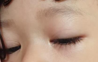
Journal of Medicine and Health                                          Atypical Kawasaki Disease in...
Vol. 3 No. 1 February 2021
e-ISSN : 2442-5257

                                          Case Report

with principal clinical findings may be diagnosed as “incomplete” or “atypical” Kawasaki
Disease. The “atypical” patients, especially those who present without eye or oral mucosal
involvement presentation and whose age is less than six months, may face substantial delay in
diagnosis and treatment.1 All mortality in patients with KD is generally caused by its cardiac
sequelae, which occurs 15 to 45 days after fever onset; the time which well-established coronary
artery vasculitis, and a notable increase in blood hypercoagulability and platelet count
concomitantly occur.1,5 Nevertheless, in children and adults with coronary sequelae (particularly
in patients with missed childhood “KD” diagnosis), myocardial infarction (MI) may cause sudden
death later in the future, underscoring the importance of early diagnosis and prompt treatment.1
The number of gastrointestinal presentation in KD is remarkably uncommon, and data are only
available from single case reports and a few case series.7
        In this paper, a case of an Indonesian child with a diagnosis of atypical KD who has a
gastrointestinal presentation were reported and reviewed.

Case Report
        A two-year-old girl was escorted by her parents to the Emergency Department with the
chief complaint of abrupt continuous fever up to 40o C accompanied by 4-5 episodes of copious,
watery, non-hemorrhagic diarrhea, mild irritability, and reduce appetite since three days prior to
admission. Physical examination revealed temperature 38.6oC, 2 cm diameter right anterior
cervical lymph enlargement, moderate dehydration, no audible cardiac murmur, no skin rashes.
Initial laboratory examination result showed platelet count 592,000/µL, leucocyte count
16,000/µL with neutrophil 84%, lymphocyte count 13%, and erythrocyte sedimentation rate
(ESR) 70mm/hour; routine fecal analysis and urinalysis were within normal limit.
        The patient was initially diagnosed with acute diarrhea with moderate dehydration.
Intravenous fluid rehydration regimen and Cefobactam antibiotic, as well as oral zinc therapy,
were delivered promptly. Her diarrhea frequency improved on the second day of hospitalization,
although she still experienced continuous fever (38.2oC). On her third day of hospitalization (day-
6 fever), she developed non-suppurative conjunctivitis with mild periorbital edema, cracked lips,
and strawberry lips (Figure 1). Further blood work results indicated high acute phase reactant C-
reactive protein (CRP) 43.35 mg/L and increased ESR 72 mm/hour. Chest X-ray and
electrocardiograph (ECG) results were within the normal limit.

J Med Health.2021;3(1):46-60                                                                   48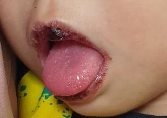
Journal of Medicine and Health                                           Atypical Kawasaki Disease in...
Vol. 3 No. 1 February 2021
e-ISSN : 2442-5257

                                          Case Report

        The patient was referred to Dr. Hasan Sadikin General Teaching Hospital for further
echocardiography examination and intravenous immunoglobulin (IVIG) administration.
Dr.Hasan Sadikin General Teaching Hospital laboratory examination revealed hypoalbuminemia
2.8 gr/dl. Echocardiography examination showed normal coronary artery: left main coronary
artery (LM) 2.6mm, left anterior descending (LAD) artery 2.2mm, left circumflex artery (LCx)
2.0mm, right coronary artery (RCA) 2.7mm, with mild posterior circumferential pericardial
effusion with a size of 0.27-0.54 mm. She was treated with oral aspirin 4 x 250mg for five days,
titrated (on her sixth day of disease) down to 1x 60mg on day 11 of the disease. Due to financial
issues, her family refused IV-IG administration and laboratory work follow-up; and decided to be
discharged from Dr.Hasan Sadikin General Teaching Hospital.

Discussion
        Kawasaki disease is an idiopathic vasculitis that becomes the most common etiology of
acquired heart disease in childhood, especially in developed countries.1,7It mainly affects children
under five years of age and has a boy to girl ratio of 1.5-1.7:1. Its diagnosis is based on the
presence of fever (lasting more than five days) and of four of the five diagnostic criteria
(oropharyngeal changes, bilateral bulbar conjunctival injection without exudate, rash, change of
the extremities, cervical lymphadenopathy ≥1.5 cm diameter).1,2,5 However, diagnosis can be
made with incomplete features if fewer than 4 of the criteria mentioned before are present.5
        In its classic form, Kawasaki disease is usually diagnosed by experienced clinicians with
no significant challenges. However, it is not uncommon that KD patients come with subtle
presentations that clinicians are not aware of, especially when they present as atypical KD. The
terminology of ‘atypical Kawasaki Disease” initially pertained to the patients whose clinical
presentations failed the classic Kawasaki Disease criteria but were found to have coronary artery
abnormalities. However, many cases revealed cardiac abnormalities without meeting the strict
classic KD criteria, and the "incomplete" case can progress in time into a "complete" case.
Therefore, most clinicians nowadays use the atypical term to describe the patients who did not
encompass the classic KD criteria but presented with compatible supportive examination findings
after excluding any other underlying disease.4,6, 8 With this approach, patients have a greater
chance of promptly initiating therapy, hopefully reducing coronary artery complication rate.8
Incomplete KD is commonly found in children aged less than two years old, and the affected
children are at greater risk of developing coronary disease; although another study by Gorczyca

J Med Health.2021;3(1):46-60                                                                    49
Journal of Medicine and Health Atypical Kawasaki Disease in...
Vol. 3 No. 1 February 2021
e-ISSN : 2442-5257
Case Report
et al. showed no significant difference in the age group between complete versus incomplete KD.7,
9, 10
Our patient experienced an abrupt onset of fever in the first three days of illness along
with gastrointestinal symptoms: watery diarrhea and dehydration that initiated her admission to
the hospital. Her fever remained constant throughout her stay in the hospital and was persistent
towards antipyretic and antibiotic treatment. Only later in her third hospitalization day (day 6 of
illness), she developed bilateral conjunctivitis, cracked lips, and strawberry tongue (Fig.1) in her
acute phase. The hallmark of KD is the abrupt-onset and persistent fever. This fever is typically
non-responsive to the antipyretic agent and tends to remain above 38.5°C during most of the acute
phases of the illness (often exceeds 40oC), and sometimes preceded by symptoms of an upper
respiratory or gastrointestinal illness.5,7 Abdominal pain and vomiting are commonly reported,
and approximately one-fourth of children with KD have profuse and watery diarrhea during the
acute febrile period.5,7,11 Baker and colleagues studied 198 patients’ symptoms within ten days
before their KD diagnosis and reported that half of their patients experienced irritability, vomiting
(44%), decreased food intake (37%), diarrhea (26%), and abdominal pain (18%).12 In the next
three to four days, the patients will develop conjunctivitis, changes in the buccal and oral mucosa,
cervical adenitis, a pleomorphic rash, erythema, and edema in the upper and lower extremities
due to a systemic necrotizing vasculitis with fibrinoid necrosis of the medium-sized muscular
arteries. Moreover, the coronary arteries are the predominant sites of this systemic vasculitis
involvement, causing sequelae many years later.5,6
This patient also presented with unilateral localized right anterior cervical
lymphadenopathy during her diarrhea episodes on her admission. Cervical lymphadenopathy in
KD, usually unilateral or only affect one single node, is the least common manifestation occurring
in 50% to 75% of patients that usually shrinks with or without specific therapy after 3 or 4
days.5,13,14 However, in atypical cases, this lymphadenopathy may antecede other symptoms.6
Other clinical findings relatively specific to KD but not listed in the classical diagnostic criteria
are induration and erythema at the BCG immunization sites.4,15 The presence of high and
persistent fever, conjunctival injection, strawberry tongue, and cervical lymphadenopathy in our
patient suggested the diagnosis of suspected Kawasaki disease with diarrhea presentation.
J Med Health.2021;3(1):46-60 50Journal of Medicine and Health Atypical Kawasaki Disease in...
Vol. 3 No. 1 February 2021
e-ISSN : 2442-5257
Case Report
The most common classic manifestations are non-suppurative bulbar conjunctivitis
bilateral, cervical lymphadenopathy >1.5 cm, a polymorphous rash with no vesicles or crusts
found, changes of lips or oral mucosa (such as red, cracked lips, "strawberry" tongue, or diffuse
erythema of oropharynx), as well as changes of extremities (seen as erythema and edema of palms
and soles in the initial stage, and manifest as peeling of skin from fingertips in convalescent
stage).1,4,6, 16 These manifestations may change in a fluctuant mode and present in no particular
pattern in the first 7 to 10 days of the disease course.5,12 Other common clinical signs in KD
include pneumonitis, aseptic meningitis, arthritis, uveitis, otitis, mastitis, dysuria, and
gastroenteritis. Clinicians may mistakenly attribute fever and pyuria findings in an infant or young
child to a urinary tract infection; and the subsequent evolution of red eyes, rash, and red lips in
patients to an antibiotic reaction.1,8
(b)
(c)
(
(a)
(d)
Figure 1 (a) Physical findings on the 6th day of fever. (b) Bilateral non-suppurated bulbar
conjunctivitis. (c) Minor bilateral palpebral edema. (d) Cracked lips and strawberry tongue.
J Med Health.2021;3(1):46-60 51Journal of Medicine and Health Atypical Kawasaki Disease in...
Vol. 3 No. 1 February 2021
e-ISSN : 2442-5257
Case Report
Our patient experienced mild irritability that might be a part of her KD manifestation
other than a clinical symptom of dehydration. In some literature, children with prolonged fever,
irritability, and laboratory finding of cerebrospinal fluid’s culture-negative pleocytosis suggestive
of aseptic meningitis (or if antibiotics have been given, partially treated meningitis) may lead
clinicians to overlook the diagnosis of KD.1,4,8 Although in our case, we decided not to test her
cerebrospinal fluid due to financial issues to prioritize other more important supportive
examinations.
Clinically, the patient only fulfills 3 out of the 5 classic symptoms required for a complete
Kawasaki Disease. However, once we suspect our patient for Kawasaki Disease, we immediately
transferred her to a higher health system to receive further examination and therapy to comply
with the investigation and evaluation algorithm for incomplete KD-suspected patients by
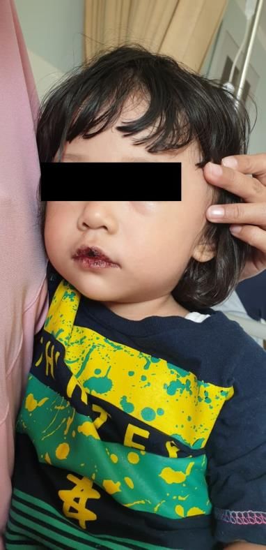
Baumer17(Please see Fig.2).
Initial laboratory work in our hospital indicated the patient to have an infection shown by
leukocytosis with a "shift to the left" and high ESR counts, so she was treated with the antibiotic.
However, suspicion was raised due to her elevated platelet count. Only when her following classic
KD signs developed, the ESR and CRP counts were promptly checked, and the results supported
her diagnosis as suspected Kawasaki Disease. Until recently, no specific diagnostic tests are
available for KD,5,14 but at the beginning of the disease, clinicians may find indicators of ongoing
inflammation such as leukocytosis with a “shift to the left” trend of white blood cell (WBC)
differential count, 4,5,11 as well as elevated C-reactive protein (CRP) and ESR count.4,10,15 (Table
1). However, there are no differences between complete and atypical KD in laboratory
investigation results statistically.10,14
J Med Health.2021;3(1):46-60 52Journal of Medicine and Health Atypical Kawasaki Disease in...
Vol. 3 No. 1 February 2021
e-ISSN : 2442-5257
Case Report
Figure 2. Kawasaki Disease Diagnosis Algorithm. Kawasaki Disease patients’ investigation and
evaluation flowchart. *Echocardiogram positive if any of criteria are fulfilled: (1) left anterior descending
(LAD) or right coronary artery (RCA) Z-score of 2.5 or more; (2) positive coronary arteries aneurysms
criteria from Japanese Ministry of Health; (3) three or more presence of suggestive features [decreasing left
ventricular function, perivascular brightness, pericardial effusion, mitral regurgitation, lack of tapering, or
z scores in LAD or RCA of 2–2.5. +If the echo result is positive, start treatment within ten days of fever
onset and beyond ten days for patients with clinical and laboratory signs of an existing infection. ≠Classic
peelings start under the fingers’ nail bed and toes. A pediatric KD expert should always be consulted when
fever persists or anytime needed. Alanine transaminase [ALT], C-reactive protein [CRP], erythrocyte
sedimentation rate [ESR], white blood cells [WBC]. Modified from: Baumer HJ 17
J Med Health.2021;3(1):46-60 53Journal of Medicine and Health Atypical Kawasaki Disease in...
Vol. 3 No. 1 February 2021
e-ISSN : 2442-5257
Case Report
Table 1 Initial investigations for suspected Kawasaki Disease.
Modified from: Brogan PA 4
During the acute stage of illness, leukocytosis with predominant immature and mature
granulocytes is classically found.1,18 Children with KD have more common positive neutrophils’
toxic granulation laboratory result compared to those with other febrile diseases. However,
clinicians should keep in mind that sometimes significant neutropenia can be detected in the early
stage, and this should be referred as a marker for other specific serious disease.5 Although
thrombocytosis is another typical feature of KD laboratory workup in most cases it usually does
not appear until the second week, reaches its highest level in the third week (mean ≈700,000 per
mm3), and returns to its normal level by four to six weeks after onset of illness.1,5 Other literature
stated that in the most severe KD cases, secondary thrombocytes might escalate to
1,000,000/mm3, a phenomenon called “reactive thrombocytosis”.1,18 No evidence shows that this
phenomenon may lead to immediate thrombosis. Thus, experts recommend that treatment should
only be given to KD patients who have other risk factors for thrombosis.19 Normochromic and
normocytic anemia is commonly discovered during KD laboratory workup, but it resolves along
with the subsidence of KD's inflammation process. Forty to sixty percent of KD patients have
J Med Health.2021;3(1):46-60 54Journal of Medicine and Health Atypical Kawasaki Disease in...
Vol. 3 No. 1 February 2021
e-ISSN : 2442-5257
Case Report
mild to moderate elevations of serum transaminases or gamma-glutamyl transpeptidase, and only
10 % of KD patients have mild hyperbilirubinemia.1,18
Clinicians should consider other illnesses if the suspected patient's ESR, CRP, and platelet
count are within normal range after the 7th day of illness since Kawasaki disease diagnosis is least
likely.1 Persistent increasing ESR level after the fever recedes is a KD laboratory hallmark and
may help doctors discerning KD from other infectious illnesses.11 Elevated CRP (≥30 mg/l) and/or
ESR (≥40 mm/hour) plus several other auxiliary laboratory tests (full blood count, ALT, plasma
albumin, urinary white cell count) and echocardiogram results can provide information for doctors
to make a clinical decision whether to treat KD patients with IVIG.17 It is also important to
remember that an increase in ESR count without any increase in CRP value may happen due to
immunoglobulin therapy. Thus, ESR monitoring is not recommended for patients on the IVIG
regiment.18 Other blood chemistry parameters changes commonly found in KD are low serum
protein and albumin, low serum sodium, elevated liver enzymes (ALT), and abnormal lipid profile
(can be aggravated by IVIG treatment).1,15 Our patient’s lab result from Dr. Hasan Sadikin
Teaching Hospital showed mild hypoalbuminemia (2.8 g/dl). She also experienced mild
periorbital edema that might be a consequence of her hypoalbuminemia state (Fig.1c).
Hypoalbuminemia and noncardiogenic edema in KD patient result from vascular leakage
accumulation due to increased microvascular permeability and the role of vascular endothelial
growth factor.6 Moreover, hypoalbuminemia in KD is typical and associated with a more serious
and more prolonged acute phase of the illness.1,18
Common ECG findings in KD patients (almost always reversible) are tachycardia, flattened
T waves, decreased QRS voltages, and prolonged rate corrected QT intervals.11,15 Arrhythmias,
including heart block, may also occur. Electrocardiography may also show signs of MI due to
coronary thrombosis in untreated large coronary artery aneurysms.1,15 After transferred to Dr.
Hasan Sadikin Hospital, our patient’s ECG result was within the normal limit.
However, echocardiography (ECHO) is a more valid and sensitive supplementary
examination to assess coronary artery aneurysms in the illness's acute and subacute stages.2,11
Clinicians may reveal mitral regurgitation, decreased left ventricular function, and pericardial
effusion from KD patients’ ECHO results.15 Moreover, ECHO can diagnose coronary aneurysms,
which generally develop within two weeks of Kawasaki disease’s onset. Coronary changes (z-
score ≥ 2.5) can be seen in these KD patients, most commonly occurring in the LAD or RAC.
The development of coronary aneurysms raises clinical suspicion for the diagnosis of KD.16,18 In
J Med Health.2021;3(1):46-60 55Journal of Medicine and Health Atypical Kawasaki Disease in...
Vol. 3 No. 1 February 2021
e-ISSN : 2442-5257
Case Report
classic KD, an ECHO should be performed in the hospital at the time of diagnosis, followed by a
repeat ECHO within two weeks. Doctors should also perform further ECHOs to assess illness
improvement or progression at around 6–8 weeks.11,16,18 Echocardiography follow-ups are also
more frequently done in high-risk children with prolonged fever, pericardial effusion, coronary
artery abnormalities, ventricle dysfunction, or valve regurgitation.16,18 Echocardiography in our
patient was done on her seventh day of fever (hospitalization day 4) in the transfer hospital. Our
patient’s echocardiography result showed mild posterior circumferential pericardial effusion with
0.27–0.54 mm size. Echocardiographic and cardiac angiographic data showed that around 20–
40% of untreated KD patients develop coronary artery abnormalities. Approximately half of these
lesions receded within five years, and in most patients with mild CAA (3–4 mm), regression
occurs within two years.4
Occasionally, clinicians need to depict the coronary arteries' anatomy to investigate the
extent of the vessel involvement in KD, making the additional imaging with a CT angiogram
(CTA) or cardiac MRI as crucial examination in this step. However, a study suggested that in
monitoring coronary aneurysms and their progression, CTA was equally invasive as an
angiogram.16 Common cardiac complications in Kawasaki disease are cardiac failures, cardiac
tamponade, pericarditis, myocarditis, and coronary arterial abnormalities.4,18 During the acute
phase of Kawasaki disease, the universal findings of myocarditis and frequent diminished left
ventricular function and contractility records should rise he clinicians' concern about KD's long-
term effects on myocardial function.11 Our patient was planned to received IVIG and aspirin as
prompt management for incomplete Kawasaki Disease to prevent further risk.
The main objective of Kawasaki disease management strategy is to avoid long-term
sequelae. The clinical error of inadequately managing a child with KD has a severe repercussion
that, within reason and after cautious evaluation, the failure on unnecessary or premature therapy
is preferable to missing or delaying therapy for a child whose KD diagnosis is vague. American
Academy of Pediatrics and AHA recommend that aspirin and IVIG should be administered during
the first ten days of the illness to those children diagnosed with Kawasaki. 1,15 Clinicians need to
keep in mind that aspirin's administration as a single treatment for KD has not been subjected to
randomized controlled clinical trials. Aspirin is administered in two different dosages. In the
acute phase of the illness, it is given at relatively high “anti-inflammatory” doses (30–100
mg/kg/day)4,17 till the time of defervescence and in “antiplatelet” dosages (2-5 mg/kg/ once daily)
for at least 6-8 weeks,9,11,19 or dependent on findings on echocardiography.4 In the UK practices,
J Med Health.2021;3(1):46-60 56Journal of Medicine and Health Atypical Kawasaki Disease in...
Vol. 3 No. 1 February 2021
e-ISSN : 2442-5257
Case Report
pediatricians prescribe aspirin dose as 30 mg/kg/day during the acute phase of the illness for more
tolerance of the gastrointestinal and other side effects.4 In children with cardiac complications,
particularly for children with small to medium aneurysms (Journal of Medicine and Health Atypical Kawasaki Disease in...
Vol. 3 No. 1 February 2021
e-ISSN : 2442-5257
Case Report
be administered after the tenth day of illness.1 Subsequent studies have shown that IVIG therapy
is most significantly beneficial if administered in the early illness course. An epidemiological
survey of more than 5000 patients in Japan treated with 2 g/kg IVIG proved that cardiac
complications at one month after the beginning of disease were fewer in the group treated before
day 6 of illness than another group treated later.2
Due to its possible mechanisms of action such as augmentation of regulatory T-cell activity,
modulation of cytokine production, suppression of antibody synthesis, neutralization of toxins or
other pathogenic agents, and provision of anti-idiotypic antibodies, the experts concluded that
IVIG appeared to have a generalized anti-inflammatory effect.1,20 The theories behind that
mechanism of actions include the macrophages’ Fc-II and Fc-III receptors cross-linking,
endothelial cells and natural killer cells interaction blockage, selective induction of interleukin-1-
receptor antagonist, interleukin-8 binding to complement fragments, induction of immune
inhibitory receptors, and provision of specific antibody to the causative agent or a toxin. In-vitro
study findings show that IVIG exerts blocking effects on endothelial-cell proliferation and
synthesis process of adhesion molecules, cytokines, and chemokines.2,20 Experts recommend that
IVIG 2 g/kg be administered together with acetylsalicylic acid as a single infusion over 10 to 12
hours.1,11,19
Less severe adverse reactions related to the IVIG regiment, including fever, chills, and
hypotension, are variously linked with the specific product infused. Due to the immunogenicity
reduction of the passive antibodies in IVIG preparation which are “vaccine-related”, clinicians
should defer injections of live virus vaccines (mumps, measles, varicella and rubella) at least 11
months after IVIG administration.2,11 In all USA health care system, high-dose IVIG usage is
considered cost-effective. In contrast, some health centers in Japan only indicate IVIG therapy
for children with a predicted high risk for CAA development.11 Similar treatment limitation
happens in Indonesia; the high cost of IVIG treatment in Indonesia makes it difficult for most
patients without proper health insurance coverage to afford it. Thus, our patient did not receive
IVIG treatment due to this financial issue.
Eosinophil count is one of the essential parameters of Kawasaki disease's IVIG treatment
failure. Eosinophil count alteration after IVIG treatment was positively relevant with interleukin
(IL)-5 levels change and inversely correlated with treatment failure. Eosinophils and IL-5 levels
increments after the IVIG regiment were inversely correlated with coronary artery lesion
formation.20 Ten to fifteen percent of KD patients treated with high-dose aspirin plus 2 g/kg IVIG
J Med Health.2021;3(1):46-60 58Journal of Medicine and Health Atypical Kawasaki Disease in...
Vol. 3 No. 1 February 2021
e-ISSN : 2442-5257
Case Report
will have a recrudescent or persistent fever.11,20 If the fever is persistent, studies have provided
other successfully tested treatment options in small case series, including pulsed intravenous
methylprednisolone (30 mg/kg for three days), plasmapheresis, cyclosporin, cyclophosphamide
plus prednisone, and monoclonal antibodies to TNF for clinicians as alternatives.2,20 Since our
patient’s parents decided to discontinue hospitalization; the Dr. Hasan Sadikin Hospital staffs had
to discharge her and educate on KD complication and the importance of follow up.
The first year recurrence rate for Kawasaki Disease is around 2%.16 There are uncertain
significances of these clinical findings in patients with no coronary aneurysms in KD acute phase
such as coronary and peripheral arteries vasodilatory capacity impairment up to 15 years after the
illness; higher blood pressure (both diastolic and systolic) and blood triglyceride concentrations,
as well as increased adiposity, compared to control group up to 11 years after the onset of illness,
and the myocardial fibrosis findings on endomyocardial biopsy up to 11 years after disease.
Therefore, due to the unclear description of the illness potential sequelae without coronary
aneurysms, identifying the proper and adequate follow-ups for these KD patients is still
considered arduous.2 Experts recommend that clinicians should acquire the initial echocardiogram
when suspecting a diagnosis of KD and repeat the echocardiography at two weeks and six weeks
following the disease for every KD patient. Clinicians should also conduct clinical re-
examinations during the first two months of the illness to detect the presence of cardiac
abnormalities such as valvular insufficiency, myocarditis, dysrhythmias, or congestive heart
failure.5 Partial Kawasaki Disease should importantly keep it in mind as a differential diagnosis
in prolonged fever patients. Careful diagnosis and prompt KD management are crucial to prevent
further morbidity and mortality.
Conclusion
Atypical Kawasaki Disease can be a puzzling diagnosis due to its uncommon presentations.
Clinicians should importantly keep it in mind as a differential diagnosis in patients with prolonged
fever.
References
1. McCrindle BW, Rowley AH, Newburger JW, Burns JC, Bolger AF, Gewitz M, et al. Diagnosis, treatment, and
long-term management of Kawasaki Disease-A Scientific Statement for Health Professionals From the American
Heart Association. Circulation. 2017;135:e927–e99.
2. Burns JC, Glode MP. Kawasaki Syndrome. Lancet. 2004;364:533–44.
J Med Health.2021;3(1):46-60 59Journal of Medicine and Health Atypical Kawasaki Disease in...
Vol. 3 No. 1 February 2021
e-ISSN : 2442-5257
Case Report
3. Nakamura Y, Yashiro M, Uehara R, Oki I, Watanabe M, Yanagawa H, et al. Epidemiologic features of Kawasaki
disease in Japan: results from the nationwide survey in 2005-2006. J Epidemiol. 2008;18:167-72.
4. Brogan PA, Bose A, Burgner D, Shingadia D, Tulloh R, Michie C, et al. Kawasaki disease: an evidence based
approach to diagnosis, treatment, and proposals for future research. Arch Dis Child. 2002;86:286–90.
5. Sundel RP, Petty RE. Kawasaki Disease. In: Cassidy JT PR, Laxer R, Lindsley C, editor. Textbook of Pediatric
Rheumatology. 6th ed. Philadephia: Elsevier-Sunders; 2011. p. 505-20.
6. Fang L-C, Shyur S-D, Peng C-C, Jim W-T, Chu S-H, Kao Y-H, et al. Unusual manifestations of Kawasaki disease
with retropharyngeal edema and shock syndrome in a Taiwanese child. J Microbiol Immunol Infect. 2014;47:152-
57.
7. Colomba C, La Placa S, Saporito L, Corsello G, Ciccia F, Medaglia A, et al. Intestinal Involvement in Kawasaki
Disease. J Pediatr. 2018;202:186-93.
8. Barone SR, Pontrelli LR, Krilov LR. The differentiation of classic Kawasaki disease, atypical Kawasaki disease,
and acute adenoviral infection: use of clinical features and a rapid direct fluorescent antigen test. Arch Pediatr
Adolesc Med. 2000;154(5):453-56.
9. De Almeida RG, Goldenzon AV, Rodrigues MCF, Sztajnbok FR, Elsas MICG, De Oliveira SKF. Profile of
Kawasaki disease in children referred to two pediatric rheumatology services in Rio de Janeiro, Brazil. Bras J
Rheumatol. 2010;50(5):529-38.
10. Gorczyca D, Postepski J, Olesinska E, Lubieniecka M, Lachor-Motyka I, Opoka-Winiarska V, et al. The clinical
profile of Kawasaki disease of children from three Polish centers: A retrospective study. Rheumatol Int.
2014;34:875-80.
11. Newburger JW, Burns JC. Kawasaki Disease. Vasc Med. 1999(4):187-202.
12. Baker AL, Lu M, Minich LL, Atz AM, Klen GD, Korsin R, et al. Pediatric Heart Network Investigators. Associated
symptoms in the ten days before diagnosis of Kawasaki disease. J Pediatr. 2009;154:592-5.
13. Yoskovitch A, Tewfik TL, Duffy CM, Moroz B. Head and neck manifestations of Kawasaki disease. Int J Pediatr
Otorhinolaryngol. 2000;52:123-9.
14. Fukushige J, Takahashi N, Ueda Y, Ueda K. Incidence and clinical features of incomplete Kawasaki disease. Acta
Pediatr. 1994;83:1057-60.
15. Harnden A, Takahashi M, Burgner D. Kawasaki Disease. BMJ. 2009;338:1133-8.
16. Goswami N, Marzan K, De Oliveira E, Wagner-Lees S, Szmuszkovicz J. Recurrent Kawasaki Disease: A case
report of three separate episodes at >4-year intervals. Children. 2018;5:155-9.
17. Baumer HJ. Kawasaki Disease: What to do with incomplete cases?. Arch Dis Child Educ Pract Ed. 2005;90:ep102–
ep4.
18. Budiyanto N. Problem Jantung pada Penyakit Kawasaki. J Kardiol Ind. 2007;28:285-96.
19. Arora K, Guleria A, Jindal AK, Rawat A, Singh S. Platelets in Kawasaki disease: Is this only a numbers game or
something beyond? . Genes Dis. 2020;7(1):62-6.
20. Kuo HC, Yang DK, Chiao WC, Ger LP, Hsieh KS. Kawasaki Disease: An update on diagnosis and treatment.
Pediatr Neonatol 2012;53:4-11.
J Med Health.2021;3(1):46-60 60You can also read