Interpreting pathologies in extant and extinct archosaurs using micro-CT
←
→
Page content transcription
If your browser does not render page correctly, please read the page content below
Interpreting pathologies in extant and
                                  extinct archosaurs using micro-CT
                                  Jennifer Anné1 , Russell J. Garwood1 , Tristan Lowe2 , Philip J. Withers2 and
                                  Phillip L. Manning1
                                 1 School of Earth, Atmospheric and Environmental Sciences, University of Manchester, Manchester, UK
                                 2 Manchester X-ray Imaging Facility, School of Materials, University of Manchester, Manchester, UK

                                     ABSTRACT
                                     Palaeopathology offers unique insight to the healing strategies of extinct organ-
                                     isms, permitting questions concerning bone physiology to be answered in greater
                                     depth. Unfortunately, most palaeopathological studies are confined to external
                                     morphological interpretations due to the destructive nature of traditional meth-
                                     ods of study. This limits the degree of reliable diagnosis and interpretation possible.
                                     X-ray MicroTomography (micro-CT, XMT) provides a non-destructive means of
                                     analysing the internal three-dimensional structure of pathologies in both extant
                                     and extinct individuals, at higher resolutions than possible with medical scanners.
                                     In this study, we present external and internal descriptions of pathologies in extant
                                     and extinct archosaurs using XMT. This work demonstrates that the combination of
                                     external/internal diagnosis that X-ray microtomography facilitates is crucial when
                                     differentiating between pathological conditions. Furthermore, we show that the use
                                     of comparative species, both through direct analysis and from the literature, provides
                                     key information for diagnosing between vertebrate groups in the typical pathological
                                     conditions and physiological processes. Micro-CT imaging, combined with com-
                                     parative observations of extant species, provides more detailed and reliable interpre-
                                     tation of palaeopathologies. Micro-CT is an increasingly accessible tool, which will
                                     provide key insights for correctly interpreting vertebrate pathologies in the future.

Submitted 5 May 2015
                                  Subjects Paleontology, Veterinary Medicine, Zoology, Histology
Accepted 6 July 2015
Published 28 July 2015            Keywords Palaeopathology, Micro-CT, Archosaur

Corresponding author
Jennifer Anné,
jennifer.anne@manchester.ac.uk    INTRODUCTION
Academic editor                   Palaeopathology is the study of ancient diseases and trauma, and is usually limited in
Marı́a Ángeles Esteban           vertebrates to lesions that affect the skeleton, as soft tissues are generally lost over time
Additional Information and        (Rothschild & Martin, 2006; Rühli et al., 2007; Straight et al., 2009; Waldron, 2009; Roth-
Declarations can be found on      schild & Depalma, 2013). The identification and classification of palaeopathologies can
page 11
                                  be difficult due to complex internal morphology and the loss of detail during fossilisation
DOI 10.7717/peerj.1130
                                  due to taphonomic overprint. Despite these problems, most palaeopathological studies
    Copyright                     rely on external gross morphological descriptions rather than examining internal features.
2015 Anné et al.
                                  Although some palaeopathologies can be identified by external examination (e.x. fracture
Distributed under                 callus) a lack of internal morphological information may lead to misidentification and
Creative Commons CC-BY 4.0
                                  over interpretation. Internal structure can be identified using thin section analysis, but
OPEN ACCESS                       this method is destructive and therefore cannot be applied to specimens that are rare or

                                  How to cite this article Anné et al. (2015), Interpreting pathologies in extant and extinct archosaurs using micro-CT. PeerJ 3:e1130;
                                  DOI 10.7717/peerj.1130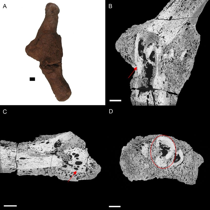
Table 1 External descriptions of specimens. External pathological description for extant and extinct archosaurs used in this study.

 Species                                            Skeletal element                            External pathological description
 Sagittarius serpentarius (secretary bird)          pedal phalanx NHM S/1869.2.16.1             Extreme bone growth; most of the digit is obscured
 Struthio camelus (ostrich)                         cervical vertebra BHI 6241                  Bony growth on the postzygapothoses resulting
                                                                                                in pseudofusion of two vertebrae
 Tyrannosaurus rex                                  cervical rib BHI 3033                       Extensive reactive bone throughout
 Edmontosaurus annectens                            metacarpal BHI 6191                         Rough fracture callus caused by angular displacement
 Edmontosaurus annectens                            dorsal rib BHI 6184                         Large “pursed” bony growth near rib head

                                             fragile. Additionally, the two-dimensional nature of this technique does not capture the
                                             entire complexity of pathological tissue unless serial sections are made. The application
                                             of medical scanners to such material has allowed for non-destructive three-dimensional
                                             investigation of internal histologic superstructures, though the resolution may be low
                                             (Straight et al., 2009; Particelli et al., 2012; Sutton, Rahman & Garwood, 2014), and medical
                                             X-ray sources struggle to penetrate dense fossils.
                                                Since the 1990s, XMT has revolutionised the study of fine and complex bone structures
                                             in medical (Englke et al., 1999; Particelli et al., 2012), forensic (Thali et al., 2003) and
                                             palaeontological studies (Straight et al., 2009; Sutton, Rahman & Garwood, 2014; Bishop
                                             et al., 2015; Foth et al., 2015). Such scanners can attain sub-micron resolution (depending
                                             on sample size) compared to the tenths of a millimetre in conventional medical scans
                                             (Englke et al., 1999; Sutton, Rahman & Garwood, 2014). XMT is usually applied to
                                             smaller samples, as the size of the area of interest dictates spatial resolution. However,
                                             by compromising the resolution of the scan (to tens of microns versus sub-micron), this
                                             technique can be applied to larger scan areas while maintaining higher resolution scans
                                             than is possible with medical scanners (which also struggle with large specimens due to
                                             their medium energy (20–150 kV)). For example, the Nikon custom bay microtomography
                                             system in the Manchester X-ray Imaging Facility (MXIF) houses a high energy source
                                             (225–320 keV) that, coupled with a 2,000 × 2,000 pixel detector, allows decimetre-scale
                                             specimens of fossil bone to be scanned at sub-100 µm resolution. This level of detail
                                             allows us to distinguish key internal morphological features required for an accurate
                                             diagnosis of palaeopathologies. Here we present XMT data from two extant and three
                                             extinct archosaurs exhibiting a variety of pathologies, further demonstrating that XMT is a
                                             powerful tool for the field of palaeopathology.

                                             MATERIALS & METHODS
                                             Specimens consisted of extant and extinct archosaur material with various pathological
                                             conditions that were identified based on external observations (Table 1).
                                                Extant taxa were included to improve palaeopathological diagnosis as medical
                                             terminology, physiology of skeletal elements, and likelihood of certain diseases can differ,
                                             even between human and mammalian companion animals (Huchzermyer & Cooper, 2000;
                                             Rothschild & Panza, 2005; Mader, 2006; Rothschild & Martin, 2006; Kranenburg, Hazewinkel
                                             & Meij, 2013; Foth et al., 2015).

Anné et al. (2015), PeerJ, DOI 10.7717/peerj.1130                                                                                               2/14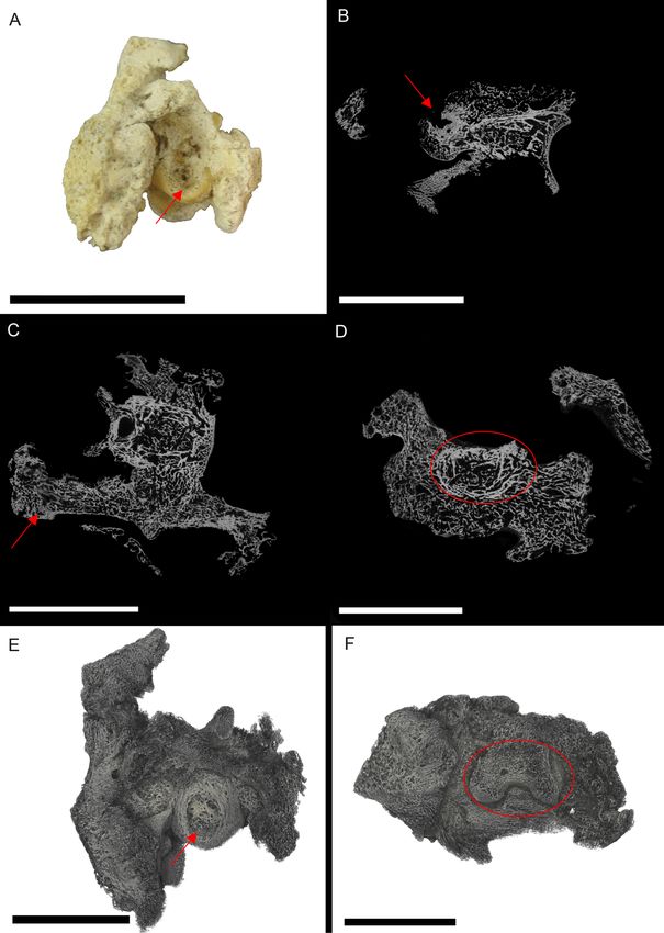
Table 2 Table of scanning parameters. Experimental parameters used for scanning with the Nikon Metris Custom Bay (MXIF).

 Species                                        kV           µA           Filter          Exposure           Voxel size      Total scan
                                                                          (mm)            (ms)               (µm)            time (min)
 Sagittarius serpentarius                       50           150          none            1,000              16.6            55
 Struthio camelus                               60           170          1.5 Al          1,000              44.9            55
 Tyrannosaurus rex                              60           170          1.5 Al          1,000              15.3            55
 Edmontosaurus annectens—metacarpal             115          115          0.25 Cu         500                47.9            30
 Edmontosaurus annectens—rib                    115          85           0.25 Cu         708                78              40

                                          XMT scanning was conducted at the Manchester X-ray Imaging Facility (MXIF)
                                       using the Nikon Metris Custom Bay, a system which can accommodate large specimens
                                       (maximum field of view of 410 mm) and provide spatial resolutions between 100 and
                                       ∼3.5 µm (Table 2). The system has a 225 kV static multi-metal anode target, which was set
                                       to tungsten in order to achieve the maximum energy, to improve X-ray penetration of the
                                       scanned material (fossil and extant bone). The source voltage was set to 225 kV, and auto
                                       conditioned for 30 min prior to scanning to decrease the likelihood of the source cutting
                                       out while scanning. Specimens were mounted on a manipulation stage using a variety
                                       of plates and clamps depending on their size, weight, and geometry. Previous scans of
                                       this type of material have demonstrated that 15–20% transmission through the specimen
                                       relative to the flat field provide excellent scans. To achieve this aim, the source voltage and
                                       current were modified coupled with changing thicknesses of Cu or Al filters to minimise
                                       beam hardening but achieve an optimal spectral width and intensity. Exposure was selected
                                       to minimise scan times while collecting clean data (between 0.5 and 1.0 s exposure, sample
                                       dependent), and for all scans counts on the detector panel outside the sample were kept
                                       below 65,000 counts for the selected gain, which was set to minimum to reduce noise. For
                                       each sample, scanning parameters were selected to fit these requirements while using the
                                       lowest voltage possible to maximise the attenuation from photoelectric absorption and
                                       thus maximise contrast from compositional differences in the sample (Sutton, Rahman &
                                       Garwood, 2014). 3,142 projections were collected; a number based on the optimise option
                                       for the CT Pro acquisition software.
                                          Volumes were reconstructed using the Nikon CT Pro software. Processed scans were
                                       converted to TIFF stacks using the HMtool in MATLAB® , which is an in-house script used
                                       by the MXIF. TIFFs or VGI/VOL files (the former for Volume Graphics’ VGStudio MAX)
                                       were opened using Fiji (Schindelin et al., 2012) for initial analysis of slice stacks. TIFFs were
                                       then opened in Avizo® to construct orthoslices, while VGI files were opened in Drishti for
                                       volume rendering (Limaye, 2006).

                                       RESULTS AND DISCUSSION
                                       NHM S/1869.2.16.1: Extensive bone growth persists through the interior of the S. serpen-
                                       tarius phalanx. In many locations, this makes distinguishing normal cancellous struts from
                                       pathological growth difficult (Fig. 1). Despite the degree of pathological intrusion, both
                                       articular surfaces maintain shape and texture. A large, circular lesion is located on the

Anné et al. (2015), PeerJ, DOI 10.7717/peerj.1130                                                                                  3/14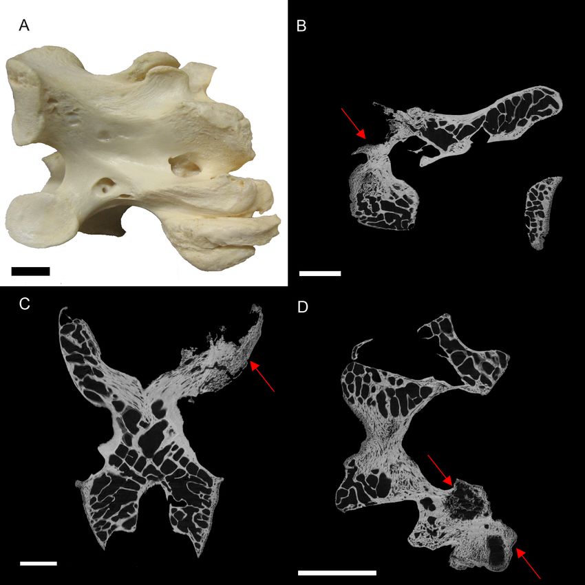
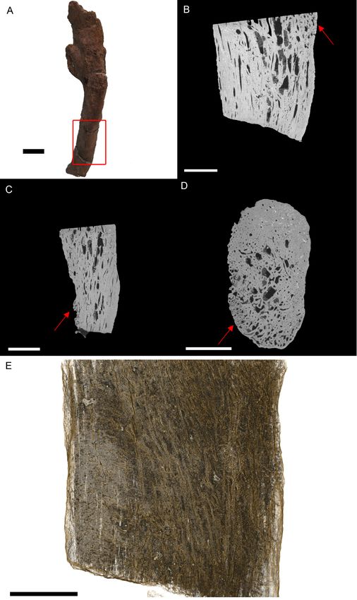
Figure 1 S. serpentarius (NHM S/1869.2.16.1) pedal phalanx; photograph of the specimen in plantar
                                       view (A), XMT slices in medial-lateral (B), dorsal-ventral (C) and transverse (D) views, and 3D render-
                                       ing of the plantar (E) and distal (F) surfaces. A large, circular lesion is seen on the plantar surface (red
                                       arrows; A, E), with small necrotic spaces persisting throughout the phalanx (red arrows B, C). Extensive
                                       reactive bone growth persists both internally and externally. The outline of the normal bone cortex is
                                       barely visible in some areas and indistinguishable in others (red circles; D, F). The extent of the growth
                                       makes it difficult to identify any possible indicators of trauma. Both articulation surfaces are relatively
                                       untouched. Scale bar is 1 cm.

Anné et al. (2015), PeerJ, DOI 10.7717/peerj.1130                                                                                               4/14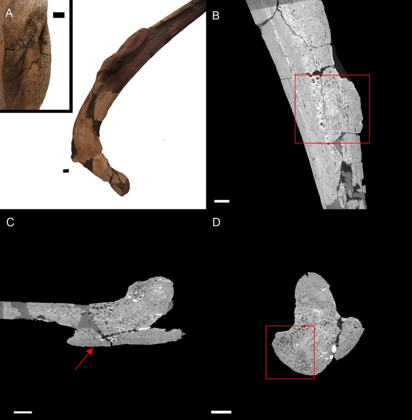
plantar surface, with signs of necrosis internally. The concentric ring appearance within
                                     the necrotic area matches the description for a fibricess; a localised inflammatory process
                                     caused by the incomplete elimination of pathogens in archosaurs (Harmon, 1998; Huchz-
                                     ermyer & Cooper, 2000). The most likely cause is osteitis (inflammation of the bone by in-
                                     fection) or osteomyelitis (inflammation of bone marrow by infection; Ritchie, Harrison &
                                     Harrison, 1994; Berners, 2002). This diagnosis is based on the lesion on the plantar surface
                                     of the bone (Fig. 1E) and internal necrosis (Figs. 1B–1D). In avians, bacterial osteomyelitis
                                     is identified based on severe necrosis, with minimal periosteal reaction (Ritchie, Harrison
                                     & Harrison, 1994). However, periosteal change can occur in chronic infections, and in
                                     fungal osteomyelitis, the periosteal reaction is pronounced (Ritchie, Harrison & Harrison,
                                     1994). Additionally, there seems to be discrepancies between veterinary diagnoses as some
                                     characterise osteomyelitis as having a pronounced periosteal reaction (Doneley, 2011).
                                         Due to the extent of the new growth, it is impossible to distinguish if the infection
                                     was a result of a fracture, or another complication such as ulcerative pododermatitis
                                     (bumblefoot; Herman, Locke & Clark, 1962; Keymer, 1972; Remple & Al-AShbal, 1993;
                                     Ritchie, Harrison & Harrison, 1994; Gentz, 1996; Huchzermyer & Cooper, 2000; Berners,
                                     2002; Wyss et al., 2015), which can cause osteomyelitis in later stages. Pododermatitis is
                                     caused by a number of bacteria and is more common in captive individuals with poor
                                     perching surfaces. However, it has been documented in wild individuals, usually as a result
                                     of a puncture (Herman, Locke & Clark, 1962; Keymer, 1972; Remple & Al-AShbal, 1993;
                                     Gentz, 1996; Berners, 2002; Wyss et al., 2015). Additionally, most studies on pododermatisis
                                     concerning birds of prey focus on species that hunt ‘on the wing.’ Secretary birds spend
                                     most of their time foraging on hard ground, using their feet to stamp their prey. Thus
                                     pododermatisis is a reasonable hypothesis for the cause of osteomyelitis.
                                         Excluded conditions include gout, osteopetrosis and neoplasia. Gout is a common
                                     metabolic disease in archosaurs caused by concentration of urate crystals (Berners, 2002;
                                     Mader, 2006). Although common in wild birds (Morishita, Aye & Brooks, 1997), especially
                                     within the pedal phalanges, gout affects the joint surfaces, which in this specimen are
                                     unaffected (Mader, 2006). Osteopetrosis causes thickening of the bone through prolific
                                     bone deposition, resulting in the loss of the medullary cavity (Ritchie, Harrison &
                                     Harrison, 1994; Doneley, 2011); however, this is has only been noted in the femur, ulna,
                                     radius, pectoral girdle and vertebrae (in captive birds). Neoplasia is the most likely of the
                                     alternative, as it resembles osteomyelitis; however neoplasia is rare in wild individuals
                                     (Ritchie, Harrison & Harrison, 1994).
                                         BHI 6241: The outer appearance of the S. camelus cervical matches descriptions
                                     for early stages of Diffuse Idiopathic Skeletal Hyperostosis (DISH). DISH is described
                                     (externally) as fused vertebrae with a ‘melted candle wax’ appearance, where fusion may be
                                     asymmetrical and the articulated surfaces appear unaffected (Fig. 2A; Rothschild & Martin,
                                     2006; Waldron, 2009). However, DISH has not been found in avians. Newcastle disease is a
                                     common ailment of captive ratites resulting in the inability for the individual to lift their
                                     head (Stewart, 1994; Huchzermyer, 2002). However, this is a neurological disease and does
                                     not affect the bone. Alternative conditions that affect vertebrae in avians include arthritis,

Anné et al. (2015), PeerJ, DOI 10.7717/peerj.1130                                                                             5/14
Figure 2 S. camelus (BHI 6241) cervical vertebra; photograph of the specimen in medial-lateral view
(A) and XMT slices in medial-lateral (B), dorsal-ventral (C) and transverse (D) views. The affected
zygapophysis shows large necrotic cavities (red arrows) surrounded by relatively dense reactive bone,
which spreads both internally and externally to form osteophyte ‘hooks.’ Scale bar is 1 cm.
osteopetrosis (viral infection in avians) and vertebral osteomyelitis (bacterial or fungal)
(Julian, 1998; Stalker et al., 2010; Doneley, 2011).
Internal inspection provided further evidence for infection of the zygapophysis due to
the presence of lesions surrounded by densely compact bone as compared to the normal
zygapophysis (Figs. 2B–2D). Reactive arthritis has been identified in avians, but within
long bone joints (MacLean et al., 2013), and the lack of soft tissue makes it difficult to
diagnose in this specimen. Osteopetrosis is characterised by the proliferation of porous
subperiosteal bone, which is not seen in BHI 6241 (Ritchie, Harrison & Harrison, 1994).
Osteomylitic infection of the vertebral column has been noted in broiler chickens; however,
cases affect the centrum and not the processes as seen in this specimen (Julian, 1998;
Stalker et al., 2010). In snakes, vertebral osteomyelitis can cause anklyosing (fusion) of the
Anné et al. (2015), PeerJ, DOI 10.7717/peerj.1130 6/14vertebrae similar to what is seen in BHI 6241 (Stacy & Pessier, 2007). Although this is a
squamate comparison, the description is the closest to what is seen in BHI 6241. Thus, the
suggested diagnosis is partial ankylosing due to a form of vertebral osteomyelitis.
BHI 3033: The T. rex cervical vertebrae associated with this rib exhibit severe reactive
bone growth, most likely due to a complication of healing after trauma, which caused two
of the cervicals to become fused (Larson & Donnan, 2002; PL Larson, pers.comm., 2013).
The complication derived from the vertebral injury appears to have spread to the cervical
rib, giving it a frothy appearance, and in some areas it is enlarged (Fig. 3). The most likely
cause is osteitis or osteomyelitis as a result of complications during healing.
Internal examination reveals high levels of porosity, with canals lined parallel to the long
axis of the rib, which is the normal condition (Figs. 3B, 3C and 3E). There are no signs of
necrosis to suggest an exudative reaction like that seen in bacterial infection (Huchzermyer,
2003); however, there is reactive bone growth present towards the distal end of the section
(red arrows). The fusion of the cervical vertebrae associated with this rib suggests a similar
condition as seen in BHI 6241, where an infection of the cervicals results in inflammation
of the bone/marrow cavity and anklyosing of the vertebrae. Although osteomyelitis
does not always result in periosteal new bone growth in birds and reptiles, it can cause
anklyosing between affected vertebrae similar to what is seen in the associated cervicals
(Mader, 2006). Other conditions that could cause bone inflammation are not applicable as
they typically affect the joints (gout, arthritis; Berners, 2002; Mader, 2006) or are rare in ribs
(osteopetrosis; Ritchie, Harrison & Harrison, 1994). However, as osteomyelitis is difficult
to diagnose in extant reptile using X-ray techniques (such as radiographs), we can only
tentatively diagnosis this as a form of osteomyelitis.
BHI 6191: The E. annectens metacarpal displays a rough fracture callus (poorly
remodelled) that surrounds a badly displaced fracture (Fig. 4). The pathological tissue
is very porous and includes several large lesions continuing past the callus and through the
metacarpal (Figs. 4B and 4C). The original morphology of the metacarpal can be seen in
the transverse view (Fig. 4D) including some laminar histological features, though there is
severe angular compaction and displacement (Figs. 4B and 4D). The metacarpal becomes
increasingly hard to discriminate from the internal pathological growth moving distally
from the apparent fracture plane, with the bone’s ends completely encompassed into the
pathological mass (Figs. 4B and 4C). The diagnosis for this pathology is osteomyelitis
caused by fracture complications based on the misalignment of the fractured pieces,
internal necrosis and islands of ‘normal’ tissue (Rothschild & Martin, 2006; Stacy & Pessier,
2007; Gál, 2008; Waldron, 2009).
Other common degenerative conditions known to affect archosaur digits include
arthritis, gout, neoplasia, and fibrous osteodystrophy (Mader, 2006). Arthritis and gout,
which are the most common of these conditions (within archosaurs), can be excluded
as both affect the joints (Mader, 2006). Fibrous osteodystrophy is known to weaken long
bones, increasing the occurrence of fractures, as well as cause massive bone turnover
(Mader, 2006). However, it is also marked by thinning of bone tissue, which is not seen in
BHI 6191. Neoplasia could be an alternative diagnosis, as it is known to cause both reactive
Anné et al. (2015), PeerJ, DOI 10.7717/peerj.1130 7/14Figure 3 T. rex (BHI 3033) cervical rib; photograph of the specimen in rostral-caudal view (A),
XMT slices in medial-lateral (B), rostral-caudal (C) and transverse (D) views, and 3D rendering in
medial-lateral view (E). Reactive bone is observed in some concentrated areas (red arrows). The high
porosity consists of long canals running parallel to the long axis of the specimen (E in yellow). Scale bar
is 5 mm.
Anné et al. (2015), PeerJ, DOI 10.7717/peerj.1130 8/14Figure 4 E. annectens (BHI 6191) metacarpal; photograph of the specimen in medial-lateral view (A)
and XMT slices in medial-lateral (B), dorsal-ventral (C) and transverse (D) views. A fracture probably
caused by crushing is seen in the centre of the element, with severe angular misalignment (B). Reactive
bone persists throughout the entire metacarpal, with a large rough fracture callus (poorly remodelled).
Several necrotic areas are seen throughout the specimen (red arrows B, C). The ends of the metacarpal
are almost completely resorbed and replaced with reactive bone (C). Despite the extent of resorption and
reactive bone growth, some of the original laminar features can still be seen (red circle D). Scale bar is
1 cm.
bone growth and destroy original bone tissue (Mader, 2006; Doneley, 2011). However, as
cancers are fairly rare in wild archosaurs (Effron, Griner & Benirschke, 1977; Siegfried, 1982;
Garner, Hernandez-Divers & Raymond, 2004; Rothschild & Martin, 2006), we maintain the
diagnosis as osteomyelitis caused by complications of a fracture.
BHI 6184: Externally, the exostosis expands radially from the E. annectens rib with
no distinguishable boundary between the pathological and normal tissues (Fig. 5). The
‘pursed’ external morphology is seen internally as a secondary protrusion in medial-lateral
view (Fig. 5C) and as a simple outgrowth of reactive bone in dorsal-ventral and transverse
Anné et al. (2015), PeerJ, DOI 10.7717/peerj.1130 9/14Figure 5 E. annectens (BHI 6184) dorsal rib; photograph of the specimen in rostral-caudal view with
magnified image of the ‘folded tissue’ (A) and XMT slices in rostral-caudal (B), medial-lateral (C) and
transverse (D) views. The reactive bone growth is localised to one side of the rib (red boxes B, D). There
are no signs of trauma, though smaller fractures may be concealed within the pathological mass. The
“folded” morphology of the pathological mass is seen as an outgrowth of bone (red arrow C). Scale bar
is 1 cm.
views (Figs. 5B and 5D). The original hypothesis suggested that the growth of bone
occurred around an embedded foreign object such as a tooth, which has been seen in
other hadrosaurians (DePalma et al., 2013). However, there is no indication here of an
embedded fragment.
Another possibility is periostitis, which results in irregular periosteal reactive bone
growth (Waldron, 2009). Periostitis can be remodelled, giving a smooth outside texture
with time; however, in this example there is no definition between the pathological growth
and normal bone surface as expected in periostitis (Figs. 5B–5D). Soft bone diseases such
as rickets/osteomalacia can also cause such deformities. Osteomalacia is the softening of
Anné et al. (2015), PeerJ, DOI 10.7717/peerj.1130 10/14the bones seen in young crocodilians as a result of an inability to ossify osteoid, and is
usually caused by poor calcium intake (Huchzermyer, 2003; Waldron, 2009). However, the
lack of preservation of osteoid in fossilised tissues makes it difficult to assess osteomalacia
in the fossil record (Rothschild & Martin, 2006). Finally, the protrusion could be a fracture
callus as reptile rib fractures usually show no fracture line, but rather an increase in rib
diameter (Mader, 2006). However, the other characteristics of reptilian rib fracture, such
as a thinning of the cortex and widening of the medullary cavity is not observed (Mader,
2006). Therefore, we cannot diagnose the pathological condition beyond an abnormal
growth of folded ossified tissue.
CONCLUSIONS
Although palaeopathological interpretation is restricted, to a degree, due to loss of
information during fossilisation, detailed internal microstructural information can
drastically improve the characterisation of pathological tissues. High-resolution CT
(specifically XMT) provides a non-destructive means to view and aid in the diagnosis of
complex internal morphologies of paleopathologies. In this study, XMT revealed fine detail
morphological features that were necessary for a more informative diagnosis, including
the correction of misinterpretations (ostrich, Edmontosaurus rib). Thus, for future studies,
we suggest the application of both internal and external morphological descriptions when
diagnosing palaeopathologies through the use of X-ray microtomography, especially when
the application of thin section analysis is either not available or possible (due to rarity of
sample).
ACKNOWLEDGEMENTS
We would like to thank the Black Hills Institute and the Natural History Museum (Tring)
for the loan of specimens, the reviewers for the insightful comments, and the Manchester
X-ray Imaging Facility.
ADDITIONAL INFORMATION AND DECLARATIONS
Funding
This project was partially funded by the Jurassic Foundation. Jennifer Anné is funded by
the Dean’s Award (University of Manchester). The MXIF facilities are partially funded by a
number of EPSRC grants, including EP/F007906/1, EP/F001452/1 and EP/I02249X/1. The
funders had no role in study design, data collection and analysis, decision to publish, or
preparation of the manuscript.
Grant Disclosures
The following grant information was disclosed by the authors:
The University of Manchester Dean’s Award.
Jurassic Foundation.
EPSRC: EP/F007906/1, EP/F001452/1, EP/I02249X/1.
Anné et al. (2015), PeerJ, DOI 10.7717/peerj.1130 11/14Competing Interests
Russell Garwood is an 1851 Royal Commission Research Fellow and a Scientific Associate
at the Natural History Museum, London. Phillip Manning is a Research Associate at
the American Museum of Natural History, New York. All authors are members of the
Interdisciplinary Centre for Ancient Life (ICAL).
Author Contributions
• Jennifer Anné conceived and designed the experiments, performed the experiments,
analyzed the data, wrote the paper, prepared figures and/or tables, reviewed drafts of the
paper.
• Russell J. Garwood analyzed the data, wrote the paper, prepared figures and/or tables,
reviewed drafts of the paper.
• Tristan Lowe performed the experiments, contributed reagents/materials/analysis tools,
reviewed drafts of the paper.
• Philip J. Withers contributed reagents/materials/analysis tools, reviewed drafts of the
paper.
• Phillip L. Manning conceived and designed the experiments, reviewed drafts of the
paper.
REFERENCES
Berners JD. 2002. Foot conditions. In: Cooper JE, ed. Birds of prey: health and disease. 3rd ed.
Oxford: Blackwell Science Ltd, 122–131.
Bishop PJ, Walmsley CW, Phillips MJ, Quayle MR, Boisvert CA, McHenery CR. 2015. Oldest
pathology in a tetrapod bone illuminates the origin of terrestrial vertebrates. PLoS ONE
10(5):e0125723 DOI 10.1371/journal.pone.0125723.
DePalma II RA, Burnham DA, Martin LD, Rothschild BM, Larson PL. 2013. Physical evidence of
predatory behaviour in Tyrannosaurus rex. Proceedings of the National Academy of Sciences of the
United States of America 110(31):12560–12564 DOI 10.1073/pnas.1216534110.
Doneley B. 2011. Avian medicine and surgery in practice: companion and aviary birds. Boca Raton:
Taylor & Francis Group, LLC.
Effron M, Griner L, Benirschke K. 1977. Nature and rate of neoplasia in captive wild mammals,
birds and reptiles at necropsy. Journal of the National Cancer Institute 59(1):185–198.
Englke K, Karolczak M, Lutz A, Shelbert U, Schaller S, Kalender W. 1999. Mikro-CT:
technologie un application zur erfassung von knochenartchitektur. Radiologe 39:203–212
DOI 10.1007/s001170050497.
Foth C, Evers SW, Pabst B, Mateus O, Flisch A, Patthey M, Rauhut OWM. 2015. New insights
into the lifestyle of Allosaurus (Dinosauria: Theropoda) based on another specimen with
multiple pathologies. PeerJ 3:e940 DOI 10.7717/peerj.940.
Gál E. 2008. Bone evidence of pathological lesions in domestic hen (Gallus domesticus Linnaeus,
1758). Veterinarija Ir Zootechnika 41(63):42–48.
Garner MM, Hernandez-Divers SM, Raymond JT. 2004. Reptile neoplasia: a retrospective study
of case submissions to a speciality diagnostic services. Veterinary Clinics of North America: Exotic
Animal Practice 7:653–671.
Anné et al. (2015), PeerJ, DOI 10.7717/peerj.1130 12/14Gentz EJ. 1996. Fusobacterium necrophorum associated with bumblefoot in a wild great horned
owl. Journal of Avian Medicine and Surgery 10(4):258–261.
Harmon BG. 1998. Avian heterophils in inflammation and disease resistance. Poultry Science
77:972–977 DOI 10.1093/ps/77.7.972.
Herman CM, Locke LN, Clark GM. 1962. Foot abnormalities of wild birds. Bird-Bending
33(4):191–198 DOI 10.2307/4510960.
Huchzermyer FW. 2002. Diseases of farmed crocodiles and ostriches. Scientific and Technical
Review of Office International des Epizooties 21(2):265–276.
Huchzermyer FW. 2003. Crocodiles: biology, husbandry and diseases. Wallingford: CABI
Publishing.
Huchzermyer FW, Cooper JE. 2000. Fibriscess, no abscess, resulting from localised
inflammatory response to infection in reptiles and birds. Veterinary Record 147:515–517
DOI 10.1136/vr.147.18.515.
Julian RJ. 1998. Rapid growth problems: ascites and skeletal deformities in broilers. Poultry Science
77:1773–1780 DOI 10.1093/ps/77.12.1773.
Keymer IF. 1972. Diseases of birds of prey. Veterinary Record 90:579–594
DOI 10.1136/vr.90.21.579.
Kranenburg HC, Hazewinkel HAW, Meij BP. 2013. Spinal hyperostosis in humans and
companion animals. Veterinary Quarterly 33(1):30–42 DOI 10.1080/01652176.2013.770181.
Larson PL, Donnan K. 2002. Rex appeal. Montpelier: Invisible Cities Press.
Limaye A. 2006. Drishti—volume exploration and presentation tool. In: Poster presentation, Vis,
Baltimore. Piscataway: IEEE.
MacLean R, Beaufrère H, Heggem-Perry B, Field C, Garner M. 2013. Presumed reactive
polyarthritis and granulomatous vasculitis in a Mississippi sandhill crane (Grus canadensis
pulla). Journal of Avian Medicine and Surgery 27(4):309–314 DOI 10.1647/2012-078.
Mader DR. 2006. Reptile medicine and surgery. St. Louis: Saunders Elsevier.
Morishita TY, Aye PP, Brooks DL. 1997. A survey of diseases of raptorial birds. Journal of Avian
Medicine and Surgery 11(2):77–92.
Particelli F, Mecozzi L, Beraudi A, Montesi M, Baruffaldi F, Viceconti M. 2012. A comparison
between micro-CT and histology for the evaluation of cortical bone: effect of
polymethylmethacrylate embedding on structural parameters. Journal of Microscopy
245(3):302–310 DOI 10.1111/j.1365-2818.2011.03573.x.
Remple JD, Al-AShbal AA. 1993. Raptor bumblefoot: another look at histopathology and
pathogens. In: Redig P, Cooper J, Remple JD, Hunter B, eds. Raptor biomedicine. Minneapolis:
University of Minnesota Press, 92–153.
Ritchie BW, Harrison GJ, Harrison LR. 1994. Avian medicine: principals and application. Lake
Worth: Wingers publishing, Inc.
Rothschild B, Depalma R. 2013. Skin pathology in the Cretaceous: evidence for probable failed
predation in a dinosaur. Cretaceous Research 42:44–47 DOI 10.1016/j.cretres.2013.01.005.
Rothschild B, Martin L. 2006. Skeletal impact of disease. Albuquerque: New Mexico Museum of
Natural History & Science.
Rothschild BM, Panza RK. 2005. Epidemiologic assessment of trauma-independent skeletal
pathology in non-passerine birds from museum collections. Avian Pathology 34(3):212–219
DOI 10.1080/03079450500096455.
Anné et al. (2015), PeerJ, DOI 10.7717/peerj.1130 13/14Rühli FJ, Kuhn G, Evison R, Müller R, Schultz M. 2007. Diagnostic value of micro-CT
in comparison with histology in the qualitative assessment of historical human
skull bone pathologies. American Journal of Physical Anthropology 133(4):1099–1111
DOI 10.1002/ajpa.20611.
Schindelin J, Arganda-Carreras I, Frise E, Kaynig V, Longair M, Pietzsch T, Preibisch S,
Rueden C, Saalfeld S, Schmid B, Tinevez J-Y, White DJ, Hartenstein V, Eliceiri K,
Tomancak P, Cardona A. 2012. Fiji: an open-source platform for biological-imaging analysis.
Nature Methods 9(7):676–682 DOI 10.1038/nmeth.2019.
Siegfried LM. 1982. Neoplasms identified in free-flying birds. Avian Diseases 27(1):86–99
DOI 10.2307/1590375.
Stacy BA, Pessier AP. 2007. Host response to infectious agents and identification of pathogens in
tissue section. In: Jacobson E, ed. Infectious diseases and pathology of reptiles: color atlas and text.
Boca Raton: CRC Press, Taylor & Francis Group, LLC, 257–298.
Stalker MJ, Brash ML, Weisz A, Ouckama RM, Slavic D. 2010. Arthritis and osteomyelitis
associated with Enterococcus cecorum infection in broiler and broiler breeder
chickens in Ontario, Canada. Journal of Veterinary Diagnostic Investigation 22:643–645
DOI 10.1177/104063871002200426.
Stewart J. 1994. Ritchie BW, Harrison GJ, Harrison LR, eds. Avian medicine: principals and
application. Lake Worth: Wingers publishing, Inc., 1284–1326.
Straight W, Davis G, Skinner C, Haims A, McClennan B, Tanke D. 2009. Bone lesions in
Hadrosaurus: computed tomographic imaging as a guide for paleohistolological and stable-
isotope analysis. Journal of Vertebrate Paleontology 29(2):315–325 DOI 10.1671/039.029.0211.
Sutton M, Rahman I, Garwood R. 2014. Techniques for virtual palaeontology. Chichester: John
Wiley & Sons, Ltd.
Thali MJ, Taubenreuther U, Karolczak M, Braun M, Brueschweiler W, Kalender WA,
Dirnhofer R. 2003. Forensic microradiology: Micro-Computed Tomography (Micro-CT) and
analysis of patterned injuries inside of bone. Journal of Forensic Science 48(6):1336–1342.
Waldron T. 2009. Paleopathology. New York: Cambridge University Press.
Wyss F, Schumacher V, Wenker C, Hoby S, Gobeli S, Arnaud A, Engels M, Friess M, Lange CE,
Stoffek MH, Robert N. 2015. Pododermatitis in captive and free-ranging greater
flamingos (Phoenicopterus Roseus). Veterinary Pathology. Epub ahead of print Jan 23 2015
DOI 10.1177/0300985814568359.
Anné et al. (2015), PeerJ, DOI 10.7717/peerj.1130 14/14You can also read