Review Paper: Role of the Popliteal Fossa in Knee Problems: Theoretical Considerations and Practical Implications
←
→
Page content transcription
If your browser does not render page correctly, please read the page content below
Journal of
Modern Rehabilitation                                                                                                                July 2020, Volume 14, Number 3

Review Paper: Role of the Popliteal Fossa in Knee Problems:
Theoretical Considerations and Practical Implications
Maghsoud Eivazi Gh1, Amin Alilou2, Sara Fereydounnia3*                   , James Selfe4, Sahar Zamani5

1. Faculty of General Medicine, Azerbaijan Medical University, Baku, Azerbaijan.
2. Faculty of Dentistry, Azerbaijan Medical University, Baku, Azerbaijan.
3. School of Rehabilitation, Tehran University of Medical Sciences, Tehran, Iran.
4. Department of Allied Health Professions, Faculty of Health, University of Central Lancashire, Preston, England.
5. School of Rehabilitation Sciences, Shahid Beheshti University of Medical Sciences, Tehran, Iran.

  Use your device to scan
 and read the article online
                               Citation: Eivazi Gh M, Alilou A, Fereydounnia S, Selfe J, Zamani S. Role of the Popliteal Fossa in Knee Problems: Theoretical
                               Considerations and Practical Implications. Journal of Modern Rehabilitation. 2020; 14(3):131-140. http://dx.doi.org/10.32598/
                               JMR.14.3.4
                                    :   http://dx.doi.org/10.32598/JMR.14.3.4

                                                       ABSTRACT
Article info:
Received: 13 Jan 2020
                                                       The popliteal fossa is located at the back of the knee joint and it is an area where blood vessels
                                                       and nerves and also lymph nodes pass. Popliteal fossa injuries includes nearly 2% of acute knee
Accepted: 05 May 2020
                                                       injuries. The treatment of chronic injuries are always more difficult than acute ones, because its
Available Online: 01 Jul 2020                          diagnosis would depend on careful interpretation of specific clinical exams. In this review, we
Keywords:                                              describe our current understanding of role of popliteal fossa in knee problems, and summarize
                                                       the anatomy and functional role of popliteal fossa and popliteomeniscal fibers, and mechanism
Posterolateral corner of
                                                       of popliteomeniscal fibers injuries, and discuss strategies for diagnosis of popliteomeniscal
the knee, Popliteus Fossa,                             fibers lesions, differential diagnosis, and treatment of the posterolateral corner injuries.
Popliteomeniscal fascicles,
Injury

                               1. Introduction                                          fossa is covered by the knee joint capsule and the ceiling

       T
                                                                                        is covered by superficial fascia and skin [1, 2].
                  he popliteal fossa is an important area
                  between the thigh and leg which is the                                  The popliteus muscle contains many muscle spindles
                  main area for passing the neurovascu-                                 which are arranged in complex form [3].The most part of
                  lar structures from thigh to leg and vice                             its kinesthesia is monitoring the locking and unlocking
                  versa. This fossa is a lozenge shaped                                 of human knee joint [4]. Therefore, it can be concluded
                  space which is located at the posterior                               that in case of any injury in the ligaments insertion of
aspect of the knee joint between the posterior compart-                                 popliteus muscle, the locking and unlocking mechanism
ment muscles of thigh and leg. The main contents of                                     is likely to be disturbed. In other words, it may disrupt
the popliteal fossa include the popliteal artery, popliteal                             the overall function of the knee.
vein, tibial and common fibular nerves. The floor of the

* Corresponding Author:
Sara Fereydounnia, PhD. & PT.
Address: School of Rehabilitation, Tehran University of Medical Sciences, Tehran, Iran.
Tel: +98 (21) 77533939
E-mail: s-fereydounnia@razi.tums.ac.ir

                                                                                                                                                                 131
Journal of
July 2020, Volume 14, Number 3 Modern Rehabilitation
Popliteal fossa injuries account for nearly 2% of acute They also have shown the presence of correlation with
knee injuries [5]. However damages to the posterolateral the amount of meniscal subluxation and joint space nar-
elements of the knee are relatively rare, but are becoming rowing in the medial or lateral part of the knee. The con-
progressively diagnosed in isolation or more commonly nection of the meniscus with bony and surrounding soft
as a part of combined injuries in this region. Since, the tissues has a complex arrangement and may have a role
treatment of chronic injuries are always more difficult in prevention of the meniscal subluxation [15]. Based on
than acute [6], but diagnosis and treatment is not so easy the above observations, popliteus muscle hypofunction
because it would depend on careful interpretation of spe- or its ligamentous shortness and inactivity may have a
cific clinical exam [7-9] which it seems difficult as well role in contributing to meniscal subluxation and knee
when posterolateral injuries occur in combined formats. osteoarthritis development. Therefore, we aimed to in-
vestigate the role of popliteomeniscal fascicles in devel-
Diamntopoulos et al. claimed that clinically undetected oping of the knee problems.
posterolateral corner injuries have been responsible for sur-
gical failure following ACL or PCL repair/reconstruction or 2. Anatomy and the Functional Role of the
chronic knee discomfort after traumatization [10] (Figure 1). Popliteal Fossa and Popliteomeniscal Fibers
Brandt states that ligamentous lesions may help promote According to the report of Grood et al., the upper wall
the development of osteoarthrosis by the creating insta- of popliteus fossa consists of superior popliteomeniscal
bility and joint laxity [11]. Medial and lateral collateral fascicles and its lower wall is made of lower popliteo-
ligaments protect the knee joint against valgus and varus meniscal fascicles. The Medial wall is shaped by the
stresses respectively [11]. Knee collateral ligament inju- lateral meniscus itself and the lateral wall is completed
ries are considered a risk factor for development of osteo- by the popliteus tendon [16]. Popliteomeniscal fibers are
arthritis in human subjects [11-14]. Ligamentous laxity in composed of two fascicles which are combined with the
associated with hypermobility syndrome was considered lateral meniscus. The first fascicle which make the floor
another recognized risk factor in the knee osteoarthritis of popliteus fossa and mostly covered by synovial tis-
[11]. According to Gale’s study, meniscal subluxation in sue [17] is connected inferiorly with the middle third of
patients with knee osteoarthritis has been reported as sig- the lateral meniscus and ventrally linked to the popliteus
nificantly higher than asymptomatic control subjects. tendon. The second fascicle in the upper section is com-
bined with posterior horn of lateral meniscus and builds
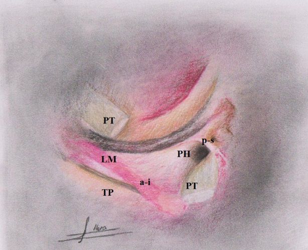
Figure 1. Schematic representation from superior view of top of right tibia
1. Anterior cruciate ligament; 2. Posterior cruciate ligament; 3. Popliteus tendon; 4. Arcuate ligament; 5. Posterior 1/3 lateral
capsular ligament; 6. Lateral meniscus; 7. Medial meniscus.
Eivazi Gh M, et al. Role of the Popliteal Fossa in Knee Problems. JMR. 2020; 14(3):131-140.
132Journal of
Modern Rehabilitation July 2020, Volume 14, Number 3
Figure 2. The anterio-inferior popliteomeniscal fascicle
A-I: Makes the floor of the popliteal hiatus and the posterior-superior popliteomeniscal fascicle; P-S: makes the roof of the
popliteal hiatus. Popliteus Tendon (PT) had been cut and reflected to illustrate clearly the role of 2 popliteomeniscal fascicles.
LM: Lateral Meniscus; TP: Tibial Plateau; PH: Popliteal Hiatus. Redrawn after Diamantopoulos et al. [10].
the roof of popliteus fossa. It seems that anterior-inferior able peripheral vascular near the popliteus tendon. This
popliteomeniscal fascicles are stronger and thicker than part of lateral meniscus gains nutrition via a ventral ped-
postero-superior popliteomeniscal fascicles (Figure 2). icle of synovial tissue which is provided through circular
According to the report of Suganuma et al., abnormal micro-vascular to the lateral septum of meniscus. This
popliteomeniscal fibers increase the risk of locking and area is also feed by the micro vascular branches which
recurrent subluxation of the lateral meniscus [17]. are carried by the popliteomeniscal fascicles [22, 23].
These fascicles are composed of three layers: a dense
Ullrich et al. and Terry and Laprade have also reported collagen layer, a vascular layer and a capsular or syno-
the existence of third popliteomeniscal fascicle in their vial layer. These fascicles are involved in stabilization
anatomic studies which is connected to the posterior of lateral meniscus furthermore they help the popliteus
horn of the lateral meniscus. This fascicle is called “pos- tendon in retracting the lateral meniscus [21].
terio-inferior popliteomeniscal” [18-20]. Sussmann et
al. believed that the fascicle’s task is to provide a vascu- Based on their view, this area of the meniscus is known
lar supply to the lateral meniscus in the area which there as the “bare area”. Bare area is limited by two tendons
is no capsular connectivity. In addition these researchers of the popliteomeniscal ligament; upper tendon (supero-
showed that the structural relationship between the pop- medial) which is connected to the upper edge of lateral
liteus tendon and lateral meniscus can help in stabiliza- meniscus and the lower tendon (infero-lateral) which
tion of the area [21]. is joined to the popliteofibular ligament, posterior horn
of meniscus and coronary ligament [22, 23]. Jae-hynk
According to the report of Arnoczky and Warren the
posterolateral part of the lateral meniscus has no perme-
Eivazi Gh M, et al. Role of the Popliteal Fossa in Knee Problems. JMR. 2020; 14(3):131-140.
133Journal of
July 2020, Volume 14, Number 3 Modern Rehabilitation
Yang et al. reported that the bare part of the lateral me- 3. Mechanism of Popliteomeniscal Fibers Injuries
niscus in their study samples was more than 1.5 cm [24].
Most likely a combination of the hyper-extension and
Simonian et al. illustrated that the functions of popliteo- varus force exertion is involved in damage of the pos-
meniscal ligaments are as the stabilizer of the lateral me- terolateral structures of the knee. Other mechanism is
niscus and in the case of ligament rupture, the mobility of simultaneously exertion of force in the direction of tibial
the lateral meniscus would increase. They suggested that external rotation and hyperextension [31].
disturbances in the amount of mobility of the lateral me-
niscus with hypofunction, adherence and shortness of the Posterolateral injuries can occur in isolation but they
ligaments could lead to a disorder in function of the knee. are often associated with the Posterior Cruciate Ligament
The differences in the form of attachment and the amount (PCL) and Anterior Cruciate Ligament (ACL) injuries or
of thickness of postero-superior and antero-inferior pop- both [31]. Sport injuries, car accidents and falls are the
liteomeniscal fascicles may explain their different roles most common causes of injures [31]. The presence of
in making the lateral meniscus stability. Anterio-inferior Trigger Points (TrPs) in the popliteus muscle may also be
fascicles which attached to the base of the meniscus com- an aggravating factor which increases in playing football,
pared with postero-superior fascicles are narrower and running, twisting, and slipping especially on ski slopes.
may play the more significant role in the lateral meniscus TrPs in this muscle often results from or are perpetuated
stability. Therefore, the amount of the lateral meniscus dis- by an acute or chronic overuse of the muscle, excessive
placement is controlled by anterio-inferior fascicles more foot subtalar joint pronation, wearing high- heeled shoes,
than postero-superior fascicles [25]. and a torn posterior cruciate ligament. TrPs in the poplit-
eus may produce knee pain when crouching or walking/
Sta and Birrer [26] recommended that three types of running downhill or down- stairs, or decreased lateral ro-
popliteomeniscal fascicles (antero-inferior, postero-su- tation or extension of the leg at the knee joint.
perior and postero-inferior) in combination with poplite-
us tendon form a ring-like peripheral connection with the The referral patterns of popliteus TrPs must be distin-
lateral meniscus in the popliteus fossa zone. At least one guished from the referral patterns of TrPs in the gastrocne-
part of popliteus complex has less movement through mius, soleus, plantaris, hamstrings, and gluteus minimus.
the popliteomeniscal fascicles compared with other ar- TrPs in this muscle are often incorrectly assessed as Bak-
eas of lateral meniscus which is inside of the popliteus er’s cyst, instability of the knee joint, popliteus tendinitis/
fossa so this enables additional stability. The necessity of tenosynovitis, meniscal tear, or torn plantris muscle. The
normal and safe static and dynamic connection between popliteus belly and its proximal attachment should be pal-
lateral meniscus and popliteus tendon is the role of pop- pated for examination of trigger points [4, 32, 33]. The
liteomeniscal fascicles in control of lateral meniscus mo- popliteomeniscal ligaments maybe particularly overload-
tion during knee flexion and extension [26-30]. ed with the braking forward movement of the femur on
tibia during the rotation of the trunk during weight bearing
Due to Suganuma et al. there is not a specific definition on the semi-flexed knee on the rotation side. Trauma or
for degree of normality of the popliteomeniscal fiber in strain which causes PCL injury may also cause overload
Magnetic Resonance Imaging (MRI) and arthroscopy. and strain of these ligaments [4].
These researchers also speculated that repeated submax-
imal traumas created by the knee varus along with me- Brody et al. have reported a correlation between exces-
dial unicompartmental knee osteoarthritis and the aging sive foot pronation in weight bearing activity, and the
process, may affect its structure and physical properties aggravation of damage and signs of popliteus tendinitis
[17]. According to these researchers the existence of any [34]. Additional pronated foot causes the tibia to be in
abnormality may predispose the patient to the symptoms external rotation stressing the popliteus, so it could in-
of knee locking. Hence, they considered abnormality tensify trigger points of popliteus muscle. Therefore, any
of inferior popliteomeniscal fibers as a precondition for abnormalities or imbalances in the lower extremity, leg
knee instability. According to their opinion insufficiency length discrepancy and biomechanical imbalance should
in popliteomeniscal fibers can be the result of the lateral be considered and be corrected by using orthotic devices
meniscus locking or recurrent subluxation of the lateral [34]. Holmes et al. have reported that leg length discrep-
meniscus and not vice versa [17]. ancy more than a quarter of an inch (6.4 mm) has clinical
significance [35].
Eivazi Gh M, et al. Role of the Popliteal Fossa in Knee Problems. JMR. 2020; 14(3):131-140.
134Journal of
Modern Rehabilitation July 2020, Volume 14, Number 3
4. Diagnosis of Popliteomeniscals Fiber Lesions of the femur is identified and marked first. By using the
pads of fingers, the insertion of popliteus muscle with
Signs using the landmark of the lateral epicondyle and palpat-
ing about 0.5 cm distal and 0.5 cm anterior toward this
Most of the patients with the popliteus complex disor- prominence is palpable. The tendon inserts between the
ders are suffering from pain in the posterior aspect of the Lateral Collateral Ligament (LCL) and capsule and it is
knee during bending, running, hiking and specially walk- rarely separable from other adjunct structures. There-
ing down and up the stairs. Patients who suffer from trig- fore, there is a possibility of localization by using slight
ger point’s pain in the popliteus muscle rarely complain active rhythmic flexion and extension of the knee. In this
of knee pain during the night. They have slight limitation case, the muscle contraction can be felt under the palpat-
in the range of flexion and extension of the knee or some- ing finger. Another structure which could be identified
times there is weakness in medial rotation of the leg [4]. for more localization is the head of fibula.
Examination The anterior, posterior and proximal part of the head of
fibula using a vertical palpation technique is identified. It
Distinguishing between single popliteomeniscal dam- is necessary to identify the top head of fibula which is the
age and other knee injuries are often very difficult. The location of LCL insertion and part of the biceps tendon.
patients who are often referred to physiotherapy clinics The knee should be in the Patrick test position for exact
have chronic disorders. Disease presence may be associ- palpation [38]. It means that the leg must be in flexion,
ated with joint line pain of the knee near the medial or abduction and lateral rotation and be over the other leg.
lateral aspects or may be associated with instability [7, With a valgus force on the joint the exerted tension on
31]. This instability is often associated with hyper ex- LCL increases and the prominence becomes bigger. The
tension problems and will be intensified while walking third structure which should be used for localization and
down and up the stairs. Instability is also determined by palpating of popliteomeniscal ligaments is the lateral joint
external rotation that causes posterior displacement of line of the knee and lateral meniscus [38].We can palpate
the lateral tibial plateau. This movement is called ‘pos- the Fibular Collateral Ligament (FCL) and then touch a
terolateral rotatory instability by Hughston et al. [7, 31]. localized tenderness exactly posterior to the FCL along
with the tendon and its junction with lateral joint line.
Observation
The knee must be examined specially in the posterolat- 5. The Method of Slight Knee Flexion Diag-
eral aspect for any swelling, soreness, bruising and ten- nosis (Bent Knee)
derness [31, 36]. Based on the experience of the authors,
the patients are more sensitive to deep palpation in this For this purpose, the patient lies supine on the table and
area rather than having tenderness. keeps his/her knees in the maximally pulled and extend-
ed position. The therapist stands at the end of the table
Patient’s gait near the patient’s feet. He/she puts both palms of hands
on both patellas on the way that the fingers tip is cranial.
In acute injuries when the patient has an antalgic gate the Pressure of both hands should be gentle and should be
examination of the patient is not necessary. However, in avoided from applying heavy pressure on the both knees.
chronic cases the examination of gait and alignment of the In most cases, slight flexion of the knee can be detected
lower extremity is considered as an important part of the by using height difference of both hand (Figure 4A).
examination. Some patients may have a slight knee flexion
during walking which usually cannot be detected without 6. The Diagnosis Test of Limitation in Knee
a careful examination. The antalgic position of semi-flex- Flexion at the end of Flexion in Prone Posi-
ion during walking may be for avoiding the pain, the oc- tion
currence of instability in the knee or further stress on the
joint and joint capsule in the hyper extension position [37]. The patient lies in the prone position. A pillow is
placed under the pelvis to avoid low back hyper ex-
Palpation tension. Then the patient is asked to bend his/her both
knees in flexion simultaneously. The therapist who is
The simple way to palpate popliteomeniscal ligaments standing at the end of table puts his/her both hands
is that the patient sits in the position in which the ankle is palm on the posterior aspect of heels. The hands are
on the opposite knee (Figure 3). The Lateral epicondyle
Eivazi Gh M, et al. Role of the Popliteal Fossa in Knee Problems. JMR. 2020; 14(3):131-140.
135Journal of
July 2020, Volume 14, Number 3 Modern Rehabilitation
Figure 3. Schematic representation of manner of palpation
1. Lateral Collateral Ligament (LCL); and 2. Popliteus tendon as immerged in the front of anterior aspect of LCL in the figure of
4-position. Redrawn based on Reichert B, Stelzenmueller W.: Palpation Techniques: Surface Anatomy for Physical Therapists:
Thieme; 2011 [38].
placed on the back of heels so that the dorsum of hand However, we cannot be sure that lack of resolution of
is cranial. The slight limitation of flexion of both knees popliteomeniscal fascicle in MRI is the result of its rup-
could be realized (Figure 4B). ture or edema [39].
7. Magnetic Resonance Imaging (MRI) Pavlov and Goldman argue that edema and adhesion
may be the cause of difficulty in observation of popliteo-
Superior popliteomeniscal fascicle may be abnormal meniscal fascicles in knee arthrography [40]. The de-
because of rupture or the lack of it in MRI imaging. tection of abnormally in the fascicles may indicate the
A B
Figure 4. A simple technique for diagnosing of mild knee limitation
A: Created following knee problem due to some structural injuries around the knee including popliteus tendon complexes
(supine); B: Prone. In this figure, the therapist’s hands are not shown to better show the limitation of the right knee flexion.
Eivazi Gh M, et al. Role of the Popliteal Fossa in Knee Problems. JMR. 2020; 14(3):131-140.
136Journal of
Modern Rehabilitation July 2020, Volume 14, Number 3
existence of rupture in the lateral meniscus. It was also of them [31, 46]. Considering the fact that the majority
claimed that abnormal superior popliteomeniscal fasci- of referring cases are non-surgical cases, therefore this
cles proposed as a cause for rupture of lateral meniscus, paper focuses on non-surgical procedures. In this section
however, it is not pathogenomic [41, 42]. conventional conservative methods which are applicable
in manual therapy are briefly described:
8. Popliteomeniscal Ligament Involvement
11. Dimensional Myofascial Release Tech-
In the case of strain or injury of Popliteomeniscal liga- nique
ment, it is possible to palpate and friction massaged the
ligament in the interface of popliteus tendon and lateral Giammatteo has explained the therapeutic procedures
joint line of the knee in the posterior aspect of the fibular as follows:
collateral ligament (FCL). For this purpose while ankle
is on the opposite knee, the index finger is placed under 1- The patient lies in prone position. Both hands of
the middle finger and the area is friction massaged trans- therapist are placed on the belly of popliteus muscle and
versely with a short-acting reciprocating motion [43]. To lateral condyle of femur in the vicinity of each other.
find the exact position of ligaments the patient is asked Shift the muscle in three directions (and in three planes)
to resist against to medial rotation force of the hand on and displace in indirect manner.
the distal side of the foot. The exact position of injury is
easily palpable with this functional test [43]. 2- First plane: move the muscle in inferior and superior
direction. Move to the easer side.
9. Differential Diagnosis
3- Second plane: “stack” motion of second plane to it.
The patients who present the complaints of the pop- Move the muscle in medial or lateral direction. Move to
liteomeniscal fiber lesions, are likely to have one of the the easer side.
four conditions in acute phase: popliteal vein thrombo-
sis, antero-medial and postero-lateral instability, avul- 4- Third plane: “add” the third plane now. Move the
sion of popliteus tendon, and meniscus or posterior muscle in a clockwise direction and vice versa. Move to
capsule rupture of knee joint. Also, the therapist should the easer side.
consider the Baker’s cyst, biceps femoris tendinitis, ilio-
tibial band friction syndrome, and peroneal nerve entrap- 5- Release: maintain the created fulcrum in step1-4.
ment as the differential diagnosis of the popliteomenis- When firmness of tissue is unwind, the motion is hap-
cals fiber lesions in chronic conditions. Therefore, this pen in the body’s internal environment. Keep your hands
injury is complex and has a wide differential diagnosis. fixed furthermore and avoid release fulcrum [30].
An awareness of the certain patterns and a good exami-
nation can help the therapist identify the dysfunction and 12. Articular Myofascial Release Technique
its underlying causes [4, 44]. in Popliteomeniscal Ligaments
10. Treatment of Postero-Lateral Corner Injuries This technique is used in the hypo mobility of knee joint
in flexion or extension. Put both hands on both sides of the
In posterolateral corner of the knee injuries it is not an knee joint line and its posterolateral aspect. Hands must
easy task to decide which patient should be treated by be put next to each other to keep control over the two
surgery and which one with conservative methods [45]. joint surfaces. Avoid distraction or approximation of joint
If the popliteomeniscal ligament injuries are in the form surfaces. Move the hands in opposite directions in three
of the single type and have sustained a low grade injury, planes of sagittal, coronal and horizontal. In sagittal plane,
it may respond to conservative treatment [31]. Accord- move on joint surface, superior-inferior direction. Then
ing to some studies, non-surgical treatment of the mild move the joint surface in opposite direction. Move a joint
to moderate damage in the posterolateral corner of the surface in medial rotation direction and the other joint
knee have had a good outcome but in severe damages surface in lateral rotation direction (medial-lateral). Move
the outcome and result are not very satisfactory [12, 31]. the joint surface in adduction direction and the other joint
surface in abduction direction in coronal plane [47].
According to Davies et al., single injury of the postero-
lateral corner of the knee is rare and almost all their in-
juries were associated with ACL or PCL injuries or both
Eivazi Gh M, et al. Role of the Popliteal Fossa in Knee Problems. JMR. 2020; 14(3):131-140.
137Journal of
July 2020, Volume 14, Number 3 Modern Rehabilitation
13. Discussion Ethical Considerations
According to the arthroscopic study of staübli and Birrer, Compliance with ethical guidelines
popliteus tendon and poplitomeniscal fascicles which are
connected to the lateral meniscus form a functional unit All ethical principles are considered in this article. The
that participates in the controlling of valgus and varus an- participants were informed about the purpose of the
gulation [26]. When a heavy trauma occurs to the knee research and its implementation stages; they were also
area, secondary structures are sacrificed first to avoid the assured about the confidentiality of their information;
main structures lesions. It means that these structures act moreover, they were free to leave the study whenever
as a kind of buffer or safety valve. They believe that the they wished, and if desired, the research results would be
extent of structural damages in popliteomeniscal fascicles available to them.
and their effect on controlling of the meniscal movement
are determinant factor in posterolateral pain of the knee. Funding
The rupture and complete damage of popliteomeniscal
This research did not receive any specific grant from fund-
and popliteofibular cause the loss of their inhibitory and
ing agencies in the public, commercial, or non- profit sectors.
controlling function. This rupture is sometimes associated
with popliteus tendon downward movement and medial
Authors' contributions
displacement of lateral meniscus [26].
All authors were equally contributed in preparing this
Garrick and Webb believe that excessive tiredness of
article.
quadriceps is the underlying cause of popliteus muscle
components and elements involvements [48]. The au- Conflict of interest
thors believe this is the same phenomenon that occurs
while walking with bent knee position. It means that The authors declared no conflict of interest.
quadriceps muscle is engaged in continues activity in
all walking phases because of physiological unlocking Acknowledgements
of the knee. This phenomenon could lead to excessive
tiredness of quadriceps and finally could lead to the insuf- We would like to express our special thanks to all au-
ficiency of popliteus muscle and its components [40]. thors who have contributed in this article.
Physical therapy for the postero-lateral corner injuries
involves a thorough subjective and objective evaluation
and assessment of the entire lower extremity from the
hip to the foot. Then the physical therapist can prescribe References
the right treatments- including manual therapy, exercis-
es, and modalities- to help decrease the knee pain and [1] Drake R, Vogl W, Mitchell A. Gray’s anatomy for students
Elsevier. London: Churchill Livingstone; 2005.
improve the overall mobility and gait.
[2] Gray H, Standring S. Gray’s anatomy: The anatomical basis
14. Conclusions of clinical practice. London: Churchill Livingstone; 2008.
[3] Amonoo-Kuofi H. Morphology of muscle spindles in the hu-
The damage of posterolateral structures of the knee is man popliteus muscle. Cells Tissues Organs. 1989; 134(1):48-53.
common and often they are associated with ACL and [DOI:10.1159/000146732] [PMID]
PCL ruptures. The damage of individual and singular [4] Simons DG, Travell J, Simons LS. Myofascial pain and dys-
posterolateral aspects of the knee is rare and uncommon. function: The trigger point manual. volume 1. 2nd edition.
Philadelphia: Williams & Wilkins; 1999. [DOI:10.1016/S0161-
According to clinical experiences of the authors the aim
4754(99)70079-5]
of the conservative treatment must be the treatment of
all elements and components in which there is probabil- [5] Haslam P, Bickerstaff D. Postero-lateral rotatory instabil-
ity. Current Orthopaedics. 2007; 21(6):451-6. [DOI:10.1016/j.
ity of lesion in this zone. In this paper we have focused cuor.2007.07.008]
on the constituent elements of popliteus fossa due to the
importance of the subject. The patient could return to [6] Djian P. Posterolateral knee reconstruction (Ligamentoplas-
ties postéro-latérales du genou) [French]. In: Conférences
normal functional activities after treatment. d’enseignement 2014: Amsterdom: Elsevier; 2014. pp.225-39.
[DOI:10.1016/B978-2-294-74506-5.00018-7]
Eivazi Gh M, et al. Role of the Popliteal Fossa in Knee Problems. JMR. 2020; 14(3):131-140.
138Journal of
Modern Rehabilitation July 2020, Volume 14, Number 3
[7] Hughston JC, Jacobson K. Chronic posterolateral rotatory [20] Veltri DM, Deng XH, Torzilli PA, Maynard MJ, Warren RF.
instability of the knee. Journal of Bone and Joint Surgery: JBJS. The role of the popliteofibular ligament in stability of the hu-
1985; 67(3):351-9. [DOI:10.2106/00004623-198567030-00001] man knee: A biomechanical study. The American Journal of
Sports Medicine. 1996; 24(1):19-27. [DOI:10.1177/0363546596
[8] LaPrade RF, Wentorf FA, Fritts H, Gundry C, Hightower 02400105] [PMID]
CD. A prospective magnetic resonance imaging study of the
incidence of posterolateral and multiple ligament injuries in [21] Sussmann PS, Simonian PT, Wickiewicz TL, Warren RF.
acute knee injuries presenting with a hemarthrosis. Arthros- Development of the popliteomeniscal fasciculi in the fe-
copy: The Journal of Arthroscopic & Related Surgery. 2007; tal human knee joint. Arthroscopy: The Journal of Arthro-
23(12):1341-7. [DOI:10.1016/j.arthro.2007.07.024] [PMID] scopic & Related Surgery. 2001; 17(1):14-8. [DOI:10.1053/
jars.2001.19653] [PMID]
[9] LaPrade RF, Terry GC. Injuries to the posterolateral aspect of
the knee: association of anatomic injury patterns with clinical [22] Arnoczky SP, Warren RF. Microvasculature of the human
instability. The American Journal of Sports Medicine. 1997; meniscus. The American Journal of Sports Medicine. 1982;
25(4):433-8. [DOI:10.1177/036354659702500403] [PMID] 10(2):90-5. [DOI:10.1177/036354658201000205] [PMID]
[10] Diamantopoulos A, Tokis A, Tzurbakis M, Patsopoulos I, [23] Arnoczky SP, Warren RF. The microvasculature of the
Georgoulis A. The posterolateral corner of the knee: Evalua- meniscus and its response to injury: An experimental study
tion under microsurgical dissection. Arthroscopy: The Jour- in the dog. The American Journal of Sports Medicine. 1983;
nal of Arthroscopic & Related Surgery. 2005; 21(7):826-33. 11(3):131-41. [DOI:10.1177/036354658301100305] [PMID]
[DOI:10.1016/j.arthro.2005.03.021] [PMID]
[24] Yang J-H, Jeong H-I, Kim T-S, Park S-C, Yoon J-R. The
[11] Brandt K D, Radin E L, Dieppe P A, van de Putte L. Yet management of the popliteus hiatus during lateral meniscal
more evidence that osteoarthritis is not a cartilage disease. transplantation. The Knee. 2012; 19(6):959-61. [DOI:10.1016/j.
Annals of the Rheumatic Diseases. 2006; 65(10):1261-4. knee.2012.04.007] [PMID]
[DOI:10.1136/ard.2006.058347] [PMID] [PMCID]
[25] Simonian PT, Sussmann PS, Wickiewicz TL, Potter HG,
[12] Kannus P. Nonoperative treatment of grade II and III van Trommel M, Weiland-Holland S, et al. Popliteomenis-
sprains of the lateral ligament compartment of the knee. The cal fasciculi and the unstable lateral meniscus: Clinical cor-
American Journal of Sports Medicine. 1989; 17(1):83-8. [DOI:1 relation and magnetic resonance diagnosis. Arthroscopy: The
0.1177/036354658901700114] [PMID] Journal of Arthroscopic & Related Surgery. 1997; 13(5):590-6.
[DOI:10.1016/S0749-8063(97)90185-7]
[13] Burr D, Radin E. Trauma as a factor in the initiation of os-
teoarthritis. Cartilage changes in osteoarthritis: Indiana Uni- [26] Sta H-U, Birrer S. The popliteus tendon and its fascicles at
versity. School of Medicine Press, Indianapolis, IN; 1990. pp. the popliteal hiatus: Gross anatomy and functional arthro-
73-80. scopic evaluation with and without anterior cruciate liga-
ment deficiency. Arthroscopy: The Journal of Arthroscopic
[14] Kannus P. Long-term results of conservatively treated & Related Surgery. 1990; 6(3):209-20. [DOI:10.1016/0749-
medial collateral ligament injuries of the knee joint. Clini- 8063(90)90077-Q]
cal Orthopaedics and Related Research. 1988; (226):103-12.
[DOI:10.1097/00003086-198801000-00015] [27] Hughston J, Andrews J, Cross M, Moschi A. Classification
of knee ligament instabilities. Part II. The lateral. The Journal
[15] Gale D, Chaisson C, Totterman S, Schwartz R, Gale M, Fel- of Bone and Joint Surgery. American Volume. 1976; 58:173-9.
son D. Meniscal subluxation: Association with osteoarthritis [DOI:10.2106/00004623-197658020-00002] [PMID]
and joint space narrowing. Osteoarthritis and Cartilage. 1999;
7(6):526-32. [DOI:10.1053/joca.1999.0256] [PMID] [28] Bousquet G, Charmion L, Passot J, Girardin P, Relave M.
Stabilisation du condyle externe du genou dans les laxités
[16] Grood ES, Stowers SF, Noyes FR. Limits of movement in antérieures chroniques: Importance du muscle poplité [Stabi-
the human knee: Effect of sectioning the posterior cruciate lization of the external condyle of the knee in chronic anterior
ligament and posterolateral structures. Journal of Bone and laxity: Importance of the popliteal muscle (French)]. Revue de
Joint Surgery: JBJS. 1988; 70(1):88-97. [DOI:10.2106/00004623- Chirurgie Orthopédique et Réparatrice de L’appareil Moteur.
198870010-00014] 1986; 72(6):427-34. [PMID]
[17] Suganuma J, Mochizuki R, Inoue Y, Yamabe E, Ueda Y, [29] Shino K, Horibe S, Ono K. The voluntarily evoked postero-
Kanauchi T. Magnetic resonance imaging and arthroscopic lateral drawer sign in the knee with posterolateral instability.
findings of the popliteomeniscal fascicles with and with- Clinical Orthopaedics and Related Research. 1987; (215):179-86.
out recurrent subluxation of the lateral meniscus. Arthros- [DOI:10.1097/00003086-198702000-00026]
copy: The Journal of Arthroscopic & Related Surgery. 2012;
28(4):507-16. [DOI:10.1016/j.arthro.2011.08.311] [PMID] [30] Staubli H, Jakob R. Posterior instability of the knee near ex-
tension. A clinical and stress radiographic analysis of acute
[18] Terry GC, LaPrade RF. The posterolateral aspect of the injuries of the posterior cruciate ligament. The Journal of
knee: Anatomy and surgical approach. The American Journal Bone and Joint Surgery. British Volume. 1990; 72(2):225-30.
of Sports Medicine. 1996; 24(6):732-9. [DOI:10.1177/03635465 [DOI:10.1302/0301-620X.72B2.2312560] [PMID]
9602400606] [PMID]
[31] Davies H, Unwin A, Aichroth P. The posterolateral cor-
[19] Ullrich K, Krudwig WK, Witzel U. Posterolateral aspect and ner of the knee: Anatomy, biomechanics and management
stability of the knee joint. I. Anatomy and function of the pop- of injuries. Injury. 2004; 35(1):68-75. [DOI:10.1016/S0020-
liteus muscle-tendon unit: An anatomical and biomechani- 1383(03)00094-9]
cal study. Knee Surgery, Sports Traumatology, Arthroscopy.
2002; 10(2):86-90. [DOI:10.1007/s00167-001-0268-5] [PMID]
Eivazi Gh M, et al. Role of the Popliteal Fossa in Knee Problems. JMR. 2020; 14(3):131-140.
139Journal of
July 2020, Volume 14, Number 3 Modern Rehabilitation
[32] Woodley, Mercer SR. Anatomy in practice: The Popliteus
muscle. New Zealand Journal of Physiotherapy. Era 2010-
2018; 34(1):25-9.
[33] Muscolino JE. The muscle and bone palpation manual with
trigger points, referral patterns, and stretching, 1st edition.
Mosby, Elsevier Health Sciences; 2008.
[34] Brody D. Techniques in the evaluation and treatment of
the injured runner. The Orthopedic Clinics of North America.
1982; 13(3):541-58. [PMID]
[35] Holmes JC, Pruitt AL, Whalen NJ. Iliotibial band syndrome
in cyclists. The American Journal of Sports Medicine. 1993;
21(3):419-24. [DOI:10.1177/036354659302100316] [PMID]
[36] Delee JC, Riley MB, Rockwood Jr CA. Acute posterolat-
eral rotatory instability of the knee. The American Journal of
Sports Medicine. 1983; 11(4):199-207. [DOI:10.1177/03635465
8301100403] [PMID]
[37] Flynn JM, Wiesel SW. Operative techniques in pediatric or-
thopaedics: Philadelphia: Lippincott Williams & Wilkins; 2010.
[38] Reichert B, Stelzenmueller W. Palpation Techniques: Sur-
face anatomy for physical therapists, 1st edition. New York, :
Thieme Medical Publishers; 2011.
[39] De Smet AA, Asinger DA, Johnson RL. Abnormal superior
popliteomeniscal fascicle and posterior pericapsular edema:
Indirect MR imaging signs of a lateral meniscal tear. American
Journal of Roentgenology. 2001; 176(1):63-6. [DOI:10.2214/
ajr.176.1.1760063] [PMID]
[40] Pavlov H, Goldman AB. The popliteus bursa: An indica-
tor of subtle pathology. American Journal of Roentgenology.
1980; 134(2):313-21. [DOI:10.2214/ajr.134.2.313] [PMID]
[41] Johnson RL, De Smet AA. MR visualization of the poplit-
eomeniscal fascicles. Skeletal Radiology. 1999; 28(10):561-6.
[DOI:10.1007/s002560050619] [PMID]
[42] Dawson D-S. Estimating and comparing proportions. Basic
and clinical biostatics 2nd edition. Norwalk, Conn.: Appleton
and Lange. 1990:142-60.
[43] Hammer WI. Functional soft-tissue examination and treat-
ment by manual methods. Massachusetts: Jones & Bartlett
Learning; 2007.
[44] Veltri D, Warren R. Anatomy, biomechanics, and physical
findings in posterolateral knee instability. Clinics in Sports
Medicine. 1994; 13(3):599-614. [PMID]
[45] Suganuma J, Inoue Y, Tani H, Sugiki T, Sassa T, Shibata
R. Reconstruction of the popliteomeniscal fascicles for treat-
ment of recurrent subluxation of the lateral meniscus. Ar-
throscopy Techniques. 2017; 6(2):e283-e90. [DOI:10.1016/j.
eats.2016.09.034] [PMID] [PMCID]
[46] Fanelli GC, Larson RV. Practical management of postero-
lateral instability of the knee. Arthroscopy: The Journal of Ar-
throscopic & Related Surgery. 2002; 18(2):1-8. [DOI:10.1053/
jars.2002.31779] [PMID]
[47] Weiselfish-Giammatteo S. Integrative manual therapy: For
the upper and lower extremities, Revised edition. Berkeley,
CA: North Atlantic Books; 1998.
[48] Webb DR. Sports injuries: Diagnosis and management.
Philadelphia: Saunders; 1999.
Eivazi Gh M, et al. Role of the Popliteal Fossa in Knee Problems. JMR. 2020; 14(3):131-140.
140You can also read