Homeobox genes in axolotl lateral line placodes and neuromasts
←
→
Page content transcription
If your browser does not render page correctly, please read the page content below
Dev Genes Evol (1997) 207:287–295                                                                         © Springer-Verlag 1997

    O R I G I NA L A RT I C L E

&roles:Brian D. Metscher · R. Glenn Northcutt
       David M. Gardiner · Susan V. Bryant

Homeobox genes in axolotl lateral line placodes and neuromasts

&misc:Received: 2 April 1997 / Accepted: 2 July 1997

&p.1:Abstract Gene expression has been studied in consider-         &kwd:Key words Sense organ embryology · Ambystoma
     able detail in the developing vertebrate brain, neural              embryology · Homeobox genes · Vertebrate genetics ·
     crest, and some placode-derived organs. As a further in-            Evolution&bdy:
     vestigation of vertebrate head morphogenesis, expression
     patterns of several homeobox-containing genes were ex-
     amined using whole-mount in situ hybridization in a sen-       Introduction
     sory system primitive for the vertebrate subphylum: the
     axolotl lateral lines and the placodes from which they         There is now an extensive literature on the involvement
     develop. Axolotl Msx-2 and Dlx-3 are expressed in all of       of homeobox-containing genes in the morphogenesis of
     the lateral line placodes. Both genes are expressed            the vertebrate head and of organs specific to vertebrates.
     throughout development of the lateral line system and          The embryonic central nervous system, neural crest mes-
     their expression continues in the fully developed neuro-       enchyme, and tissues in later organogenesis are known to
     masts. Expression within support cells is highly polar-        express genes of the Hox, Msx and Dlx families during
     ized. In contrast to most other observations of Msx genes      morphogenesis, and genes of all of these groups have
     in vertebrate organogenesis, expression of Msx-2 in de-        been characterized in a number of animal lineages (Ak-
     veloping lateral line organs is exclusively epithelial and     imenko et al. 1994, 1995; Holland 1992; Krumlauf 1994;
     is not associated with epithelial-mesenchymal interac-         Ma et al. 1996; Robinson and Mahon 1994). The phylo-
     tions. A Hox-complex gene, Hoxb-3, is shown to be ex-          genetic distribution of conserved sequences within and
     pressed in the embryonic hindbrain and in a lateral line       outside of the homeobox suggests that these genes may
     placode at the same rostrocaudal level, but not in other       have functions important in animal development general-
     placodes nor in mature lateral line organs. A Hox gene of      ly, and the similarities of expression patterns in various
     a separate paralog group, Hoxa-4, is expressed in a more       vertebrates may be indicative of some developmental
     posterior hindbrain domain in the embryo, but is not ex-       roles retained in the different lineages from their com-
     pressed in the lateral line placode at that rostrocaudal       mon chordate ancestor (Holland et al. 1994; Sharman
     level. These data provide the first test of the hypothesis     and Holland 1996). Thus these same genes might also be
     that the neurogenic placodes develop in two rostrocaudal       expected to be associated with the development of a sen-
     series aligned with the rhombomeric segments and pat-          sory system primitive for the vertebrates, such as lateral
     terned by combinations of Hox genes in parallel with the       lines (Northcutt 1989).
     central nervous system.                                            Genes of the Hox complex are known to be instrumen-
                                                                    tal in patterning two major constituents of the vertebrate
                                                                    embryo, the central nervous system and the neural crest
Edited by R. Elinson                                                (Godsave et al. 1994; Keynes and Krumlauf 1994). The
                                                                    lateral line system develops from a third component of the
B.D. Metscher (✉)1 · D.M. Gardiner · S.V. Bryant
Developmental Biology Center and Department of Developmental        vertebrate embryo, the ectodermal placodes (Northcutt
and Cell Biology, University of California, Irvine, CA 92697, USA   and Gans 1983; Northcutt et al. 1994; and see Fig. 1). The
R.G. Northcutt
                                                                    lateral line placodes develop in a dorsolateral series paral-
Dept. of Neurosciences, School of Medicine,                         lel to the main body axis, and it has been hypothesized
University of California, La Jolla, CA 92093, USA                   (Northcutt 1996) that the dorsolateral and ventrolateral
Present adress:                                                     placode series may be patterned by a mechanism similar
1Department of Palaeontology, The Natural History Museum,           to the Hox code described for the head and branchial re-
Cromwell Road, London SW7 5BD, UK&/fn-block:                        gions of amniote embryos (Hunt and Krumlauf 1992).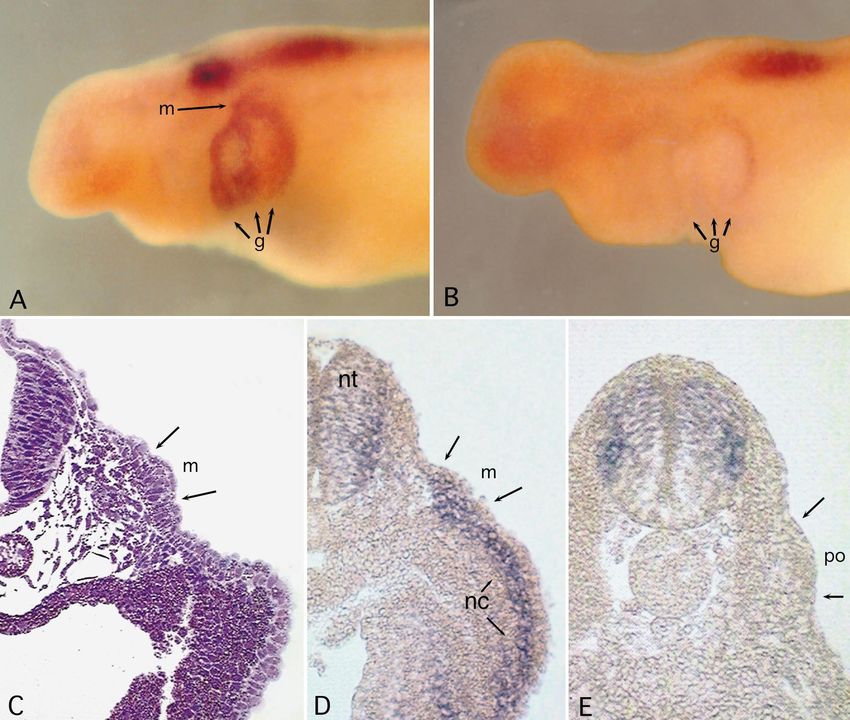
288

   While a critical test of this placode patterning hypoth-            Flat skin mounts of whole-mount in situ hybridization specimens
esis will ultimately require extensive investigation of nu-
                                                                       After fixation overnight in 10% formalin (see above), whole-
merous Hox genes in the developing ectodermal pla-                     mount specimens were dehydrated to methanol to harden the tis-
codes, the data presented here offer the first evidence for            sues and render the stain bluer. The head was removed and bisect-
Hox gene expression associated with lateral line pla-                  ed midsagittally. Internal tissues were then removed using sharp
codes. We examined expression of Hoxb-3 and Hoxa-4                     forceps. The remaining skin was rehydrated to PBS with 0.2% so-
                                                                       dium azide and any remaining tissues removed. The skin was
in the axolotl lateral line placodes, and we discuss some              transferred to a drop of AquaMount on a 24 × 30-mm cover glass
refinements of Northcutt’s hypothesis. We also describe                and spread flat using a pair of small (no. 505) artist’s brushes. A
the expression of two non-Hox homeobox genes, Msx-2                    second cover glass containing a drop of AquaMount was placed
and Dlx-3, in the placodes, and their continued expres-                over the specimen. Flat mounts were allowed to set overnight at
sion in mature lateral line sensory organs.                            room temperature and could then be examined and photographed
                                                                       under a compound microscope.

Materials and methods                                                  Sectioning of whole-mount in situ specimens

                                                                       For cryosectioning embryonic specimens, well-stained embryos
Specimens
                                                                       were fixed overnight as above and infiltrated with 7% sucrose in
                                                                       PBS, 15% sucrose in PBS, and then 15% sucrose and 7% gelatin
Axolotls, Ambystoma mexicanum, were spawned either at UCI or
                                                                       in PBS at 37–40°C. Specimens were embedded in Tissue Tek
at the Indiana University Axolotl Colony and were maintained at
                                                                       O.C.T. compound, frozen in a dry ice-ethanol bath, and sectioned
20–22°C in 20% Holtfreter’s solution. All embryos and early lar-
                                                                       at 30 µm (nominal) on a Slee HR cryostat at approximately
vae used for in situ hybridization were albinos staged according to
                                                                       –20°C. Slides were dried for several hours to overnight, then
the tables of Bordzilovkaya et al. (1989). Prior to fixation animals
                                                                       soaked in PBS and mounted in Aqua-Mount.
were anaesthetized in 0.1% MS222 (tricaine methanesulfonate;
                                                                           Later larval tissues were embedded and sectioned in polyester
Sigma).
                                                                       wax as described by Mullen et al. (1996). Blocks were sectioned
                                                                       at 8–12 µm using an AO 820 microtome in an ambient tempera-
                                                                       ture of about 20°C. Slides were dried overnight, dewaxed in two
RNA probes
                                                                       changes of 100% ethanol, and mounted in Aqua-Mount. Sections
                                                                       were counterstained in Safranin O (0.5% aqueous; Kiernan 1989).
Several homeobox-containing clones isolated from a screen of re-
generating axolotl limb blastema cDNA libraries by Gardiner et al.
(1995) were used as templates to prepare digoxygenin-labelled          Photography and computer enhancement of images
RNA probes for in situ hybridization. Information about the
probes is given in Table 1.                                            Embryos were photographed whole in PBS using FujiColor 200
    Probes were synthesized with digoxygenin-labelled uridine tri-     print film in a camera mounted on a Wild dissecting microscope.
phosphate (UTP) according to the manufacturer’s protocol (Boeh-        Flat skin mounts and thin sections were photographed with the
ringer), except that reagents were mixed at room temperature, not      same camera and film on a compound microscope using Nomarski
on ice, and probes were not hydrolyzed.                                (flat mounts) or Koehler illumination (thin sections).
                                                                           All photographs were digitized either by scanning a photo-
                                                                       graphic print on a flatbed scanner (HP ScanJet IICX), or by direct
Whole-mount in situ hybridization                                      image capture from a microscope-mounted color CCD (charge-
                                                                       coupled device) camera. Images were cropped, enhanced, labelled,
Our procedure for whole-mount in situ hybridization is as de-          and assembled using Adobe Photoshop.
scribed by Gardiner et al. (1995), with the following modifications
to optimize results for embryos and for lateral line organs. Embry-
os were treated with 10 µg/ml proteinase K at room temperature
for 3–15 min. Early larvae (B40–B43) were treated with 10 µg/ml        Results
proteinase K at 37°C for 20–30 minutes, and later larvae with
30 µg/ml at 37°C for 30–60 min. Before hybridization, probes           The complete series of ectodermal placodes from which
were diluted to approximately 10 µg/ml in hybridization buffer
and heated to 80°C for 2 min before adding to samples. Hybridiza-
                                                                       the lateral line system develops, the dorsolateral pla-
tion was carried out overnight at 54–65°C. Post-hybridization          codes, can first be recognized in axolotls starting at em-
washes were carried out at temperatures 4–5° higher than the hy-       bryonic stage 35 (about the end of somitogenesis;
bridization temperature.                                               Bordzilovskaya et al. 1989) as discrete epithelial thick-
    Tissues were blocked in blocking solution (maleic acid buffer      enings lateral to the hindbrain and most anterior portion
with 0.2% bovine serum albumin and 20% heat-inactivated sheep
serum) for at least 4 h at 4°C with gentle rotation. Samples were      of the spinal cord (Northcutt et al. 1994; Stone 1922; and
incubated in 500 µl blocking solution with antibody overnight at       see Fig. 1). The expression patterns of five homeobox-
4°C with gentle rotation. The next day tissues were washed 6 or        containing genes were examined in placode-stage em-
more times in MAB over at least 3 h, then twice in AP buffer           bryos: Hoxb-3, Hoxa-4, and Hoxa-5, Msx-2, and Dlx-3
(100 mM Tris, pH 9.5, 100 mM NaCl, 50 mM MgCl2), all at room
temperature. Samples were stained in AP buffer containing a chro-      (see Table 1). Hoxb-3 is expressed in the hindbrain and
mogenic AP substrate (340 µg/ml 4-Nitro blue tetrazolium               middle lateral line placode, which lies adjacent to the re-
chloride and 175 µg/ml 5-Bromo-4-chloro-3-indolyl-phosphate;           gion of hindbrain expression; Hoxa-4 is expressed in a
Boehringer) for 20 min to 2 h at room temperature. When staining       more posterior hindbrain domain, but not in the lateral
was complete, specimens were rinsed twice in PBS with 0.1%             line placode at that axial level (the posterior placode).
Tween-20 and fixed overnight in phosphate-buffered formalde-
hyde (3.7%), then stored at 4°C in phosphate-buffered saline           Subdivision of the hindbrain into rhombomeres has not
(PBS) with 0.2% sodium azide.                                          been described in axolotls, and classical studies of neu-
                                                                       romery in amphibians record that Ambystoma lacks the
289
Fig. 1 The six dorsolateral placodes in a stage 35 (Bordzilovs- Hox gene expression in early placodes
kaya et al. 1989) axolotl embryo, which give rise to the lateral line
system (five placodes in black) and the inner ear (gray; no longer a
placode by this stage). By stage 35, the anterodorsal and antero- We examined the expression of Hoxb-3, Hoxa-4 and
ventral placodes have begun to elongate into the sensory ridges Hoxa-5in placodal stage embryos, and all three genes
from which the supraorbital, suborbital, and mandibular lateral have hindbrain expression patterns consistent with those
line receptors will differentiate. The middle division of the posteri- observed in other vertebrates (Krumlauf 1994).
or placode has begun to migrate caudally to form the middle trunk
and tail line. (Modified from Northcutt et al. 1995) (Bar 0.5 mm,
The anterior boundary of Hoxb-3 expression in the
ad anterodorsal lateral line placode, av anteroventral lateral line neural tube is aligned with the rostral edge of the first
placode, b limb bud, e eye, g gill primordia, j lower jaw primordi- gill primordium and with the center of the otic vesicle
um, m middle lateral line placode, n olfactory organ, otv otic vesi- (presumably at the anterior edge of rhombomere 5, based
cle, pld dorsal division of the posterior lateral line placode, plm on embryonic anatomy of other hindbrains). By stage 35
middle division of the posterior lateral line placode, plv ventral di-
vision of the posterior lateral line placode, st supratemporal lateral and later, Hoxb-3 expression is also detected as three
line placode)&ig.c:/f dorsoventral bands, one in the center of each of the three
gill arch primordia (Fig. 2A). Based upon comparison of
whole-mount in situ hybridization specimens with serial
Table 1 RNA probes used for in situ hybridization&/tbl.c:& histological sections of axolotl embryos of the same
Genea Size Part of geneb stage (R. G. Northcutt, unpublished data), the locations
NvMsx-1 ~400 bp 5′ of these bands of expression correspond to the positions
Msx-2 ~800 bp 5′ H 3′ of migrating neural crest cells in the branchial arches. A
Dlx-3 ~500 bp 5′ (5) patch of Hoxb-3 expression is detected dorsal to the gill
Hoxb-3 ~1.6 kb 5′ H 3′
Hoxa-4 ~800 bp 5′ H 3′ primordia, at the position of the middle lateral line pla-
Hoxa-5 ~1.1 kb 5′ H 3′ code (Fig. 2A). Transverse sections through whole-
mount in situ specimens show that expression is lacking
a All probes were made from axolotl clones except NvMsx-1
b
in the cuboidal epithelium overlying the placode, but that
Relative to the homeobox; 5′ H 3′ means the homeobox plus 3′
and 5′ regions were included in the probe it is present in the columnar cells of the placode itself,
c From a newt (Notophthalmus) clone, kindly provided by J. and also in the mesenchyme (Fig. 2D). Sections of axo-
Brockes; see Crews et al. 1995 lotl embryos after in situ hybridization do not show indi-
d M. Carlson et al., in prep.
e See Mullen et al. 1996
vidual cells clearly. Thus the conclusion that the Hoxb-3
f See Gardiner et al. 1995 staining is in the middle placode is based upon: examina-
&/tbl.: tion of numerous whole-mount in situ hybridization
specimens similar to the ones shown in Fig. 2; upon the
boundary between rhombomeres 2 and 3 (von Kupffer complete series of sections from which the one shown in
1906, cited in Kuhlenbeck 1973). Given this uncertainty Fig. 2D was taken, and from which can be determined
about rhombomere numbers and identities in axolotls, the anteroposterior position of that section; and finally
other landmarks such as the otic vesicle and the gill arch upon comparison of those sections with a histological se-
primordia, rather than rhombomeres, were used to locate ries for the same embryonic stage, from which Fig. 2C
Hox gene expression boundaries in the dorsolateral pla- was taken (R. G. Northcutt, unpublished). The expres-
codes that develop adjacent to the hindbrain. sion pattern of Hoxb-3 contrasts with the pattern of Msx-290
Fig. 2 A Hoxb-3 in a stage 35 embryo. Expression in the hind- corresponds to a region of Hoxa-4 expression within the
brain is consistent with results for other vertebrates, with the ante- neural tube, and it migrates caudally during stages 35 to
rior border at the level of the otic vesicle. Hoxb-3 is expressed in
the middle lateral line placode (m), but not in the posterior pla- 40 through axial levels at which Hoxa-5 is expressed.
code. B Hoxa-4 in a stage 35 embryo. The posterior lateral line However, at stage 35 the posterior placode is negative for
placode is at the same rostrocaudal level as the Hoxa-4 expression Hoxa-4 expression (Fig. 2B, E), and as the dorsal, mid-
domain, but it does not show Hoxa-4 expression. C Transverse dle, and ventral divisions (see Fig. 3C) of the posterior
histological section through a stage 35 axolotl embryo, at the level
of the second gill arch primordium and the middle lateral line pla-
code. Note the columnar cells of the middle placode (m) in the in-
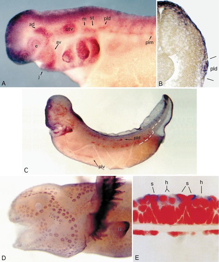
ner layer of skin epithelium and the migratory neural crest cells in Fig. 3 A Msx-2 expression in an axolotl embryo at stage 35, when
the mesenchyme of the gill arch primordium. (glycol methacrylate the lateral line placodes are present. Dlx-3 expression is similar at
section, cresyl violet). D Transverse cryosection (30 µm) through this stage. B Transverse cryosection through the embryo in A,
a specimen similar to A, showing Hoxb-3 in the neural tube (nt), showing Msx-2 expression in the posterior placode. Staining for
in neural crest mesenchyme (nc), and in the middle lateral line Msx-2 is in the columnar placodal cells of the inner epithelium as
placode (m). E Transverse cryosection through a specimen similar well as in the overlying epithelial layer. C Stage 38 embryo (gills
to B, showing expression of Hoxa-4 in the hindbrain but not in the trimmed) showing Msx-2 expression in the three migrating sec-
posterior placode (po). (For other abbreviations see Fig. 1)&ig.c:/f tions of the posterior placode, and in the neuromast primordia of
the trunk lateral lines. Dashed line marks the path of the middle
posterior lateral line. The middle section of the posterior placode
has already reached the tail tip by this stage. D Stage 43 feeding
2 (described below), and presumably also of Dlx-3, larva showing Msx-2 expression in the neuromasts of all the ce-
which are in the epithelial layers but not the mesen- phalic lateral lines. Expression is also apparent in the forelimb bud
(b) at this stage. Dlx-3 shows a similar pattern of expression in the
chyme. Also unlike Msx-2 and Dlx-3, no Hoxb-3 expres- lateral lines at this stage. E Polyester wax section through a prima-
sion is detected after about stage 37 in cells of the devel- ry and secondary neuromast showing Msx-2 expression (dark
oping or mature lateral line system (data not shown). blue) in the support cells (s), and less in the hair cells (h). Cell nu-
Expression of neither Hoxa-4 nor Hoxa-5 shows any clei are stained red (Safranin O). Based on examination of numer-
ous such sections and flat skin mounts, the cytoplasm of the sup-
association with the supratemporal or posterior placodes port cells extends apically over the nuclei of the hair cells, and the
comparable to that of Hoxb-3 with the middle placode. staining between the support cell and hair cell nuclei is in the sup-
The posterior placode originates at an axial level that port cell cytoplasm. (For other abbreviations see Fig. 1)&ig.c:/f291 Fig. 3A–E for legend see page 290
292

placode migrate caudally, they do not express Hoxa-5 ei-          as neuromast development takes place entirely within the
ther (not shown). Hox gene expression was not detected            epithelium and does not involve interactions with mesen-
during differentiation of neuromasts, nor in mature later-        chyme. Second, expression of Msx and Dlx genes in oth-
al line organs (not shown).                                       er organs is usually confined to the developmental stag-
                                                                  es, whereas the expression described here in neuromasts
                                                                  continues in the mature, functioning organs.
Msx-2 is expressed in early and migrating placodes                   Both Msx-2 and Dlx-3 are expressed continuously in
                                                                  cells of the lateral lines from placodal stages through dif-
The Msx-2 expression domains during placode stages                ferentiation of the neuromast organs. Both genes are ex-
correspond well with the location and extent of the dor-          pressed at sites of neuromast differentiation in the senso-
solateral placodes as described by Northcutt et al.               ry ridges (Fig. 3C), and expression then becomes local-
(1994). Msx-2 is expressed in the anterodorsal placode as         ized to the support cells and sensory hair cells of the
early as stage 30 (data not shown), and expression can be         neuromast organs as they develop (Figs. 3D, E; 4). Ex-
seen in the anteroventral placode and in the neural crest         pression is similar in all neuromasts, from the cephalic
of the first visceral arch at that stage. At stage 35, all five   lines to the posterior line neuromasts at the tip of the tail.
of the lateral line placodes show similar levels of expres-       Dlx-3 expression is similar in pattern (Fig. 4E, F).
sion (Fig. 3A). The middle division of the posterior pla-            In fully formed primary neuromasts, Msx-2 and Dlx-3
code has begun to migrate caudally at this stage, and             transcripts are detected most strongly in support cells,
Msx-2 expression continues during its migration. The ce-          and in the regions of the support cells’ cytoplasm that are
phalic sensory organs of placodal origin – the eye, olfac-        directly in contact with a central hair cell (Figs. 3E, 4),
tory vesicle, and otic vesicle – also express Msx-2.              revealing a polarity in these cells. Expression in mantle
    Transverse sections through stage 35 embryos stained          cells is lower and less consistent. In some neuromasts, an
as whole-mounts for Msx-2 expression show that expres-            area of intense expression can be seen in the cytoplasm
sion is restricted to the placodal epithelium and is not de-      in the center of the array of hair cells, possibly associat-
tected in the underlying mesenchyme (Fig. 3B). At this            ed with an extra hair cell disturbing the order of the array
stage, Msx-2 is also expressed in the forebrain, dorsal           (Fig. 4B; both genes show this expression pattern, con-
neural tube, jaws and gill arches (not shown). Neural             trary to an earlier report of differential expression by
crest expression was not detected at stage 35.                    Metscher et al. 1995). Jones and Corwin (1996) have re-
    Expression of Dlx-3 at stage 35 is similar to that of         ported that during regeneration in response to laser abla-
Msx-2 (not shown). Dlx-3 appears in the placodal epithe-          tion of all hair cells in a neuromast, new hair cells differ-
lium during the same stages as Msx-2, as well as in the           entiate from the remaining support cells. New hair cells
dorsal neural tube and branchial arches.                          in normal neuromasts probably differentiate from the
    Msx-2 and Dlx-3 expression continues in the cephalic          surrounding support cells; this patch of gene expression
placodes as they elongate to form sensory ridges at stag-         may correspond with a site of hair cell differentiation.
es 36 to 38, and expression becomes localized to the                 Expression in secondary neuromasts is similar to that
sites of neuromast differentiation and to the three subdi-        in primary neuromasts. At about 1 week posthatching,
visions of the posterior placode as they migrate caudally         secondary neuromasts begin to develop from cells of the
to deposit the neuromast primordia of the trunk and tail          mantle zones surrounding primary neuromasts (Stone
lines from stage 35 to about stage 40. The middle divi-           1933). The secondaries express Msx-2 as soon as they
sion turns dorsally at the level of the anus, then contin-        are morphologically recognizable, and expression con-
ues caudally to form the tail line (see Fig. 3C). Neuro-          tinues in the same pattern as in primary neuromasts
mast primordia first appear as rosettes of cells in the pla-      (Fig. 4D).
codal epithelium; sensory hair cells differentiate in the            The pit organ mechanoreceptors of the lateral lines,
center of the rosette, and their apical ends erupt through        which are developmentally and morphologically similar
the outer epithelial layer starting at stage 37 (Northcutt et     to neuromasts (Northcutt and Bleckmann 1993), also ex-
al. 1994).                                                        press Msx-2 (Fig. 3D, dots) and Dlx-3 (not shown). The
                                                                  electroreceptive ampullary organs of the lateral line form
                                                                  along the margins of the same placodal epithelium from
Msx-2 and Dlx-3 expression continues in mature lateral            which mechanoreceptors develop (except the posterior
line organs                                                       placode, which does not produce electroreceptors), start-
                                                                  ing after most of the primary neuromasts have already
Given the associations of Msx and Dlx genes with the de-          erupted, at about stage 42 (Northcutt et al. 1995). The
velopment of many vertebrate organs, it is not surprising         ampullary organs show some expression of Msx-2 and
that they should also be found in the developing lateral          Dlx-3, but it is less intense and does not show the same
lines. However, two features of the expression of Dlx-3,          localization within the organ as expression in neuromasts
Msx-2, and Msx-1 in the lateral line neuromasts stand out         (data not shown). The non-sensory ciliated epithelial
in contrast to their expression in other organs. First, the       cells of the body surface also express both genes, and in
expression of these genes in other organs is associated           contrast to expression in support cells of the neuromasts,
mainly with sites of interactions between tissues, where-         transcripts appear to be uniformly distributed within the
293
Fig. 4 A Mature neuromast in a flat-mounted skin from a hatch- pressed in hair cells and support cells of mature neuro-
ling larva showing Msx-2 expression in the support cells (s), with masts (Fig. 4C). Its expression is not as strong as Msx-2
weaker staining in hair cells (h). The visible outlines correspond to
individual cell nuclei. The cytoplasm of the support cells extends and in contrast to both Msx-2 and Dlx-3, it is not local-
inward toward the hair cells, whose cytoplasm is concentrated di- ized within the cytoplasm. Therefore the characteristic
rectly above the center of the neuromast organ. The staining that ring of expression that surrounds the central hair cells is
appears between the support and hair cell nuclei is in the support not seen with Msx-1.
cells’ cytoplasm. B A mature neuromast showing a ring of Msx-2
expression in the support cells’ (s) cytoplasm surrounding the hair
cells, and a patch of expression in the hair cells themselves (h). C
Msx-1 expression in mature neuromasts. D Msx-2 expression in a Discussion
secondary (2) neuromast, showing a similar pattern to that seen in
primary neuromasts (1). E Dlx-3 expression in a mature neuro- The described pattern of Hoxb-3 expression is consistent
mast, at a slightly later stage than A–C (note the larger number of
hair cells). In this example, Dlx-3 transcripts are most abundant in with the hypothesis that the ectodermal placode series
the support cells surrounding the sensory hair cells, with only lim- are patterned in parallel with the developing central ner-
ited expression in the hair cells themselves. Again the stained cy- vous system and by a similar genetic mechanism (North-
toplasm belongs to the support cells. F Section through a neuro- cutt 1993, 1996). The Hox-defined segments of the neu-
mast expressing Dlx-3 both in the support cells and in a patch in
the hair cells. (8 µm polyester wax section, Safranin O counter- rula may form developmental compartments within
stain) (For other abbreviations see Fig. 1)&ig.c:/f which the lateral walls of the neural folds and migratory
neural crest function to form the structures characteristic
of each rostrocaudal level (Couly and Le Douarin 1990;
Hunt and Krumlauf 1992). Because the lateral line pla-
cytoplasm (not shown). The remaining epithelial cells of codes arise from the lateral walls of the hindbrain neural
the body surface of hatchling larvae did not express ei- folds at different rostrocaudal levels, their individual
ther Msx-2 or Dlx-3 at detectable levels. identities might also be specified by differential expres-
Expression of a second msh-like gene, Msx-1, was ex- sion of Hox genes (Northcutt 1996). The expression of
amined using an RNA probe made from a newt (Not- Hoxb-3 in the middle lateral line placode and not in more
ophthalmus) Msx-1 clone, kindly provided by J. Brockes. anterior placodes tends to support this hypothesis of pla-
Since the expression patterns of Msx-1 and Msx-2 were codal patterning.
different from each other in limb buds (not shown), If there is a developmental registry between dorsolat-
where the normal expression of homologs of these genes eral placodes and hindbrain Hox domains, the posterior
has been described for other vertebrates, it can be con- placode would be expected to express Hoxa-4, provided
cluded that the Msx-1 probe is capable of detecting Msx- that it originates within the Hoxa-4 domain. The absence
1 expression specifically. Like Msx-2, Msx-1 is ex- of Hoxa-4 expression in the placode might be due to its294
alignment with a more anterior segment: the posterior in normal neuromasts is not known, but in axolotl neuro-
placode could originate in the gap between the Hoxb-3 masts in which all the hair cells have been ablated by a
and Hoxa-4 expression domains (see Keynes and microscopic laser beam, new hair cells differentiate from
Krumlauf 1994). the remaining support cells (Jones and Corwin 1996). It
The difference in expression of Hoxb-3 and the two is possible that the patches of expression among the hair
Hoxa genes in placodes suggests another question ame- cells in unperturbed neuromasts coincide with sites of
nable to experimental testing: the absence of Hoxa-4 ex- new hair cell differentiation, either in response to cell
pression in the posterior placode may be indicative of death or as a component of neuromast growth.
different derived roles for paralogous Hoxb and Hoxa The heterogeneous gene expression within the cyto-
genes in the mechanism specifying placode identities. plasm of support cells in the neuromasts may reflect in-
The phylogenetic appearance of the lateral line system volvement in patterning or polarizing the neuromast or-
coincides with the divergence of the vertebrate lineage gans. Hair cells are aligned within each neuromast for
from non-vertebrate chordates (Northcutt 1989), and maximum sensitivity to particle waves travelling along
therefore with at least part of the expansion of the Hox the direction of the major axis of the neuromast organ
gene complex (Holland and Garcia-Fernandez 1996; (Lannoo and Smith 1989), and secondary neuromasts
Pendleton et al. 1993; Schubert et al. 1993). It should be differentiate among the mantle cells along the major axis
noted however that the involvement of other Hox para- of the primary neuromast (Northcutt et al. 1994, and see
logs and the possibility of Hox paralogs having been lost Fig. 4D). It is unlikely that the Msx and Dlx genes are
in the ambystomid (or urodele) lineage cannot be ruled themselves responsible for establishing this polarity,
out with data available for so few genes (see Maconochie since the neuromasts in any line have characteristic
et al. 1996, for a review of paralogous Hox genes). The alignments and expression correlates with the structure
critical tests of these possibilities will have to await clon- within each neuromast, not with the pattern or alignment
ing of a more complete set of axolotl Hox genes and a of the neuromasts within a line. However, they may be
more extensive study of their expression in the embryon- part of the genetic mechanism that regulates growth of
ic neural tube and placodal ectoderm. the neuromast so as to produce an elongate, polarized or-
gan.
Msx and Dlx genes in lateral line organs &p.2:Acknowledgements We thank the members of the S. Bryant lab-
oratory for invaluable technical assistance, and Quynh Phan and
Msx genes have been associated mainly with sites of epi- Judy Harbertson for assistance with animal care and other aspects
thelial-mesenchymal interactions during development of the laboratory work. This research was supported by PHS
(Thesleff et al. 1995, and references therein), and their grants HD25620 and HD33465 (SB & DG); ONR grant N00014-
92-J-1967(SB & DG) and NIH grant NS24669 (RGN).
expression has been shown to be induced by tissue inter-
actions in developing limbs (Davidson et al. 1991; Ros et
al. 1994), teeth (Jowett et al. 1993), and other organs
(Davidson 1995; Noveen et al. 1995). In contrast to these References
other organ systems, the axolotl lateral line system de- Akimenko MA, Ekker M, Wegner J, Lin W, Westerfield M (1994)
velops exclusively from ectoderm, at least once the pla- Combinatorial expression of three zebrafish genes related to
codes have formed. Experiments involving transplanta- distal-less: part of a homeobox gene code for the head. J Neu-
tion of placodal epithelium to ectopic locations on living rosci 14:3475–3486
Akimenko MA, Johnson SL, Westerfield M, Ekker M (1995) Dif-
embryos (Northcutt et al. 1995; Parichy 1996) have indi- ferential induction of four Msx homeobox genes during fin de-
cated that the development of the lateral line sensory or- velopment and regeneration in zebrafish. Development 121:
gans from placodes does not depend on epithelial-mes- 347–357
enchymal interactions. Unlike other ectodermal sensory Bordzilovskaya NP, Dettlaff TA, Duhon ST, Malacinski GM
(1989) Developmental-stage series of axolotl embryos. In:
placodes, the lateral line placodes do not invaginate into Armstrong JB, Malacinski GM (eds) Developmental biology
the underlying mesoderm, and Msx-2 is expressed only of the Axolotl. Oxford University Press, Oxford New York, pp
in the placode and overlying epithelial layer, not in the 201–219
mesenchymal tissue underlying the placode, at least at Couly G, Le Douarin NM (1990) Head morphogenesis in embry-
stage 35. The mature lateral line organs also do not ap- onic avian chimeras: evidence for a segmental pattern in the
ectoderm corresponding to the neuromeres. Development 108:
pear to depend on interaction with their underlying tis- 543–558
sues, and Msx-2 expression in neuromasts is also exclu- Crews L, Gates PB, Brown R, Joliot A, Foley C, Brockes JP, Gann
sively epithelial. These observations are consistent with a AA (1995) Expression and activity of the newt Msx-1 gene in
functional role for Msx-2 in processes within the lateral relation to limb regeneration. Proc R Soc London Ser B 259:
161–171
line ectoderm, rather than in interactions between tissues Davidson D (1995) The function and evolution of Msx genes:
of different germ layer origins. pointers and paradoxes. Trends Genet 11:405–411
Once the individual neuromast organs become recog- Davidson DR, Hill RE (1991) Msh-like genes: a family of homeo-
nizable, expression of both Msx-2 and Dlx-3 continues in box genes with wide-ranging expression during vertebrate de-
velopment. Semin Dev Biol 2:405–412
the mature organs, mainly in a ring around the sensory Davidson DR, Crawley A, Hill RE, Tickle C (1991) Position-de-
hair cells, with patches of expression in the hair cells pendent expression of two related homeobox genes in develop-
themselves. The specific locus of origin of new hair cells ing vertebrate limbs. Nature 352:429–431295 Gardiner DM, Blumberg B, Komine Y, Bryant SV (1995) Regula- Northcutt RG (1989). The phylogenetic distribution and innerva- tion of HoxA expression in developing and regenerating axo- tion of craniate mechanoreceptive lateral lines. In: Coombs S, lotl limbs. Development 121:1731–1741 Görner P, Münz H (eds) The mechanosensory lateral line: neu- Godsave S, Dekker EJ, Holling T, Pannese M, Boncinelli E, Durs- robiology and evolution. Springer, Berlin Heidelberg New ton A (1994) Expression patterns of Hoxb genes in the Xeno- York, pp 17–78 pus embryo suggest roles in anteroposterior specification of Northcutt RG (1993) A reassessment of Goodrich’s model of cra- the hindbrain and in dorsoventral patterning of the mesoderm. nial nerve phylogeny. Acta Anat 148:71–80 Dev Biol 166:465–476 Northcutt RG (1996) The origin of craniates: neural crest, neurogen- Holland PWH (1992) Homeobox genes in vertebrate evolution. ic placodes and homeobox genes. Isr J Zool 42 Suppl:273–313 Bioessays 14:267–273 Northcutt RG, Bleckmann H (1993) Pit organs in axolotls: a second Holland PWH, Garcia-Fernandez J (1996) Hox genes and chordate class of lateral line neuromasts. J Comp Physiol 172:439–446 evolution. Dev Biol 173:382–395 Northcutt RG, Gans C (1983) The genesis of neural crest and epi- Holland PWH, Garcia-Fernandez J, Williams NA, Sidow A dermal placodes: a reinterpretation of vertebrate origins. Q (1994a) Gene duplications and the origins of vertebrate devel- Rev Biol 58:1–28 opment. Development Suppl 1994:125–133 Northcutt RG, Brändle K, Fritzsch B (1995) Electroreceptors and Hunt P, Krumlauf R (1992) Hox codes and positional specification mechanosensory lateral line organs arise from single placodes in vertebrate embryonic axes. Annu Rev Cell Biol 8:227–256 in axolotls. Dev Biol 168:358–373 Jones JE, Corwin JT (1996) Regeneration of sensory cells after la- Northcutt RG, Catania KC, Criley BB (1994) Development of lat- ser ablation in the lateral line system – hair cell lineage and eral line organs in the axolotl. J Comp Neurol 340:480–514 macrophage behavior revealed by time-lapse video microsco- Noveen A, Jiang TX, Ting-Berreth SA, Chuong CM (1995) Ho- py. J Neurosci 16:649–662 meobox genes Msx-1 and Msx-2 are associated with induction Jowett AK, Vainio S, Ferguson MWJ, Sharpe PT, Thesleff I and growth of skin appendages. J Invest Dermatol 104:711–719 (1993) Epithelial-mesenchymal interactions are required for Parichy DM (1996) When neural crest and placodes collide – in- Msx1 and Msx2 gene expression in the developing murine mo- teractions between melanophores and the lateral lines that gen- lar tooth. Development 117:461–470 erate stripes in the salamander Ambystoma tigrinum tigrinum Keynes R, Krumlauf R (1994) Hox genes and regionalization of (Ambystomatidae). Dev Biol 175:283–300 the nervous system. Annu Rev Neurosci 17:109–132 Pendleton JW, Nagai BK, Murtha MT, Ruddle FH (1993) Expan- Kiernan JA (1989) Histological and histochemical methods: Theo- sion of the Hox gene family and the evolution of chordates. ry and Practice. Pergammon Press, Oxford New York Proc Natl Acad Sci USA 90:6300–6304 Krumlauf R (1994) Hox genes in vertebrate development. Cell 78: Robinson GW, Mahon KA (1994) Differential and overlapping ex- 191–201 pression domains of Dlx-2 and Dlx-3 suggest distinct roles for Kuhlenbeck H (1973) The central nervous system of vertebrates, Distal-less homeobox genes in craniofacial development. vol. 3. Part II: Overall morphologic pattern. S. Karger, Basel Mech Dev 48:199–215 New York Ros MA, Lyons GE, Mackem S, Fallon JF (1994) Recombinant Lannoo M, Smith S (1989) The lateral line system. In: Armstrong limbs as a model to study homeobox gene regulation during JB, Malacinski GM (eds) Developmental biology of the axo- limb development. Dev Biol 166:59–72 lotl. Oxford University Press, Oxford New York, pp 176– Schubert FR, Nieselt-Struwe K, Gruss P (1993) The Antennape- 186 dia-type homeobox genes have evolved from three precursors Ma L, Swalla BJ, Zhou J, Dobias SL, Bell JR, Chen J, Maxson separated early in metazoan evolution. Proc Natl Acad Sci RE, Jeffery WR (1996) Expression of an Msx homeobox gene USA 90:143–147 in ascidians: Insights into the archetypal chordate expression Sharman AC, Holland PWH (1996) Conservation, duplication, and pattern. Dev Dyn 205:308–318 divergence of development genes during chordate evolution. Maconochie M, Nonchev S, Morrison A, Krumlauf R (1996) Para- Neth J Zool 46:47–67 logous Hox genes: function and regulation. Annu Rev Genet Stone LS (1922) Experiments on the development of the cranial 30:529–556 ganglia and the lateral line sense organs in Amblystoma punc- Metscher B, Mullen L, Gardiner D, Bryant SV (1995) Differential tatum. J Exp Zool 35:421–496 expression of homeobox genes in axolotl lateral line organs. Stone LS (1933) The development of lateral-line sense organs in Dev Biol 170:752 amphibians observed in living and vital-stained preparations. J Mullen LM, Torok MA, Bryant SV, Gardiner DM (1996) Nerve Comp Neurol 57:507–540 dependency of regeneration: the role of Distal-less and FGF Thesleff I, Vaahtokari A, Partanen AM (1995) Regulation of orga- signalling in amphibian limb regeneration. Development 122: nogenesis. Common molecular mechanisms regulating the de- 3487–3497 velopment of teeth and other organs. Int J Dev Biol 39:35–50
You can also read