Viruses Run: The Evasion Mechanisms of the Antiviral Innate Immunity by Hantavirus
←
→
Page content transcription
If your browser does not render page correctly, please read the page content below
MINI REVIEW
                                                                                                                                             published: 30 September 2021
                                                                                                                                          doi: 10.3389/fmicb.2021.759198

                                              Viruses Run: The Evasion
                                              Mechanisms of the Antiviral Innate
                                              Immunity by Hantavirus
                                              Yusi Zhang 1†, Ruixue Ma 2†, Yutong Wang 3, Wenjie Sun 2, Ziwei Yang 2, Mingwei Han 3,
                                              Tixin Han 3, Xing-an Wu 2* and Rongrong Liu 2*

                                              Department of Immunology, School of Basic Medicine, Fourth Military Medical University, Xi΄an, China, 2 Department of
                                              1

                                              Microbiology, School of Basic Medicine, Fourth Military Medical University, Xi΄an, China, 3 School of Basic Medicine, Fourth
                                              Military Medical University, Xi΄an, China

                                              Hantavirus can cause hemorrhagic fever with renal syndrome (HFRS) in Eurasia and
                                              hantavirus pulmonary syndrome (HPS) in America, with high mortality and unknown
                                              mechanisms. Innate immunity is the host’s first-line defense to bridge the acquired
                                              immunity against viral infections. However, hantavirus has evolved various strategies
                                              in both molecular and cellular aspects to evade the host’s natural immune surveillance.
                              Edited by:
                                              The Interferon-I (IFN-I) signaling pathway, a central link of host defense, induces various
                          Chunfu Zheng,
           University of Calgary, Canada      antiviral proteins to control the infection. This paper summarizes the molecular
                        Reviewed by:          mechanisms of hantavirus evasion mechanisms of the IFN signaling pathway and
                        Hongjuan You,         cellular processes such as regulated cell death and cell stress. Besides, hantavirus
     Xuzhou Medical University, China
                           Chenhe Su,
                                              could also evade immune surveillance evasion through cellular mechanisms, such as
        Wistar Institute, United States       upregulating immune checkpoint molecules interfering with viral infections.
                    *Correspondence:          Understanding hantavirus’s antiviral immune evasion mechanisms will deepen our
                           Xing-an Wu
                                              understanding of its pathogenesis and help us develop more effective methods to
                wuxingan@fmmu.edu.cn
                          Rongrong Liu        control and eliminate hantavirus.
                   rong4713@163.com
                                              Keywords: hantavirus, immune evasion, IFN, PRR, cell death, antiviral innate immunity
     †
         These authors have contributed
                    equally to this work

                      Specialty section:      BRIEF INTRODUCTION OF HANTAVIRUS
            This article was submitted to
                                 Virology,    In recent years, the repeated outbreaks of diseases caused by hantavirus have seriously threatened
                  a section of the journal    human health. Hantavirus syndrome is caused by hantaviruses infection and is a type of
                Frontiers in Microbiology     emerging zoonosis. Hantavirus has been found in Europe, Asia, North America, and parts of
         Received: 16 August 2021             South America. It is a pathogen sometimes transmitted from animals to humans. Through
     Accepted: 10 September 2021              gene sequence alignment and serodiagnosis, hantaviruses are categorized into more than 20
     Published: 30 September 2021             types; some have different geographical distributions and unique rodent hosts. Old World
                             Citation:        Hantavirus, including Hantaan virus (HTNV), Dobrava virus (DOBV), Puumala virus (PUUV),
     Zhang Y, Ma R, Wang Y, Sun W,            and Seoul virus (SEOV), mainly targets human kidneys, causing hemorrhagic fever with renal
    Yang Z, Han M, Han T, Wu X and
                                              syndrome (HFRS). New World Hantavirus, including Sin Nombre virus (SNV) and Andes
        Liu R (2021) Viruses Run: The
                                              virus (ANDV), mainly targets the lungs, causing hantavirus cardiopulmonary syndrome (HCPS).
  Evasion Mechanisms of the Antiviral
       Innate Immunity by Hantavirus.         However, more and more studies have shown that many clinical manifestations overlap between
         Front. Microbiol. 12:759198.         HFRS and HCP (Clement et al., 2019). Hantavirus is mainly transmitted by rodents, which
    doi: 10.3389/fmicb.2021.759198            pollute the surrounding food and water sources through their excreta and saliva. It then

Frontiers in Microbiology | www.frontiersin.org                                       1                                        September 2021 | Volume 12 | Article 759198
Zhang et al. Hantavirus Evades Antiviral Innate Immunity
spreads through the respiratory tract, digestive tract, and skin line of defense against hantavirus infection. IFN signaling is
damage caused by biting and scratching (Clement et al., 2019). activated by different mechanisms. It has been demonstrated
It is also reported that the virus can be transmitted vertically that the virus RNA rather than the virion proteins acts as
from mother to child. The clinical symptoms of infected pregnant pathogen-associated molecular patterns (PAMPs) to trigger innate
women and their fetuses will be more severe, and the prognosis immune activation during hantavirus infection (Zhang et al.,
will worsen (Lu et al., 2021). Compared with adults, children 2014; Kell et al., 2020). In the significant target cells, like
have a milder course of hantavirus infection (Echterdiek et al., endothelial cells and epithelial cells, hantavirus was recognized
2019). People susceptible to infection mainly include field by RNA helicase retinoic acid-inducible gene I (RIG-I; Lee et al.,
workers, outdoor explorers, agriculture, construction workers, 2011; Zhang et al., 2014; Kell et al., 2020), TLR3 (Handke et al.,
and rodent veterinarians easily exposed to rodent excrement 2009; Zhang et al., 2014), and MDA5 (Zhang et al., 2014) and
(Duggan, 2019). Rodent control and public health education induced the following interferon signaling pathway.
and promotion play a significant role in preventing hantavirus Nucleotide-binding oligomerization domain-like receptors
infection (Dheerasekara et al., 2020). (NLRs) were also reported to be involved in anti-hantavirus
Hantavirus is in the Bunyaviridae family. Elliott and McGregor infection. Ye et al. (2015) described that in the THP-1 cells,
(1989) determined the complete genome sequence of the the formation of the NLRP3 inflammasome was responsible for
Bunyaviridae family (Elliott, 1989). All members of the the induction of IL-1β. NLRC3 is a negative regulator, which
Bunyaviridae share a similar genome structure. Hantaviruses attenuates the reaction of type I interferon (IFN-I) by isolating
are enveloped, negative-sense, single-stranded RNA viruses and attenuating the stimulation of the interferon gene (STING).
(ssRNA), with three segments named small (S), medium (M), Ma et al. (2021) have shown that after attacking the virus,
and large (L; Elliott, 1990). L segment encodes the viral NLRC3−/− mice can show symptoms similar to patients
RNA-dependent RNA polymerase (RdRp), while M and S segments characterized by thrombocytopenia, renal tubular dilatation, and
encode the precursor (GPC) for two viral surface glycoproteins hemorrhage, which can be a potential disease research model.
(GnP and GcP), and the nucleocapsid protein (NP), respectively Besides the classical pathogen recognition receptors (PRRs), long
(Elliott, 1990). The S segment of many bunyaviruses also encodes noncoding RNAs (lncRNAs), and miRNAs also regulate innate
non-structural proteins (NSs; Hart et al., 2009). immunity. Ma et al. (2017) identified that the lncRNA NEAT1
The incubation period of HFRS is approximately 12–16 days; served as a positive modulator for RIG-I signaling and acted
however, this can vary from 5 to 42 days. Typical HFRS clinical antiviral function in endothelial cells. MiR-145-5p, packaged into
courses include five phases: febrile, hypotensive, oliguric, diuretic, exosomes released from hantavirus infected endothelial cells, was
and convalescent. Stages may occasionally overlap (Liu et al., found to transmit the signal to the recipient cells, induce the
2021). Although hantavirus infections have a worldwide type I interferon response, and inhibit hantavirus infection (Wang
distribution with a high mortality rate, there are no currently et al., 2020). The innate immunocytes were another weapon to
safe and effective vaccines or therapeutics for hantavirus-related defend against hantavirus infection. Dendritic cells infected by
diseases (Engdahl and Crowe, 2020; Liu et al., 2020). Endothelial hantavirus produced pro-inflammatory cytokines, such as TNF-α
cells and monocyte/macrophages are generally considered the and IFN-α, and activated T cells efficiently (Raftery et al., 2002).
principal target cells of hantaviruses (Sacks et al., 2018). However, Monocytes infected by hantavirus resulted in activation of the
recent breakthroughs have been made in the pathogenic oxygen-dependent metabolism and NO-synthase, which were
mechanism of HTNV. Liu et al. (2021) showed for the first correlated with its phagocytosis activity (Plekhova et al., 2005).
time that a significant portion of CD8+ T cells in patients at Monocytes exposed to PUUV also induced IFN-α and MxA
the acute phase of severe HFRS harbored HTNV nucleocapsid production to mediate resistance to this virus (Temonen et al.,
protein, and then confirmed that primary human CD8+ T cells 1995). Furthermore, a recent study has described that primary
were not only permissive to HTNV infection in vitro but also monocytes and endothelial cells infected by PUUV could even
supported the complete viral replication cycle using electoral activate mucosal-associated invariant T (MAIT) cells, which might
microscopy. The study revealed a cross-talk between virus and increase the cytolytic potential of MAIT cells and exert its
host factors and suggested some observed changes related to antiviral effect (Maleki et al., 2021). Understanding the anti-virus
innate immune response. Exemplified by the current COIVD-19 immune escape mechanisms of hantavirus will deepen our
pandemic, zoonotic infections can cause havoc to human society. understanding of the pathogenesis of hantavirus and help us
We reasoned that lessons learned from studying a specific develop more effective methods to control and eliminate hantavirus.
zoonotic viral infection, hantavirus infection, can be helpful
in the understanding of virus-host interaction in general.
MOLECULAR MECHANISMS OF
HANTAVIRUS TO EVADE INNATE
THE LATEST RESEARCHES RELATED ANTIVIRAL IMMUNE RESPONSES
TO INNATE ANTI-HANTAVIRUS IMMUNE
RESPONSES Virus Proteins Inhibit IFN Signaling
Virus-induced interferon expression is not only a critical part
The innate immune system, characterized by interferon (IFN) of the innate cellular immune response but also the first defense
responses and innate immunocyte activation, provides the first against virus invasion, thus limiting viral replication (Samuel, 2007).
Frontiers in Microbiology | www.frontiersin.org 2 September 2021 | Volume 12 | Article 759198Zhang et al. Hantavirus Evades Antiviral Innate Immunity
Studies have shown that pathogenic New World Hantavirus Cell Death During HTNV Infection
and non-pathogenic hantavirus modulate the innate immune Hantavirus escapes the immune response by regulating cell
response and evade interferon-mediated antiviral signals in death, mainly including autophagy, apoptosis, and pyroptosis.
different ways (Smith and Ward, 2006). It has been confirmed Cells and organisms can utilize autophagy to remove damaged
that pathogenic New York-1 virus (NY-1V) and HTNV replicate organelles, degrade toxic proteins, and achieve bioenergetic
in human endothelial cells and regulate early IFN response materials recycling, which is necessary for survival (Li et al.,
(Alff et al., 2006). Co-expression of the NY-1V Gn cytoplasmic 2021a). With constant evolution, some viruses gained a capacity
tail inhibited TBK-1-directed IFN-β transcriptional responses to hijack, evade, or manipulate the host autophagy process to
and inhibited RIG-I-directed IFN-stimulated response elements complete their life cycle (Mao et al., 2019). Muhammad et al.
(ISRE) transcription. Their following study illustrated that the found that SNV Gn can be rapidly degraded by autophagy
NY-1V Gn tail interacted with TRAF3 and disrupted the to decrease the intrinsic steady-state levels in the early replication
formation of the TBK1-TRAF3 complex, which affected the and assembly stages, indicating that autophagy clearance of
IFN-β transcription and then prevented IFN-β induction (Alff Gn is necessary for efficient hantavirus replication (Hussein
et al., 2008). In 2014, the same group demonstrated that besides et al., 2012; Ganaie and Mir, 2014). Wang et al. (2019) showed
the Gn proteins from NY-1V, Gn proteins of ANDV and Tula that the HTNV could hijack the host autophagy machinery
virus (TULV) containing elements in their 142-residue to manipulate a complete mitophagy at the early replication
cytoplasmic tails could inhibit RIG-1/mitochondria antiviral stage and incomplete autophagy at the packaging and assembly
signaling protein (MAVS)/TBK-1-TRAF3-directed IFN-β stage. Gn-induced mitosis promoted MAVS degradation and
induction by binding with TRAF3 in the TRAF-N domain delayed host IFN response. NP prevents the autophagy-dependent
(Matthys et al., 2014). Furthermore, as with NY-1 V, SEOV clearance of Gn by binding to LC3B and SNAP29. Inhibition
infection damped the antiviral responses and dramatically of autophagy in the early stage of infection can limit HTNV
suppressed the IFN-β induction (Au et al., 2010). In ANDV replication (Wang et al., 2019).
infection, both NP and GPC were found to inhibit the induction There are accumulating studies showing that hantavirus
of IFN-β and block the downstream JAK/STAT signaling (Levine infection inhibits apoptosis in the infected cell through extrinsic
et al., 2010). Although NP’s precise inhibition mechanism or intrinsic pathways to support viral replication and survival.
remains to be clarified, Levine et al. (2010) speculated that For example, Jonas Klingström et al. demonstrated that six
the NP of hantavirus might interact with proteins responsible different orthohantaviruses all showed an inhibitory effect of
for the posttranslational modification, such as small ubiquitin- apoptosis and function of cytotoxicity of lymphocyte on infected
related modifier 1 (SUMO-1). Besides the NP and GP, the cells, and the NP of different orthohantaviruses can inhibit
ANDV NSs protein also has suppressive properties to modulate granzyme B and caspase-3 activity (Solà-Riera et al., 2019a).
the immune response. By interacting with MAVS, ANDV-NSs In addition, hantaviruses could inhibit apoptosis of infected
protein suppressed IFN-β promoter activity when the signaling cells in an intrinsic manner that manifests at the mitochondrial
pathway was activated by ectopic expression of MDA5, RIG-I, level by upregulating the pro-survival factor BCL-2 expression
or TBK1. However, their study did not unravel the precise and subsequent activation caspases 3, 8, and 9 (Solà-Riera
molecular mechanism on how ANDV-NSs protein inhibited et al., 2020). They also proved that by promoting ubiquitination
the MAVS signaling pathway. Further studies are still needed of death receptor 5 (DR5), hantavirus inhibited TNF-related
(Vera-Otarola et al., 2020). Similar results were seen during apoptosis-inducing ligand (TRAIL)-mediated extrinsic apoptosis
PUUV infection. GPC and NSs proteins of PUUV were found induction in infected cells (Solà-Riera et al., 2019b). It is also
as inhibitors to suppress RIG-I-mediated IFN-β production showed that the NP of TULV inhibited apoptosis by binding
and ISRE activation (Gallo et al., 2021). with, and sequestered caspase-3C (Davies et al., 2019).
In contrast to pathogenic hantaviruses, the non-pathogenic However, some researches showed that hantaviruses could
hantaviruses-Prospect Hill (PHV) elicits robust interferon also induce apoptosis in some cell lines. Whether induction
response by inducing IRF-3 activation early after infection of apoptosis is for virus evasion of host anti-viral effect is
within human endothelial cells simultaneously. It directs still inconclusive. For example, cultured Vero E6 cells exhibited
high-level ISG56 and MxA compared with NY-1 V or HTNV, characteristic features of apoptosis, including condensation and
and the degree of STAT-1/2 phosphorylation in PHV-infected segmentation of nuclei and internucleosomal cleavage of nuclear
cells was considerably higher than that in ANDV (Smith DNA when infected by the HTNV or the PHV (Kang et al.,
and Ward, 2006), which are in line with reduced PHV 1999). Hantavirus infection also induced apoptosis in human
replication (Alff et al., 2006). However, as for TULV, embryonic kidney cell line HEK293, which might be linked
low-pathogenic hantaviruses replicate successfully in human to the persistence and pathogenesis in hantavirus infections
endothelial cells, suggesting that TULV can regulate cellular (Markotic et al., 2003). Xu et al. (2005) demonstrated that
IFN responses. Alff et al. (2006) pointed out that expression under the influence of cordycepin (Cor), HTNV infection of
of the cytoplasmic tail of TULV Gn protein suppressed the human embryonic pulmonary fibroblasts (HEPF) could
IFN response at the level of regulating TBK1-directed induce apoptosis by detecting caspase-3 activity, annexin V
transcriptional ISRE and IFN-β responses, which was the binding, and cell cycle. Their results also indicated that with
same as NY-1V, yet TULV protein was unable to bind TRAF3 the induction of apoptosis, a reduced and slowed viral maturation
(Alff et al., 2008). occurred in HEPF. Another study showed that the NP and
Frontiers in Microbiology | www.frontiersin.org 3 September 2021 | Volume 12 | Article 759198Zhang et al. Hantavirus Evades Antiviral Innate Immunity
GP of HTNV could induce TRAIL expression in HUVECs differentially expressed by promoting or inhibiting virus
and promote cells apoptosis, leading to IFN-β production and replication (Lu et al., 2020). These studies give us a hint
exhibiting an antiviral effect (Chen et al., 2020). Further studies regarding the small RNA as a novel therapeutic target for
are needed to reveal the role of apoptosis in hantavirus HTNV infection.
target cells.
Only one study has reported pyroptosis associated with Virus-Induced Cell Stress
hemorrhagic fever disease, but that was in Zebrafish Larvae. Virus infection could induce cell stress in both target cells
Using zebrafish larvae as a viral hemorrhagic diseases model, and lymphocytes. However, whether the hantavirus would
Varela et al. (2014) found that pyroptosis and IL-1β release benefit from cells is still controversial. It might be relevant to
could be observed in the macrophages after rhabdovirus different cell models. The study confirmed that TULV infection
spring viremia of carp virus (SVCV) infection. Ye et al. caused ER stress, which mediated the death program in Vero
(2015) reported that HTNV induces the formation of the E6 cells (Li et al., 2005). They thought that ER stress might
NLRP3 inflammasome in THP-1 cells and this may cause the production of pro-inflammation cytokines. Similarly,
be responsible for the elevated IL-1β levels in HFRS patients. recent research reported that HTNV infection could induce
It has been reported in the literature that after the ER stress in differentiated THP-1 (dTHP-1) cells (Li et al.,
inflammasome is activated, caspase1 cleaves IL-1β and IL-18 2021b). They believed that ER stress is the process of self-
precursors into mature forms, and cleaves Gasdermin D compensation and self-protection. In HUVECs, Christ et al.
(GSDMD) to induce cell membrane perforation and pyroptosis (2020) found that hantavirus infection could inhibit stress
(Pan et al., 2021). However, whether hantavirus causes granule formation mediated by protein kinase R (PKR) and
pyroptosis or not needs further study. PKR-like ER kinase (PERK), which sensitively respond to cell
The ability of the virus to cause cell death is directly related stress. These mechanisms help the hantavirus escape the
to its pathogenicity. Studying whether and how cells die after detrimental effects of host stress signaling (Christ et al., 2020).
virus infection is important for us understanding the interaction The role of the induction of cell stress in different cells needs
between virus and cells and host immunity and providing further exploration.
unique insights for potential therapeutic intervention. In 2019,
Kanneganti et al. proposed the concept of “PANoptosis.”
PANoptosis is an interplay of three different cell death pathways –
pyroptosis, apoptosis, and necroptosis. When pathogens or CELLULAR MECHANISMS OF
other blockers destroy one or more programmed death pathways, HANTAVIRUS TO EVADE INNATE
PANoptosis provides an alternative cell death defense mechanism ANTIVIRAL IMMUNE RESPONSES
for the host to coordinate adaptive immune responses to
promote pathogen elimination (Christgen et al., 2020; Malireddi The battle between viruses and host cells is complex and
et al., 2020; Samir et al., 2020). In turn, inflammation is fierce. Au et al. (2010) infected both dendritic cells (DCs)
triggered by cell death, leading to the release of more cytokines and macrophages with SEOV and found that the expression
and inflammatory molecules, which are closely related to SARS- of MHC-II, CD80, IL-6, IL-10, TNF-α, and IFN-β was reduced.
COV2, MERS, IAV, and other viral infectious diseases (Malireddi Their results indicated that hantavirus infection suppressed
et al., 2019; Christgen et al., 2020; Lee et al., 2020). Based the innate immune response potential of antigen-presenting
on some researches, we speculate that “PANoptosis” would cells (APCs), which connected the adaptive immune response
also occur after hantavirus infects different kinds of cells, which to the innate immune response. Li et al. stated that SEOV
needs further researches. infection could also increase the level of PD-L1 (Li and Klein,
2012). They infected lung microvascular endothelial cells
miRNA Released From Virus Target Cells (LMVECs) of Norway rats with SEOV. SEOV infection failed
Promote Virus Infection to induce antiviral pro-inflammatory cytokines and promoted
The link between miRNAs and viruses has been shown in the expression of TGF-β and PD-L1 in LMVECs. These may
recent studies. miRNA released from virus target cells is reported help the SEOV to replicate and evade immune surveillance.
to regulate virus infection and replication. Studies have found Raftery et al. (2018) described an imperfect immune evasion
that HTNV infection and HTNV NP/GP can promote the of hantavirus. Hantavirus infection induced surface expression
production of miR-146a in human umbilical vein endothelial of PD-L1 and PD-L2 on the endothelial cells and monocyte-
cells (HUVECs), which can negatively regulate the NF-κB derived DCs. The upregulation of PD-L1 and PD-L2 could
pathway. Therefore, using miR-146a mimic could reduce the have been used as a way to evade immune surveillance.
expression of pro-inflammatory cytokines, thus escaping the However, their study found that costimulatory markers on
host immune response. It was also discovered that viral proteins DCs, such as CD80 and CD86, were also upregulated. These
(NP/GP) could increase the transcriptional activity of the caused bystander CD8+T cells activation is bypassing the
miR-146a promoter (Chen et al., 2017). Latest research reports checkpoint inhibition. However, they did not test the cytokines
that RNA sequencing of HTNV infection and mock infection production and cell killing ability of the activated T cells in
HUVEC showed that in the process of HTNV infection, a their study. The outcome of their battle needs to
total of 70 circRNAs, 66 miRNAs, and 788 mRNAs were be further determined.
Frontiers in Microbiology | www.frontiersin.org 4 September 2021 | Volume 12 | Article 759198Zhang et al. Hantavirus Evades Antiviral Innate Immunity
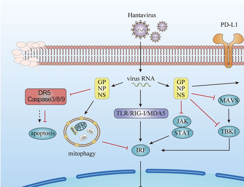
FIGURE 1 | Evasion of the innate immune response by hantavirus. Viral proteins from hantavirus evolving escape strategies involving the inhibition of Interferon
(IFN) signaling pathways, the prevent on the host cell apoptosis, the production of microRNA, and the increasing of immune check point molecules.
FUTURE PERSPECTIVE various immune responses induced by hantavirus infection will
enhance our understanding of hantavirus pathogenesis and develop
This review summarizes the molecular and cellular mechanisms more effective methods to control infection and fine-tune the
on how hantavirus engages several strategies to evade innate activation, strength, and duration of the antiviral immunity.
immune responses, as illustrated in Figure 1. These mechanisms
influence each other. When infection occurs, the cells undergo
a series of physiological and pathological responses. Virus AUTHOR CONTRIBUTIONS
infection leads the cells into a stress condition and induces
the production of inflammatory cytokines. By regulating target YZ, YW, ZY, WS, and RM wrote the manuscript. MH and
cell death, the virus could also struggle its way to survive. TH drew a diagram. RL, YZ, and XW edited, reviewed, and
During these processes, the miRNA expression profile is also approved the manuscript. All authors contributed to the article
changed. In turn, the miRNAs regulate the immune response and approved the submitted version.
induced by virus infection. The IFN signaling is the end pathway
of most processes. The cytokines and surface molecules changes
in DCs and monocytes lead to the subsequent cellular mechanisms. FUNDING
Although the host cells defeat the hantavirus finally, in most
cases, the hantaviruses struggled their way to survive. In our This work was supported by the National Natural Science
perspective, these strategies used to regulate hantavirus immune Foundation Grants (Nos. 81772167, 81971563) and Key
response negatively could also help maintain a balance between Research and Development Project of Shaanxi Province
immune activation and inhibition. Further research focusing on (No. 2019ZDLSF02-03).
REFERENCES Alff, P. J., Sen, N., Gorbunova, E., Gavrilovskaya, I. N., and Mackow, E. R.
(2008). The NY-1 hantavirus Gn cytoplasmic tail coprecipitates TRAF3 and
Alff, P. J., Gavrilovskaya, I. N., Gorbunova, E., Endriss, K., Chong, Y., Geimonen, E., inhibits cellular interferon responses by disrupting TBK1-TRAF3 complex
et al. (2006). The pathogenic NY-1 hantavirus G1 cytoplasmic tail inhibits formation. J. Virol. 82, 9115–9122. doi: 10.1128/JVI.00290-08
RIG-I- and TBK-1-directed interferon responses. J. Virol. 80, 9676–9686. Au, R. Y., Jedlicka, A. E., Li, W., Pekosz, A., and Klein, S. L. (2010). Seoul
doi: 10.1128/JVI.00508-06 virus suppresses NF-kappaB-mediated inflammatory responses of antigen
Frontiers in Microbiology | www.frontiersin.org 5 September 2021 | Volume 12 | Article 759198Zhang et al. Hantavirus Evades Antiviral Innate Immunity
presenting cells from Norway rats. Virology 400, 115–127. doi: 10.1016/j. Lee, M.-H., Lalwani, P., Raftery, M. J., Matthaei, M., Lütteke, N., Kirsanovs, S.,
virol.2010.01.027 et al. (2011). RNA helicase retinoic acid-inducible gene I as a sensor of Hantaan
Chen, Q.-Z., Luo, F., Lu, M.-X., Li, N., Teng, Y., Huang, Q.-L., et al. (2017). virus replication. J. Gen. Virol. 92, 2191–2200. doi: 10.1099/vir.0.032367-0
HTNV-induced upregulation of miR-146a in HUVECs promotes viral infection Levine, J. R., Prescott, J., Brown, K. S., Best, S. M., Ebihara, H., and Feldmann, H.
by modulating pro-inflammatory cytokine release. Biochem. Biophys. Res. (2010). Antagonism of type I interferon responses by new world hantaviruses.
Commun. 493, 807–813. doi: 10.1016/j.bbrc.2017.08.073 J. Virol. 84, 11790–11801. doi: 10.1128/JVI.00916-10
Chen, Q.-Z., Wang, X., Luo, F., Li, N., Zhu, N., Lu, S., et al. (2020). HTNV Li, W., He, P., Huang, Y., Li, Y.-F., Lu, J., Li, M., et al. (2021a). Selective
sensitizes host toward TRAIL-mediated apoptosis-A pivotal anti-hantaviral autophagy of intracellular organelles: recent research advances. Theranostics
role of TRAIL. Front. Immunol. 11:1072. doi: 10.3389/fimmu.2020.01072 11, 222–256. doi: 10.7150/thno.49860
Christ, W., Tynell, J., and Klingström, J. (2020). Puumala and Andes Li, W., and Klein, S. L. (2012). Seoul virus-infected rat lung endothelial cells
orthohantaviruses cause transient protein kinase R-dependent formation of and alveolar macrophages differ in their ability to support virus replication
stress granules. J. Virol. 94, e01168–e01119. doi: 10.1128/JVI.01168-19 and induce regulatory T cell phenotypes. J. Virol. 86, 11845–11855. doi:
Christgen, S., Zheng, M., Kesavardhana, S., Karki, R., Malireddi, R. K. S., 10.1128/JVI.01233-12
Banoth, B., et al. (2020). Identification of the PANoptosome: a molecular Li, X.-D., Lankinen, H., Putkuri, N., Vapalahti, O., and Vaheri, A. (2005). Tula
platform triggering pyroptosis, apoptosis, and necroptosis (PANoptosis). hantavirus triggers pro-apoptotic signals of ER stress in Vero E6 cells.
Front. Cell. Infect. Microbiol. 10:237. doi: 10.3389/fcimb.2020.00237 Virology 333, 180–189. doi: 10.1016/j.virol.2005.01.002
Clement, J., LeDuc, J. W., McElhinney, L. M., Reynes, J.-M., Van Ranst, M., Li, Z., Shen, Y., Song, Y., Zhang, Y., Zhang, C., Ma, Y., et al. (2021b). ER
and Calisher, C. H. (2019). Clinical characteristics of ratborne Seoul hantavirus stress-related molecules induced by Hantaan virus infection in differentiated
disease. Emerg. Infect. Dis. 25, 387–388. doi: 10.3201/eid2502.181643 THP-1 cells. Cell Stress Chaperones 26, 41–50. doi: 10.1007/s12192-020-01150-9
Davies, K., Afrough, B., Mankouri, J., Hewson, R., Edwards, T. A., and Barr, J. N. Liu, R., Ma, R., Liu, Z., Hu, H., Shu, J., Hu, P., et al. (2021). HTNV infection
(2019). Tula orthohantavirus nucleocapsid protein is cleaved in infected of CD8+ T cells is associated with disease progression in HFRS patients.
cells and may sequester activated caspase-3 during persistent infection to Commun. Biol. 4:652. doi: 10.1038/s42003-021-02182-2
suppress apoptosis. J. Gen. Virol. 100, 1208–1221. doi: 10.1099/jgv.0.001291 Liu, R., Ma, H., Shu, J., Zhang, Q., Han, M., Liu, Z., et al. (2020). Vaccines
Dheerasekara, K., Sumathipala, S., and Muthugala, R. (2020). Hantavirus and therapeutics against hantaviruses. Front. Microbiol. 10:2989. doi: 10.3389/
infections-treatment and prevention. Curr. Treat. Options Infect. Dis. 1–12. fmicb.2019.02989
doi:10.1007/s40506-020-00236-3 [Epub ahead of print] Lu, D.-H., Jiang, H., and Lian, J.-Q. (2021). Hantavirus infection during pregnancy.
Duggan, J. M. (2019). Prevalence of Seoul hantavirus in UK wild rats: an Virol. Sin. 36, 345–353. doi: 10.1007/s12250-020-00300-8
emerging public health problem? Vet. Rec. 184, 523–524. doi: 10.1136/vr.l1163 Lu, S., Zhu, N., Guo, W., Wang, X., Li, K., Yan, J., et al. (2020). RNA-seq revealed
Echterdiek, F., Kitterer, D., Alscher, M. D., Schwenger, V., Ruckenbrod, B., a circular RNA-microRNA-mRNA regulatory network in hantaan virus infection.
Bald, M., et al. (2019). Clinical course of hantavirus-induced nephropathia Front. Cell. Infect. Microbiol. 10:97. doi: 10.3389/fcimb.2020.00097
epidemica in children compared to adults in Germany-analysis of 317 Ma, H., Han, P., Ye, W., Chen, H., Zheng, X., Cheng, L., et al. (2017). The
patients. Pediatr. Nephrol. 34, 1247–1252. doi: 10.1007/s00467-019-04215-9 long noncoding RNA NEAT1 exerts antihantaviral effects by acting as positive
Elliott, R. M. (1989). Nucleotide sequence analysis of the large (L) genomic feedback for RIG-I signaling. J. Virol. 91, e02250–e02216. doi: 10.1128/
RNA segment of Bunyamwera virus, the prototype of the family Bunyaviridae. JVI.02250-16
Virology 173, 426–436. doi: 10.1016/0042-6822(89)90555-2 Ma, R., Zhang, X., Shu, J., Liu, Z., Sun, W., Hou, S., et al. (2021). Nlrc3
Elliott, R. M. (1990). Molecular biology of the Bunyaviridae. J. Gen. Virol. 71, knockout mice showed renal pathological changes after HTNV infection.
501–522. doi: 10.1099/0022-1317-71-3-501 Front. Immunol. 12:692509. doi: 10.3389/fimmu.2021.692509
Elliott, R. M., and McGregor, A. (1989). Nucleotide sequence and expression Maleki, K. T., Tauriainen, J., García, M., Kerkman, P. F., Christ, W., Dias, J.,
of the small (S) RNA segment of Maguari bunyavirus. Virology 171, 516–524. et al. (2021). MAIT cell activation is associated with disease severity markers
doi: 10.1016/0042-6822(89)90621-1 in acute hantavirus infection. Cell Rep. Med. 2:100220. doi: 10.1016/j.
Engdahl, T. B., and Crowe, J. E. (2020). Humoral immunity to hantavirus xcrm.2021.100220
infection. mSphere 5, e00482–e00420. doi: 10.1128/mSphere.00482-20 Malireddi, R. K. S., Kesavardhana, S., and Kanneganti, T.-D. (2019). ZBP1 and
Gallo, G., Caignard, G., Badonnel, K., Chevreux, G., Terrier, S., Szemiel, A., TAK1: master regulators of NLRP3 inflammasome/pyroptosis, apoptosis, and
et al. (2021). Interactions of viral proteins from pathogenic and low or necroptosis (PAN-optosis). Front. Cell. Infect. Microbiol. 9:406. doi: 10.3389/
non-pathogenic orthohantaviruses with human type i interferon signaling. fcimb.2019.00406
Viruses 13:140. doi: 10.3390/v13010140 Malireddi, R. K. S., Tweedell, R. E., and Kanneganti, T.-D. (2020). PANoptosis
Ganaie, S. S., and Mir, M. A. (2014). The role of viral genomic RNA and components, regulation, and implications. Aging 12, 11163–11164. doi:
nucleocapsid protein in the autophagic clearance of hantavirus glycoprotein 10.18632/aging.103528
Gn. Virus Res. 187, 72–76. doi: 10.1016/j.virusres.2013.12.034 Mao, J., Lin, E., He, L., Yu, J., Tan, P., and Zhou, Y. (2019). “Autophagy and
Handke, W., Oelschlegel, R., Franke, R., Krüger, D. H., and Rang, A. (2009). viral infection,” in Autophagy Regulation of Innate Immunity Advances in
Hantaan virus triggers TLR3-dependent innate immune responses. J. Immunol. Experimental Medicine and Biology. ed. J. Cui (Singapore: Springer Singapore),
182, 2849–2858. doi: 10.4049/jimmunol.0802893 55–78.
Hart, T. J., Kohl, A., and Elliott, R. M. (2009). Role of the NSs protein in Markotic, A., Hensley, L., Geisbert, T., Spik, K., and Schmaljohn, C. (2003).
the zoonotic capacity of Orthobunyaviruses. Zoonoses Public Health 56, Hantaviruses induce cytopathic effects and apoptosis in continuous human
285–296. doi: 10.1111/j.1863-2378.2008.01166.x embryonic kidney cells. J. Gen. Virol. 84, 2197–2202. doi: 10.1099/vir.0.19090-0
Hussein, I. T. M., Cheng, E., Ganaie, S. S., Werle, M. J., Sheema, S., Haque, A., Matthys, V. S., Cimica, V., Dalrymple, N. A., Glennon, N. B., Bianco, C., and
et al. (2012). Autophagic clearance of sin nombre hantavirus glycoprotein Mackow, E. R. (2014). Hantavirus GnT elements mediate TRAF3 binding
Gn promotes virus replication in cells. J. Virol. 86, 7520–7529. doi: 10.1128/ and inhibit RIG-I/TBK1-directed beta interferon transcription by blocking
JVI.07204-11 IRF3 phosphorylation. J. Virol. 88, 2246–2259. doi: 10.1128/JVI.02647-13
Kang, J.-I., Park, S.-H., Lee, P.-W., and Ahn, B.-Y. (1999). Apoptosis is induced Pan, P., Shen, M., Yu, Z., Ge, W., Chen, K., Tian, M., et al. (2021). SARS-
by hantaviruses in cultured cells. Virology 264, 99–105. doi: 10.1006/ CoV-2 N protein promotes NLRP3 inflammasome activation to induce
viro.1999.9896 hyperinflammation. Nat. Commun. 12:4664. doi: 10.1038/s41467-021-25015-6
Kell, A. M., Hemann, E. A., Turnbull, J. B., and Gale, M. (2020). RIG-I-like Plekhova, N. G., Somova, L. M., Slonova, R. A., Companets, G. G., Luk’yanova, V. V.,
receptor activation drives type I IFN and antiviral signaling to limit Hantaan and Yakubovich, N. V. (2005). Metabolic activity of macrophages infected
orthohantavirus replication. PLoS Pathog. 16:e1008483. doi: 10.1371/journal. with hantavirus, an agent of hemorrhagic fever with renal syndrome.
ppat.1008483 Biochemistry 70, 990–997. doi: 10.1007/s10541-005-0214-0
Lee, S., Channappanavar, R., and Kanneganti, T.-D. (2020). Coronaviruses: Raftery, M. J., Abdelaziz, M. O., Hofmann, J., and Schönrich, G. (2018).
innate immunity, inflammasome activation, inflammatory cell death, and Hantavirus-driven PD-L1/PD-L2 upregulation: an imperfect viral immune
cytokines. Trends Immunol. 41, 1083–1099. doi: 10.1016/j.it.2020.10.005 evasion mechanism. Front. Immunol. 9:2560. doi: 10.3389/fimmu.2018.02560
Frontiers in Microbiology | www.frontiersin.org 6 September 2021 | Volume 12 | Article 759198Zhang et al. Hantavirus Evades Antiviral Innate Immunity Raftery, M. J., Kraus, A. A., Ulrich, R., Krüger, D. H., and Schönrich, G. Wang, X., Chen, Q.-Z., Zan, Y.-X., Wang, M.-R., Yan, J., Guo, W.-W., et al. (2002). Hantavirus infection of dendritic cells. J. Virol. 76, 10724–10733. (2020). Exosomal miR-145-5p derived from orthohantavirus-infected doi: 10.1128/JVI.76.21.10724-10733.2002 endothelial cells inhibits HTNV infection. FASEB J. 34, 13809–13825. doi: Sacks, D., Baxter, B., Campbell, B. C. V., Carpenter, J. S., Cognard, C., Dippel, D., 10.1096/fj.202001114R et al. (2018). Multisociety consensus quality improvement revised consensus Wang, K., Ma, H., Liu, H., Ye, W., Li, Z., Cheng, L., et al. (2019). The glycoprotein statement for endovascular therapy of acute ischemic stroke. Int. J. Stroke and nucleocapsid protein of hantaviruses manipulate autophagy flux to 13, 612–632. doi: 10.1177/1747493018778713 restrain host innate immune responses. Cell Rep. 27, 2075–2091.e5. doi: Samir, P., Malireddi, R. K. S., and Kanneganti, T.-D. (2020). The PANoptosome: 10.1016/j.celrep.2019.04.061 a deadly protein complex driving pyroptosis, apoptosis, and necroptosis Xu, F. L., Lee, Y. L., Tsai, W. Y., Lin, S. J., Yang, Z. Q., Yang, C. C., et al. (PANoptosis). Front. Cell. Infect. Microbiol. 10:238. doi: 10.3389/fcimb.2020.00238 (2005). Effect of cordycepin on Hantaan virus 76-118 infection of primary Samuel, C. E. (2007). Innate immunity minireview series: making biochemical human embryonic pulmonary fibroblasts--characterization of apoptotic effects. sense of nucleic acid sensors that trigger antiviral innate immunity. J. Biol. Acta Virol. 49, 183–193. Chem. 282, 15313–15314. doi: 10.1074/jbc.R700013200 Ye, W., Lei, Y., Yu, M., Xu, Y., Cao, M., Yu, L., et al. (2015). NLRP3 inflammasome Smith, S. L., and Ward, P. (2006). Behavioral interventions to improve performance is responsible for hantavirus inducing interleukin-1β in THP-1 cells. Int. J. in collegiate football. J. Appl. Behav. Anal. 39, 385–391. doi: 10.1901/jaba.2006.5-06 Mol. Med. 35, 1633–1640. doi: 10.3892/ijmm.2015.2162 Solà-Riera, C., García, M., Ljunggren, H.-G., and Klingström, J. (2020). Hantavirus Zhang, Y., Liu, B., Ma, Y., Yi, J., Zhang, C., Zhang, Y., et al. (2014). Hantaan inhibits apoptosis by preventing mitochondrial membrane potential loss virus infection induces CXCL10 expression through TLR3, RIG-I, and MDA-5 through up-regulation of the pro-survival factor BCL-2. PLoS Pathog. pathways correlated with the disease severity. Mediat. Inflamm. 2014:697837. 16:e1008297. doi: 10.1371/journal.ppat.1008297 doi: 10.1155/2014/697837 Solà-Riera, C., Gupta, S., Ljunggren, H.-G., and Klingström, J. (2019a). Orthohantaviruses belonging to three phylogroups all inhibit apoptosis in Conflict of Interest: The authors declare that the research was conducted in infected target cells. Sci. Rep. 9:834. doi: 10.1038/s41598-018-37446-1 the absence of any commercial or financial relationships that could be construed Solà-Riera, C., Gupta, S., Maleki, K. T., González-Rodriguez, P., Saidi, D., as a potential conflict of interest. Zimmer, C. L., et al. (2019b). Hantavirus inhibits TRAIL-mediated killing of infected cells by downregulating death receptor 5. Cell Rep. 28, 2124–2139.e6. Publisher’s Note: All claims expressed in this article are solely those of the doi: 10.1016/j.celrep.2019.07.066 authors and do not necessarily represent those of their affiliated organizations, Temonen, M., Lankinen, H., Vapalahti, O., Ronni, T., Julkunen, I., and Vaheri, A. or those of the publisher, the editors and the reviewers. Any product that may (1995). Effect of interferon-alpha and cell differentiation on Puumala virus be evaluated in this article, or claim that may be made by its manufacturer, is infection in human monocyte/macrophages. Virology 206, 8–15. doi: 10.1016/ not guaranteed or endorsed by the publisher. S0042-6822(95)80014-X Varela, M., Romero, A., Dios, S., van der Vaart, M., Figueras, A., Meijer, A. H., Copyright © 2021 Zhang, Ma, Wang, Sun, Yang, Han, Han, Wu and Liu. This is et al. (2014). Cellular visualization of macrophage pyroptosis and interleukin-1 an open-access article distributed under the terms of the Creative Commons Attribution release in a viral hemorrhagic infection in zebrafish larvae. J. Virol. 88, License (CC BY). The use, distribution or reproduction in other forums is permitted, 12026–12040. doi: 10.1128/JVI.02056-14 provided the original author(s) and the copyright owner(s) are credited and that Vera-Otarola, J., Solis, L., Lowy, F., Olguín, V., Angulo, J., Pino, K., et al. (2020). the original publication in this journal is cited, in accordance with accepted academic The Andes orthohantavirus NSs protein antagonizes the type I interferon response practice. No use, distribution or reproduction is permitted which does not comply by inhibiting MAVS signaling. J. Virol. 94, e00454–e00420. doi: 10.1128/JVI.00454-20 with these terms. Frontiers in Microbiology | www.frontiersin.org 7 September 2021 | Volume 12 | Article 759198
You can also read