Epigenetic silencing directs expression heterogeneity of stably integrated multi transcript unit genetic circuits - Nature
←
→
Page content transcription
If your browser does not render page correctly, please read the page content below
www.nature.com/scientificreports
OPEN Epigenetic silencing directs
expression heterogeneity of stably
integrated multi‑transcript unit
genetic circuits
Jan Zimak1,2,7, Zachary W. Wagoner1,2,7, Nellie Nelson1,2, Brooke Waechtler1,2,
Hana Schlosser1,2, Morgan Kopecky1,2, Jie Wu3,6 & Weian Zhao1,2,3,4,5,6*
We report that epigenetic silencing causes the loss of function of multi-transcript unit constructs that
are integrated using CRISPR-Cas9. Using a modular two color reporter system flanked by selection
markers, we demonstrate that expression heterogeneity does not correlate with sequence alteration
but instead correlates with chromosomal accessibility. We partially reverse this epigenetic silencing
via small-molecule inhibitors of methylation and histone deacetylation. We then correlate each
heterogeneously-expressing phenotype with its expected epigenetic state by employing ATAC-
seq. The stability of each expression phenotype is reinforced by selective pressure, which indicates
that ongoing epigenetic remodeling can occur for over one month after integration. Collectively,
our data suggests that epigenetic silencing limits the utility of multi-transcript unit constructs that
are integrated via double-strand repair pathways. Our research implies that mammalian synthetic
biologists should consider localized epigenetic outcomes when designing complex genetic circuits.
Abbreviations
PCR Polymerase chain reaction
TU Transcription unit
HEK293T Human embryonic kidney cell line 293 T
DN-late Double negative “late”
DN-Early Double negative “early”
DP Double positive
G GFP only
R RFP only
DNMT1 DNA methyltransferase 1
5-Aza-dc 5-AZA-2′-deoxycytidine
TSA Trichostatin A
ATAC-seq Assay for transposase-accessible chromatin using sequencing
CTCF CCCTC-binding factor
The ability to engineer the genomes of mammalian cells with exogenous genetic circuits is central to the goal
and applications of mammalian synthetic b iology1. Many upcoming therapeutic applications of synthetic biol-
ogy employ designer cells engineered with genetic circuits2–4. Genetic circuits in synthetic biology are generally
composed of multiple transcription units (TUs). Recently, standardized modular DNA assembly methods have
been used to construct multi-TU vectors5 that enable multiple different genes to be expressed under the control
of multiple different promoters. While these methods have been extensively used in prokaryotes and simple
eukaryotes6,7, multi-gene circuits have been used in transient e xpression8, genomically-integrated modular gene
1
Sue and Bill Gross Stem Cell Research Center, University of California, Irvine, Irvine, CA 92697, USA. 2Department
of Pharmaceutical Sciences, University of California, Irvine, Irvine, CA 92697, USA. 3Chao Family Comprehensive
Cancer Center, University of California, Irvine, Irvine, CA 92697, USA. 4Edwards Life Sciences Center for Advanced
Cardiovascular Technology, University of California, Irvine, Irvine, CA 92697, USA. 5Department of Biomedical
Engineering, University of California, Irvine, Irvine, CA 92697, USA. 6Department of Biological Chemistry,
University of California, Irvine, Irvine, CA 92697, USA. 7These authors contributed equally: Jan Zimak and Zachary
W. Wagoner. *email: weianz@uci.edu
Scientific Reports | (2021) 11:2424 | https://doi.org/10.1038/s41598-021-81975-1 1
Vol.:(0123456789)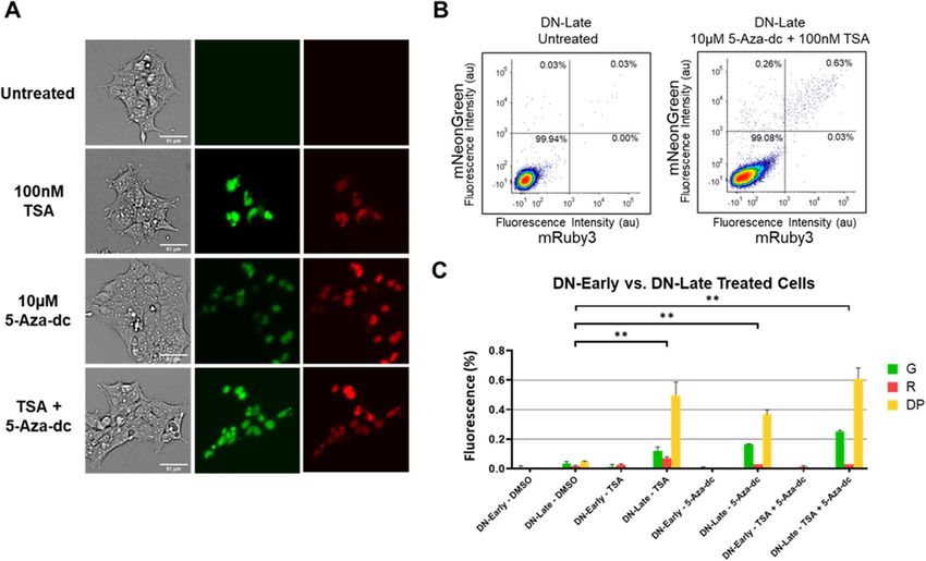
www.nature.com/scientificreports/
circuits have not been widely adopted within the mammalian synthetic biology community since their initial
development. Duportet et al.9 developed a site-specific recombinase-mediated method of integrating large modu-
lar multi-TU genetic circuits into mammalian cells. Each TU is separated by chromatin insulators designed to
block adjacent TUs from affecting each other as well as maintaining the whole genetic circuit in a euchromatin
state10,11. Such circuits can be integrated into monoclonal chassis cell lines, termed “landing pads,” using the Bxb1
recombinase at high efficiency. Landing pad cell lines first require the integration of recombinase sites at only
one chromosomal location, achieved by causing double-stranded breaks using a targeted nuclease (such as zinc
fingers or CRISPR/Cas9) and relying on the cells’ endogenous double-stranded break repair pathways to integrate
the vector. Thus, two sequential steps are required to engineer new cell lines. Given the progress in improving
CRISPR/Cas9 editing e fficiency12–14, these modular systems could be integrated in one step. However, Duportet
et al.9 showed that modular constructs integrated in this manner heterogeneously express each TU, rendering any
resulting genetic circuits non functional. This expression heterogeneity could be caused by sequence alterations to
the constructs during integration9, chromatin changes imparted during integration15, locus-mediated inhibition
of expression16, or insufficient chromatin insulation17. While silencing of transgene expression in mammalian
cells has been extensively studied18, it has not been studied in the context of large multi-gene constructs.
The function of stably integrated genetic circuits breaks down when any circuit element is silenced and thus
these systems are extremely sensitive to variations in gene expression or time-dependent silencing18. Transgene
silencing and variations in expression have been linked to epigenetic mechanisms, such as DNA methylation,
histone deacetylation and chromatin accessibility at the site of integration18. While regulatory elements such
as chromatin insulators have been used to activally remodel local chromatin interactions19 and passively block
heterochromatin spread20. These regulatory elements have been improved in recent years, by targeting sequences
from ubiquitous chromatin opening elements (UCOE)21,22, as well as discovery of novel CTCF-binding motifs
with 10 times the insulating capacity of the canonical cHS4 chromatin i nsulator17.
Here we report an approach to identify the causes of expression heterogeneity in multi-TU genetic circuits
integrated using endogenous DNA-repair pathways. To enable this, we developed a modular multipart assembly
method utilizing a hierarchical cloning method. Using this method, we constructed a four TU vector comprised
of two fluorescent reporters flanked by two independent selection markers each under the control of separate
constitutive promoters. After integration of our two color reporters, we isolated each expression phenotype
and performed genetic and epigenetic analyses to identify possible mechanisms. We also show that position-
dependent use of selection markers controls which expression phenotype dominates and that repetitive screen-
ing reinforces each expression phenotype, indicating that continued epigenetic remodeling occurs for over one
month after integration. Then we examined how expression phenotype matches the local epigenotype using a
genome-wide chromosomal accessibility assay (ATAC-seq). Lastly, we show that phenotypes are partially reversed
using small-molecule inhibitors of methylation and histone deacetylation. Our results indicate that widespread
application of multi-TU genetic circuits will likely require strategies that can mitigate local epigenetic effects of
integrated genetic circuits.
Results
Expression heterogeneity stabilizes into distinct expression phenotypes. We set out to build a
model system to better understand expression heterogeneity in a multi-TU system. This was constructed using
a hierarchical Gibson assembly method similar to previous methods23. Our model system contained four TUs
in the following order: (1) PuromycinR, (2) nuclear localized-mNeonGreen, (3) nuclear localized mRuby3, and
(4) ZeocinR (Fig. 1A). Using this system, we can functionally test for the expression of all four transcripts using
either resistance to selection or the visual presence of fluorescent reporters.
Following two weeks of puromycin selection (2 µg/ml), we categorized cells into one of four distinct expres-
sion phenotypes based on their expression of fluorescent markers mRuby3-NLS and mNeonGreen-NLS: Double
Negative (DN), Green only (G), Red only (R), and Double Positive (DP; Fig. 1B). While these show intracolony
homogeneous expression, we also observed colonies with intracolony heterogeneity at the end of week 2, suggest-
ing that silencing occurs post integration. We further performed FACS analysis which showed the vast majority
of cells existed in the DN state, while less than 1% of cells expressed our desired DP phenotype even after cells
had been subjected to antibiotic selection (Fig. 2A). High-transfection efficiency is observed in HEK293T, and
these cells initially exist in the double positive state (> 99% from days 1–4, data not shown). However, as transient
expression decreases from days 4–14, we see the emergence of the four distinct expression phenotypes shown
above. Next, we set out to interrogate potential sources of this expression heterogeneity.
Expression phenotype silencing dynamics are controlled by screening and selection pressure.
After sorting cells into the four distinct expression phenotypes, we set out to characterize how silencing
dynamics are affected by screening pressure and selective markers. After performing a sorting step at the end of
week 2 post-transfection, using fluorescent gates of 1 04 au, biological duplicates of each expression phenotype
were cultured for an additional 3 weeks and sorted once a week (Fig. 2A). For example, the DP cells were sorted
again at the end of week 3 and only the DP population from that sample was continued in culture. Crucially, the
fraction of DP cells that transition into the DN state at week 3 would represent the fraction silenced per week.
We refer to a cell transition as the temporal changes in the fraction of cells in each state from one sort to the
next. Figure 2A shows representative plots from one biological replicate for the DP state at week 3, 4 and 5. We
see the proportion of cells that are silenced (transition from the DP state to the DN state) decreases from 21.37%
in week 3, to 7.64% in week 4 and 3.12% in week 5.
The silencing dynamics for the other expression phenotypes are summarized in Fig. 2B. We see that while the
DN state is very stable (no visible reversion to a fluorescent state), the G and R state follow similar dynamics to
the DP, although from different initial conditions. Refer to Fig. S1 for weekly sorts from each replicate for each
Scientific Reports | (2021) 11:2424 | https://doi.org/10.1038/s41598-021-81975-1 2
Vol:.(1234567890)
www.nature.com/scientificreports/
Figure 1. Heterogeneous expression of multi-gene construct. (A) Schematic showing our multi-gene construct
design in which expression of four genes (PuromycinR, mNeonGreen-NLS, mRuby3-NLS, and ZeocinR) is
constitutively driven by independent CAG promoters. Genes are flanked by CTCF insulators (orange) and
WPRE-bGHpolyA (pA) sequences. The multi-gene transcripts are then flanked by 5′ and 3′ homology arms
(HA) directing insertion into the AAVS1 loci. (B) Fluorescent imaging representative of the four stable and
distinct phenotypic populations observed after undergoing multiple rounds of puromycin selection at 14 days
post-transfection. DN Double negative, G mNeonGreen-NLS only, R mRuby3-NLS only, DP Double Positive.
expression phenotype. Additionally, the rate of change follows a first order trajectory. Thus assuming a first order
reaction rate, we calculated the silencing rate constant or “half-life of the active state” by taking the ratio of new
cells silenced from week to week (Fig. 2C). For the DP cells, this constant is 2.2 ± 0.1 per week. Additionally,
the same asymptotic trend is shown in Fig. 2B between all three fluorescent expression phenotypes, along with
similar silencing rate constants in Fig. 2C.
We also measured the reversibility of our model system during this time frame. DN-Early cells, initially sorted
at week 2, demonstrated a return to the fluorescent states of only 0.23% cells after one week (week 3; Fig. 2B)
and had less than 0.02% fluorescing cells at week 5. We also sub-cultured DN cells taken from the week 4 DP
sort that had transitioned into the DN state, termed DN-late. These DN-Late cells showed around 2% fluoresc-
ing cells during the following sorting session (week 5; Fig. 2D) exhibiting increased reversibility of silencing.
Additionally, to see if the use of selective markers (without screening pressure) can also affect our model
system, we used the antibiotic selection markers that flank our fluorescent reporters separately to impart selective
pressure onto our model system. Figure 2E shows that when using puromycin to select, the Green only expres-
sion phenotype dominates, while when using zeocin to select, the Red only expression phenotype dominates.
This effect has not previously been observed in modular systems.
Expression phenotypes are controlled by epigenetic factors.
Based on this work, we might expect multiple epigenetic states to be active in our model system including
methylation and histone modifications. However, to definitively rule out the silencing of individual TUs on
the basis of any sequence alterations as a result of integration errors, we examined sorted fluorescent and non-
fluorescent cell lines for the presence of the fluorescent marker sequences. Genomic PCRs showed the presence
of fluorescence marker sequences in the non-fluorescent cells (Fig. S2). Additionally, using RNA-FISH, we found
that fluorescent marker RNA was absent in the silenced cells, while being present within the fluorescent cells in
the same field of view (Fig. S2). These assays were performed on a 3-TU construct shown in Fig. S2A.
These results suggest that while the correct DNA sequence is present, any subsequent silencing may be
occurring through epigenetic mechanisms. To conclusively demonstrate that silencing is epigenetically caused,
we used small-molecule inhibitors of these epigenetic processes to partially reverse the silencing of expression
phenotype in DN cells both in the early and late phase samples. We used two small molecule inhibitors involved
in these pathways: 5-AZA-2′-deoxycytidine (5-Aza-dc) and Trichostatin A (TSA). 5-Aza-dc integrates in place of
cytidine within newly formed DNA strands and forms covalent bonds with DNMT1 when the enzyme attempts
to methylate it24. DNMT1 proteins, which become trapped on DNA, can no longer function and eventually are
Scientific Reports | (2021) 11:2424 | https://doi.org/10.1038/s41598-021-81975-1 3
Vol.:(0123456789)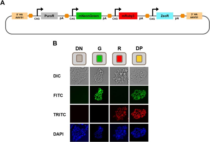
www.nature.com/scientificreports/

                                            Figure 2.  Characterization of expression phenotype dynamics using screening and selective markers. (A)
                                            Transfected HEK293T cells were selected with puromycin for two weeks following transfection with our model
                                            system construct prior to sorting via FACS into four expression phenotypes (DN, G, R, and DP). Each phenotypic
                                            state was further sorted each following week until 5 weeks where cells were gated and isolated for their respective
                                            phenotype (Ex: Green only cells at week 3 were sorted for the 88.62% mNeonGreen- population and replated for
                                            sorting the following week). (B) Summary of temporal data for the DN, G, R, and DP populations, generated from
                                            biological duplicates, over 5 weeks of FACS sorting. Fluorescent percentage refers to percent of cells in specific
                                            expression phenotype. (C) Average ratios of newly silenced DN cells (from week 3 to week 4 and from week 4
                                            to week 5). (D) Example FACS plot of a new phenotypic population isolated during the week 4 sorting process:
                                            DN-Late (refer to Fig. S1). During the week 4 sorts, the DN population of the DP cells (7.64% DN population)
                                            were additionally sorted. These cells were FACS analyzed along with the other intended cell populations during
                                            week 5. (E) Barchart displaying cytometry data for puromycin and zeocin selected cell lines detailing percentages
                                            of each phenotypic state, performed in duplicates, where cells were initially gated for all fluorescent populations at
                                            week 2 (G. R, and DP) which were plated and resorted the following week.

          Scientific Reports |   (2021) 11:2424 |                https://doi.org/10.1038/s41598-021-81975-1                                                      4

Vol:.(1234567890)
www.nature.com/scientificreports/
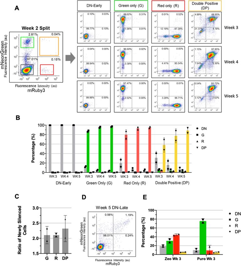
Figure 3. Reversibility of the epigenetically silenced state via epigenetic intervention through small molecule
inhibition. (A) Representative microscopy images of treated DN-Late cells with either 10 µM 5-Aza-dc,
100 nM TSA, or a combination of both molecules at similar concentrations. (B) Flow cytometry comparing the
expression of mNeonGreen-NLS and mRuby3-NLS within untreated DN-Late cells to DN-Late cells treated
over the duration of 72hrs with a combination of 5-Aza-dc and TSA. (C) Summary of treatment conditions
comparing the response of both DN-Early and DN-Late cells to a variety of inhibitory conditions using either
Dimethyl Sulfoxide (DMSO), 5-Aza-dc only, TSA only, or a combination of both 5-Aza-dc and TSA. All
conditions were performed in triplicate with significant changes (**p < 0.01, Bonferroni-corrected multiple
t-Test) represented above.
degraded as part of the DNA damage repair response. This downregulation of DNMT1 activity results in genome
wide hypomethylation and the reactivation of previously silenced genes which now remain unmethylated. TSA
acts as a selective inhibitor of Histone Deacetylase (HDAC) proteins which normally function to promote
chromatin wrapping by removing acetyl groups on h istones25. By inhibiting HDAC proteins, acetyl groups will
persist, DNA will not be bound, and chromatin should shift towards a more open and accessible state allowing
for increased expression.
Early and late phase DN cells were incubated with either DMSO, 10 µM 5-Aza-dc, 100 nM TSA, or a com-
bination of 10 µM 5-Aza-dc and 100 nM TSA. 5-Aza-dc was added fresh every 24 h due to its short half-life26,
while TSA was added only in the last 24 h due to its high toxicity27. These inhibitor concentrations are similar
to those used previously in literature22,27. We attempted to culture cells continuously in the presence of both
inhibitors; however, we found that doing so resulted in drastic loss of viability and the inability to collect enough
cells for FACS analysis over the 72 h timeline. Cells were analyzed by flow cytometry after 72 h of being cultured
with these inhibitors. Using fluorescent microscopy in Fig. 3A, we show that DN-Late cells form individual
fluorescent colonies upon treatment with either small molecule inhibitor. These results were quantified by flow
cytometry in triplicate (Fig. 3B and Fig. S3). Figure 3B shows that both TSA and 5-Aza-dc can partially reverse
expression phenotypes in approximately 1% of cells, indicating that both histone deacetylation and methyla-
tion are causal mechanisms to the silencing of multi-TU transgenes. We found that only late phase cells exhibit
significant phenotype reversal into the DP state (p < 0.01 for all treatments in comparison to DMSO control) for
all treatment regimes, while early phase silenced cells were only reversed into either G or R states at extremely
low frequency (less than 1 in a 1000 cells) (Fig. 3C). Interestingly, the principal reversible transition in the late
phase is the DN → DP transition (Fig. 3B), suggesting that late silencing tends to silence more than one TU. The
observed differences between early and late silenced cells suggest that changes in local DNA structure at the scale
of a single TU (~ 4 kb) can be affected by epigenetic silencing motifs.
ATAC-seq shows expression phenotypes correlate with epigenotype.
After confirming epigenetic silencing of our modular circuits, we wanted to assess how epigenetic motifs
constrain individual TUs. To assess this, we profiled chromatin accessibility on each expression phenotype using
ATAC-seq. Generally, genome-wide epigenetic profiling methods, such as bisulfite-sequencing, are inherently
Scientific Reports | (2021) 11:2424 | https://doi.org/10.1038/s41598-021-81975-1 5
Vol.:(0123456789)www.nature.com/scientificreports/
difficult to apply to modular vectors used in mammalian synthetic biology, such as our model system, due to
the presence of repetitive elements (promoters, chromatin insulators, UNS barcodes, etc.). Hence, any next
generation sequencing-based analysis is restricted to assessing the gene bodies within each TU and not the pro-
moters that are typically epigenetically modified28,29. We chose ATAC-seq as it evaluates chromatin accessibility
(the functional outcome of multiple epigenetic mechanisms) and not specific epigenetic mechanisms (such as
methylation or histone modifications).
ATAC-seq was performed on our five expression phenotypes generated through our long-term stabilization
study (refer to Fig. 2). Reads generated through ATAC-seq which did not align to the human genome were
instead aligned to our plasmid as a template. We then filtered for reads only present within the four gene bod-
ies in our model system (PuromycinR, mNeonGreen, mRuby3, ZeocinR) for each sample (replicates shown in
Fig. S4 and procedure explained in Fig. S5). We specifically excluded repetitive regions in our construct such
as terminators, insulators and promoters due to the inability to ascertain which gene body a short NGS read
would be associated with. Representative replicates for each expression phenotype are shown in Fig. 4A (also
refer to Fig. S3). In Fig. 4A, we see there are observable differences in gene body accessibility (measure by reads
per base) between different expression phenotypes. In Fig. 4A we see the presence of some mRuby3 reads in the
Green only phenotype and the presence of some mNeonGreen reads in the Red only sample. This further rein-
forces the while the fluorescent phenotype is absent the genotype is present suggesting an underlying epigenetic
mechanism is causing the silencing.
To allow for statistical comparisons, ATAC-seq data needs to be normalized. Normalization of reads within
samples was performed by comparing reads with the gene bodies to plasmid segments in the flanking regions
that were equally accessible in all samples (Fig. S6), no differences were observed when data was normalized to
endogenous loci (data not shown). After normalization, the percentage accessibility is calculated, enabling com-
parison between samples. Figure 4B shows that ATAC-seq accessibility, or epigenotype, matches the expression
phenotype, as expected. When comparing accessibility within a sample, both the G and R expression pheno-
types show decreases in accessibility for the non-fluorescing marker, though they are not statistically significant
(p = 0.092 and 0.080 respectively). Comparisons of fluorescent marker accessibility across samples also show
statistically significant changes in accessibility (p < 0.006). For example, both fluorescent markers have significant
decrease in accessibility between the DP and DN-early phenotypes (Fig. 4B).
The only significant difference between the DN-early and DN-late samples is in the selection marker accessi-
bility (p < 0.016, refer to Fig. 4A and Fig. S7). This shows that the DN-late phenotype has all gene bodies decrease
in accessibility, while the DN-early phenotype only has decrease in the fluorescent phenotypes. This reinforces
the previous observation that early silencing has short range implications (single TU), while late silencing targets
the entire integrated construct.
Discussion
Herein, we used a model multi-gene system to understand expression heterogeneity in integrated multi-gene
genetic circuits. Our data suggests this differential expression phenotypes can in part can be explained by epi-
genetic silencing after integration. This demonstrates the need to understand the resulting localized genomic
environment that arises after the integration of multi-gene transcripts.
The dramatic changes in expression heterogeneity emerged during the second week of culture after transfec-
tion. We associated these transient expression changes with the degradation of transient plasmids, any remaining
mRNA, and proteins in the second week after transfection, as the phenotypic state of each cell was actualized.
Then by applying constant screening pressure (one sort per week), we observed changes in silencing dynamics
over time. The expression of selected fluorescent states appears to stabilize in our model system over the course
of a month through a constant decrease in the proportion of newly silenced states with each sort. Given that
puromycin selection occurred for two weeks and that all expression phenotypes were stably integrated and
fluorescent when sorted, we expect that any subsequent silencing is likely not caused by sequence alterations due
to integration. Hence, this suggests other silencing mechanisms are active from week 2 to week 5. We tried to
perform clonal analysis on these samples however, due to instability of these expression phenotypes we weren’t
able to culture enough cells to assess these on a clonal level. The lack of clonal analysis means it is not completely
possible to rule out other alternative explanations such as non-allelic homologous recombination or genetic drift.
Further studies using long-read sequencing on the entire integrated construct will resolve this.
The plasticity (ability to change phenotypic state) of the DN-late cells when compared to the DN-early cells
suggests that the permanence of specific epigenetic states might be linked to the time from the establishment of
these states as well as the time since integration of the genetic constructs. Previously, different epigenetic silencing
states have been shown to have varying reversibility characteristics30. In particular, it is known that methylation
and histone modifications can dominate different stages of silencing, which we will investigate below. Addition-
ally, selection markers were shown to favor expression phenotypes associated with proximal genes, indicating that
selective pressure appears to change the distribution of expression phenotypes. This suggests that both selection
and screening pressure can force the local epigenetic environment to remodel over time. As the changes in expres-
sion are not occurring at the transcriptional or translational level, but rather, epigenetic modifications at the DNA
level and histone level are likely dictating local chromatin conformation and transcription factor accessibility.
This was seen in the expression plasticity observed in DN-late cells and could result from an incomplete
stabilization of epigenetic mechanisms, which have not had time to become fully established. Because of this,
we were able to intervene with inhibitors of these mechanisms to partially restore fluorescent expression. Mean-
while, DN-early cells had several weeks to properly reinforce these silencing mechanisms wherein no recovery
of fluorescence was observed. While our model system employed chromatin insulators upstream of every TU,
this was not sufficient to isolate our TU from being silencing. Insulators have previously been shown to play a
Scientific Reports | (2021) 11:2424 | https://doi.org/10.1038/s41598-021-81975-1 6
Vol:.(1234567890)www.nature.com/scientificreports/
Figure 4. ATAC-seq analysis of genomic DNA derived from sorted and enriched phenotypic populations.
(A) Representative images displaying ATAC-seq reads associated with each of the constructs’ gene bodies
(PuromycinR, mNeonGreen, mRuby3, and ZeocinR) generated for each of the distinct phenotypic states. (B)
Comparison of fold change as it relates to chromatin accessibility looking at mNeonGreen and mRuby3 between
samples. Performed in duplicate with significant changes (**p < 0.01, Bonferroni-corrected multiple t-Test)
represented above.
Scientific Reports | (2021) 11:2424 | https://doi.org/10.1038/s41598-021-81975-1 7
Vol.:(0123456789)www.nature.com/scientificreports/
significant role in regulating the epigenetic silencing of transgenes through the control of local DNA structure
and accessibility15,22,31,32. Hence, we employed the A2 chromatin insulator17 that has previously been shown to
have a greater insulating capacity over the canonical cHS4 chromatin insulator17. While such CTCF-binding
motifs can themselves be methylated33, this will necessitate future modifications of chromatin insulators to be
resistant to epigenetic modifications.
While integration-mediated recombination is not expected to affect our samples (as transient Cas9 RNP
expression is minimal after two weeks) we have not definitely ruled out that the observed silencing effects are
not caused by non-allelic homologous recombination (NAHR). We intend to perform long-read sequencing on
our samples in the future to assay this mechanism. NAHR occurs between repeated transposable elements (such
as the 300 bp Alu sequences)34 are inversely correlated to inter-repeat d istance35. As NAHR is usually studied
using endogenous sequences, our modular constructs could provide a unique exogenous model system to study
this genetic mechanism.
Subsequent studies of these model systems should target specific epigenetic mechanisms. One example would
be using SMRT sequencing (Pacific Biosciences) to assess the DNA methylation status of promoter and chromatin
regions in our modular system. We anticipate that by modifying chromatin insulator sequences and d esign17,22,28
or changing their number and o rientation31,36, we could be able to positively impact the performance of inte-
grated modular genetic circuits. Additionally, we expect the ability to screen for integration using splice acceptor
systems37, also known as promoter t raps38, should increase circuit performance by imparting selective pressure
on cells where the circuit is integrated and not just being transiently expressed. This work suggests the ability to
select or screen for integration from both ends of the modular system will lead to significant decreases in TU
silencing and consequent increases in the performance of integrated genetic circuits.
Conclusion
In this study, we developed a model system to investigate the causes of expression heterogeneity in stably inte-
grated multi-transcript unit genetic circuits. Our model system was used to show that expression heterogeneity
is not caused by alteration to the coding sequence during integration but instead results from local changes in
chromosomal accessibility leading to epigenetic silencing of individual transcript units. We demonstrated that
expression phenotypes are clearly reflected in local changes in chromatin accessibility within the integrated
genetic constructs. This work suggests that new mammalian synthetic biology approaches are required to resist
epigenetic silencing mechanisms that degrade the performance of modular genetic circuits and further stresses
the need for synthetic biology to engineer the epigenome.
Methods
Cloning of plasmid constructs. Unique Nucleotide Sequences (UNS) were designed by Casini et al.39 and
used to assemble various chromatin insulator, promoter, genes, terminators and other genetic elements in a hier-
archical assembly using the UNS as overlaps for Gibson assembly similar to previous methods described23. All
DNA fragments were either chemically synthesized from primers (designed by D NAworks40 or PCR-amplified
from existing plasmids). All DNA syntheses were performed by Integrated DNA Technologies (Coralville, IA).
Plasmids were sequence-verified by Genewiz (San Diego, CA). Annotated plasmids maps for relevant sequences
are provided in Figs. 1A and S2A and in Supplementary information.
Cell culture, transfections, and selection. HEK293T cells obtained from ATCC were cultured in Dul-
becco’s modified Eagle’s medium (DMEM; Corning) containing 10% Fetal Bovine Serum (FBS; Seradigm) with
1% Penicillin and Streptomycin (GenClone) and incubated at 37 °C and 5% C O2. 20,000 cells were seeded into
each well of a 24 well plate 24 h before being transfected. Cells were transfected in duplicates as per Promega
Fugene HD manufacturer’s protocol. Each transfection was performed using 2 µg of DNA consisting of 1 µg of
our multi-gene construct plasmid (pJZ1506), 0.33 µg of Cas9 plasmid (pJZ0387), and 0.66 µg of gRNA plasmid
(pJZ0338). Within various experiments, cells were either selected with puromycin (Invivogen) at a concentra-
tion of 2 µg/mL or zeocin (Alfa Aesar) at a concentration of 100 µg/mL. Cells were gently washed with fresh
media containing antibiotics every two days over the course of two weeks (PBS was not used as we experienced
noticeable cell detachment).
Genomic PCR. Genomic DNA was extracted using the Quick-DNA Midiprep Plus Kit (Zymo Research)
according to manufacturer’s instructions immediately after FACS sorting. PCR was done using primers that
contained the first unique sequence on each gene on the 5′ end and the last unique sequence on the 3′ end that
had an annealing temperature of near 60 °C For the mNeonGreen gene, the forward and reverse primers were
5′-AGAGGACAACATGGCC-3′ and 5′-CTTGTACAGCTCGTCCA-3, respectively.
FACS sorting and analysis. Sorting was performed through the UCI Institute for Immunology Flow Core
Facility using the BD FACSAria Fusion Sorter with a 70 µm nozzle. For the first sort at two weeks, we collected
as many cells as we could, capped at 50,000 cells, for each of the phenotypic states. In the following weeks 50,000
cells were collected for each of the four phenotypic states, Double Negative (DN), Green only (G), Red only
(R), and Double Positive (DP), based on their expression of mNeonGreen and mRuby3 gated at 1 04 au. All four
distinct populations were cultured separately and resorted every 7 days for their respective phenotypes within
all samples (Ex: GFP only expressing cells were sorted and replated within the G only samples each week). Cells
treated with zeocin were screened for all four distinct populations, but only fluorescent cells (DP, G, and R) were
collected and cultured together each week. Data was analyzed using FCS Express 7.
Scientific Reports | (2021) 11:2424 | https://doi.org/10.1038/s41598-021-81975-1 8
Vol:.(1234567890)www.nature.com/scientificreports/
Flow cytometry and analysis. Flow Cytometry was performed through the UCI Stem Cell Research
Center Flow Cytometry Core using the BD Fortessa. 200,000 cells were processed for each of the samples, and
data was analyzed using FCS Express 7. Plots were designed displaying 50,000 events each and reflect identical
fluorescent compensation values.
ATAC‑sequencing (assay for transposase‑accessible chromatin using sequencing) prepara‑
tion. Cells were collected the next day following our 5-week FACS sort timepoint in which 50,000 live cells
were prepared for ATAC-sequencing following the Corces et al. Omni-ATAC p rotocol41. Briefly, the cells were
washed with PBS and counted to isolate 50,000 live cells. The nuclei were isolated and then pelleted by cen-
trifugation at 500×g for 5 min at 4 °C. The DNA was transposed by the UCI Genomic High-Throughput Core
Facility. The DNA was then purified using MinElute PCR Purification Kit (Qiagen), and the UCI Genomic
High-Throughput Core Facility performed the final ATAC-seq library preparations and sequencing. ATAC-seq
libraries were sequenced on 1 lane of a NovaSeq 50 bp paired-end run to an average 30 million reads.
ATAC‑seq data analysis. Raw reads from the 10 samples were first QCed (fastqQC, v0.11.7) and trimmed
using trimmomatic (v0.35). Trimmed reads were then aligned to the hg19 build of the human genome using
Bowtie2 (v2.2.7) with alignment parameters: bowtie2-X 2000-dovetail. Potential PCR duplicate reads were
marked using MarkDuplicates from the Picard tools (v1.130). Black list regions in the human genome (Stanford
version) were excluded and peaks were called using MACS2. Unmapped reads were then extracted from the
alignment and further aligned to the multi-TU plasmid sequence and reads aligned to the four regions of interest
were processed and counted using samtools (v1.9) and featureCounts from subread (v1.5.0).
Statistical methods. A minimum of two independent experiments were carried out (FACS-based pheno-
type characterization and ATAC-seq), and where possible three biological replicates were assayed. Values repre-
sent average ± SEM and were subject to the Bonferroni-corrected two-tailed Student’s t test. Values of *p < 0.05,
**p < 0.01, and ***p < 0.001 were considered statistically significant.
Small molecule inhibition. Previously transfected HEK293T cells that were FACS sorted into the pheno-
typic population of Late-DN and Early-DN were seeded at 40,000 cells on a 6 well plate. Cells were treated with
either 5-Aza-dc (SelleckChem) at 10 µM, TSA (Apexbio Technology) at 100 nM, or a combination of both at
the same concentrations. Cells treated with either 5-Aza-dc only or TSA only were treated for 72 h with a media
change and reintroduction of the respective inhibitor every 24 h. When used in combination, cells were treated
with 10 µM 5-Aza-dc for 72 h and 100 nM TSA was additionally added for the last 24 h. Media was changed
every 24 h similar to the single inhibitor treated wells. Experiment was run in triplicates.
Received: 5 August 2020; Accepted: 8 January 2021
References
1. Ceroni, F. & Ellis, T. The challenges facing synthetic biology in eukaryotes. Nat. Rev. Mol. Cell Biol. https://doi.org/10.1038/s4158
0-018-0013-2 (2018).
2. Kitada, T., DiAndreth, B., Teague, B. & Weiss, R. Programming gene and engineered-cell therapies with synthetic biology. Science
359, 1067 (2018).
3. Xie, M. & Fussenegger, M. Designing cell function: Assembly of synthetic gene circuits for cell biology applications. Nat. Rev. Mol.
Cell Biol. 19, 507–525 (2018).
4. Black, J. B., Perez-Pinera, P. & Gersbach, C. A. Mammalian synthetic biology: Engineering biological systems. Annu. Rev. Biomed.
Eng. 19, 249–277 (2017).
5. Casini, A., Storch, M., Baldwin, G. S. & Ellis, T. Bricks and blueprints: Methods and standards for DNA assembly. Nat. Rev. Mol.
Cell Biol. 16, 568–576 (2015).
6. Smanski, M. J. et al. Functional optimization of gene clusters by combinatorial design and assembly. Nat. Biotechnol. 32, 1241–1249
(2014).
7. Lee, M. E., DeLoache, W. C., Cervantes, B. & Dueber, J. E. A highly characterized yeast toolkit for modular, multipart assembly.
ACS Synth. Biol. 4, 975–986 (2015).
8. Weinberg, B. H. et al. Large-scale design of robust genetic circuits with multiple inputs and outputs for mammalian cells. Nat.
Biotechnol. 35, 453–462 (2017).
9. Duportet, X. et al. A platform for rapid prototyping of synthetic gene networks in mammalian cells. Nucleic Acids Res. 42, 13440–
13451 (2014).
10. Phillips, J. E. & Corces, V. G. CTCF: Master weaver of the genome. Cell 137, 1194–1211 (2009).
11. Giles, K. E., Gowher, H., Ghirlando, R., Jin, C. & Felsenfeld, G. Chromatin boundaries, insulators, and long-range interactions in
the nucleus. Cold Spring Harb. Symp. Quant. Biol. 75, 79–85 (2010).
12. Vakulskas, C. A. et al. A high-fidelity Cas9 mutant delivered as a ribonucleoprotein complex enables efficient gene editing in human
hematopoietic stem and progenitor cells. Nat. Med. 24, 1216–1224 (2018).
13. Kleinstiver, B. P. et al. High-fidelity CRISPR–Cas9 nucleases with no detectable genome-wide off-target effects. Nature 529, 490–495
(2016).
14. Suzuki, K. et al. In vivo genome editing via CRISPR/Cas9 mediated homology-independent targeted integration. Nature 540,
144–149 (2016).
15. Morano, A. et al. Targeted DNA methylation by homology-directed repair in mammalian cells. Transcription reshapes methylation
on the repaired gene. Nucleic Acids Res. 42, 804–821 (2014).
16. Ordovás, L. et al. Efficient recombinase-mediated cassette exchange in hpscs to study the hepatocyte lineage reveals AAVS1 locus-
mediated transgene inhibition. Stem Cell Rep. 5, 918–931 (2015).
Scientific Reports | (2021) 11:2424 | https://doi.org/10.1038/s41598-021-81975-1 9
Vol.:(0123456789)www.nature.com/scientificreports/
17. Liu, M. et al. Genomic discovery of potent chromatin insulators for human gene therapy. Nat. Biotechnol. 33, 198–203 (2015).
18. Alhaji, S. Y., Ngai, S. C. & Abdullah, S. Silencing of transgene expression in mammalian cells by DNA methylation and histone
modifications in gene therapy perspective. Biotechnol. Genet. Eng. Rev. 35, 1–25 (2019).
19. Emery, D. W. The use of chromatin insulators to improve the expression and safety of integrating gene transfer vectors. Hum. Gene
Ther. 22, 761–774 (2011).
20. Chung, J. H., Bell, A. C. & Felsenfeld, G. Characterization of the chicken beta-globin insulator. Proc. Natl. Acad. Sci. USA. 94,
575–580 (1997).
21. Saunders, F., Sweeney, B., Antoniou, M. N., Stephens, P. & Cain, K. Chromatin function modifying elements in an industrial
antibody production platform–comparison of UCOE, MAR, STAR and cHS4 elements. PLoS ONE 10, e0120096 (2015).
22. Rudina, S. S. & Smolke, C. D. A novel chromatin-opening element for stable long-term transgene expression. bioRxiv https://doi.
org/10.1101/626713 (2019).
23. Guye, P., Li, Y., Wroblewska, L., Duportet, X. & Weiss, R. Rapid, modular and reliable construction of complex mammalian gene
circuits. Nucleic Acids Res. 41, e156 (2013).
24. Navada, S. C., Steinmann, J., Lübbert, M. & Silverman, L. R. Clinical development of demethylating agents in hematology. J. Clin.
Invest. 124, 40–46 (2014).
25. Yoshida, M., Horinouchi, S. & Beppu, T. Trichostatin A and trapoxin: novel chemical probes for the role of histone acetylation in
chromatin structure and function. BioEssays 17, 423–430 (1995).
26. Kim, S. H., Heeb, R. M. & Krämer, I. Physicochemical stability of reconstituted decitabine (Dacogen®) solutions and ready-to-
administer infusion bags when stored refrigerated or frozen. Pharm. Technol. Hosp. Pharm. 2, 145–157 (2017).
27. Yang, X. et al. Synergistic activation of functional estrogen receptor (ER)-alpha by DNA methyltransferase and histone deacetylase
inhibition in human ER-alpha-negative breast cancer cells. Cancer Res. 61, 7025–7029 (2001).
28. Müller-Kuller, U. et al. A minimal ubiquitous chromatin opening element (UCOE) effectively prevents silencing of juxtaposed
heterologous promoters by epigenetic remodeling in multipotent and pluripotent stem cells. Nucleic Acids Res. 43, 1577–1592
(2015).
29. Yeo, N. C. et al. An enhanced CRISPR repressor for targeted mammalian gene regulation. Nat. Methods 15, 611–616 (2018).
30. Bintu, L. et al. Dynamics of epigenetic regulation at the single-cell level. Science 351, 720–724 (2016).
31. Li, C. L. & Emery, D. W. The cHS4 chromatin insulator reduces gammaretroviral vector silencing by epigenetic modifications of
integrated provirus. Gene Ther. 15, 49–53 (2008).
32. Mutskov, V. & Felsenfeld, G. Silencing of transgene transcription precedes methylation of promoter DNA and histone H3 lysine
9. EMBO J. 23, 138–149 (2004).
33. Wang, H. et al. Widespread plasticity in CTCF occupancy linked to DNA methylation. Genome Res. 22, 1680–1688 (2012).
34. Belancio, V. P., Deininger, P. L. & Roy-Engel, A. M. LINE dancing in the human genome: Transposable elements and disease.
Genome Med. 1, 97 (2009).
35. Liu, P. et al. Frequency of nonallelic homologous recombination is correlated with length of homology: Evidence that ectopic
synapsis precedes ectopic crossing-over. Am. J. Hum. Genet. 89, 580–588 (2011).
36. Benabdellah, K., Gutierrez-Guerrero, A., Cobo, M., Muñoz, P. & Martín, F. A chimeric HS4-SAR insulator (IS2) that prevents
silencing and enhances expression of lentiviral vectors in pluripotent stem cells. PLoS ONE 9, e84268 (2014).
37. Hockemeyer, D. et al. Efficient targeting of expressed and silent genes in human ESCs and iPSCs using zinc-finger nucleases. Nat.
Biotechnol. 27, 851–857 (2009).
38. Kao, T. et al. GAPTrap: A simple expression system for pluripotent stem cells and their derivatives. Stem Cell Rep. 7, 518–526
(2016).
39. Casini, A. et al. R2oDNA designer: Computational design of biologically neutral synthetic DNA sequences. ACS Synth. Biol. 3,
525–528 (2014).
40. Hoover, D. M. & Lubkowski, J. DNAWorks: An automated method for designing oligonucleotides for PCR-based gene synthesis.
Nucleic Acids Res. 30, e43 (2002).
41. Corces, M. R. et al. An improved ATAC-seq protocol reduces background and enables interrogation of frozen tissues. Nat. Methods
14, 959–962 (2017).
Acknowledgements
We thank Xutao Liu, Xuesi (Kathy) Ruan for their assistance on the project. Dr. Timothy Downing, Dr. Aude
Segaliny, Dr. Egest Pone, Jayapriya Jayaraman, Josh Gu for helpful discussions. We thank Dr. Melanie Oakes
at the UCI Genomics High-Throughput Facility for her assistance with ATAC-seq study design and sequenc-
ing, Dr. Jennifer Atwood at the UCI Institute for Immunology Flow Core Facility for her assistance with FACS
sorting, as well as Vanessa Scarfone at the UCI Sue and Bill Gross Stem Cell Research Center Flow Cytometry
Core for training and assisting with flow cytometry. This work was funded by the NIH (1DP2CA195763-01 and
R21CA219225 to W.Z.), the DOD (W81XWH-17-1-0522 to W.Z.). This research also received funding from the
Alliance for Regenerative Rehabilitation Research & Training (AR3T) under Award Number P2CHD086843 to
W.Z., which is supported by the Eunice Kennedy Shriver National Institute of Child Health and Human Devel-
opment (NICHD), National Institute of Neurological Disorders and Stroke (NINDS), and National Institute of
Biomedical Imaging and Bioengineering (NIBIB) of the National Institutes of Health. The content is solely the
responsibility of the authors and does not necessarily represent the official views of the National Institutes of
Health. J.Z. was supported by the National Center for Research Resources and the National Center for Advancing
Translational Sciences, National Institutes of Health, through Grant TL1TR001415. Z.W. was supported by the
California Institute of Regenerative Medicine EDUC 2-08383.
Author contributions
J.Z., Z.W. and W.Z. designed the research; J.Z., Z.W., N.N., B.W., H.S., M.K. performed the research; J.W. per-
formed the ATAC-seq analysis; J.Z., Z.W., N.N., B.W., H.S. analysed and interpreted the data; J.Z. and Z.W. wrote
the paper; N.N., B.W. and W.Z. helped with editing the paper.
Competing interests
The authors declare the following competing financial interests: W.Z. is a (co)founder of Velox Biosystems Inc.
and Amberstone Biosciences Inc.
Scientific Reports | (2021) 11:2424 | https://doi.org/10.1038/s41598-021-81975-1 10
Vol:.(1234567890)www.nature.com/scientificreports/
Additional information
Supplementary Information The online version contains supplementary material available at https://doi.
org/10.1038/s41598-021-81975-1.
Correspondence and requests for materials should be addressed to W.Z.
Reprints and permissions information is available at www.nature.com/reprints.
Publisher’s note Springer Nature remains neutral with regard to jurisdictional claims in published maps and
institutional affiliations.
Open Access This article is licensed under a Creative Commons Attribution 4.0 International
License, which permits use, sharing, adaptation, distribution and reproduction in any medium or
format, as long as you give appropriate credit to the original author(s) and the source, provide a link to the
Creative Commons licence, and indicate if changes were made. The images or other third party material in this
article are included in the article’s Creative Commons licence, unless indicated otherwise in a credit line to the
material. If material is not included in the article’s Creative Commons licence and your intended use is not
permitted by statutory regulation or exceeds the permitted use, you will need to obtain permission directly from
the copyright holder. To view a copy of this licence, visit http://creativecommons.org/licenses/by/4.0/.
© The Author(s) 2021
Scientific Reports | (2021) 11:2424 | https://doi.org/10.1038/s41598-021-81975-1 11
Vol.:(0123456789)You can also read