The Efficacy of Phototherapy for the Treatment of Onychomycosis: An Observational Study - MDPI
←
→
Page content transcription
If your browser does not render page correctly, please read the page content below
hv
               photonics

Article
The Efficacy of Phototherapy for the Treatment of
Onychomycosis: An Observational Study
Nadia Dembskey and Heidi Abrahamse *

                                          Laser Research Centre, Faculty of Health Sciences, University of Johannesburg, P.O. Box 17011,
                                          Doornfontein 2028, South Africa; nadia.dembskey@gmail.com
                                          * Correspondence: habrahamse@uj.ac.za; Tel.: +27-11-559-6550

                                          Abstract: (1) Background: Onychomycosis accounts for 50% of nail pathologies and is a therapeutic
                                          challenge due to an increase in resistance to antifungal agents. This study aimed to explore the
                                          effectiveness of 1064 nm diode laser irradiation for the treatment of Onychomycosis and establish
                                          a new set of laser parameters for effective and safe treatment; (2) Methods: An exploratory, single-
                                          blinded study was conducted on forty-five patients with toenail Onychomycosis. Digital images and
                                          nail clippings were taken for Periodic Acid-Schiff (PAS) staining and fungal microscopy and culture
                                          (MC&S). Group 1 received 5% topical Amorolfine lacquer to apply to affected nails. Group 2 received
                                          1064 nm diode laser treatment at 10 mW/s, hallux 790 J/cm2 and lesser digits 390 J/cm2 (standard
                                          treatment). Group 3 received 1064 nm diode laser treatment at 10 mW/s, hallux 1 100 J/cm2 and lesser
                                          digits 500 J/cm2 (new treatment parameters). After laser treatment, nail temperatures were taken with
                                          a surface thermometer; (3) Results: PAS staining was more sensitive in identifying Onychomycosis
                                          (91.1%), compared to Fungal Microscopy (44.4%). Comparing treatment requirements over a period
                                          of 24 weeks, there was a statistical significance, p ≤ 0.01 (**), for standard laser treatment and,
                                          p ≤ 0.001 (***), for new laser parameter treatment, indicating treatment needed over time decreased.
                                          No adverse effects were noted with new laser therapy. An 86.7% visual improvement was noted in
                                Group 3 after 24 weeks; (4) Conclusions: Phototherapy, or photo thermolysis, was the best treatment

                                          option for Onychomycosis. A new protocol for the standardization of laser irradiation with the
Citation: Dembskey, N.; Abrahamse,        possible inclusion into the Scoring Clinical Index for Onychomycosis treatment plan, was proposed.
H. The Efficacy of Phototherapy for
the Treatment of Onychomycosis: An        Keywords: Onychomycosis; phototherapy; Scoring Clinical Index for Onychomycosis
Observational Study. Photonics 2021,
8, 350. https://doi.org/10.3390/
photonics8090350

                                          1. Introduction
Received: 20 July 2021
Accepted: 21 August 2021
                                               Onychomycosis (OM), a chronic fungal infection of the toenails, is a common condition
Published: 25 August 2021
                                          that accounts for 50% of all nail pathologies [1–7]. Currently, one-third of the world
                                          population has this condition, and is of great concern [8,9]. The term OM is derived from
Publisher’s Note: MDPI stays neutral
                                          the Greek word ‘onyx’ which means a nail and ‘mykes’ which denotes a fungus [10]. It’s
with regard to jurisdictional claims in
                                          primarily caused by dermatophytes but can also be caused by yeasts and non-dermatophyte
published maps and institutional affil-   molds [5,7,8,11–13]. Previously regarded as contaminants, yeasts and some moulds are
iations.                                  now increasingly recognised as pathogens [10]. The most noticeable among these are
                                          Candida albicans, Candida parapsilosis, Candida glabrata, Candida krusei (yeasts) and Aspergillus
                                          fumigates (mould) [5,11,14,15]. These moldy fungi are especially difficult to cure in OM
                                          using standard treatment modalities as they focus on dermatophyte organisms [16,17].
                                          Also, the clinical presentation of OM should be taken into consideration when therapeutic
Copyright: © 2021 by the authors.
Licensee MDPI, Basel, Switzerland.
                                          decisions are being made [18]. Table 1 details the clinical picture of OM.
This article is an open access article
                                               This provides an outline for diagnosis and expected response to treatment. It can also
distributed under the terms and           predict the possible prognostic outcomes [20].
conditions of the Creative Commons
Attribution (CC BY) license (https://
                                          1.1. The Scoring Clinical Index for OM (SCIO)
creativecommons.org/licenses/by/               The Scoring Clinical Index for OM (SCIO) attempts to present the severity of OM
4.0/).                                    as a composite score, and aims to identify the type of OM, the area and thickness of nail

Photonics 2021, 8, 350. https://doi.org/10.3390/photonics8090350                                            https://www.mdpi.com/journal/photonics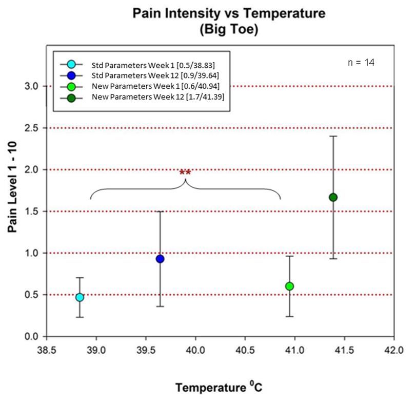
Photonics 2021, 8, 350                                                                                                       2 of 16

                                involvement, the age of the patient and the location of the digit affected. It is calculated
                                using the clinical index component and growth component. The higher the SCIO, the more
                                severe the OM, which in turn requires a higher dosage and more prolonged treatment [21].
                                The treatment dosage is for oral and topical antifungals only; laser treatment was a novel
                                treatment in this study with a SCIO attached. An electronic version of the SCIO is available
                                at http://www.onychoindex.com; accessed on 2 July 2021. Table 2 describes the current
                                proposed treatment guidelines according to the SCIO.

                                    Table 1. Clinical picture of Onychomycosis [8,11,19,20].

             Classification                           Description                                  Causative Organism

                                    •    Distal free edge (hyponychium) of nail and
                                         spreads to nail plate and bed
                                    •    Hyperkeratotic debris accumulate and            •     Trichophyton rubrum
                                         result in onycholysis                           •     Trichophyton mentagrophytes
                                    •    Nails become dystrophic                         •     Epidermophyton floccosum
         Distal Subungual           •    Turn yellow-white or brown-black                •     Candida albicans
       Onychomycosis (DSO)          •    Can spread proximally causing linear            •     Fusarium species
                                         spikes                                          •     Scopulariopsis brevicaulis
                                    •    Associated with paronychia                      •     Scytalidium species
                                    •    Mode of Infection: Through break in skin at
                                         the distal under surface of nail

                                    •    Continuation from DSO
                                    •    Pathogen migrates from hyponychium at
                                         side proximally                                 •     Trichophyton rubrum
                                    •    Hyperkeratotic debris accumulate and            •     Trichophyton mentagrophytes
     Lateral and Distolateral            result in onycholysis                           •     Epidermophyton floccosum
    Subungual Onychomycosis         •    Brownish-yellow                                 •     Candida albicans
             (DLSO)                 •    Can spread proximally causing linear            •     Fusarium species
                                         spikes                                          •     Scopulariopsis brevicaulis
                                    •    Associated with paronychia                      •     Scytalidium species
                                    •    Mode of Infection: Through break in skin
                                         at the distal under surface of nail

                                    •    Rare
                                    •    Debris accumulate under the eponychium,
                                         causing onycholysis and spreads distally
                                                                                         •     Trichophyton rubrum
                                    •    Found in immunosuppressed patients
        Proximal Subungual                                                               •     Candida albicans
                                    •    Hyperkeratotic
        Onychomycosis (PSO)                                                              •     Aspergillus species
                                    •    White
                                                                                         •     Fusarium species
                                    •    Mode of Infection: Invasion of the
                                         proximal nail fold and cuticle. May cause
                                         secondary paronychia

                                    •    Superficial nail plate infection
                                                                                         •     Trichophyton rubrum
                                    •    White plaque-like layer covers the nail plate
                                                                                         •     Trichophyton mentagrophytes
         White Superficial          •    Powder-like patches of transverse striae
                                                                                         •     Acremonium species
       Onychomycosis (WSO)          •    Mode of Infection: Appear om superficial
                                                                                         •     Fusarium species
                                         nail plate. May form a deep penetration of
                                                                                         •     Scytalidium species
                                         the superficial infection

                                    •    Special variant
                                                                                         •     Trichophyton rubrum
                                    •    White discolouration underneath proximal
     Proximal White Subungual                                                            •     Candida albicans
                                         part of nail plate
      Onychomycosis (PWSO)                                                               •     Aspergillus species
                                    •    Mode of Infection: Invasion of the
                                                                                         •     Fusarium species
                                         proximal nail fold and cuticle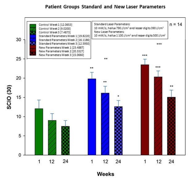
Photonics 2021, 8, 350                                                                                                     3 of 16

                                                         Table 1. Cont.

             Classification                           Description                                Causative Organism

                                   •     “Glacier nail”
                                   •     Longitudinal yellow streaks stretching
                                                                                       •    Can result from any of the other
                                         medially and laterally reaching the nail
         Total Dystrophic                                                                   classes
                                         matrix
       Onychomycosis (TDO)                                                             •    Most often from severe DSO or
                                   •     Complete destruction of the nail from
                                                                                            DLSO infections
                                         longstanding infection
                                   •     Nail structure is lost

                              Table 2. Proposed treatment guidelines for Onychomycosis treatment according to Scoring Clinical
                              Index for Onychomycosis values [21].

                                  Scio                                      Treatment Approach

                                             •    Topical treatment
                                  1–3        •    Remove affected minor parts of nail
                                             •    Use topical antifungals until healthy nail re-grows

                                             •    Topical treatment with lower success
                                             •    Depends on nail growth rate
                                  3–6        •    Systemic treatment recommended in slower-growing nails or proximal
                                                  Onychomycosis types

                                             •    Systemic treatment
                                  6–9        •    Use dosage purported for fingernails: For example: Itraconazole 200 mg BD
                                                  pulse treatment for 2 months

                                             •    Systemic treatment
                                  9–12       •    Use dosage purported for toenails: For example: Itraconazole 200 mg BD pulse
                                                  treatment for 3 months

                                             •    Systemic treatment
                                 12–16       •    Use dosage purported for fingernails with any antifungal of the clinicians’
                                                  choice: For example: Itraconazole 200 mg BD pulse treatment for 4–5 months

                                             •    Combination treatment (systemic and topical)
                                 16–20       •    Adequate keratolytic treatment recommended

                                             •    Consider nail avulsion
                                 20–30       •    Continue with systemic treatment

                                   This may prove an accurate indicator of therapeutic effectiveness, but further clinical
                              studies must be done before definitive claims can be made [22]. As we aimed to identify
                              the effectiveness of laser therapy for OM compared to topical treatment, this may bridge
                              the gap of clinical effectiveness by adding laser therapy as a possible route of treatment,
                              depending on the SCIO.

                              1.2. Diagnosis
                                    Only 50% of nail pathologies are caused by OM, so adequate diagnosis is essential
                              for treatment [12]. Clinical examination may not always be precise and laboratory testing
                              should be included for accurate diagnosis [20]. Nail clippings and scrapings are the
                              most common sampling methods for suspected OM [11,12,23]. The simplest method for
                              detecting fungi is by way of 20% potassium hydroxide (KOH) preparations. However,
                              it shows only 40–60% sensitivity [5,14]. Fungi can also be grown in culture form, using
                              Sabouraud 2% dextrose agar; however, a 70% sensitivity failure rate is seen, possibly due to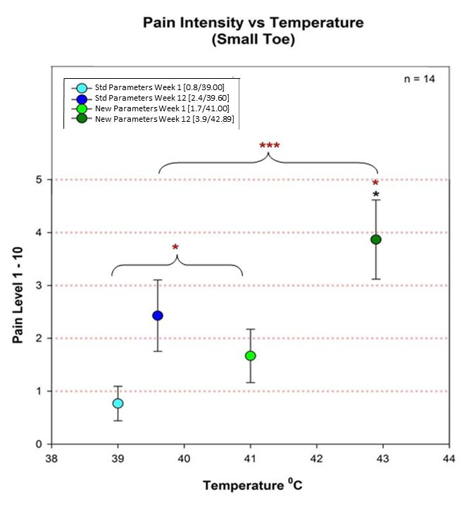
Photonics 2021, 8, 350                                                                                            4 of 16

                         antifungal agent use [7,13]. Recently, histological fungal detection i.e., Periodic Acid-Schiff
                         (PAS) stain, has shown high sensitivity (92%) in the detection of fungal elements; however,
                         specificity is low as there is no indication of fungal genus or species [8,14]. Wilsmann-
                         Theis, et al., writes that to date, the gold standard in the diagnosis of OM remains direct
                         microscopy and culture [23]. They compared the current gold standard in OM diagnosis
                         with histological PAS staining in a large cohort. A total of 631 samples revealed a positive
                         result in at least 1 test. They found that the most sensitive single test for the diagnosis
                         of OM was PAS staining (82%), followed by culture (53%) and direct microscopy (48%).
                         In 64 cases where prediagnostic antimycotic treatment was implemented, PAS staining
                         had the highest sensitivity (88%) in comparison with culture (33%) or direct microscopy
                         (50%) [23]. Although PAS staining is shown to be the single method with the highest
                         sensitivity in terms of detecting fungal hyphae (especially in cases with prior antimycotic
                         therapy), it still has a very low specificity rate [8,14,23]. Kaur, et al., recommends that PAS
                         stain and culture should be done together [10].

                         1.3. Current Treatment Modalities for OM
                              Several factors e.g., infecting organism, clinical presentation, severity of infection and
                         co-morbidities should be taken into consideration when treating OM [12,16]. Oral antifun-
                         gal agents (terbinafine hydrochloride, itraconazole and fluconazole) are the medications of
                         choice for this condition, as either continuous or “pulse” therapies [11,14,24]. Recently, there
                         has been an increase in resistance shown to these medications and various questions remain
                         as to their safety for patients and their potential to cause hepatotoxicity [3,4,9,11,15–17].
                         Topical antifungal treatments (amorolfine and ciclosporox) are preferred by most patients
                         as they have less serious side effects. These agents can only be administered to those
                         who have OM without matrix involvement and requires extensive patient monitoring [9].
                         Serious questions also arise on the effectiveness of nail penetration as well as issues relating
                         to increased organism resistance [1,9,11,15,25].

                         1.4. Phototherapy
                              Limitations in outcomes in both these antifungal agents have led to the investigation of
                         laser and light as a possible new way of treating OM [3,18,24]. This treatment modality has
                         been in the spotlight since 2010, although mentioned in 1980 [14]. Studies show lasers of
                         near-infrared light range with wavelengths of 780–3000 nm being used, but most commonly
                         the Neodymium Yttrium Aluminium Garnet (Nd: YAG) 1064 nm laser [13,21]. Preliminary
                         studies show good clinical and microscopical cure rates using the Nd: YAG 1064 nm laser in
                         the short term [2,4,12,18,25]. In 2012, the Food and Drug Administration (FDA) approved
                         the use of lasers for the “temporary increase in clear nail in Onychomycosis” [12]. This
                         is based on a cosmetic outcome that differs from the medical efficacy approvals granted
                         to topical and oral antifungal agents and may be partially due to poor published study
                         designs with small study samples. Device-based therapies are promising alternatives for
                         OM treatment, as they can mitigate some of the negative factors associated with treatment
                         failure [26]. Authors described the Laser therapy for OM as based on the “principle
                         of selective photo thermolysis”, for which the pathogen absorbs the light energy and
                         converted it into heat. Fungi are considered by the authors [2,4,22,25] sensitive to heat
                         above 55 ◦ C, so the absorption generates photothermal heating of the fungal structures
                         with fungicidal effects. However, heating dermal tissue to above 40 ◦ C produces pain and
                         necrosis [2,4,22,25]. The authors correctly point out that in the literature it is considered
                         necessary to pulsate or keep “low” the levels of radiated energy, in laser treatment, to
                         avoid overheating that is harmful to the patient. Currently, lasers of different wavelengths,
                         pulse duration and spot size are used throughout literature [22,26–30]. In studies found, no
                         standard podiatric treatment was done prior to laser treatment, not enough time was given
                         between treatments in order to see clear nail growth, with limited output, and no clear
                         protocol could be attained [14,31]. With onychomycosis in progress, urgent intervention is
                         required, decided by a doctor, a podiatrist, or in any case a specialist, who must choose
Photonics 2021, 8, 350 5 of 16
the antifungal drug treatment with topical or systemic action deemed most appropriate
for the subject. It must be considered that this treatment is usually long since the nails
take from eight, nine months, up to a year to fully regrow. For this reason, the search for
alternative treatments to conventional antifungals is useful and necessary. For this purpose,
the authors wanted to investigate the effectiveness of the 1064 nm diode laser irradiation
for the treatment of OM and establish a new set of laser parameters for effective and safe
treatment [32].
2. Materials and Methods
Ethical clearance was granted from the University of Johannesburg Faculty Ethics
Committee (REC-01-176-2015) and participating patients gave written consent.
We obtained the use of the 1064 nm Nd: YAG Fox diode laser form 1360 Moderno
predgradie, Sofia, Bulgaria, for the two study groups. Wavelength was set to 10 W (re-
gardless of fluence) with a pulse length of 0.1 ms, pulse interval of 0.1 ms and spot size of
2 mm.
Three groups were used in a single-blinded study performed with 45 toenail Ony-
chomycosis patients for 12 weeks of treatment. The investigation techniques used were
image acquisition by Periodic Acid-Schiff (PAS) staining on the nail. clippings and fungal
microscopy and culture. Of the 3 groups, Group 1 was treated with 5% topical Amorolfine
lacquer, Group 2 was subjected to treatment using the 1064 nm diode laser (10 mW/s,
hallux 790 J/cm2 , and lesser digits 390 J/cm2 ) as standard treatment, while Group 3 re-
ceived 1064 nm diode laser treatment at 10 mW/s, hallux 1 100 J/cm2 and lesser digits
500 J/cm2 , come new treatment parameters to investigate. Correctly, at the end of the laser
treatment, nail temperatures were measured with a surface thermometer. Really, despite
the use of light and laser as treatment of mycosis, was considered in scientific literature
since the early ‘80, as correctly reported by authors, when in 2012, the Food and Drug
Administration (FDA) approved the use of lasers for the “Temporary increase in clear nail
in Onychomycosis” this decision was based on a cosmetic point of view only and not from
the medical efficacy. Table 3 outlines the different laser parameters used for Groups 2 and
3.
Table 3. Laser parameter settings for Group 2 (Standard Laser Treatment) and Group 3 (New Laser Treatment).
Group Wavelength Frequency Pulse Length Pulse Interval Aiming Beam Spot Size
Hallux; 790 J/cm2 0.1 ms 0.1 ms Green; 532 nm 2 mm
2 10 mW/s
Lesser Digits; 390 J/cm2 0.1 ms 0.1 ms Green; 532 nm 2 mm
Hallux; 1 100 J/cm2 0.1 ms 0.1 ms Green; 532 nm 2 mm
3 10 mW/s
Lesser Digits; 500 J/cm2 0.1 ms 0.1 ms Green; 532 nm 2 mm
At Stage 1 (Week 1), we classified the type of OM and noted this on the Patient
Care Form. Afterwards, patients were randomly grouped into three groups (single-blind).
Digital images of the toenails were taken at 10 cm distance to establish the severity of
fungal infection before treatment. All patients received standard podiatric treatment
(cutting and drilling down of nails to 2–3 mm thickness and post-drilling cleaning of nails
with 50% Hibitaine solution to get grid of any contaminants [18,33]) before treatment
with laser or control. Nail clippings were taken at the site of infection for PAS staining
and fungal MC&S to identify fungal elements and isolate the causative pathogen prior to
treatment. Clippings were placed into two separate specimen bottles—one that contained
formalin to fix the organism (PAS staining); the other in an empty specimen bottle to
allow for fungal culture and microscopic analysis [5,34]. Patients received a Numeric
Pain Scale form to document their level of discomfort during each treatment, omitting
the control group. Group 1 received 5% topical Amorolfine lacquer to apply to affected
nails once weekly. Group 2 received 1064 nm diode laser energy treatment in a gridPhotonics 2021, 8, 350 6 of 16
pattern on each toenail, regardless of infection at 10 mW/s wavelength, which is the
visible colour spectrum, hallux 790 J/cm2 frequency, which is the oscillation frequency of
the corresponding electromagnetic wave, or the laser mode, and lesser digits 390 J/cm2
frequency, pulse length 0.1 ms, pulse interval 0.1 ms, aiming beam green 532 nm, and
spot size 2 mm. Group 3 received 1064 nm laser energy treatment in a grid pattern on
each toenail, regardless of infection at 10 mW/s wavelength, which is the visible colour
spectrum, hallux 1 100 J/cm2 frequency and lesser digits 500 J/cm2 frequency, which is
the oscillation frequency of the corresponding electromagnetic wave, or the laser mode,
pulse length 0.1 ms, pulse interval 0.1 ms, aiming beam green 532 nm, and spot size 2 mm.
Immediately after laser treatment within each group, nail temperature reached was taken
with a surface thermometer. Specimen bottles with tissue were sent to a private laboratory
where diagnostic tests were conducted.
At Stage 2 (Week 12), digital images of the toenails were taken to establish the severity
and/or improvement of fungal infection. Standard podiatric treatment was given again.
This time, nail scrapings were then taken at the site of the eponychium (or most proximal
site of infection) for PAS staining fungal MC&S to identify whether fungal elements and the
causative pathogen were still present at the site of new nail growth. All data was recorded
on the second Patient Care Form. Patients were again given a Numeric Pain Scale form. All
3 groups received the same treatment as they did in week 1 and nail temperature reached
was taken with a surface thermometer.
At Stage 3 (Week 24), digital images of the toenails were taken to establish the severity
and/or improvement of fungal infection. All patients received final standard podiatric
treatment. Nail scrapings were then taken at the site of the eponychium for final PAS
staining and fungal MC&S to identify whether fungal elements and the causative pathogen
were still present at the site of new nail growth. Data was recorded on the last Patient Care
Form.
Data and Analysis
Data recorded on the Patient Care Form and Numeric Pain Scale as well as the labora-
tory reports were grouped, sorted, and recorded on a Microsoft Excel® 2016 spreadsheet.
All digital photography data was recorded on a Microsoft PowerPoint® 2016 presentation.
Infection patterns were traced on the photos by using Microsoft Paint® 2016. Data were
analysed using Sigmaplot, Version 13. Bar charts and line graphs were utilized to indicate
the statistical significance between intra- and intergroup results. Statistical significance of
all analysed data was recorded as p ≤ 0.05 (*); p ≤ 0.01 (**) and p ≤ 0.001 (***) at Week 1,
Week 12, and Week 24 for all groups. These hypotheses tests were used to test the validity
of the results. A small p-value (Photonics 2021, 8, x FOR PEER REVIEW 7 of 16
Photonics 2021, 8, 350 7 of 16
consideration by comparing the results obtained with the PAS staining, which allowed to
comparing the results obtained with the PAS staining, which allowed to better identify
better identify Onychomycosis (91.1%), with the microscopical approach (44.4%).
Onychomycosis (91.1%), with the microscopical approach (44.4%).
Different organisms identified at Week 1: 1 (2.2%) yeast infection (Candida albicans),
Different organisms identified at Week 1: 1 (2.2%) yeast infection (Candida albicans), 9
9(20%)
(20%)dermatophyte
dermatophyteinfections
infections(Trichophyton
(Trichophytonrubrum)
rubrum)and
and10 10(22.2%)
(22.2%) non-dermatophyte
non-dermatophyte
mould
mould infections (Fusarium and Curvularia species), which is in contrast to literature,
infections (Fusarium and Curvularia species), which is in contrast to literature, where
where
most
most infections are dermatophyte in nature. Identifying organisms after one
infections are dermatophyte in nature. Identifying organisms after one laser
laser treat-
treat-
ment
ment (standard
(standardandandnewnewparameters)
parameters)and control
and therapy
control therapyat Week 12: 412:
at Week (9.09%) dermato-
4 (9.09%) der-
phyte infections
matophyte (Trichophyton
infections rubrum)
(Trichophyton and one
rubrum) and(2.2%) had ahad
one (2.2%) combination yeastyeast
a combination and non-
and
dermatophyte
non-dermatophytemould infection
mould (Fusarium
infection species
(Fusarium andand
species Candida
Candidaparapsilosis). At At
parapsilosis). Week
Week 24,24,
1
(2.2%) patient had a positive PAS stain and no positive fungal MC&S
1 (2.2%) patient had a positive PAS stain and no positive fungal MC&S results. results.
3.2. Overall
Overall Pain and Temperature Differences
Differences between
between Groups
Groups 22 and
and 33
For the laser treatment groups, patients were
were asked
asked to
to record
record their
their pain
pain during
during treat-
treat-
ment between 1 (no pain) and 10 (excruciating pain). Temperatures
Temperatures reached directly after
therapy
therapy were analysed
analysed and
and compared
compared to pain experienced
experienced during
during treatment
treatment at
at Weeks
Weeks 1
and 12 for both halluces (Figure 1) and lesser digits (Figure 2).
Figure 1. Comparing the halluces, both treatment groups were compared to each other, and each
Figure 1. Comparing the halluces, both treatment groups were compared to each other, and each
group between
group between treatments
treatments (weeks). Statistical significances
(weeks). Statistical significances were
were indicated
indicated with
with aa (*). Pain increases
(*). Pain increases
were indicated by black (*) and temperature increases were indicated by red (*). p-values of pp =≤Photonics 2021, 8, x FOR PEER REVIEW 8 of 16
Photonics 2021, 8, 350 8 of 16
Figure 2. Comparing the lesser digits, both treatment groups were compared to each other, and each
Figure 2. Comparing the lesser digits, both treatment groups were compared to each other, and each
group between treatments (weeks). p-values of p ≤0.05 (*) and p ≤0.001 (***) were noted. In Group 3,
group between treatments (weeks). p-values of p =Photonics 2021, 8, 350 9 of 16
Photonics 2021, 8, x FOR PEER REVIEW 9 of 16
significant difference in SCIO and 26.7% of patients failed to respond to conventional
(topical 5% Amorolfine) therapy for OM for the duration of this study (6 months).
Figure 3. All treatment groups were compared to the control group (Group 1), to each other (Group
Figure
2 and 3),3. All
andtreatment
each group groups weretreatments
between compared(weeks).
to the control group
Statistical (Group 1), to
significances each
were other (Group
indicated with a
2 and 3), and each group between treatments (weeks). Statistical significances were indicated with
(*). p-values of p ≤ 0.05 (*); p ≤ 0.01 (**) and p ≤ 0.001 (***) were noted between treatments. When
a (*). p-values of p =Photonics 2021, 8, x FOR PEER REVIEW 10 of 16
Photonics 2021, 8, 350 10 of(la-
OM infection cure rates are classified into clinical cure (visual), mycological cure 16
boratory) and complete cure (visual and mycological). The overall results after 24 weeks
show an improvement in infection of 33 patients (75%). This improvement is based on the
show an improvement
combined in stain
negative PAS infection of 33
results patients
and (75%).
the visual This improvement
improvement is based (Figure
after 6 months on the
combined
4). negative PAS stain results and the visual improvement after 6 months (Figure 4).
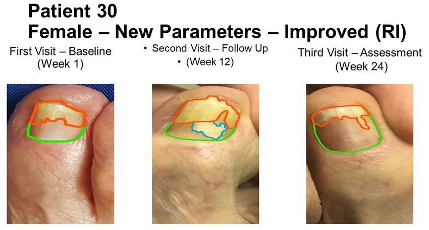
Figure4.4.Objective
Figure Objectiveimage
imageanalysis
analysisof
ofinfected
infecteddigits.
digits.The
Theoverall
overallresults
resultsindicate
indicatethat
thatthe
theparameters
parameters
set in this study was the most successful in treating OM infections. Example of a Group
set in this study was the most successful in treating OM infections. Example of a Group 3 patient3 patient
(fe-
(female, 85 years of age, infection duration 10 years, positive PAS stain) indicating overall improve-
male, 85 years of age, infection duration 10 years, positive PAS stain) indicating overall improvement
ment over 24 weeks.
over 24 weeks.
Enteringthe
Entering theresults
resultsofofthe
theexperiment,
experiment,the thedata
dataobtained
obtained would
would show
show that
that there
there waswas
a
a statistical significance, p = < 0.01 (**), for standard laser treatment compared
statistical significance, p ≤ 0.01 (**), for standard laser treatment compared with, p ≤ 0.001 with, p =<
0.001new
(***), (***),laser
newparameter
laser parameter treatment,
treatment, drawingdrawing as an indication
as an indication that the treatment
that the treatment needed
needed over time decreased. No adverse effects were noted
over time decreased. No adverse effects were noted with new laser therapy. with new laser therapy.
An 86.7%An
86.7% visual improvement was recorded in Group 3 after 24 weeks;
visual improvement was recorded in Group 3 after 24 weeks; In conclusion, the authors In conclusion, the
authors consider
consider phototherapy phototherapy
as “the bestas “the best treatment
treatment option foroption for Onychomycosis”
Onychomycosis” and considerand
consider establishing
establishing a “newfor
a “new protocol protocol for the standardization
the standardization of laser irradiation
of laser irradiation with the
with the possible
possible inclusion
inclusion into theClinical
into the Scoring ScoringIndex
Clinical
for Index for Onychomycosis
Onychomycosis treatmenttreatment
plan”. plan”.
4.4.Discussion
Discussion
ItItisisevident
evidentthatthatOMOMisisincreasing
increasingininresistance
resistanceagainst
againstoral
oraland
andtopical
topicalantifungal
antifungal
agents.
agents. Laser
Laser therapy
therapy andand light
light has
has been
been the
the topic
topic of
of discussion
discussion for
for the
the treatment
treatment of of OM;
OM;
more specifically, the 1064 nm ND: YAG laser. Preliminary studies show
more specifically, the 1064 nm ND: YAG laser. Preliminary studies show good clinical and good clinical and
microscopical
microscopical cure cure rates
ratesusing
usingthetheNd:
Nd: YAG
YAG10641064nmnmlaser
laserininthe
theshort
shortterm.
term.Published
Published
studies
studies revealed
revealed poor,
poor,small
smallstudy
studysamples;
samples;no nostandard
standardPodiatric
Podiatrictreatment
treatmentwas wasdone
done
prior
priortotolaser
lasertreatment;
treatment; not enough
not enough time given
time givenbetween
betweentreatments in order
treatments to seetoclear
in order nail
see clear
growth;
nail growth;limited output
limited and no
output and clear protocol
no clear couldcould
protocol be attained during
be attained literature
during review.
literature re-
Aview.
clearAprotocol with three separate study groups were designed and
clear protocol with three separate study groups were designed and proposed forproposed for the
purpose
the purpose of this
of study.
this study.
4.1. Standard Podiatric Treatment Efficacy
4.1. Standard Podiatric Treatment Efficacy
Standard podiatric treatment is important when deciding to treat OM, as the fungi
Standard podiatric treatment is important when deciding to treat OM, as the fungi
denatures the nail keratin, causing increased nail thickness. This thickness decreases
denatures the nail keratin, causing increased nail thickness. This thickness decreases treat-
treatment outcomes as the product/therapy cannot penetrate the desired structures. A
ment outcomes as the product/therapy cannot penetrate the desired structures. A study
study conducted by Malay looked at the different outcomes in 55 patients between nail
conducted by Malay looked at the different outcomes in 55 patients between nail debride-
debridement alone and nail debridement with a topical antifungal nail lacquer. The
ment alone and nail debridement with a topical antifungal nail lacquer. The primary
primary outcome was mycological cure. Of the 55 patients, 27 (49.09%) were allocated
to the debridement only group, and 28 (50.91%) were allocated to the debridement and
antifungal group. At 10 months and 2 weeks follow-up, 76.74% of patients achievedPhotonics 2021, 8, 350 11 of 16
mycological cure in the antifungal group. None experienced mycological cure in the
control group [35]. Recently, Fernández et, al. did a study where they combined laser
and ozone therapy for OM in vitro and ex vivo. In the ex vivo model experiments, with
the same duration and ozone concentration, Penicillium chrysogenum and Epidermophyton
floccosum showed total inhibition; Trichophyton mentagrophytes and Trichophyton rubrum
showed a 75% growth inhibition; Microsporum canis showed a delay in sporulation; and
Microascus brevicaulis and Aspergillus terreus did not show growth inhibition. This combined
laser and ozone treatment may be developed as a fast therapy for human onychomycosis,
as a potential alternative to the use of antifungal drugs with potential side effects and
long duration treatments [36]. This supports our reasoning for doing standard podiatric
treatment for all patients and likely contributed to the results, as the nails were etched thin
enough for treatment to penetrate.
4.2. Specimen Sampling and Analysis
Reported studies support the findings in our study where PAS stain identified OM
infections in 91.1% of samples, compared to 44.4% in culture form [7,8,13,14,35]. This
was a culture failure rate of 55.6%. Although PAS staining shows good sensitivity rates,
the specificity of this test is low. This is a clinical concern when prescribing an oral or
topical antifungal agent, as these drugs act against certain fungal elements. Culture results
need to be present when prescribing a specific antifungal agent. However, laser therapy
works on the principle of denaturing a fungal elements membrane potential through heat.
When a practitioner is considering laser therapy as a treatment option, culture results are
not necessary. It is recommended to do both PAS staining (as primary test) and Fungal
Microscopy (as secondary test) when doing combination therapy.
4.3. Infective Organisms
In earlier published studies, OM was primarily caused by dermatophytes (60–90%),
followed by yeasts and rarely by non-dermatophyte moulds [5,7,8,11–13], which is incon-
sistent with our study where the primary infective agent was non-dermatophyte moulds
(22.2%), followed by dermatophyte infections (20%) and yeasts (2.2%). As reported by
Kaur, et al., these previously regarded contaminants are now increasingly recognized as
primary causative pathogens [10]. However, careful diagnostic attention is required when
identifying non-dermatophyte moulds as aetiologic agents. The biggest difference between
a non-dermatophyte mould infection and others, is that the mould should be the sole (or
primary) aetiologic agent in culture and can only be achieved by repeated reproducibility
growths at different time points and that dermatophytes do not appear on repeated culture
attempts [37].
4.4. SCIO Decrease
In Group 1, there was an overall infection measurement decrease of 4.578, which
correspond with literature [28,38]. Group 2 showed an overall infection measurement
decrease of 7.2270, which was statistically significant (p ≤ 0.01 (**)) and may be due to the
higher SCIO at Week 1. Finally, in Group 3, there was an overall infection measurement
decrease of 8.4227, and it was the biggest statistically significant (p ≤ 0.001 (***)) decrease
compared to standard laser and control groups. This may prove an accurate indicator
of therapeutic effectiveness, but further clinical studies must be done before definitive
claims can be made [30]. As we aimed to identify the effectiveness of laser therapy for
OM compared to topical treatment, this may bridge the gap of clinical effectiveness by
adding laser therapy as a possible route of treatment, depending on the SCIO. Ethical
clearance was granted. Helou et, al. concluded in their study of 105 patients, where they
looked at the response of big toenail OM to 1064 nm Nd: YAG laser treatment that the SCIO
decrease after 3 sessions of Nd: YAG laser was significantly more important in women and
in patients with positive mycology culture, smaller affected area of the nail, no subungual
hyperkeratosis, and no nail matrix involvement. Age, smoking, hypertension, and sidePhotonics 2021, 8, 350 12 of 16
effects were not shown to significantly correlate with the decrease of the SCIO score [39],
which is similar to the findings of Kandpal et, al. [40]. Both of the findings in these studies
correlate with the findings in our study where there was a significant decrease in the SCIO
with the proposed new laser parameter settings.
4.5. Temperature and Pain Correlation
When looking at laser therapy as a potential alternative treatment for OM, the probable
pain associated with it has to be taken into consideration. The type of laser chosen to make
the comparison is justifiable for their intrinsic characteristics and for the literature data that
report the use of 1064 nm ND: YAG laser. “Preliminary studies show good clinical and
microscopical cure rates using the Nd: YAG 1064 nm laser in the short term” [2,4,12,18,20].
In Group 2, the mean hallux temperature reached was 38.83 degrees Celsius at Week 1 and
39.64 at Week 12, and the mean hallux pain experienced at Week 1 was 0.5/10 and 0.9/10
for Week 12. Both increases were not significant for standard laser therapy. In Group 3, the
mean hallux temperature reached was 40.94 degrees Celsius at Week 1 and 41.39 at Week
12, and the mean hallux pain experienced at Week 1 was 0.6/10 and 1.7/10 for Week 12.
Also, both increases were not significant for new laser therapy. When comparing Week 1
temperatures between Groups 2 and 3, there was a statistically significant increase (p ≤ 0.01
(**)) and may be due to the increase in energy fluence for the new laser parameters set by
us as well as the inactivity of fungal elements after the initial treatment. As for the lesser
digits in Group 2, the mean toe temperature reached was 41.00 degrees Celsius at Week 1
and 42.89 at Week 12 and was a statistically significant increase (p ≤ 0.05 (*)). There was
also a significant increase (p ≤ 0.05 (*)) in temperature between Groups 2 and 3 at Week
1. When comparing Groups 2 and 3, there was a bigger statistically significant increase in
temperature (p = 0.001 (***)) at Week 12. The mean lesser digit pain experienced at Week 1
was 1.7/10 and 3.9/10 for Week 12, which was also statistically significant (p ≤ 0.05 (*))
when compared to Week 1 in Group 3. Our findings are supported by a study conducted
by Kozarev, where 72 patients were treated with a long-pulse 1064 nm Nd: YAG laser, with
a fluence of 35–40 J/cm2 ; 4 mm spot size; pulse duration 35 ms. During treatment, the nails
reached 50 degrees Celsius and was at 40 degrees Celsius after one minute. Patient pain
was reported as follows: 24 (33.33%) no pain; 35 (48.61%) mild pain; 13 (18.06%) moderate
pain; 0% severe pain; 0% intolerable. Kozarev also reported that the desired average tissue
temperature for laser irradiation is between 43–50 degrees Celsius, which support the
findings in our study [41]. Tolerating higher temperatures is in correlation with increased
blood flow and may explain why patients experienced less pain tolerance at the second
treatment. Heating the fungal colonies does not instantly kill them, but it results in the
disability of them to replicate or survive according to an apoptotic mechanism. Killing of
the fungal colonies may be caused by superheating and exploding or rupturing the fungal
cell membranes [41].
4.6. Overall Findings
In the control group, 11 (73.3%) patients showed improvement and 4 (26.7%) failed
to respond and this could be due to patient non-adherence to protocol given regarding
adequate decontamination of shoes, socks, etc. at home, or failure to apply the Amorolfine
once weekly. In another study conducted by Gupta, et al., they did an evaluation of
26 studies and found that 5% topical Amorolfine was effective in mild to moderate OM,
and they also had to apply the treatment to the affected nails once weekly. Here, two
open, randomized studies compared the efficacy and safety between once-weekly applica-
tions and twice-weekly applications. Both studies found slightly higher cure rates in the
twice-weekly groups; however, there was no statistical significance between the dosage
regimens [42]. Another study conducted by Ghannoum and Isham stated that low efficacy
rates can mainly be attributed to the inability of the drug to penetrate the nail plate and bed
where the infection resides [19]. This study’s 5% topical Amorolfine outcomes are better,Photonics 2021, 8, 350 13 of 16
when compared to another study by Roberts, et al., which indicate only a 50% efficacy for
fingernail and toenail OM [20].
In the standard laser therapy group, 9 (64.3%) patients showed improvement and 5
(35.7%) failed to respond. Interestingly, this indicates that standard laser therapy was not
a better treatment option for OM infections compared to conventional therapy, although
the SCIO decreased more between standard laser therapy patients compared to control
patients. However, this could be due to patient non-adherence to decontamination protocol
given for shoes, socks, etc. This was the original research problem—that standard (industry
set) laser parameters were not effective in treating OM. Our findings are supported by
Hollmig, et al., where the primary end point was a negative mycological cure from all
clinically involved nails. Laser parameters (recommended by manufacturer, as was the
case with standard laser settings in our study) included: 1064 nm; fluence of 5 J/cm2 ; pulse
width 0,3 ms; spot size 6 mm and rate of 6 Hz to achieve measured target temperatures
of between 40–42 degrees Celsius. 12 laser patients completed the study at 3 months, and
only 50% had negative fungal cultures. At 12 months follow up, no modest improvement
of nail plate clearance was sustained [43–46].
In the final group, who had new laser parameters set by us, 13 (86.7%) patients
showed improvement and 2 (13.3%) failed to respond. These results also indicate that new
laser therapy was the best treatment option for OM infections compared to conventional
and standard laser therapies. The overall SCIO for this group decreased the most. Our
results were more effective when compared to a study conducted by Zhang, et al., who
used long-pulse 1064 nm laser therapy on 33 patients with clinically and mycologically
proven OM. All patients were given 8 sessions at 1-week intervals. At week 8, 68%
of nails showed mycological cure [43]. Our study results are also higher than those of
Wantiphakdeedecha, et al., where 35 patients demonstrated an overall mycological cure
rate of 51.9% at 6 months [31,44,47,48].
The results at week 24 show an overall improvement in infection of 33 patients (75%).
This improvement is based on the combined negative PAS stain results and the visual
improvement after 6 months. There was no difference in overall improvement regardless
of treatment between week 12 and 24; however, there was a difference between individual
group improvement rates. All patients who received standard and new laser therapy
reported no adverse effects during or after treatment. When asked whether the laser
parameters set for this study was tolerable, both group’s patients indicated that it was.
Summarizing, during the treatments of the three groups which lasted for 12 weeks, with
the use of lasers, a temperature higher than 50 ◦ C was never reached, nor a pain index
higher than mild pain. The authors, based on the results and literature data, propose as
the antifungal mechanism of the laser treatment used the fact that the “Heating the fungal
colonies does not instantly kill them, but it results in the disability of them to replicate
or survive according to an apoptotic mechanism. The killing of the fungal colonies may
be caused by superheating and exploding or rupturing the fungal cell membranes [33].
From the findings, a new protocol for the standardization of laser therapy with the possible
inclusion into the SCIO treatment plan, has been proposed.
5. Conclusions
In conclusion, the authors state that onychomycosis, due to the new pharmacological
resistance and the risk of hepatotoxicity of oral therapies, is a current problem from
a therapeutic point of view. Thus, new non-pharmacological or non-pharmacological
therapeutic approaches must be taken into consideration. We aimed to set new laser
parameters that were effective in treating OM and we also wanted to ascertain whether
these parameters were tolerable and safe for patients. The proposed new laser parameters
were subjected to clinical verification and the data obtained gave indications of a better
therapeutic activity both of the parameters previously used with the same type of laser,
and of the standard topical antifungal therapy. The findings indicate that new parameters
were more effective at 6 months, compared to other methods topical antifungal therapyPhotonics 2021, 8, 350 14 of 16
and standard laser therapy. A new protocol for the standardization of laser therapy has
been proposed with the possible inclusion into the SCIO treatment plan.
Author Contributions: Conceptualization, N.D. and H.A.; methodology, formal analysis, N.D.;
writing—original draft preparation, N.D.; writing—review and editing, N.D. and H.A.; supervision,
H.A.; project administration, H.A.; funding acquisition, H.A. All authors have read and agreed to the
published version of the manuscript.
Funding: This work is based on the research supported by the South African Research Chairs
Initiative of the Department of Science and Technology and National Research Foundation of South
Africa (Grant No 98337). The authors sincerely thank the University of Johannesburg, the National
Laser Centre, and the National Research Foundation of South Africa for their financial grant support.
Institutional Review Board Statement: The study was conducted according to the guidelines of the
Declaration of Helsinki, and approved by the University of Johannesburg Research Ethics Committee
(Approval number: REC-01-176-2015 Date: 3 June 2015).
Informed Consent Statement: Informed consent was obtained from all subjects involved in the
study.
Data Availability Statement: The data presented in this study are available on request from the
corresponding author. The data are not publicly available due to privacy issues.
Acknowledgments: The authors would like to acknowledge the Institution, University of Johannes-
burg, for the opportunity to conduct this study under its auspices.
Conflicts of Interest: The authors declare no conflict of interest.
Ethical Approval: Ethical approval has been granted for studies involving human subjects by the
University of Johannesburg Faculty of Health Sciences Faculty Ethics Committee on 3 June 2015,
reference number REC-01-176-2015. Furthermore, all patients who participated in this study gave
formal consent for the outcomes of this study to be published, with images.
Abbreviations
OM Onychomycosis
DSO Distal Subungual Onychomycosis
DLSO Distolateral Subungual Onychomycosis
PSO Proximal Subungual Onychomycosis
PWSO Proximal White Superficial Onychomycosis
TDO Total Dystrophic Onychomycosis
WSO White Superficial Onychomycosis
SCIO Scoring Clinical Index for Onychomycosis
Nd: YAG Neodymium Yttrium Aluminium Garnet
FDA Food and Drug Administration
KOH Potassium Hydroxide
PAS Periodic Acid-Schiff
MC&S Fungal microscopy and culture
References
1. Baran, R.; Kaoukhov, A. Topical antifungal drugs for the treatment of onychomycosis: An overview of current strategies for
monotherapy and combination therapy. J. Eur. Acad. Dermatol. Venereol. 2005, 19, 21–29. [CrossRef]
2. Kimura, U.; Takeuchi, K.; Kinoshita, A.; Takamor, K.; Hirum, M.; Suga, Y. Treating onychomycosis of the toenail: Clinical efficacy
of the sub-millisecond 1064 nm Nd. YAG laser using a 5 mm spot diameter. J. Drugs Dermatol. 2012, 11, 496–504. [PubMed]
3. Ortiz, A.E.; Avram, M.M.; Wanner, M.A. A review of lasers and light for the treatment of onychomycosis. Lasers Surg. Med. 2014,
46, 117–124. [CrossRef]
4. Suga, Y.; Kimura, U.; Hiruma, M. Can persistent toenail fungus be successfully treated with laser? J. Med. Mycol. 2014, 55, J65–J71.
[CrossRef] [PubMed]
5. Tcherney, G.; Penev, P.K.; Nenoff, P.; Zisova, L.G.; Cardoso, G.C.; Taneva, T.; Ginter-Hanselmayer, G.; Ananiev, J.; Gulubova,
M.; Hristova, R.; et al. Onychomycosis: Modern diagnostic and treatment approaches. Wien. Med. Wochenschr. 2013, 163, 1–12.
[CrossRef] [PubMed]Photonics 2021, 8, 350 15 of 16
6. Tosti, A. Efinaconazole solution 10%: Topical antifungal therapy for toenail onychomycosis. Cutis 2013, 92, 203–208. [PubMed]
7. Welsh, O.; Vera-Cabrera, L.; Welsh, E. Onychomycosis. Clin. Dermatol. 2010, 28, 151–159. [CrossRef]
8. Nenoff, P.; Grunewald, S.; Paasch, U. Laser therapy of onychomycosis. J. Der Dtsch. Dermatol. Ges. 2014, 12, 33–38. [CrossRef]
9. Waibel, J.; Wulkan, A.J.; Rudnick, A. Prospective efficacy and safety evaluation of laser treatments with real-time temperature
feedback for fungal onychomycosis. J. Drugs Dermatol. 2013, 12, 1237–1242.
10. Kaur, R.; Kashyap, B.; Bhalla, P. Onychomycosis—Epidemiology, diagnosis and management. Indian J. Med. Microbiol. 2008, 26,
108–116. [CrossRef]
11. Evans, E.G. Causative pathogens in onychomycosis and the possibility of treatment resistance: A review. J. Am. Acad. Dermatol.
1988, 38, S32–S36. [CrossRef]
12. Gupta, A.K.; Paquet, M.; Simpson, F.C. Therapies for the treatment of onychomycosis. Clin. Dermatol. 2013, 31, 544–554.
[CrossRef] [PubMed]
13. Noguchi, H.; Miyata, K.; Sugita, T.; Hiruma, M.; Hiruma, M. Treatment of onychomycosis using a 1064 nm Nd. YAG laser. J. Med.
Mycol. 2013, 54, 333–339. [CrossRef] [PubMed]
14. Nenoff, P.; Krűger, C.; Ginter-Hanselmayer, G.; Tietz, H.J. Mycology—An update. Part 1: Dermatomycosis: Causative agents,
epidemiology and pathogenesis. J. Ger. Soc. Dermatol. 2014, 1203, 188–210. [CrossRef]
15. Nenoff, P.; Krűger, C.; Ginter-Hanselmayer, G.; Schulte-Beerbühl, R.; Tietz, H.-J. Mycology—An update. Part 2: Dermatomycosis:
Clinical picture and diagnosis. J. Ger. Soc. Dermatol. 2014, 1209, 749–777.
16. Thomas, J.; Jacobson, G.A.; Narkowicz, C.K.; Peterson, G.; Burnet, H.; Sharpe, C. Toenail onychomycosis: An important global
disease burden. J. Clin. Pharm. Ther. 2010, 35, 497–519. [CrossRef]
17. Baudraz-Rosselet, F.; Ruffieux, C.; Lurati, M.; Bontems, O.; Monod, M. Onychomycosis insensitive to systemic terbinafine and
azole treatments reveals non-dermatophyte molds as infectious agents. Dermatology 2010, 220, 164–168. [CrossRef]
18. Westerberg, D.P.; Voyack, M.J. Onychomycosis: Current trends in diagnosis and treatment. Indian J. Clin. Pract. 2014, 25, 309–319.
19. Ghannoum, M.; Isham, N. Fungal nail infections (onychomycosis): A never-ending story? PLoS Pathog. 2014, 10, 1–5. [CrossRef]
20. Roberts, D.T.; Taylor, W.D.; Boyle, J. Guidelines for treatment of onychomycosis. Br. J. Dermatol. 2003, 148, 403–410. [CrossRef]
21. Sergeev, A.Y.; Gupta, A.K.; Sergeev, Y.V. The scoring clinical index for onychomycosis (SCIO Index). Ski. Ther. Lett. 2002, 7, 6–7.
22. Gupta, A.K.; Simpson, F.C. New therapeutic options for onychomycosis. Expert Opin. Pharmacother. 2012, 13, 1131–1142.
[CrossRef] [PubMed]
23. Wilsmann-Theis, D.; Sareika, F.; Bieber, T.; Schmid-Wendtner, M.-H.; Wenzel, J. New reasons for histopathological nail clipping
examination in the diagnosis of onychomycosis. J. Eur. Acad. Dermatol. Venereol. 2011, 25, 235–237. [CrossRef]
24. Zaias, N. Onychomycosis. Arch. Dermatol. 1972, 105, 263–274. [CrossRef]
25. Bristow, I.R. The effectiveness of lasers in the treatment of onychomycosis: A systemic review. Br. J. Foot Ankle Res. 2014, 7, 1–10.
[CrossRef] [PubMed]
26. Evans, E.G. Resistance of Candida species to antifungal agents used in the treatment of onychomycosis: A review of current
problems. Br. J. Dermatol. 1999, 141, 33–35. [CrossRef]
27. Bunert, N.; Homey, B.; Gerber, P.A. Onychomycosis. Successful treatment with a 1064-nm Nd. YAG laser. Der Hautarzt 2013, 64,
716–718. [CrossRef]
28. Gupta, A.K.; Paquet, M. A retrospective chart review of the clinical efficacy of Nd. YAG 1064-nm laser for toenail onychomycosis.
J. Dermatol. Treat. 2014, 5, 1–3.
29. Heers, H.; Jäger, M.W.; Raulin, C. Treatment of onychomycosis using the 1064 nm Nd. YAG laser: A clinical pilot study. J. Der
Dtsch. Dermatol. Ges. 2014, 12, 322–329.
30. Wantiphakdeedecha, R.; Thanomkitti, K.; Bunyaratavej, S.; Manuskiatti, W. Efficacy and safety of 1064 nm Nd: YAG laser in the
treatment of onychomycosis. J. Dermatol. Treat. 2015, 27, 75–79. [CrossRef]
31. Dembskey, N.; Abrahamse, H. Laser Therapy for the Treatment of Onychomycosis: Best Evidence Based Practice or Not? Clin.
Res. Foot Ankle 2016, 4, 3–7. [CrossRef]
32. Malay, D.S. Efficacy of debridement alone versus debridement combined with topical antifungal nail lacquer for the treatment of
pedal onychomycosis: A randomised, controlled trial. J. Foot Ankle Surg. 2009, 48, 294–308. [CrossRef]
33. Gupta, A.K.; Ryder, J.E.; Summerbell, R.C. The diagnosis of non-dermatophyte mold onychomycosis. Int. J. Dermatol. 2003, 24,
272–273. [CrossRef] [PubMed]
34. Sigurgeirsson, B.; Olafsson, J.H.; Steinsson, J.T.; Kerrouche, N.; Sidou, F. Efficacy of amorolfine nail lacquer for the prophylaxis of
onychomycosis over 3 years. J. Eur. Acad. Dermatol. Venereol. 2010, 24, 910–915. [CrossRef] [PubMed]
35. Aditya, K. Gupta, Maanasa Venkataraman, Emma M Quinlan. Efficacy of lasers for the management of dermatophyte toenail
onychomycosis. J. Am. Podiatr. Med. Assoc. 2021, 20–236. [CrossRef]
36. Ameen, M.; Lear, T.J.; Madan, V.; Mustapa, M.F.M.; Richardson, M. British Association of Dermatologists’ guidelines for the
management of onychomycosis 2014. Br. J. Dermatol. 2014, 171, 937–958. [CrossRef]
37. Gupta, A.K.; Jain, H.C.; Lynde, C.W.; Macdonald, P.; Cooper, E.A.; Summerbell, R.C. Prevalence and epidemiology of onychomy-
cosis in patients visiting physicians’ offices: A multicenter Canadian survey of 15,000 patients. J. Am. Acad. Dermatol. 2000, 43 (Pt
1), 244–248. [CrossRef]
38. Fernández, J.; Del Valle Fernández, I.; Villar, C.J.; Lombó, F. Combined laser and ozone therapy for onychomycosis in an in vitro
and ex vivo model. PLoS ONE 2021, 16, e0253979. [CrossRef]Photonics 2021, 8, 350 16 of 16
39. Kandpal, R.; Arora, S.; Arora, D. Study of Q-switched Nd: YAG Laser versus Itraconazole in Management of Onychomycosis. J.
Cutan. Aesthet. Surg. 2021, 14, 93–100.
40. Kosarev, J. Novel laser therapy in the treatment of onychomycosis. J. Laser Health Acad. 2010, 1, 1–8.
41. Gupta, A.K.; Simpson, F.C. Device-based therapies for onychomycosis treatment. Skin Ther. Lett. 2012, 17, 57–61.
42. Hollmig, T.; Rahman, Z.; Henderson, M.T.; Rotatori, R.M.; Gladstone, H.; Tang, J.Y. Lack of efficacy with 1064 nm Nd: YAG laser
for the treatment of onychomycosis: A randomised, controlled trial. J. Am. Acad. Derm. 2014, 70, 911–917. [CrossRef] [PubMed]
43. Zhang, R.N.; Wang, D.K.; Zhuo, F.L.; Duan, X.-H.; Zhang, X.-Y.; Zhao, J.-Y. Long-pulse Nd: YAG 1064 nm laser treatment of
onychomycosis. Chin. Med. J. 2012, 125, 3288–3291. [PubMed]
44. Hochman, L.G. Laser treatment of onychomycosis using a novel 0.65-milisecond pulsed Nd:YAG 1064 nm laser. J. Cosmet. Laser
Ther. 2011, 13, 2–5. [CrossRef] [PubMed]
45. Sotiriou, E.; Koussidou-Ermonti, T.; Chaidemenos, G.; Apalla, Z.; Ioannides, D. Photodynamic therapy for distal and lateral
subungual toenail onychomycosis caused by Trichophyton rubrum: Preliminary results of a single-centre open trial. Acta Derm.
Venerol. 2010, 90, 216–217. [CrossRef]
46. Shemer, A.; Davidovici, B.; Grunwald, M.H.; Lyakhovitsky, A.; Amichai, B. Onychomycosis: A simpler in-office technique for
sampling specimens. J. Fam. Pract. 2012, 61, 552–554.
47. Gupta, A.K.; Ryder, J.E.; Baran, R. The use of topical therapeutics to treat onychomycosis. Derm. Clin. 2003, 21, 481–489. [CrossRef]
48. Helou, J.; Maatouk, I.; Soutou, B. Big toenail onychomycosis features associated with response to 1064 nm Nd: YAG laser
treatment. J. Cosmet. Derm. 2021. [CrossRef]You can also read