Original Article A novel regulatory function for miR-217 targetedly suppressing fibronectin expression in keloid fibrogenesis
←
→
Page content transcription
If your browser does not render page correctly, please read the page content below
Int J Clin Exp Pathol 2018;11(4):1866-1877
www.ijcep.com /ISSN:1936-2625/IJCEP0021672

Original Article
A novel regulatory function for miR-217 targetedly
suppressing fibronectin expression
in keloid fibrogenesis
Wei Wu1, Feng Xie1, Yi Zhang2, Xiuxia Wang1, Lingling Xia1, Xiaoli Wu1, Zhen Gao1
1
 Department of Plastic and Reconstructive Surgery, Shanghai Ninth People’s Hospital, Shanghai Jiao Tong Univer-
sity School of Medicine, Shanghai, China; 2Biomedcial Research Center, Shanghai Zhong Shan Hospital, Fudan
University, Shanghai, China
Received December 11, 2015; Accepted February 18, 2016; Epub April 1, 2018; Published April 15, 2018

Abstract: Fibronectin (FN) plays a critical role in the development and progression of keloid scarring (KS). In the
present study, we hypothesized that a post-translational mechanism of microRNAs regulated the expression of FN in
keloid scarring fibroblasts (KSFBs). Here, we collected 20 KS tissues and paired corresponding adjacent normal tis-
sues from clinical patients and measured the expression of miRNA-217. First, by using PicTar, TargetScan and miR-
Base database, we found that miRNA-217 might be a regulator of FN in human species. Based on these hypotheses,
the expression of miRNA-217 and FN in KS tissues was investigated. The results demonstrated that the expression
of miRNA-217 was greatly suppressed, and FN was increased in KS tissues. Intriguingly, the expression levels of
endogenous miRNA-217 negatively correlated with the FN mRNA levels (Pearson’s correlation coefficient = -0.683,
P < 0.001). In vitro, miRNA-217 could regulate FN through the predicted binding sites in its 3’-UTR. miRNA-217
played an impact on cell proliferation and apoptosis, thereby regulating KSFBs growth. Moreover, miRNA-217 gain-
of function decreased FN, Col-1 and Col-3 protein expression, and miRNA-217 loss-of function increased FN, Col-1
and Col-3 protein expression in KSFBs. In conclusion, overexpressed miRNA-217 could inhibit KSFBs growth, and
the underlying mechanism was mediated, at least partially, through the suppression of FN expression. But above all,
miRNA-217 might play a potential therapeutic avenue for the treatment of keloid fibrogenesis.

Keywords: MiRNA-217, fibronectin, keloid fibrogenesis, fibroblasts

Introduction                                                  Fibrogenesis is characterized by ECM remod-
                                                              eling and stiffening. However, the functional
Keloid scarring (KS) is a benign dermal fibropro-             contribution of tissue stiffening to non-cancer
liferative disease due to abnormal wound heal-                pathogenesis remains largely unknown. Fib-
ing process after skin injury and is character-               ronectin (FN) is an ECM glycoprotein that exists
ized by overproduction of extracellular matrix                in blood plasma in its soluble form (plasma
(ECM) that extend beyond the area of the origi-               type) and in its insoluble form (cellular type) as
nal skin injury in predisposed individuals [1-3].             a part of the ECM of almost every tissue in an
Excess deposition of ECM such as hyalinized                   organism [6]. Previous studies indicate that
collagen bundles by fibroblasts is responsible                overexpressed FN distorts the architecture of
for keloid [4]. Previous studies indicate that                the liver and leads to hepatic cirrhosis and con-
transforming growth factor-beta1 (TGF-β1) can                 sequently hepatocellular cancer (HCC) [7, 8]. In
induce collagen production and contraction in                 early phase of wound healing, FN contributes to
scar-derived human skin fibroblasts [5, 6]. Ho-               the formation of granulation tissue, which is
wever, the underlying molecular mechanisms in                 similar to embryonic skin [9]. In the psoriatic
keloid fibrogenesis are still poorly understood.              non-lesional skin, the expression of FN is ele-
Therefore, there is an urgent need for a better               vated by fibroblasts, and the current findings
understanding of keloid pathogenesis in ord-                  imply that FN plays a pathophysiological roles in
er to develop better prevention and treatment                 fibrogenesis [10]. In KS, plasma FN and then in
approaches.                                                   situ FN are deposited at the site of injury. In situ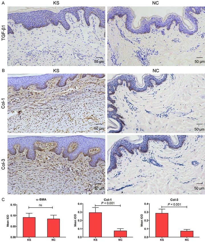
microRNA-217 in keloid fibrogenesis

FN makes by tissue cells at the injury site often   lished as described previously [1] and cultured
contains an extra domain A (EDA) insert. Multi-     in Dulbecco’s modified Eagle’s medium (DMEM,
ple wound-related signal transduction path-         Gibco Life Technologies) that contained 10%
ways control the deposition of EDA-FN, and the      fetal calf serum (Gibco Life Technologies), 10%
EDA insert can in turn trigger pathways that        L-glutamine, 0.5% penicillin/streptomycin, 10%
induce inflammation, increased extracellular        nonessential amino acids and 10% pyruvate, in
matrix molecule deposition including FN and         a 5% CO2 atmosphere and incubated at 37°C.
collagen, and activation of fibroblasts, which
can create a vicious cycle that lead to fibrosis    MTT assay
or keloid formation [11]. These results indicate
that FN may be a key target for the develop-        Cell proliferation was monitored by a 3-(4,5-
ment of novel therapeutic strategies for keloid     dimethylthiazol-2-yl)-2,5-diphenyltetrazolium
fibrogenesis. However, the underlying molecu-       bromide (MTT) Cell Proliferation/Viability As-
lar mechanisms of microRNAs by targeting FN         say kit (R&D SYSTEMS) in according to the
contribute to the induction of keloid fibrogene-    guidelines.
sis remains to be determined.
                                                    Caspase-3 activity assay
In the present study, we hypothesize that a
                                                    KSFBs lysates were prepared and incubated
post-translational mechanism may exist for FN
                                                    with anti-caspase 3. Immunocomplexes were
expression and be regulated by microRNAs in
                                                    incubated with peptide substrate in assay buf-
keloid tissues and keloid fibroblasts. By using
                                                    fer for 2 h at 37°C. Release of p-nitroaniline
PicTar, TargetScan, and miRBase database and
                                                    was measured at 405 nm using an ELISA read-
microarray assay, we indicate that miRNA-217
                                                    er (MD SpectraMax M5, USA) according to the
is a regulator of FN in human species and
                                                    manufacturer’s instructions.
decreases expression in keloid tissues and
keloid fibroblasts, and its aberrant regulation     Quantification of apoptosis by flow cytometry
has been demonstrated in a wide variety of dis-
eases [12, 13]. Furthermore, miRNA-217 will be      Quantitative assessment of apoptotic cells was
analyzed for its potential use as therapeutic       also assessed by the terminal deoxynucleotidyl
target to develop novel strategies for keloid       transferase-mediated deoxyuridine triphos-
fibrogenesis prevention and treatment.              phate nick endlabeling (TUNEL) method, which
                                                    examines DNA-strand breaks during apoptosis
Materials and methods                               by using BD ApoAlertTM DNA Fragmentation
                                                    Assay Kit. The KSFBs were trypsinized, fixed
Patients’ samples                                   with 4% paraformaldehyde, and permeabilized
                                                    with 0.1% Triton-X-100 in 0.1% sodiumcitrate.
Twenty keloid scarring tissues (KS) and paired
                                                    After being washed, the cells were incubated
corresponding adjacent normal tissues (NC)
                                                    with the reaction mixture for 60 min at 37°C.
were collected from patients who had under-
                                                    Cells were immediately analyzed using FACScan
gone surgical excision at the Shanghai Ninth
                                                    and the Cellquest program (Becton Dickinson).
People’s Hospital, Shanghai Jiao Tong University
School of Medicine between June 2012 and            Histological observation and immunohisto-
June 2015. All collected tissue samples were        chemical staining
immediately stored at liquid nitrogen until use.
Human samples were obtained with written            Formalin-fixed and paraffin-embedded HS tis-
informed consent from all patients. The study       sues were cut into about 4 μm section, which
was approved by the Ethics Committee of the         were stained with hematoxylin & eosin (H&E)
Shanghai Jiao Tong University School of Medi-       staining, and visualized under a microscope
cine (Shanghai, China).                             (Leica DM 2500).

Cell culture                                        Paraffin embedded KS tissues were cut into
                                                    about 4 μm section and mounted on glass
Human keloid scarring fibroblasts (KSFBs) and       slides and stained by immunoperoxidase. The
embryonic skin fibroblasts (ESF) were estab-        paraffin sections were baked in oven at 65°C

1867                                                   Int J Clin Exp Pathol 2018;11(4):1866-1877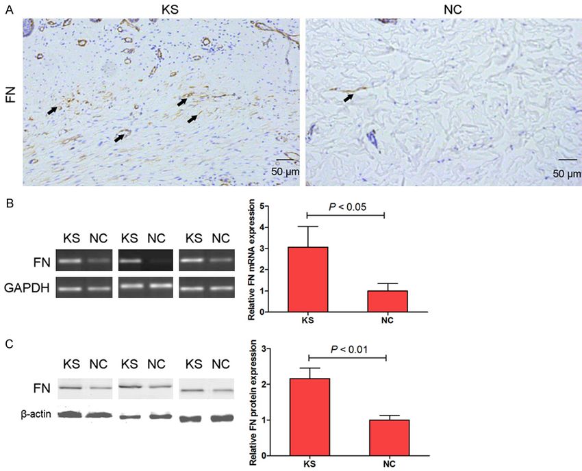
microRNA-217 in keloid fibrogenesis

for 24 h, then dewaxing to water, rinsed with            GUUCCUGAUGCAGUA-3’; and 5’-ACUUUCAG-
PBS 3 times. Well washed section was placed              AGGUCUUGACCUAG-3’. Cells were transfected
in the EDTA buffer for microwave antigen re-             using Lipofectamine2000 (Invitrogen, CA, USA)
trieval, the fire to boil, then low heat to boil after   at a final concentration of 50 nM. At 24 h post-
an interval of 10 min. After natural cooling, the        transfection, the culture medium was changed.
sections were washed with PBS 3 times. The               After 48 h, cells were harvested for analysis.
sections were put into 3% hydrogen peroxide
                                                         Real time-polymerase chain reaction
solution and incubated at room temperature for
10 min, which was to block endogenous peroxi-            RNA extraction was performed according to
dase, then washed with PBS 3 times, closed               the TRIzol manufacturer’s protocol (Invitrogen,
with 5% bovine serum albumin (BSA) for 20                Carlsbad, CA, USA). Synthesis of cDNAs was
min. After removal of BSA liquid, each section           performed by reverse transcription reactions
was added with 50 μl diluted primary antibody            with 4 μg of total RNA using moloney murine
overnight at 4°C, then washed with PBS 3                 leukemia virus reverse transcriptase (Invi-
times. After the removal of PBS liquid, each             trogen) with oligo dT (15) primers (Fermentas)
slice which added with 50-100 μl secondary               as described by the manufacturer. miRNA-217
antibody was incubated at 4°C for 50 min, then           level was quantified by the mirVana qRT-PCR
washed with PBS 3 times, each slice was added            miRNA detection kit (Ambion, Austin, USA) in
with 50-100 μl freshly prepared DAB solution             conjunction with real-time PCR with SYBR Gr-
with the help of microscope for controlling              een. After circle reaction, the threshold cycle
color. After being washed, sections were coun-           (Ct) was determined and relative miRNA-217
terstained with hematoxylin, rinsed with tap             level was calculated based on the Ct values
water, dehydrated and mounted, and visualized            and normalized to U6 level in each sample. PCR
under a microscope (Leica DM 2500).                      with the following primers: FN, Forward 5’-GC-
                                                         GCCGGCTGTGCTGCACAGG-3’ and Reverse 5’-
Luciferase reporter gene activity assay                  GCCTGGGGACAGCGGTGCCC-3’; GAPDH, For-
                                                         ward 5’-GCACCGTCAAGCTGAGAAC-3’ and Re-
The 3’-UTR of FN gene containing the predicat-           verse 5’-TGGTGAAGACGCCAGTGGA-3’.
ed target sites for miRNA-217 was obtained by
PCR amplification. The fragment was inserted             Western blotting
into the multiple cloning sites in the pMIR-
REPORT luciferase microRNA expression repor-             KS tissues and KSFBs were homogenized and
ter vector (Ambion, Austin, USA). HEK-293 cells          extracted in NP-40 buffer, followed by 5-10 min
were co-transfected with 0.1 μg of luciferase            boiling and centrifugation to obtain the super-
reporters containing FN 3’-UTR and miRNA-217             natant. Samples containing 30 μg of protein
mimics by Lipofectamine 2000 (Invitrogen, CA,            were separated on 10% SDS-PAGE gel, trans-
USA). We harvested the cell lysates after 48 h           ferred to nitrocellulose membranes (Bio-Rad
                                                         Laboratories, Hercules, CA, USA). After satura-
transfection and measured the luciferase activ-
                                                         tion with 5% (w/v) non-fat dry milk in TBS and
ity with a dual luciferase reporter assay kit
                                                         0.1% (w/v) Tween 20 (TBST), the membranes
according to manufacturer’s instruction.
                                                         were incubated with primary antibodies: FN,
Transfection of miRNA-217 mimics and inhibi-             Col-1 and Col-2 (Santa Cruz Biotechnology, CA,
tor                                                      USA). After three washes with TBST, The mem-
                                                         branes were next incubated with the appropri-
The FAM modified 2’-OMe-oligonucleotides we-             ate HRP (horseradish peroxidase)-conjugated
re chemically synthesized and purified by high-          antibody visualized with chemiluminescence
performance liquid chromatography (Gene-                 (Thermo, USA).
Pharma, Shanghai, China). The 2’-OMe-miR-217             Statistical analysis
mimics were composed of RNA duplexes with
the following sequence: 5’-UACUGCAUCAGG-                 The data from these experiments were report-
AACUGAUUGGA-3’. The sequences of 2’-OMe-                 ed as mean ± standard deviation (SD) for each
miR-217 inhibitor and 2’-Ome-scramble oligo-             group. All statistical analyses were performed
nucleotides were as follows: 5’-UCCAAUCA-                by using PRISM version 5.0 (GraphPad). Inter-

1868                                                        Int J Clin Exp Pathol 2018;11(4):1866-1877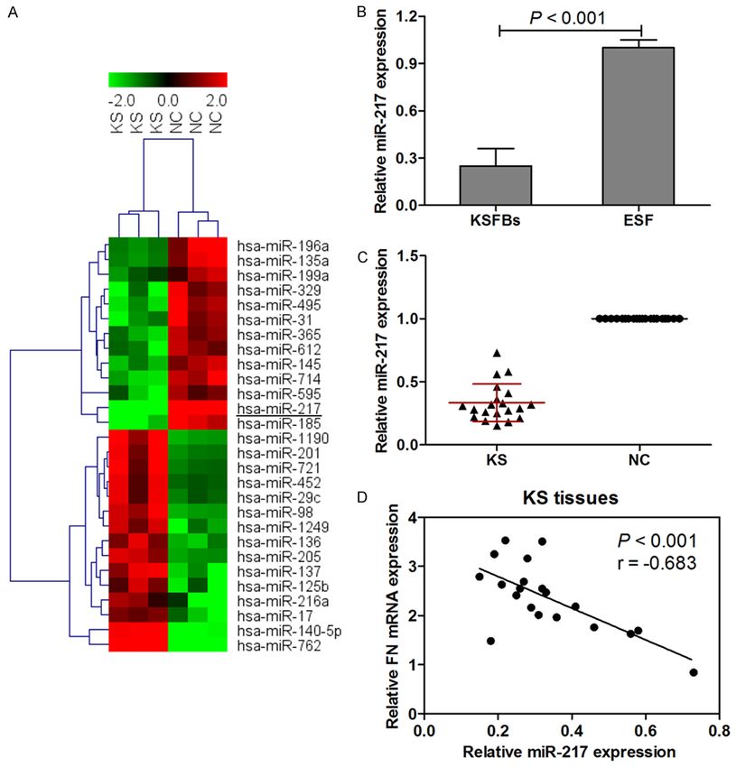
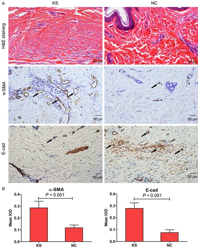
microRNA-217 in keloid fibrogenesis

Figure 1. The histological observation on keloid scarring tissues (KS) and paired corresponding adjacent normal
tissues (NC) from clinical patients by hematoxylin and eosin (H&E) staining. α-SMA and E-cad expression was mea-
sured by immunohistochemical staining in KS and NC tissues (A, 100×). Histogram represented the mean integral
optical density (IOD) of α-SMA and E-cad in KS and NC tissues (B).

group differences in genes expression between               were assessed by the Paired-Student’s t-test.
hypertrophic scarring tissues (KS) and paired               Differences with P value of < 0.05 were consid-
corresponding adjacent normal tissues (NC)                  ered statistically significant.

1869                                                            Int J Clin Exp Pathol 2018;11(4):1866-1877
microRNA-217 in keloid fibrogenesis

Figure 2. TGF-β1 (A, 100×), Col-1 and Col-3 (B, 100×) expression was measured by immunohistochemical staining
in KS and NC tissues. Histogram represented the mean integral optical density (IOD) of TGF-β1, Col-1 and Col-3 in
KS and NC tissues (C).

Results                                                      the dermis. However, the thickness and com-
                                                             plexity of the collagen bundles were diminished
Histological observation                                     in paired corresponding adjacent normal tis-
By H&E staining, we observed that keloid tis-                sues compared with keloid tissues (Figure 1A).
sues had a dense and excessive collagen depo-                To further investigate induction of fibrogenic in
sition, and the collagen bundles were running                KS, α-SMA and E-cad expression levels in le-
parallel to the epidermis in a large fraction of             sional tissues were measured by immunohisto-

1870                                                            Int J Clin Exp Pathol 2018;11(4):1866-1877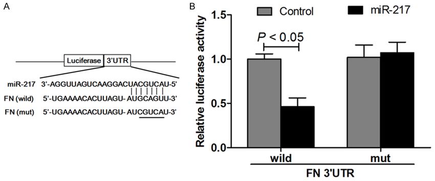
microRNA-217 in keloid fibrogenesis

                                                                                Figure 3. FN expressi-
                                                                                on was measured by
                                                                                immunohistochemi-
                                                                                cal staining in KS and
                                                                                NC tissues (A, 100×).
                                                                                mRNA (B) and protein
                                                                                (C) expression of FN
                                                                                were measured by RT-
                                                                                PCR and western blot-
                                                                                ting assay respectively.

chemical staining. α-smooth muscle actin (α-        Fibronectin (FN) expression in keloid tissues
SMA), a marker of myofibroblasts, and E-cad-
herin (E-cad), an epithelial marker, are closely    We results demonstrated that immunohisto-
related to fibrotic diseases [14]. In our study,    chemical staining (Figure 3A), RT-PCR (Figure
α-SMA was significantly increased in KS group       3B) and western blotting (Figure 3C) showed a
compared with NC group, in contrast, the le-        marked increase in FN levels in the keloid tis-
sional keloid tissues showed very dramatic          sues compared with the corresponding adja-
reduction of E-cad as compared to NC group          cent normal tissues. However, the underlying
(Figure 1A). Moreover, the immunohistochemi-        molecular mechanisms of miRNAs via targeting
cal staining was measured by integral optical       FN contribute to the induction of KS remains to
density (IOD) assay (Figure 1B).                    be determined. A follow-up study will try to iden-
                                                    tify the miRNAs expression profiles in keloid
Keloid fibrogenesis-related proteins expression     tissues.

To investigate the expression of TGF-β1, colla-     miRNAs expression in keloid tissues
gen types I and III in KS, we used immunohisto-
chemical staining to confirm this conclusion in     To explore the miRNAs expression profiles in
our study. As expected, the expression of colla-    KS tissues, we compared miRNAs expression
gen types I (Col-1) and III (Col-3) were signifi-   between KS tissues and adjacent normal tis-
cantly increased in KS group compared with NC       sues using microarray assay. The results sh-
group (Figure 2B and 2C). However, the expres-      owed that 13 miRNAs were found to be signifi-
sion of TGF-β1 had no significant differen-         cantly down-regulated, and 15 miRNAs were
ces between keloid tissues and corresponding        significantly up-regulated in the KS tissues as
adjacent normal tissues (Figure 2A and 2C).         compared to the corresponding adjacent nor-

1871                                                   Int J Clin Exp Pathol 2018;11(4):1866-1877
microRNA-217 in keloid fibrogenesis Figure 4. Microarray and hierarchical cluster analysis of the significantly regulated miRNAs in patients with keloid scarring. The figure is drawn by MeV software (version 4.2.6). Correlation similarity matrix and average linkage al- gorithms are used in the cluster analysis. Each row represents an individual miRNA, and each column represents a sample. The dendrogram at the left side and the top displays similarity of expression among miRNAs and samples individually. The color legend at the top represents the level of miRNA expression, with red indicating high expres- sion levels and green indicating low expression levels (A). miRNA-217 expression was examined by real-time PCR and normalized to U6 expression in KSFBs (B) and 20 keloid scarring tissues and paired corresponding adjacent normal tissues (C). Inverse correlation of miRNA-217 and FN mRNA expression in KS tissues (D). mal tissues, and we finally focused on miRNA- 20 KS tissues and paired corresponding adja- 217 in our study. miRNA-217 was significantly cent normal tissues. Subsequent investigation lowly expressed in KS tissues as compared to led to an intriguing conclusion that the expres- adjacent normal tissues (Figure 4A). Moreover, sion of miRNA-217 was greatly suppressed in by using PicTar, TargetScan, and miRBase data- KSFBs and KS tissues (Figure 4B and 4C). To base, we indicated that miR-217 was a poten- validate the correlation between miRNA-217 tial regulator of FN. Based on these results, the and FN, the FN mRNA and miRNA-217 expres- expression of miRNA-217 was investigated in sion levels were analyzed in 20 KS tissue sam- 1872 Int J Clin Exp Pathol 2018;11(4):1866-1877
microRNA-217 in keloid fibrogenesis
Figure 5. Schematic representation of the
putative miRNA-217 binding site in the FN
3’UTR as in Targetscan (A) and luciferase
activity assay (B).
ples by real-time PCR. As shown in Figure 4D, mutant-type 3’-UTR of FN, and the reporter
the expression levels of endogenous miRNA- activity did not show significant difference com-
217 negatively correlated with the FN mRNA pared with control group (Figure 5B).
levels (Pearson’s correlation coefficient =
-0.683; P < 0.001). These data provide further miRNA-217 regulates FN expression in keloid
evidence of a functional link between miRNA- scarring fibroblasts
217 and FN in KS tissues.
miRNA-217 levels were measured when the
miRNA-217 is a regulator of FN KSFBs were transfected with miRNA-217 mim-
ics or inhibitor. As expected, KSFBs transfected
The miRNAs were initially proposed to media- with miRNA-217 mimics increased miRNA-217
te post-translational repression of their targ- levels, and KSFBs transfected with miRNA-217
et mRNAs [15]. To further validate the miRNA inhibitor decreased miRNA-217 levels in KSFBs
microarray results, we selected miRNA-217 (Figure 6A). Next, in KSFBs, the miRNA-217
from the down-regulated miRNAs and evaluat- mimics or inhibitor was transfected to evaluate
ed the alignment predicted by the TargetScan the potential effect on cell proliferation and
algorithm. We found the potential miR-217 apoptosis of miRNA-217 in vitro. KSFBs cell
binding sites within the 3’-UTR of FN in human viability was measured by MTT assay when
sapiens (Figure 5A). The complementarity be- cells were transfected with miRNA-217 mimics
tween the seed region of miRNA and 3’-UTR of or inhibitor for 24 h, 48 h and 72 h. The viabi-
mRNAs is the most important determinant for lities of KSFBs transfected with miRNA-217
target specificity, and miRNA-mediated post- mimics was significantly lower than those of
translational repression often depends on per- untreatment group. However, miRNA-217 loss-
fect or near-perfect base pairing of a seed of function increased the growth of KSBFs as
region to its target [1]. As shown in Figure 5A, compared to the scramble cells (Figure 6B).
FN is a strong candidate that is regulated by Moreover, we examined whether miRNA-217
miRNA-217. To further investigate whether mimics or inhibitor induced apoptosis in KSFBs
miRNA-217 directly regulates the expression of through an apoptotic mechanism. Caspase-3
FN, we generated luciferase reporter plasmi- activity assay and Triphosphate nick-end lab-
ds containing the 3’-UTR of the wild-type or eling (TUNEL) staining were measured after
mutant-type FN gene in HEK293 cells. Cotr- KSFBs transfected with miRNA-217 mimics or
ansfection with this luciferase construct con- inhibitor for 48 h. The results indicated that the
taining wild-type 3’-UTR and miR-185 mimics caspase-3 activity in miRNA-217 mimics group
resulted in a lower luciferase activity than con- were significantly higher than that of the control
trol group, which resulted in more than 50% group. In the contrast to that KSFBs transfect-
decline in the luciferase activity compared with ed with miRNA-217 inhibitor suppressed cas-
control group. Cotransfection of HEK293 cells pase-3 activity (Figure 6C). TUNEL staining sh-
with this recombination structure containing owed that miRNA-217 gain-of function induced
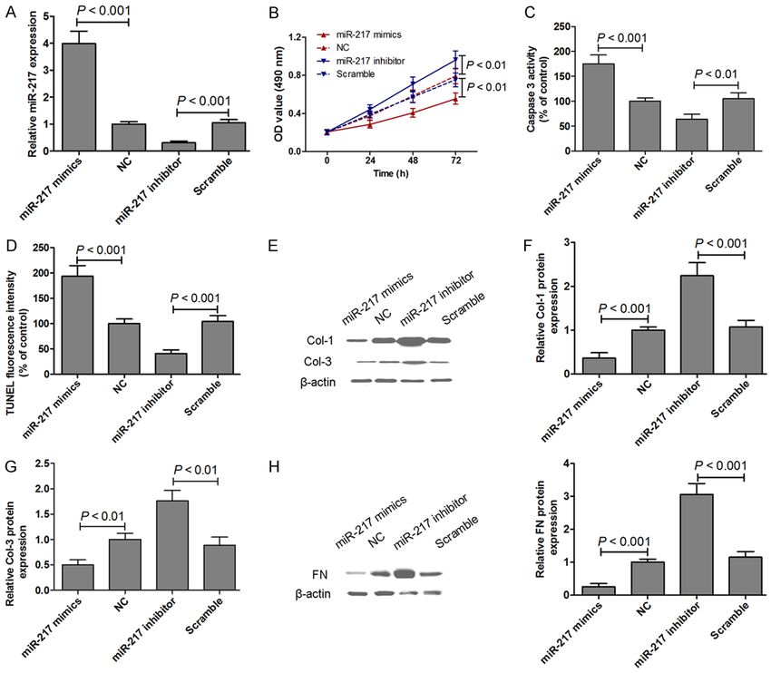
1873 Int J Clin Exp Pathol 2018;11(4):1866-1877microRNA-217 in keloid fibrogenesis Figure 6. miRNA-217 expression was examined by real-time PCR and normalized to U6 expression in KSFBs trans- fected with miRNA-217 mimics or inhibitor for 48 h (A). MTT assays were performed on KSFBs transfected with miRNA-217 mimics or inhibitor for 24 h, 48 h and 72 h (B). Caspase-3 activity assay (C) and Triphosphate nick-end labeling (TUNEL) staining (D) were measured after KSFBs transfected with miRNA-217 mimics or inhibitor for 48 h. The protein expression of Col-1 and Col-3 was measured by western blotting in KSFBs transfected with miRNA-217 mimics or inhibitor for 48 h (E-G). The protein expression of FN was measured by western blotting in KSFBs trans- fected with miRNA-217 mimics or inhibitor for 48 h (H). cell apoptosis, and miRNA-217 loss-of function RNA-217 loss-of function increased FN expres- suppressed cell apoptosis in KSFBs (Figure sion in KSFBs (Figure 6H). 6D). These results suggested that miRNA-217 played an impact on cell proliferation and apop- Discussion tosis, thereby regulating KSFBs growth. In addi- tion, the protein expression of Col-1 and Col-3 Downregulation of miRNA-217 is a frequent was measured by western blotting in KSFBs event in various cancers, such as gastric can- transfected with miRNA-217 mimics or inhibitor cer [16], colorectal cancer [12], pancreatic can- for 48 h. The results showed that miRNA-217 cer [17] and hepatocellular carcinoma [18]. gain-of function decreased Col-1 and Col-3 pro- Moreover, miRNA-217 was demonstrated to tein expression, and miRNA-217 loss-of func- modulate glomerular mesangial cell survival tion increased Col-1 and Col-3 protein expres- and hypertrophy [19]. However, the role of sion in KSFBs (Figure 6E-G). Furthermore, mi- miRNA-217 in keloid fibrogenesis remains to be RNA-217 gain-of function decreased, and mi- elucidated. Keloid is benign skin tumors char- 1874 Int J Clin Exp Pathol 2018;11(4):1866-1877
microRNA-217 in keloid fibrogenesis

acterized by collagen accumulation and hyperp-        There is increasing evidence to show that the
roliferation of fibroblasts and represents the        FN signalling is involved in keloid pathogenesis
end-point of a spectrum of abnormal wound             [6, 9, 27]. Early in healing, fibroblasts migrate
healing [20]. Previous studies demonstrate            into the wound area and rapidly produce a tran-
that miRNA-29a [21], miRNA-21 [22] and miR-           sient matrix of FN. However, as healing pro-
NA-196a [1] are involved in the fibrogenesis of       gresses, collagen content increases, the colla-
keloid. In the present study, we found that the       gen is organized into bundles, and eventually
expression of miRNA-217 was greatly sup-              the fibronectin matrix diminishes [9]. These
pressed in KSFBs and KS tissues. miRNA-217            results suggest that overexpressed FN may
could directly regulate the expression of FN          be resulting in abnormal physiology in keloid
via targeting its 3’-UTR. Moreover, the correla-      fibrogenesis. Consistent with these previous
tion between miRNA-217 and FN showed that             reports, we results demonstrated that immuno-
endogenous miRNA-217 negatively correlated            histochemical staining, RT-PCR and western
with the FN mRNA levels in KS tissues. These          blotting showed a marked increase in FN levels
data provide further evidence of a functional         in the keloid tissues compared with the corre-
link between miRNA-217 and FN in KS tissues,          sponding adjacent normal tissues. Based on
and miRNA-217 via targeting FN as a post-             these studies, we conclude that FN may be a
                                                      key target for the development of novel thera-
translational mechanism might provide new
                                                      peutic strategies for KS. Intriguingly, we also
insights into the underlying mechanism and
                                                      found that miRNA-217 affected KSFBs growth
therapeutic strategies for keloid fibrogenesis.
                                                      and apoptosis through regulating FN expres-
                                                      sion. Thus, our study provided evidence to
Fibroblasts are the effector cells in fibrotic dis-
                                                      determine that miRNA-217 might be a useful
eases including keloid and hypertrophic scars
                                                      target for management of keloid fibrogenesis.
which are characterized by their over-prolifera-
tion and over-production of collagen and ECM.         In conclusion, the data reported herein indica-
In addition to the suppression of fibroblast pro-     te that overexpressed miRNA-217 can inhibit
liferation, the inhibition of collagen and ECM        KSFBs growth, and the underlying mechanism
production and deposition by fibroblasts is an        was mediated, at least partially, through the
important goal for pharmaceutical agents used         suppression of FN expression. But above all,
in the treatment of these fibroproliferative con-     miRNA-217 may play a potential therapeutic
ditions [23]. Massive collagen synthesis and          avenue for the treatment of keloid fibroge-
changes has been regarded as a main charac-           nesis.
ter of hypertrophic scar formation. Excess
deposition of ECM, such as type I and III colla-      Acknowledgements
gen, by fibroblasts is responsible for keloid and
hypertrophic scarring [24]. Consistent with pre-      This work was supported by National Natural
vious data, we found that upregulations of            Science Foundation of China (No. 81272109).
Col-1 and Col-3 were observed in KS tissues.
                                                      Disclosure of conflict of interest
Moreover, miRNA-217 gain-of function decre-
ased Col-1 and Col-3 protein expression, and          None.
miRNA-217 loss-of function increased Col-1
and Col-3 protein expression in KSFBs. Con-           Address correspondence to: Dr. Zhen Gao, De-
tributing to the pathogenesis of keloid fibrogen-     partment of Plastic and Reconstructive Surgery,
esis are both a genetic predisposition and vari-      Shanghai Ninth People’s Hospital, Shanghai Jiao
ous local factors. Previous studies indicate that     Tong University School of Medicine, 639 Zhizaoju
collagen types I and III are the most abundant        Road, Shanghai, China. Tel: (+86) 21-23271699;
components within the abnormal ECM deposits           E-mail: gaozhen_nf@163.com
in keloids [4, 25]. Previous studies indicate that
                                                      References
miRNA-196a and miRNA-200b display the high-
est altered expression in KSFBs compared with         [1]     Kashiyama K, Mitsutake N, Matsuse M, Ogi T,
normal fibroblast and inhibit the expression of               Saenko VA, Ujifuku K, Utani A, Hirano A and Ya-
type I and III collagens, whose deposition is a               mashita S. miR-196a downregulation increas-
major manifestation in keloid pathology [1, 26].              es the expression of type I and III collagens in

1875                                                        Int J Clin Exp Pathol 2018;11(4):1866-1877
microRNA-217 in keloid fibrogenesis
keloid fibroblasts. J Invest Dermatol 2012; AEG-1 dependent mechanism. BMC Cancer
132: 1597-1604. 2015; 15: 437.
[2] Lee WJ, Ahn HM, Roh H, Na Y, Choi IK, Lee JH, [13] Vychytilova-Faltejskova P, Kiss I, Klusova S,
Kim YO, Lew DH and Yun CO. Decorin-express- Hlavsa J, Prochazka V, Kala Z, Mazanec J,
ing adenovirus decreases collagen synthesis Hausnerova J, Kren L, Hermanova M, Lenz J,
and upregulates MMP expression in keloid fi- Karasek P, Vyzula R and Slaby O. MiR-21, miR-
broblasts and keloid spheroids. Exp Dermatol 34a, miR-198 and miR-217 as diagnostic and
2015; 24: 591-597. prognostic biomarkers for chronic pancreatitis
[3] Harn HI, Wang YK, Hsu CK, Ho YT, Huang YW, and pancreatic ductal adenocarcinoma. Diagn
Chiu WT, Lin HH, Cheng CM and Tang MJ. Me- Pathol 2015; 10: 38.
chanical coupling of cytoskeletal elasticity and [14] Liu Y. Epithelial to mesenchymal transition in
force generation is crucial for understanding renal fibrogenesis: pathologic significance, mo-
the migrating nature of keloid fibroblasts. Exp lecular mechanism, and therapeutic interven-
Dermatol 2015; 24: 579-584. tion. J Am Soc Nephrol 2004; 15: 1-12.
[4] Aoki M, Miyake K, Ogawa R, Dohi T, Akaishi S, [15] Baek D, Villen J, Shin C, Camargo FD, Gygi SP
Hyakusoku H and Shimada T. siRNA knock- and Bartel DP. The impact of microRNAs on
down of tissue inhibitor of metalloproteinase-1 protein output. Nature 2008; 455: 64-71.
in keloid fibroblasts leads to degradation of [16] Wang H, Dong X, Gu X, Qin R, Jia H and Gao J.
collagen type I. J Invest Dermatol 2014; 134: The microRNA-217 functions as a potential tu-
818-826. mor suppressor in gastric cancer by targeting
[5] Fan C, Dong Y, Xie Y, Su Y, Zhang X, Leavesley GPC5. PLoS One 2015; 10: e0125474.
D and Upton Z. Shikonin reduces TGF-beta1- [17] Zhao WG, Yu SN, Lu ZH, Ma YH, Gu YM and
induced collagen production and contraction Chen J. The miR-217 microRNA functions as a
in hypertrophic scar-derived human skin fibro- potential tumor suppressor in pancreatic duc-
blasts. Int J Mol Med 2015; 36: 985-991. tal adenocarcinoma by targeting KRAS. Carci-
nogenesis 2010; 31: 1726-1733.
[6] Liang CJ, Yen YH, Hung LY, Wang SH, Pu CM,
[18] Su J, Wang Q, Liu Y and Zhong M. miR-217 in-
Chien HF, Tsai JS, Lee CW, Yen FL and Chen YL.
hibits invasion of hepatocellular carcinoma
Thalidomide inhibits fibronectin production in
cells through direct suppression of E2F3. Mol
TGF-beta1-treated normal and keloid fibro-
Cell Biochem 2014; 392: 289-296.
blasts via inhibition of the p38/Smad3 path-
[19] Kato M, Putta S, Wang M, Yuan H, Lanting L,
way. Biochem Pharmacol 2013; 85: 1594-
Nair I, Gunn A, Nakagawa Y, Shimano H, Todor-
1602.
ov I, Rossi JJ and Natarajan R. TGF-beta acti-
[7] Zeng C, Wang YL, Xie C, Sang Y, Li TJ, Zhang M,
vates Akt kinase through a microRNA-depen-
Wang R, Zhang Q, Zheng L and Zhuang SM.
dent amplifying circuit targeting PTEN. Nat Cell
Identification of a novel TGF-beta-miR-122-fi-
Biol 2009; 11: 881-889.
bronectin 1/serum response factor signaling
[20] Jumper N, Paus R and Bayat A. Functional his-
cascade and its implication in hepatic fibro- topathology of keloid disease. Histol Histo-
genesis. Oncotarget 2015; 6: 12224-12233. pathol 2015; 30: 1033-1057.
[8] Hernandez-Gea V and Friedman SL. Pathogen- [21] Zhang GY, Wu LC, Liao T, Chen GC, Chen YH,
esis of liver fibrosis. Annu Rev Pathol 2011; 6: Zhao YX, Chen SY, Wang AY, Lin K, Lin DM,
425-456. Yang JQ, Gao WY and Li QF. A novel regulatory
[9] Babu M, Diegelmann R and Oliver N. Fibronec- function for miR-29a in keloid fibrogenesis.
tin is overproduced by keloid fibroblasts during Clin Exp Dermatol 2015; [Epub ahead of print].
abnormal wound healing. Mol Cell Biol 1989; [22] Wang X, Liu Y, Chen X, Zhang M and Xiao Z.
9: 1642-1650. Impact of MiR-21 on the expression of FasL in
[10] Guban B, Vas K, Balog Z, Manczinger M, Bebes the presence of TGF-beta1. Aesthet Surg J
A, Groma G, Szell M, Kemeny L and Bata-Csor- 2013; 33: 1186-1198.
go Z. Abnormal regulation of fibronectin pro- [23] Phan TT, Lim IJ, Sun L, Chan SY, Bay BH, Tan EK
duction by fibroblasts in psoriasis. Br J Derma- and Lee ST. Quercetin inhibits fibronectin pro-
tol 2016; 174: 533-41. duction by keloid-derived fibroblasts. Implica-
[11] Kelsh RM, McKeown-Longo PJ and Clark RA. tion for the treatmcent of excessive scars. J
EDA Fibronectin in Keloids Create a Vicious Dermatol Sci 2003; 33: 192-194.
Cycle of Fibrotic Tumor Formation. J Invest Der- [24] Syed F, Ahmadi E, Iqbal SA, Singh S, Mc-
matol 2015; 135: 1714-1718. Grouther DA and Bayat A. Fibroblasts from the
[12] Wang B, Shen ZL, Jiang KW, Zhao G, Wang CY, growing margin of keloid scars produce higher
Yan YC, Yang Y, Zhang JZ, Shen C, Gao ZD, Ye levels of collagen I and III compared with intra-
YJ and Wang S. MicroRNA-217 functions as a lesional and extralesional sites: clinical impli-
prognosis predictor and inhibits colorectal cations for lesional site-directed therapy. Br J
cancer cell proliferation and invasion via an Dermatol 2011; 164: 83-96.
1876 Int J Clin Exp Pathol 2018;11(4):1866-1877microRNA-217 in keloid fibrogenesis
[25] Al-Attar A, Mess S, Thomassen JM, Kauffman [27] Chua AW, Gan SU, Ting Y, Fu Z, Lim CK, Song C,
CL and Davison SP. Keloid pathogenesis and Sabapathy K and Phan TT. Keloid fibroblasts
treatment. Plast Reconstr Surg 2006; 117: are more sensitive to Wnt3a treatment in
286-300. terms of elevated cellular growth and fibronec-
[26] Li P, He QY and Luo CQ. Overexpression of miR- tin expression. J Dermatol Sci 2011; 64: 199-
200b inhibits the cell proliferation and pro- 209.
motes apoptosis of human hypertrophic scar
fibroblasts in vitro. J Dermatol 2014; 41: 903-
911.
1877 Int J Clin Exp Pathol 2018;11(4):1866-1877You can also read