The cystine knot is responsible for the exceptional stability of the insecticidal spider toxin ω-Hexatoxin-Hv1a
←
→
Page content transcription
If your browser does not render page correctly, please read the page content below
Please do not remove this page

The cystine knot is responsible for the
exceptional stability of the insecticidal spider
toxin ω-Hexatoxin-Hv1a
Herzig, Volker; King, G F
https://research.usc.edu.au/discovery/delivery/61USC_INST:ResearchRepository/12126668760002621?l#13127222620002621

Herzig, V., & King, G. F. (2015). The cystine knot is responsible for the exceptional stability of the
insecticidal spider toxin ω-Hexatoxin-Hv1a. Toxins, 7(10), 4366–4380.
https://doi.org/10.3390/toxins7104366

Link to Published Version: https://dx.doi.org/10.3390/toxins7104366
Document Type: Published Version

USC Research Bank: https://research.usc.edu.au
research-repository@usc.edu.au
CC BY V4.0
Copyright © 2015 by the authors; licensee MDPI, Basel, Switzerland. This article is an open access article
distributed under the terms and conditions of the Creative Commons Attribution license
(http://creativecommons.org/licenses/by/4.0/).
Downloaded On 2021/05/07 17:26:33 +1000

Please do not remove this page
Toxins 2015, 7, 4366-4380; doi:10.3390/toxins7104366
                                                                                          OPEN ACCESS

                                                                                      toxins
                                                                                     ISSN 2072-6651
                                                                          www.mdpi.com/journal/toxins
Article

The Cystine Knot Is Responsible for the Exceptional Stability of
the Insecticidal Spider Toxin ω-Hexatoxin-Hv1a
Volker Herzig and Glenn F. King

Institute for Molecular Bioscience, The University of Queensland, St. Lucia QLD 4072, Australia;
E-Mails: v.herzig@imb.uq.edu.au (V.H.); glenn.king@imb.uq.edu.au (G.F.K.);
Tel.: +61-7-3346-2018 (V.H.); +61-7-3346-2025 (G.F.K.); Fax: +61-7-3346-2021 (V.H. & G.F.K)

Academic Editor: Ren Lai

Received: 25 August 2015 / Accepted: 21 October 2015 / Published: 26 October 2015

      Abstract: The inhibitor cystine knot (ICK) is an unusual three-disulfide architecture in
      which one of the disulfide bonds bisects a loop formed by the two other disulfide bridges
      and the intervening sections of the protein backbone. Peptides containing an ICK motif are
      frequently considered to have high levels of thermal, chemical and enzymatic stability due
      to cross-bracing provided by the disulfide bonds. Experimental studies supporting this
      contention are rare, in particular for spider-venom toxins, which represent the largest
      diversity of ICK peptides. We used ω-hexatoxin-Hv1a (Hv1a), an insecticidal toxin from the
      deadly Australian funnel-web spider, as a model system to examine the contribution of the
      cystine knot to the stability of ICK peptides. We show that Hv1a is highly stable when
      subjected to temperatures up to 75 °C, pH values as low as 1, and various organic solvents.
      Moreover, Hv1a was highly resistant to digestion by proteinase K and when incubated in
      insect hemolymph and human plasma. We demonstrate that the ICK motif is essential for
      the remarkable stability of Hv1a, with the peptide’s stability being dramatically reduced
      when the disulfide bonds are eliminated. Thus, this study demonstrates that the ICK motif
      significantly enhances the chemical and thermal stability of spider-venom peptides and
      provides them with a high level of protease resistance. This study also provides guidance to
      the conditions under which Hv1a could be stored and deployed as a bioinsecticide.

      Keywords: inhibitor cystine knot; physicochemical stability; spider toxin; insecticidal toxin;
      ω-hexatoxin-Hv1a; thermal stability; proteolytic degradation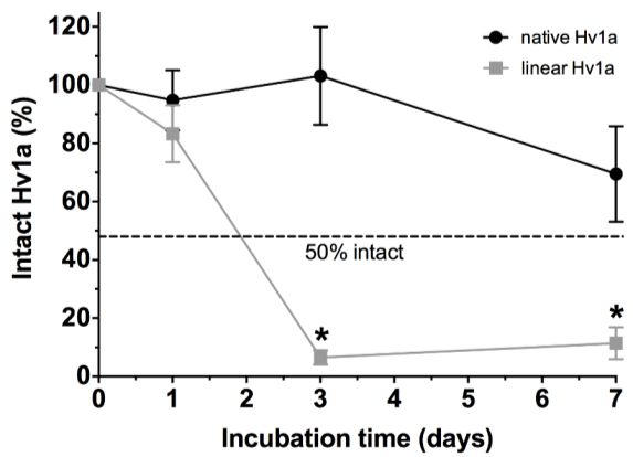
Toxins 2015, 7 4367
1. Introduction
The inhibitor cystine knot (ICK) is a protein scaffold defined as an antiparallel sheet stabilized by
a cystine knot [1,2]. The sheet typically comprises two strands, although a third N-terminal strand is
sometimes present [3] (Figure 1A). The cystine knot itself comprises a ring formed by two disulfide
bridges (Cys1–Cys4 and Cys2–Cys5) and the intervening sections of peptide backbone, with a third
disulfide bond (Cys3–Cys6) penetrating the ring to create a pseudo-knot (Figure 1B). The two central
disulfide bridges emanating from the strands are tightly packed against one another and they form the
compact hydrophobic core of ICK peptides [4].
Figure 1. Inhibitor cystine knot of the spider-venom peptide ω-hexatoxin-Hv1a.
(A) Schematic of the ICK motif, which comprises an antiparallel sheet stabilised by a
cystine knot [1]. strands are shown in green and the six cysteine residues that form the
cystine knot are labeled 1–6. In spider toxins, the sheet typically comprises only the two
strands housing Cys5 and Cys6, although a third N-terminal strand encompassing Cys2 is
sometimes present [3]. The two “outer” disulfide bonds are shown in green and the “inner”
disulfide bridge is red. (B) Schematic of the three-dimensional structure of the 37-residue
spider-venom peptide Hv1a (PDB 1AXH) [5] highlighting the ICK motif. The cystine knot
comprises a ring formed by two disulfides (Cys1–Cys4 and Cys2–Cys5, green) and the
intervening sections of polypeptide backbone (gray), with a third disulfide (Cys3–Cys6, red)
piercing the ring to create a pseudo-knot. The hydrophobic core of the toxin consists
primarily of the two central disulfide bridges connected to the strands. (C) Primary
structure of Hv1a showing the location of the three disulfide bonds and the 10 proteinase K
cleavage sites predicted by PeptideCutter [6].
Peptides containing an ICK motif are found in taxonomically diverse organisms ranging from fungi
and plants to molluscs and arthropods [1,7], although they are most abundant in the venom of spiders [8].
ICK-containing peptides are frequently considered to be highly stable over a wide range of
physicochemical conditions, but very few studies have experimentally examined their stability. The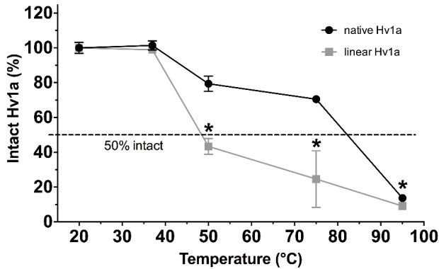
Toxins 2015, 7                                                                                          4368

best-studied example is kalata B1, a plant cyclotide whose thermal, chemical and enzymatic stability has
been examined in detail. Remarkably, it was demonstrated that the cystine knot is more important for
the stability of this cyclic peptide than its circular backbone [9].
   The ICK motif is the most common protein architecture found in spider-venom peptides [8], but data
on the contribution of this motif to the stability of spider toxins is lacking. Thus, in the present study, we
used the insecticidal spider-venom peptide ω-hexatoxin-Hv1a (Hv1a) as a model ICK peptide and
explored the contribution of the cystine knot to its physicochemical stability. Hv1a is 37-residue ICK
peptide isolated from the venom of the lethal Australian funnel-web spider Hadronyche versuta (Figure
1C). It has been used as a lead for bioinsecticide development due to its ability to selectively block
invertebrate, but not vertebrate, voltage-gated calcium (CaV) channels [5,8,10–12] and its lack of toxicity
to bees [13]. Hv1a has high level of biological stability as it is active when expressed in planta [8,14]
and it is capable of crossing the insect blood brain barrier in order to inhibit CaV channels in the insect
central nervous system [5,15]. The molecular basis of Hv1a’s inherent biological stability is presumed
to be its ICK motif but this has not been experimentally demonstrated.
   In order to determine how the cystine knot contributes to the physicochemical stability of Hv1a, we
compared native Hv1a with reduced and alkylated (linear) Hv1a over a wide range of temperatures,
solvents and pH conditions, and after incubation in proteinase K, insect hemolymph, and human plasma.
We show that Hv1a has a high level of chemical and thermal stability and that it is highly resistant to
proteolytic degradation. Reduction and alkylation of the six cysteine residues that form the cystine knot
motif of Hv1a completely abrogated the peptide’s resistance to enzymatic degradation and its resistance
to high temperature and acidic pH. Even the long-term stability of Hv1a in water and organic solvents
was compromised by loss of the ICK motif. This study reveals that the ICK motif is capable of providing
spider-venom peptides with a high level of thermal, chemical and biological stability, and it further
highlights the ICK motif as an excellent scaffold for protein engineering studies directed towards the
development of peptide drugs and bioinsecticides.

2. Results and Discussion

2.1. Thermal Stability

    Figure 2 summarises the stability of Hv1a when incubated for 24 h over a temperature range from 20
to 95 °C. There was no visible effect on toxin stability for both native and linear Hv1a up to 37 °C
(Figure 2), but peptide stability decreased at a much faster rate for linear Hv1a at temperatures exceeding
37 °C. Remarkably, ~71% of native Hv1a remained intact after incubation for 24 h at 75 °C, whereas
only ~25% of linear Hv1a remained intact under these conditions. In order to determine whether the
degradation observed at high temperature is reversible, we incubated a sample of Hv1a at 95 °C for 24 h,
then stored the sample at 20 °C for 3 days prior to HPLC analysis. There was no recovery in the level of
intact peptide over the 3 days (data not shown), indicating that the degradation observed at 95 °C is
irreversible. Nevertheless, based on the data shown in Figure 2, Hv1a should be highly stable in the field
even if employed as a bioinsecticide in climates that reach high temperatures.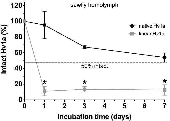
Toxins 2015, 7                                                                                         4369

      Figure 2. Thermal stability of Hv1a. Fraction of intact native Hv1a (black circles) and linear
      Hv1a (grey squares), relative to the corresponding 20 °C samples, after incubation at the
      indicated temperatures for 24 h. The dashed line indicates that the Tm under these conditions
      is ~48 °C and ~83 °C for linear and native Hv1a, respectively. Data are mean ± standard
      deviation (SD). In this and subsequent figures, asterisks indicate statistically significant
      differences between native and linear Hv1a (p < 0.05).

2.2. pH Stability

   Figure 3 shows the stability of Hv1a when incubated for 24 h in buffers with pH ranging from 1 to
13. Native Hv1a was highly stable at neutral and acidic pH, with essentially no degradation between pH
1 and 7. However, the peptide began to degrade as the pH approached the pKa of cysteine (~8.3), and
degradation became very apparent at pH 9 and above. Only 6% of the peptide remained intact after 24 h
at pH 13 (Figure 3). Additional peaks with higher retention time than Hv1a began to emerge in the
RP-HPLC chromatograms of samples incubated at pH 8 and higher (data not shown). MALDI mass
spectrometric analysis revealed that these peaks have the same mass as Hv1a and we therefore conclude
that these peaks correspond to isoforms of Hv1a resulting from disulfide-bond shuffling at alkaline pH.
In contrast with native toxin, the linear version of Hv1a was only stable at neutral pH values, with marked
degradation under both acidic and alkaline conditions. This instability presumably arises from
well-known processes that impact on protein stability such as asparagine deamidation, aspartate
isomerisation, and racemisation [16,17]. Interestingly, Hv1a contains an Asn-Gly sequence (Figure 1),
which has been shown to be highly susceptible to asparagine deamidation [18].
   The pH of the gut lumen in insects is highly variable, although there is a general trend, particularly
in exopterygous insects, towards acidic crops, neutral to mildly alkaline midguts, and neutral to acidic
hindguts [19,20]. Hv1a is stable under all of these pH conditions. However, several orders of
endopterygote insects such as lepidopterans, coleopterans, and dipterans have highly alkaline midguts
(pH > 8) [19–21]. This is especially true of lepidopteran larvae that feed on the leaves of trees, where
the average midgut pH of ~8.7 is believed to provide protection against leaf tannins and two-component
plant chemical defenses [21,22]. The instability of Hv1a at pH > 8 may therefore compromise its
effectiveness against lepidopterans and other insects with highly alkaline guts. Nevertheless, it has been
shown that transgenic expression of Hv1a in tobacco and cotton provides high levels of resistance against
larvae of the lepidopterans Helicoverpa armigera (cotton bollworms) and Spodoptera littoralis (cotton
leafworms) [23–25]. Thus, Hv1a must be sufficiently stable within the gut of these lepidopterans that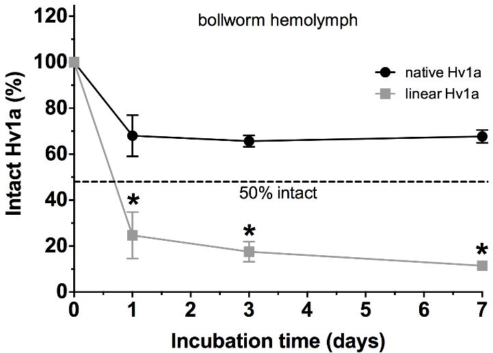
Toxins 2015, 7                                                                                          4370

enough toxin is taken up into the hemolymph to cause toxic effects at target sites (i.e., CaV channels)
within the central nervous system.

      Figure 3. pH stability of Hv1a. Fraction of intact native Hv1a (black circles) and linear Hv1a
      (grey squares), relative to the corresponding pH 7 samples, following incubation for 24 h at
      the indicated pH. Dashed line corresponds to 50% intact toxin. The pI of Hv1a and the pKa
      of free cysteine thiol groups are indicated by the arrows. Data are mean ±SD.

2.3. Chemical Stability

   Figure 4 shows that native Hv1a is highly stable during long-term incubation at room temperature
(in the dark) in water and different organic solvents (i.e., ethanol, methanol and acetonitrile). In contrast,
linear Hv1a was unstable over one week in water and all three organic solvents, although stability was
marginally higher in acetonitrile. This further highlights the importance of the cystine knot for the
long-term stability of ICK peptides. The long-term stability of Hv1a in organic solvents is useful from a
bioinsecticide development perspective since insecticides are often formulated with emulsifiers,
stabilisers, surfactants or other adjuvants that require dissolution in organic solvents [26].

2.4. Proteolytic Stability

    Proteinase K is a broad-spectrum serine protease from the fungus Engyodontium album that primarily
cleaves at peptide bonds on the C-terminal side of aliphatic and aromatic amino acid residues. It is
commonly used to remove contaminating protein from nucleic acid preparations [27], and consequently
it provides a stringent test of peptide/protein stability. Hv1a is predicted to contain 10 proteinase K
cleavage sites dispersed along the entire length of the toxin (see Figure 1C). Thus, as expected, linear
Hv1a was highly susceptible to proteinase K cleavage, with less than 25% of the peptide remaining intact
after incubation for just 2 h at 37 °C using a 1:200 molar ratio of proteinase K:Hv1a (Figure 5, grey
squares). In striking contrast, 74% of native Hv1a remained intact after incubation for 24 h under the
same conditions (Figure 5, black circles). Thus, the ICK motif of Hv1a provides a very high level of
resistance against the proteolytic activity of proteinase K.
Toxins 2015, 7 4371
Figure 4. Stability of Hv1a in various solvents. Fraction of intact native Hv1a (black
columns) and linear Hv1a (grey columns) after incubation for seven days in water and
organic solvents. Toxin amounts were quantified relative to a control sample incubated for
one day in water. Dashed line indicates 100% intact toxin. Data are mean ±SD.
Figure 5. Proteolytic stability of Hv1a. Fraction of intact native Hv1a (black circles) and
linear Hv1a (grey squares) after incubation with proteinase K (1:200 molar ratio) at pH 7.5
and 37 °C for up to 24 h. The values for native Hv1a were quantified relative to a sample of
native Hv1a incubated for 24 h under the same conditions without proteinase K. Values for
linear Hv1a were quantified relative to a sample of linear Hv1a at pH 7.5 incubated for 0 h
without proteinase K. The dashed line indicates 50% intact toxin. Data are mean ± SD.
2.5. Stability in Insect Hemolymph
Since Hv1a is considered a lead peptide for bioinsecticide development [8,13], its stability in insect
gut and hemolymph is critical in addition to its environmental stability. We therefore examined the
stability of Hv1a in hemolymph extracted from sawfly larvae and cotton bollworms (i.e., larvae of the
recalcitrant lepidopteran pest Helicoverpa armigera). Most insects carefully regulate the pH of their
hemolymph, typically in the range 6.4–6.8 [19,28,29]. The pH of the extracted sawfly hemolymph was
measured to be 6.68, which is similar to the pH of 6.99 reported for hemolymph from larvae of the
European pine sawfly Neodiprion sertifer [30]. The hemolymph extracted from H. armigera larvae had
a pH of 6.45, which is slightly lower than the pH of 6.8 reported for hemolymph from larvae of the
related lepidopteran H. zea [31]. Although native Hv1a is highly stable at neutral and acidic pH (see
Figure 3), insect hemolymph contains proteases that can degrade exogenous peptides [32]. Nevertheless,Toxins 2015, 7 4372
we found that Hv1a is highly stable in insect hemolymph, with 54%–68% of the peptide remaining intact
after seven days incubation at ambient temperature (~20 °C) in hemolymph from sawfly larvae (Figure
6A) and bollworms (Figure 6B). In contrast, only 11%–25% of linear Hv1a remained intact after one
day of incubation with sawfly or bollworm hemolymph (Figure 6A,B, grey squares), again highlighting
the importance of the cystine knot for providing resistance against chemical degradation and proteases.
Unfortunately, we were unable examine toxin stability in insect midgut solution, as we found it
difficult to isolate midgut contents without contamination from foregut, hindgut or hemolymph.
(A) (B)
Figure 6. Stability of Hv1a in insect hemolymph. Fraction of intact native Hv1a (black
circles) and linear Hv1a (grey squares) after incubation in (A) sawfly hemolymph and (B)
bollworm hemolymph for up to seven days at ambient temperature (~20 °C). Native and
linear Hv1a were quantified relative to control samples that were incubated in water for one
day. The dashed line indicates 50% intact toxin. Data are mean ±SD.
Figure 7. Stability of Hv1a in human plasma. Fraction of intact native Hv1a (black circles)
and linear Hv1a (grey squares) after incubation in human plasma for up to seven days at
ambient temperature (~20 °C). Native and linear Hv1a were quantified relative to control
samples that were incubated in water for one day. The dashed line indicates 50% intact toxin.
Data are mean ±SD.Toxins 2015, 7 4373
2.6. Stability in Human Plasma
Remarkably, we found that Hv1a is even more stable in human plasma than insect hemolymph, with
~70% of the peptide remaining intact after seven days incubation in plasma at ~20 °C (Figure 7, black
circles). In contrast, only ~7% of linear Hv1a remained intact after three days incubation in human
plasma (Figure 7, grey squares). Thus, the cystine knot of Hv1a provides exceptional resistance against
plasma proteases as well as plasma reductants such as glutathione and serum albumin that can potentially
reduce disulfides add/or cause disulfide scrambling [33].
2.7. Comparison of Hv1a Stability with Other ICK Peptides
The ICK motif is an unusual protein architecture in which a single disulfide bond is threaded through
a closed loop formed by two other disulfide bonds and the intervening sections of the polypeptide
backbone [1,3,7,34]. ICK peptides, also known as knottins [34], have evolved independently in a diverse
range of terrestrial taxa, including insects, arachnids, fungi, and plants [7], as well as marine cone snails [1],
sponges [35], horseshoe crabs [36] and sea anemones [37]. This protein fold appears to have been
evolutionarily favoured for two reasons. First, the ICK motif is highly plastic to sequence
changes [38,39], which allows it to support a wide variety of disparate functions [7]. Second, although
it is not a true knot in the mathematical sense, the inhibitor cystine knot is expected to greatly reduce the
propensity for peptide unfolding, and hence it is generally thought to imbue ICK peptides with a high
level of physicochemical stability [4,7,40].
Surprisingly, very few studies have examined the stability of native knottin peptides in detail, much
less addressed whether the cystine-knot motif is the underlying cause of unusually high levels of
chemical, thermal and biological stability. The only systematic study of this kind was performed on
kalata B1, a plant cyclotide [9]. Cyclotides are macrocyclic plant knottins containing a head-to-tail
cyclised peptide backbone in addition to a cystine knot motif. Kalata B1 was shown to be highly
thermostable and extremely resistant to chaotropic agents and proteases. These properties were largely
retained in an acyclic permutant but not when the disulfide bonds were eliminated by reduction and
alkylation, which led the authors to conclude that the cystine knot, rather than backbone cyclization, is
the primary contributor to the remarkable stability of kalata B1 [9]. The same authors demonstrated that
native conotoxin PVIIA, a knottin peptide from the venom of a marine cone snail, is impervious to
various endoproteinases but this protease resistance is lost when the cystine knot is eliminated by
disulfide reduction [9]. Engineered ICK peptides derived from plant knottins or the cystine-knot motif
of human agouti-related protein were shown to be highly resistant to pepsin and elastase, and have long
half-lives in rat plasma, but the contribution of the cystine knot to these properties was not
examined [41,42]. A variety of diverse ICK peptides from sponges [35] and spiders [32] have been
shown to be resistant to proteases and/or have high levels of stability in human plasma or insect
hemolymph, but the contribution of the cystine-knot motif to this stability was not examined.
The current work represents the first systematic study of the contribution of the inhibitor cystine knot
to the thermal, chemical, and biological stability of a native, non-cyclic knottin. We demonstrated that
the cystine knot underlies the remarkable resistance of the insecticidal toxin Hv1a to extremes of
temperature, acidic pH, organic solvents, and both human plasma and insect hemolymph. Highly alkalineToxins 2015, 7 4374 pH (>8) was the only condition under which native Hv1a was not considerably more stable than its linearised counterpart, presumably due to reduction/scrambling of the disulfide bonds in the cystine knot as the pH exceeds the pKa of the cysteine side-chain thiol groups. OAIP-1, an insecticidal knottin from tarantula venom, was also found to be susceptible to degradation under alkaline conditions [32]. Hv1a was isolated from the venom of the Australian funnel-web spider Hadronyche versuta, a close relative of the infamous Sydney funnel-web spider Atrax robustus that is lethal to rodents and primates [43]. The lethal component of A. robustus venom is the knottin peptide δ-hexatoxin-Ar1a [44–46], which potently delays inactivation of voltage-gated sodium channels [47]. Thus, data from 1961 on the stability of crude A. robustus venom [43] can be considered a proxy for the stability of δ-hexatoxin-Ar1a. Wiener found that the toxicity to mice of A. robustus venom was not altered after storage for four months at 4 °C, incubation at 37 °C for two days, incubation at 100 °C for 1 h, incubation at 100 °C for 20 min in the presence of 0.1 M HCl (pH~1), storage for 14 days in 33% acetic acid, and incubation with 0.1% pepsin and 0.2% HCl at 40 °C for 2 h [43]. However, incubation of venom for 24 h at pH 8.5 and 37 °C resulted in a slight loss of activity, incubation for 2 h at 40 °C with 0.1% trypsin and 0.1% Na2CO3 (pH ~11) resulted in a 50% loss of toxicity, and activity was completely lost when venom was incubated with 0.1 M NaOH (pH~13) for 20 min at 100 °C [43]. Finally, Wiener found that although A. robustus venom precipitates in 80% ethanol, the precipitate readily dissolves in water and retains toxicity [43]. Thus, A. robustus venom, and by extension the lethal knottin peptide δ-hexatoxin-Ar1a, is stable under the same conditions as Hv1a: it is resistant to extremes of temperature, acidic pH, ethanol, and proteases, but is susceptible to degradation under highly alkaline conditions. We conclude that these are likely to be general properties of spider-venom knottins. 3. Experimental Section 3.1. Chemicals 2-Aminoethanol and triethylphosphine were from Sigma-Aldrich (Castle Hill, NSW, Australia). 2-Iodoethanol was from Acros Organics (Thermo Fisher Scientific, Geel, Belgium). Proteinase K was from Promega (Madison, WI, USA) and synthetic Hv1a was kindly supplied by Vestaron Corporation (Kalamazoo, MI, USA). 3.2. Sample Treatment Every condition for each treatment was tested in triplicate for both native and linear Hv1a, and all data reported are mean ± SD. Unless otherwise stated, all samples were immediately frozen after treatment and then analysed via HPLC. Native Hv1a was linearised by one-step reduction and alkylation (RA) of the six Cys residues [48]. Toxin (1 mg) was dissolved in 450 µL of water prior to addition of 5 µL aminoethanol, 10 µL iodoethanol, 2 µL triethylphosphine (0.1 M solution in tetrahydrofuran), and 450 µL acetonitrile. After incubation at 37 °C for 2 h, the toxin sample was dried by vacuum centrifugation and the fully reduced and alkylated toxin was purified using RP-HPLC on a Shimadzu 20A series HPLC system (Shimadzu Scientific Instruments, Rydalmere, NSW, Australia). All native Hv1a samples were processed using a reverse-phase analytical column (Jupiter C18, 5 µm particle size, 300 Å pore size, 150 ×4.6 mm; Phenomenex, Lane Cove, NSW, Australia;) on a Shimadzu
Toxins 2015, 7                                                                                     4375

20A series HPLC system, with detection at 214 nm. Solvent A (0.1% formic acid in water) and solvent
B (0.1% formic acid in 90% acetonitrile) were used at a flow rate of 1 mL/min using a gradient of 5%
solvent B for the first 3 min, 5%–25% solvent B over the next 10 min, then 25%–80% solvent B over
0.5 min. For all linear Hv1a samples, a VisionHT HILIC column (5 µm particle size, 150 × 4.6 mm;
Grace, Columbia, MD, USA) was used on a Shimadzu 20A series HPLC system with HILIC-solvent A
(0.05% triflouracetic acid (TFA) in water) and HILIC-solvent B (90% acetonitrile, 0.043% TFA in
water). The following gradients were used for the HILIC runs: 85% HILIC solvent B for the first 3 min,
then 85%–55% HILIC solvent B for another 6 min, then 55%–5% HILIC solvent B for 1 min followed
by 5% HILIC solvent B for 1 min. Peptide peaks (identified from absorbance at 214 nm) were collected
manually and molecular masses determined using MALDI mass spectrometry on a 4700 Proteomics
Analyzer (Applied Biosystems, Foster City, CA, USA) using α-cyano-4-hydroxycinnamic acid (CHCA)
as matrix. The fractions containing native or linear Hv1a were identified based on molecular mass and
retention time, then the amount of intact toxin after each treatment was quantified based on peak area.

3.3. Thermal Stability

   Thermal stability was examined by incubating native (24.7 µM) and linear Hv1a (18.7 µM) for 24 h
at various temperatures (20, 37, 50, 75 and 95 °C). Following the respective incubation period, samples
were immediately stored at −20 °C prior to HPLC fractionation. In order to determine whether the effect
of high temperature is reversible for the native Hv1a, a sample was incubated for 24 h at 95 °C and then
stored at room temperature for three days before HPLC analysis. All native and linear Hv1a amounts
were quantified relative to their respective 20 °C sample.

3.4. pH Stability

   Stability under varying pH conditions was assessed by incubating native (49.4 µM) and linear Hv1a
(18.7 µM) for 24 h at room temperature in buffers of the following pH/composition (pKa values for the
relevant species are indicated): (i) pH 1 (50 parts 0.2 M KCl plus 134 parts 0.2 M HCl); (ii) pH 4 (0.2 M
sodium acetate, pKa 4.76); (iii) pH 7 (100 parts 0.1 M KH2PO4 plus 58.2 parts 0.1 M NaOH; pKa2 7.21);
(iv) pH 8 (100 parts 0.1 KH2PO4 plus 93.4 parts 0.1 M NaOH; pKa2 7.21); (v) pH 9 (0.1 M Tris-HCl,
pKa 8.07); (vi) pH 10 (966.4 parts 0.1 M Na2HPO4 plus 33.6 parts 0.1 M NaOH); (vii) pH 13 (50 parts
0.2 M KCl plus 132 parts 0.2 M NaOH). After the incubation period, native Hv1a samples were desalted
using a Maxi-Clean SPE column (Large Pore C18; Grace, Columbia, MD, USA) before application onto
the C18 HPLC column. For desalting, 10 mL of 5% solvent B was used to remove salts then Hv1a was
eluted with 10 mL of 45% solvent B and lyophilised prior to RP-HPLC analysis. All native and linear
Hv1a amounts were quantified relative to their respective pH 7 sample.

3.5. Chemical Stability

  The long-term stability of native (49.4 µM) and linear Hv1a (18.5 µM) in water and organic solvents
was determined by incubation in Milli-Q water, methanol, ethanol or acetonitrile in the dark for seven
days at room temperature. All samples were then lyophilised before HPLC analysis. The amounts of
Toxins 2015, 7                                                                                         4376

native and linear Hv1a were quantified relative to a respective control sample that was incubated in
Milli-Q water for one day.

3.6. Proteolytic Stability

   Analysis of the Hv1a sequence using PeptideCutter [6] indicated that it contains more potential
cleavage sites for proteinase K than any other protease examined (Figure 1C). Accordingly, native Hv1a
(24.7 µM) was incubated in a pH 7.5 buffer (0.2 M phosphate, 5 mM CaCl2) with proteinase K added at
a molar ratio of 1:200 (proteinase K:Hv1a). Samples were then incubated at 37 °C for 24 h. An identical
sample of native Hv1a without proteinase K was used as a negative control and also incubated at 37 °C
for 24 h. This sample was used as a reference (i.e., set to 100%) for the relative quantification of native
Hv1a sample incubated in the presence of proteinase K. After the incubation period, native Hv1a samples
were processed through a Maxi-Clean C18 column prior to HPLC (conditions see above under pH
stability). Linear Hv1a (6.9 µM) was incubated in a pH 7.5 buffer (0.2 M phosphate, 5 mM CaCl2) with
proteinase K added at a molar ratio of 1:200 (proteinase K:Hv1a) for a range of different time intervals
(20 and 40 min, and 1, 2, 5, 8 and 24 h). All linear Hv1a amounts were quantified relative to a sample
of linear Hv1a in the pH 7.5 buffer (without proteinase K) that was immediately processed through a
Maxi-Clean C18 column prior to HPLC. All other samples were incubated for the respective incubation
time before being cleaned through a Maxi-Clean C18 column.

3.7. Stability in Insect Hemolymph

   Sawfly larvae (Hymenoptera, Pergidae) were collected from Regency Downs, Queensland, Australia.
Fifth instar H. armigera larvae were purchased from AgBiTech Pty Ltd. (Clifford Gardens, Queensland,
Australia). A 1.0 mL Terumo Insulin syringe (B-D Ultra-Fine, Terumo Medical Corporation, Somerset,
NJ, USA) with a fixed 29 gauge needle was used to puncture the insect exoskeleton and collect the
extruded hemolymph, which was then centrifuged at 4 °C for 10 min at 10,000 rpm to remove insoluble
material. The pH of supernatant was measured using an Ultra M Micro Combination pH electrode
(Van London Co., Houston, TX, USA). Native Hv1a (367 µM) and linear Hv1a (154 µM) were dissolved
in clarified hemolymph, then the samples were incubated for 1–7 days at room temperature (~20 °C).
Prior to HPLC analysis, all samples were processed through a Maxi-Clean SPE column. A HPLC
chromatogram of a hemolymph-only control sample was subtracted from each HPLC chromatogram of
the respective native or linear Hv1a samples in order to account for peaks originating from endogenous
hemolymph compounds. All native and linear Hv1a amounts were quantified relative to a respective
control sample that was incubated in Milli-Q water for one day.

3.8. Stability in Human Plasma

   Native Hv1a (367 µM) or linear Hv1a (116 µM) were incubated for 1–7 days at room temperature
(~20 °C) in pooled human plasma (Innovative Research, Novi, MI, USA). Native and linear Hv1a
samples incubated in Milli-Q water for seven days at room temperature (~20 °C) were used as controls.
Prior to HPLC analysis, samples were processed through a Maxi-Clean SPE column. Native and linear
Hv1a were quantified relative to a respective control sample that was incubated in Milli-Q water for one day.
Toxins 2015, 7                                                                                        4377

3.9. Statistical Analyses

   GraphPad Prism 6 (GraphPad Software, La Jolla, CA, USA) was used to determine statistically
significant differences between native and linear Hv1a for each treatment condition using multiple t tests
followed by a Holm-Sidak correction. p < 0.05 was defined as statistically significant.

4. Conclusions

   We demonstrated that the insecticidal spider-venom peptide Hv1a is remarkably resistant to chemical
degradation; the peptide is stable over the pH range 1–8, at temperatures up to 75 °C, and when dissolved
in a range of organic solvents. Moreover, we found that Hv1a is highly resistant to proteolytic
degradation, with outstanding stability in insect hemolymph and human plasma, and when incubated
with proteinase K. These high levels of chemical and proteolytic resistance were completely abolished
when the toxin’s ICK motif was destroyed by reduction and alkylation of the six cysteine residues. We
conclude that the ICK motif, which is prevalent in spider-venom peptides, provides these toxins with
remarkable levels of chemical, thermal, and biological stability.
   The current study also provides a guide to the range of conditions under which Hv1a could be
formulated and used as a bioinsecticide. Our data show that formulations requiring dissolution in organic
solvents and/or acidic conditions should not adversely affect the stability of Hv1a. Moreover, we
demonstrated that Hv1a has a high level of thermal stability, which should facilitate its application in
the field in high-temperature climates.

Acknowledgments

  The authors thank Vestaron Corporation for kindly supplying the Hv1a used in this study. This work
was supported by Discovery Grant DP0878450 from the Australian Research Council.

Author Contributions

   Volker Herzig and Glenn King conceived and designed the experiments; Volker Herzig performed
the experiments and analyzed the data; Volker Herzig and Glenn King wrote the paper.

Conflicts of Interest

     The authors declare no conflict of interest.

References

1.    Pallaghy, P.K.; Nielsen, K.J.; Craik, D.J.; Norton, R.S. A common structural motif incorporating a
      cystine knot and a triple-stranded β-sheet in toxic and inhibitory polypeptides. Protein Sci. 1994, 3,
      1833–1839.
2.    Norton, R.S.; Pallaghy, P.K. The cystine knot structure of ion channel toxins and related
      polypeptides. Toxicon 1998, 36, 1573–1583.
3.    King, G.F.; Tedford, H.W.; Maggio, F. Structure and function of insecticidal neurotoxins from
      Australian funnel-web spiders. J. Toxicol.-Toxin Rev. 2002, 21, 359–389.
Toxins 2015, 7 4378
4. Saez, N.J.; Senff, S.; Jensen, J.E.; Er, S.Y.; Herzig, V.; Rash, L.D.; King, G.F. Spider-venom
peptides as therapeutics. Toxins 2010, 2, 2851–2871.
5. Fletcher, J.I.; Smith, R.; O’Donoghue, S.I.; Nilges, M.; Connor, M.; Howden, M.E.; Christie, M.J.;
King, G.F. The structure of a novel insecticidal neurotoxin, ω-atracotoxin-HV1, from the venom of
an Australian funnel web spider. Nat. Struct. Biol. 1997, 4, 559–566.
6. PeptideCutter. Available online: http://web.expasy.org/peptide_cutter (accessed on 23 October 2015).
7. Craik, D.J.; Daly, N.L.; Waine, C. The cystine knot motif in toxins and implications for drug design.
Toxicon 2001, 39, 43–60.
8. King, G.F.; Hardy, M.C. Spider-venom peptides: Structure, pharmacology, and potential for control
of insect pests. Annu. Rev. Entomol. 2013, 58, 475–496.
9. Colgrave, M.L.; Craik, D.J. Thermal, chemical, and enzymatic stability of the cyclotide kalata B1:
The importance of the cyclic cystine knot. Biochemistry 2004, 43, 5965–5975.
10. Tedford, H.W.; Gilles, N.; Ménez, A.; Doering, C.J.; Zamponi, G.W.; King, G.F. Scanning
mutagenesis of ω-atracotoxin-Hv1a reveals a spatially restricted epitope that confers selective
activity against invertebrate calcium channels. J. Biol. Chem. 2004, 279, 44133–44140.
11. Tedford, H.W.; Sollod, B.L.; Maggio, F.; King, G.F. Australian funnel-web spiders: Master
insecticide chemists. Toxicon 2004, 43, 601–618.
12. Windley, M.J.; Herzig, V.; Dziemborowicz, S.A.; Hardy, M.C.; King, G.F.; Nicholson, G.M.
Spider-venom peptides as bioinsecticides. Toxins 2012, 4, 191–227.
13. Nakasu, E.Y.; Williamson, S.M.; Edwards, M.G.; Fitches, E.C.; Gatehouse, J.A.; Wright, G.A.;
Gatehouse, A.M. Novel biopesticide based on a spider venom peptide shows no adverse effects on
honeybees. Proc. R. Soc. B. 2014, 281, 1–9.
14. Bonning, B.C.; Pal, N.; Liu, S.; Wang, Z.; Sivakumar, S.; Dixon, P.M.; King, G.F.; Miller, W.A.
Toxin delivery by the coat protein of an aphid-vectored plant virus provides plant resistance to
aphids. Nat. Biotechnol. 2014, 32, 102–105.
15. Bloomquist, J.R. Mode of action of atracotoxin at central and peripheral synapses of insects.
Invertebr. Neurosci. 2003, 5, 45–50.
16. Manning, M.C.; Chou, D.K.; Murphy, B.M.; Payne, R.W.; Katayama, D.S. Stability of protein
pharmaceuticals: An update. Pharm. Res. 2010, 27, 544–575.
17. Furman, J.L.; Chiu, M.; Hunter, M.J. Early engineering approaches to improve peptide
developability and manufacturability. AAPS J. 2015, 17, 111–120.
18. Robinson, N.E. Protein deamidation. Proc. Natl. Acad. Sci. USA 2002, 99, 5283–5288.
19. Harrison, J.F. Insect acid-base physiology. Annu. Rev. Entomol. 2001, 46, 221–250.
20. Nation, J.L. Insect Physiology and Biochemistry, 2nd ed.; CRC Press: Boca Raton, FL, USA, 2008.
21. Berenbaum, M.R. Adaptive significance of midgut pH in larval Lepidoptera. Am. Nat. 1980, 115,
138–146.
22. Pentzold, S.; Zagrobelny, M.; Roelsgaard, P.S.; Moller, B.L.; Bak, S. The multiple strategies of an
insect herbivore to overcome plant cyanogenic glucoside defence. PLoS One 2014, 9,
doi:10.1371/journal.pone.0091337
23. Khan, S.A.; Zafar, Y.; Briddon, R.W.; Malik, K.A.; Mukhtar, Z. Spider venom toxin protects plants
from insect attack. Transgenic Res. 2006, 15, 349–357.Toxins 2015, 7 4379
24. Shah, A.D.; Ahmed, M.; Mukhtar, Z.; Khan, S.A.; Habib, I.; Malik, Z.A.; Mansoor, S.; Saeed, N.A.
Spider toxin (Hvt) gene cloned under phloem specific RSs1 and RolC promoters provides resistance
against American bollworm (Heliothis armigera). Biotechnol. Lett. 2011, 33, 1457–1463.
25. Omar, A.; Ali Chatha, K. National Institute for Biotechnology and Genetic Engineering (NIBGE):
Genetically modified spider cotton. Asian J. Manag. Cases 2012, 9, 33–58.
26. Foy, C.L.; Pritchard, D.W. Pesticide Formulation and Adjuvant Technology; CRC Press: Boca
Raton, FL, USA, 1996.
27. Strauss, W.M. UNIT 2.2 Preparation of genomic DNA from mammalian tissue. Curr. Protoc. Mol.
Biol. 2001, doi:10.1002/0471142727.mb0202s42.
28. Chapman, R.F. The Insects: Structure and Function, 5th ed.; Cambridge University Press:
Cambridge, UK, 1998.
29. Kanost, M.R. Hemolymph. In Encylopedia of Insects, 2nd ed.; Resh, V.H., Cardé, R.T., Eds.;
Academic Press: Burlington, MA, USA, 2009; pp. 446–448.
30. Brown, S.E.; Patton, R.L.; Zerillo, R.T.; Douglas, S.M.; Breillatt, J.P.; Mazzone, H.M. Comparative
properties of hemolymph of the Gypsy moth and the European pine sawfly. J. N.Y. Entomol. Soc.
1977, 85, 36–42.
31. Wang, Z.; Haunerland, N.H. Storage protein uptake in Helicoverpa zea. purification of the
very high density lipoprotein receptor from perivisceral fat body. J. Biol. Chem. 1993, 268,
16673–16678.
32. Hardy, M.C.; Daly, N.L.; Mobli, M.; Morales, R.A.; King, G.F. Isolation of an orally active
insecticidal toxin from the venom of an Australian tarantula. PLoS One 2013, 8,
doi:10.1371/journal.pone.0073136
33. Armishaw, C.J.; Daly, N.L.; Nevin, S.T.; Adams, D.J.; Craik, D.J.; Alewood, P.F. α-Selenoconotoxins,
a new class of potent α7 neuronal nicotinic receptor antagonists. J. Biol. Chem. 2006, 281,
14136–14143.
34. Gracy, J.; Le-Nguyen, D.; Gelly, J.C.; Kaas, Q.; Heitz, A.; Chiche, L. KNOTTIN: The knottin or
inhibitor cystine knot scaffold in 2007. Nucleic Acids Res. 2008, 36, D314–D319.
35. Li, H.; Su, M.; Hamann, M.T.; Bowling, J.J.; Kim, H.S.; Jung, J.H. Solution structure of
a sponge-derived cystine knot peptide and its notable stability. J. Nat. Prod. 2014, 77, 304–310.
36. Fujitani, N.; Kouno, T.; Nakahara, T.; Takaya, K.; Osaki, T.; Kawabata, S.; Mizuguchi, M.;
Aizawa, T.; Demura, M.; Nishimura, S.; et al. The solution structure of horseshoe crab antimicrobial
peptide tachystatin B with an inhibitory cystine-knot motif. J. Pept. Sci. 2007, 13, 269–279.
37. Rodríguez, A.A.; Salceda, E.; Garateix, A.G.; Zaharenko, A.J.; Peigneur, S.; López, O.; Pons, T.;
Richardson, M.; Díaz, M.; Hernández, Y.; et al. A novel sea anemone peptide that inhibits
acid-sensing ion channels. Peptides 2014, 53, 3–12.
38. Olivera, B.M.; Hillyard, D.R.; Marsh, M.; Yoshikami, D. Combinatorial peptide libraries in drug
design: Lessons from venomous cone snails. Trends Biotechnol. 1995, 13, 422–426.
39. Sollod, B.L.; Wilson, D.; Zhaxybayeva, O.; Gogarten, J.P.; Drinkwater, R.; King, G.F. Were
arachnids the first to use combinatorial peptide libraries? Peptides 2005, 26, 131–139.
40. Ackerman, S.E.; Currier, N.V.; Bergen, J.M.; Cochran, J.R. Cystine-knot peptides: Emerging tools
for cancer imaging and therapy. Expert Rev. Proteomics 2014, 11, 561–572.Toxins 2015, 7 4380
41. Werle, M.; Schmitz, T.; Huang, H.L.; Wentzel, A.; Kolmar, H.; Bernkop-Schnürch, A. The potential
of cystine-knot microproteins as novel pharmacophoric scaffolds in oral peptide drug delivery.
J. Drug Target 2006, 14, 137–146.
42. Werle, M.; Kafedjiiski, K.; Kolmar, H.; Bernkop-Schnürch, A. Evaluation and improvement of the
properties of the novel cystine-knot microprotein McoEeTI for oral administration. Int. J. Pharm.
2007, 332, 72–79.
43. Wiener, S. Observations on the venom of the Sydney funnel-web spider (Atrax robustus).
Med. J. Aust. 1961, 48, 693–699.
44. Sheumack, D.D.; Claassens, R.; Howden, M.E.H.; Whitley, N.M. Complete amino acid sequence
of of a new type of lethal neurotoxin from the venom of the funnel-web spider Atrax robustus.
FEBS Lett. 1985, 181, 154–156.
45. Fletcher, J.I.; Chapman, B.E.; Mackay, J.P.; Howden, M.E.H.; King, G.F. The structure of
versutoxin (δ-atracotoxin-Hv1) provides insights into the binding of site 3 neurotoxins to the
voltage-gated sodium channel. Structure 1997, 5, 1525–1535.
46. Pallaghy, P.K.; Alewood, D.; Alewood, P.F.; Norton, R.S. Solution structure of robustoxin, the
lethal neurotoxin from the funnel-web spider Atrax robustus. FEBS Lett. 1997, 419, 191–196.
47. Nicholson, G.M.; Little, M.J.; Tyler, M.; Narahashi, T. Selective alteration of sodium channel
gating by Australian funnel-web spider toxins. Toxicon 1996, 34, 1443–1453.
48. Hale, J.E.; Butler, J.P.; Gelfanova, V.; You, J.S.; Knierman, M.D. A simplified procedure for the
reduction and alkylation of cysteine residues in proteins prior to proteolytic digestion and mass
spectral analysis. Anal. Biochem. 2004, 333, 174–181.
© 2015 by the authors; licensee MDPI, Basel, Switzerland. This article is an open access article
distributed under the terms and conditions of the Creative Commons Attribution license
(http://creativecommons.org/licenses/by/4.0/).You can also read