Cholecystokinin-2 Receptor Targeting with Novel C-terminally Stabilized HYNIC-Minigastrin Analogs Radiolabeled with Technetium-99m - MDPI
←
→
Page content transcription
If your browser does not render page correctly, please read the page content below
pharmaceuticals

Article
Cholecystokinin-2 Receptor Targeting with Novel
C-terminally Stabilized HYNIC-Minigastrin Analogs
Radiolabeled with Technetium-99m
Maximilian Klingler , Christine Rangger, Dominik Summer , Piriya Kaeopookum ,
Clemens Decristoforo and Elisabeth von Guggenberg *
 Department of Nuclear Medicine, Medical University of Innsbruck, Anichstrasse 35, A-6020 Innsbruck, Austria;
 maximilian.klingler@i-med.ac.at (M.K.); christine.rangger@i-med.ac.at (C.R.);
 dominik.summer@i-med.ac.at (D.S.); piriya.kaeopookum@student.i-med.ac.at (P.K.);
 clemens.decristoforo@i-med.ac.at (C.D.)
 * Correspondence: elisabeth.von-guggenberg@i-med.ac.at; Tel.: +43-512-504-80960

 Received: 29 November 2018; Accepted: 10 January 2019; Published: 15 January 2019

 Abstract: The high overexpression of cholecystokinin-2 receptors (CCK2R) in tumors, such as
 medullary thyroid carcinoma, allows for highly specific diagnostic and therapeutic targeting with
 radiolabeled peptide probes derived from natural ligands for the receptor. Based on the ideal imaging
 characteristics, high availability and low cost of technetium-99m (99m Tc)-labeled radiopharmaceuticals
 we have developed two hydrazinonicotinic acid (HYNIC) conjugated minigastrin analogs allowing
 labeling at high specific activity. The CCK2R targeting peptide conjugates show specific amino acid
 substitutions in the C-terminal receptor-specific sequence with the aim to increase stability and tumor
 targeting. The CCK2R affinity and the cell uptake of the new radioligands were analyzed using A431
 human epidermoid carcinoma cells stably transfected with human CCK2R and mock transfected
 cells. Metabolic studies in BALB/c mice revealed a high resistance against enzymatic degradation
 for both radioligands. Biodistribution studies in tumor-xenografted athymic BALB/c nude mice at
 1 h and 4 h p.i. showed that the two 99m Tc-labeled compounds showed varying uptake in receptor
 expressing organs, stomach and pancreas (1.3–10.4% IA/g), as well as kidneys, the main route of
 excretion (7.8–19.9% IA/g). The tumor uptake in A431-CCK2R xenografts was 24.75 ± 4.38% IA/g for
 [99m Tc]Tc-HYNIC-MGS5 and 42.48 ± 6.99% IA/g for [99m Tc]Tc-HYNIC-MGS11 at 4 h p.i., whereas the
 tumor-to-kidney ratio was comparable (2.6–3.3). On demand availability and potential application
 for radioguided surgery of a 99m Tc-labeled minigastrin analog support the further evaluation of these
 highly promising new compounds.

 Keywords: cholecystokinin-2 receptor; minigastrin; molecular imaging; radiometals; technetium-99m;
 hydrazinonicotinic acid (HYNIC)

1. Introduction
     Regulatory peptides exhibiting high target specificity are suitable lead structures, particularly
in the field of oncology, for the development of analogs for radionuclide imaging and therapy [1,2].
Such peptide analogs have the advantage of an easy production via well-established solid phase
peptide synthesis (SPPS) allowing the conjugation of a chelator for radiolabeling and the introduction
of different modifications into the peptide sequence to optimize pharmacokinetics. Additionally,
peptide analogs show low immunogenicity and toxicity and are therefore preferable candidates as new
targeting agents [3,4]. A highly promising molecular target to develop radiopeptides for diagnostic
imaging and targeted radiotherapy (TRT) of medullary thyroid carcinoma (MTC), small cell lung cancer
(SCLC), astrocytoma, stromal ovarian cancer, as well as carcinoids and other tumors of neuroendocrine

Pharmaceuticals 2019, 12, 13; doi:10.3390/ph12010013                   www.mdpi.com/journal/pharmaceuticals
Pharmaceuticals 2019, 12, 13 2 of 15
origin is the cholecystokinin-2 receptor (CCK2R) as an increased level of expression is observed in
these malignancies [5,6].
The most promising CCK2R targeting radiopeptides developed so far, are based on the peptide
sequence of minigastrin (MG), a naturally occurring ligand for this receptor [7]. MG and its analogs
bind to CCK2R with their bioactive C-terminal region (Trp-Met-Asp-Phe-NH2 ). In the last 20 years
a variety of MG analogs conjugated to different chelators for nuclear medicine procedures have been
reported [7–9]. Most of the preclinically [10–16] and clinically [17,18] investigated MG analogs have
been conjugated to the bifunctional chelator 1,4,7,10-tetraazacyclododecane-1,4,7,10-tetraacetic acid
(DOTA) allowing stable radiolabeling with trivalent radiometals such as Gallium-68, Indium-111 or
Lutetium-177 suitable for positron emission tomography (PET), single photon emission computed
tomography (SPECT) and TRT. Due to radioprotection issues and regulatory requirements for the
in-house production of radiopharmaceuticals, such as the need of hot cells, automated synthesis
modules, aseptic processing and trained personnel, the availability of these radiopeptides seems to be
restricted to a limited number of clinics.
A cost-effective and broadly available alternative for CCK2R imaging in hospitals without
PET would be a kit-based MG analog suitable for 99m Tc-labeling. Such a kit is already available for
somatostatin receptor targeting using [99m Tc]Tc-EDDA/HYNIC-Tyr3 -octreotide (Tektrotyd, Polatom,
Otwock, Poland). Technetium-99m can be easily eluted from a licensed 99 Mo/99m Tc-generator and is
used in the major part of nuclear medicine procedures. Due to its ideal physical properties (half-life of
6 h, monoenergetic gamma photons of 140 keV, low radiation burden) Technetium-99m remains the
most attractive radionuclide for SPECT applications as well as for gamma probe detection during
radioguided surgery [19,20].
Different attempts have been made to develop MG analogs with high tumor accumulation and
a biodistribution profile suitable for gastrin receptor scintigraphy [19,21,22]. However, only two
99m Tc-labeled MG analogs have been investigated in clinical studies. [99m Tc]Tc-Demogastrin
2 ([99m Tc]Tc-N4 -Gly-dGlu-(Glu)5 -Ala-Tyr-Gly-Trp-Met-Asp-Phe-NH2 ) contains an open chain
tetraamine chelator forming a monocationic complex [23,24]. [99m Tc]Tc-EDDA/HYNIC-MG11
([99m Tc]Tc-EDDA/HYNIC-dGlu-Ala-Tyr-Gly-Trp-Met-Asp-Phe-NH2 ) conjugated to the monodentate
ligand hydrazinonicotinic acid (HYNIC) needs additional coligands such as tris(hydroxymethyl)-
methylglycine (tricine) or ethylenediamine-N,N’-diacetic acid (EDDA) to complete the coordination
sphere [25]. The administration of these two 99m Tc-labeled MG analogs to patients was well tolerated
showing no to only mild side effects. With [99m Tc]Tc-Demogastrin 2 all known lesions in nine MTC
patients could be visualized. In a comparative study with [99m Tc]Tc-EDDA/HYNIC-MG11 and
[99m Tc]Tc-EDDA/HYNIC-TOC the potential additional information which can be obtained in MTC
patients using gastrin receptor scintigraphy was pointed out. The same MG analogs conjugated to
DOTA showed drawbacks related to high kidney uptake or low in vivo stability, requiring further
improvement to develop a CCK2R targeting peptide analog with optimal tumor targeting and
biodistribution profile [8].
Various research groups have worked on the development of metabolically stable MG analogs.
Different strategies such as cyclization, dimerization and substitutions of amino acids mainly
in the N-terminal part of the peptide sequence were investigated [8]. However, due to rapid
C-terminal enzymatic degradation the need of alternative stabilization strategies was suggested [26].
Recently we could present different amino acid substitutions introduced into the C-terminal
receptorspecific sequence of MG analogs improving the stability against enzymatic degradation
and the biodistribution profile [15,16]. After an intense preclinical evaluation of different
substitutions, we discovered that most promising results in terms of improved in vivo stability
and enhanced tumor targeting could be achieved when substituting methionine (Met) with
N-methyl-norleucine ((N-Me)-Nle) and phenylalanine (Phe) with 1-naphtyl-alanine (1-Nal). With the
new MG analog DOTA-dGlu-Ala-Tyr-Gly-Trp-(N-Me)Nle-Asp-1-Nal-NH2 (DOTA-MGS5) radiolabeled
with Gallium-68, Indium-111 and Lutetium-177 a very promising targeting profile was achieved [16].Pharmaceuticals 2019, 12, 13 3 of 15
Pharmaceuticals
In the present 2018, 11, x we have conjugated the clinically well-established HYNIC ligand to MGS5
study 3 of to
16
develop a 99m Tc-labeled MG analog, suitable for SPECT and radioguided surgery. Furthermore the
HYNIC‐conjugate dGlu‐Ala‐Tyr‐Gly‐Trp‐(N‐Me)Nle‐Asp‐(N‐Me)1‐Nal‐NH2 (HYNIC‐MGS11) with
HYNIC-conjugate dGlu-Ala-Tyr-Gly-Trp-(N-Me)Nle-Asp-(N-Me)1-Nal-NH2 (HYNIC-MGS11) with
additional N‐methylation of the peptide bond between Asp and 1‐Nal was synthesized to evaluate if
additional N-methylation of the peptide bond between Asp and 1-Nal was synthesized to evaluate
a further stabilizing effect can be achieved. The two 99m99m Tc‐labeled MG analogs were characterized in
if a further stabilizing effect can be achieved. The two Tc-labeled MG analogs were characterized
vitro and in vivo, including receptor affinity and cell uptake assays, as well as metabolic and
in vitro and in vivo, including receptor affinity and cell uptake assays, as well as metabolic and
biodistribution studies in tumor‐xenografted BALB/c nude mice.
biodistribution studies in tumor-xenografted BALB/c nude mice.
2. Results
2. Results and
and Discussion
Discussion
2.1. Peptide Synthesis and Radiolabeling
Following straightforward
straightforwardSPPS
SPPSHYNIC‐MGS5
HYNIC-MGS5and andHYNIC‐MGS11
HYNIC-MGS11 were
weresynthesized using
synthesized 30
using
μmol
30 of resin,
µmol 150 150
of resin, μmolµmol
of each Fmoc‐protected
of each aminoamino
Fmoc-protected acid and
acid 90 μmol
and of HYNIC.
90 µmol After
of HYNIC.
purification
After by RP‐HPLC
purification and lyophilization
by RP-HPLC the peptide
and lyophilization conjugates
the peptide were obtained
conjugates in ~10%
were obtained yield
in ~10%
with with
yield a chemical purity
a chemical ≥95%
purity as confirmed
≥95% by by
as confirmed RP‐HPLC
RP-HPLC and
andMALDI‐TOF
MALDI-TOFMS. MS.The
The amino
amino acid
sequences and chemical structures of both MGMG analogs
analogs are
are presented
presented in
in Figure
Figure 1.
1.
(a) NH2
HYNIC-DGlu-Ala-Tyr-Gly-Trp-(N-Me)Nle-Asp-1-Nal-NH2
NH
(HYNIC-MGS5)
N
O O O O
O NH NH NH NH NH2
NH NH N NH
O O O O
OH
O
O OH OH N
H
(b) NH2
NH
HYNIC-DGlu-Ala-Tyr-Gly-Trp-(N-Me)Nle-Asp-(N-Me)1-Nal-NH2
(HYNIC-MGS11)
N
O O O O
O NH NH NH NH NH2
NH NH N N
O O O O
OH
O
O OH OH N
H
Amino acid sequence and chemical structure of (a) HYNIC-MGS5
Figure 1. Amino HYNIC‐MGS5 and (b) HYNIC-MGS11.
HYNIC‐MGS11.
Radiolabeling
Radiolabeling with
withtechnetium-99m
technetium‐99musing usingthe theexchange
exchangelabeling approach
labeling approach from tricine,
from used
tricine, as an
used as
intermediate coligand, to EDDA yielded in [ 99m Tc]Tc-HYNIC-MGS5 and [99m Tc]Tc-HYNIC-MGS11 at
an intermediate coligand, to EDDA yielded in [ Tc]Tc‐HYNIC‐MGS5
99m and
high molar activity of 35–40 GBq/µmol comparable to previously published
[ Tc]Tc‐HYNIC‐MGS11 at high molar activity of 35–40 GBq/μmol comparable to previously
99m results [22,27]. The main
peak occurring
published resultsin[22,27].
the radio-HPLC
The main peak chromatogram
occurring inafter labeling indicates
the radio‐HPLC completeafter
chromatogram conversion
labeling
of the initial tricine complex into the EDDA complex, as already described
indicates complete conversion of the initial tricine complex into the EDDA complex, as already previously [28,29].
Minor hydrophilic impurities, related to free pertechnetate and 99m Tc-coligands, could be efficiently
described previously [28,29]. Minor hydrophilic impurities, related to free pertechnetate and
removed by solid
99mTc‐coligands, phase
could extraction removed
be efficiently (SPE) resultingby solid inphase
labeling with radiochemical
extraction (SPE) resultingpurity >95%.
in labeling
Peptide related side products with relative retention of 0.9–1.1 were below 5%.
with radiochemical purity >95%. Peptide related side products with relative retention of 0.9–1.1 were Representative
radiochromatograms
below 5%. Representative are displayed in Figure 2. are displayed in Figure 2.
radiochromatogramsPharmaceuticals 2018, 11, x 4 of 16
Pharmaceuticals 2019, 12, 13 4 of 15
Pharmaceuticals 2018, 11, x 4 of 16
(a)
(a)
mVmV
[99mTc]Tc-HYNIC-MGS5
[99mTc]Tc-HYNIC-MGS5
5 10 15 20 25 min
(b)
5 10 15 20 25 min
(b)
mVmV
[99mTc]Tc-HYNIC-MGS11
[99mTc]Tc-HYNIC-MGS11
5 10 15 20 25 min
5 10 15 20 25 min
Figure 2. Radiochromatograms of (a) [99mTc]Tc‐HYNIC‐MGS5 and (b) [99mTc]Tc‐HYNIC‐MGS11.
Figure 2. Radiochromatograms of (a) [99m Tc]Tc-HYNIC-MGS5 and (b) [99m Tc]Tc-HYNIC-MGS11.
Figure 2. Radiochromatograms of (a) [99mTc]Tc‐HYNIC‐MGS5 and (b) [99mTc]Tc‐HYNIC‐MGS11.
2.2. Characterization
2.2. Characterization in in Vitro
Vitro
2.2. Characterization
The stability
stability of ofinthe Vitro
two radiopeptides
radiopeptides was was analyzed
analyzed in in PBS,
PBS, confirming
confirming aa high high complex
complex stability
stability
The the two
obtained
The by exchange
stability of the labeling.
two After
radiopeptides 24 h incubation,
was analyzed still
in a high
PBS, percentage
confirming
obtained by exchange labeling. After 24 h incubation, still a high percentage of intact radiopeptide was a of
highintact radiopeptide
complex stability
was present
obtained
present by (Tc]Tc-HYNIC-MGS5:
([99m [99mTc]Tc‐HYNIC‐MGS5:
exchange labeling. After 95.1%;
24
95.1%; h[99m [99mTc]Tc‐HYNIC‐MGS11:
incubation, still a high percentage
Tc]Tc-HYNIC-MGS11: 94.4%).
94.4%). MG ofMG analogs
intact
analogs missing
radiopeptide
missing the
the penta‐Glu
was present
penta-Glu motif motif
([99m are are generally
Tc]Tc‐HYNIC‐MGS5:
generally known known95.1%;
for for [their
their low low
in in vitro
99mTc]Tc‐HYNIC‐MGS11:
vitro serumserum stability
stability94.4%). regardless
MG analogs
regardless of whether
of whether missing
they
theyconjugated
the
are are conjugated
penta‐Glu motif
to DOTA to DOTA
are [26] or[26]
generally HYNIC or HYNIC
known [30].can
for This
[30]. their This
low beincan beserum
vitro
changed changed by introducing
stability
by introducing regardless site‐specific
of whether
site-specific amino
amino
they
acid are acid substitutions
conjugated
substitutions into intoC-terminal
to their
DOTA their
[26] C‐terminal
or HYNIC
peptide peptide
[30]. Thissequence
sequence can [15,16].
For [ For
be changed
[15,16]. 99m [ Tc]Tc‐HYNIC‐MGS5
99m
byTc]Tc-HYNIC-MGS5
introducing site‐specific
and
and
99m
amino [ 99mTc]Tc‐HYNIC‐MGS11 showing such substitutions, a high stability
acid substitutions into their C‐terminal peptide sequence
[ Tc]Tc-HYNIC-MGS11 showing such substitutions, a high stability with no evidence of enzymatic [15,16]. For [ 99mwith no evidence
Tc]Tc‐HYNIC‐MGS5 of
enzymatic
99m
degradation degradation
and [ Tc]Tc‐HYNIC‐MGS11
was found in human was found in
showing human
serum (>95% serum
such intact (>95%
substitutions, intact
radiopeptide radiopeptide
a highafterstability after 24 h
with no These
24 h incubation). incubation).
evidence of
results
These
enzymatic
are results
in accordance are with
degradation in accordance
was with
foundresults
previous previous
in human serum
reported results
(>95%
for 111reported
68 Ga-
intact
In-, for
or 177In‐,
111
radiopeptide 68Ga‐ or 177Lu‐labeled
after 24 h
Lu-labeled incubation).
DOTA-MGS5
DOTA‐MGS5
These
(96–98% results (96–98%
intactare radiopeptide)intact radiopeptide)
in accordance with previous
incubated incubated
under results
the same under the for
reported
conditions same conditions
111In‐,
[16]. the[16].
68Ga‐ or
From From the
177Lu‐labeled
octanol/PBS
octanol/PBS
DOTA‐MGS5
distribution distribution
(96–98%
a log D value a log
intact D value
radiopeptide)
of −2.91 of −2.91
± 0.06 wasincubated ± 0.06
calculated underwas calculated
99m
for [ the for [ 99mTc]Tc‐HYNIC‐MGS5.
same conditions [16].
Tc]Tc-HYNIC-MGS5. From the
The additional
The additional
octanol/PBS methyl99m
distribution group
a login D[ 99mTc]Tc‐HYNIC‐MGS11 did not change the
value of −2.91 ± 0.06
methyl group in [ Tc]Tc-HYNIC-MGS11 did not change the hydrophobicity (log D value was calculated for [ 99mhydrophobicity
Tc]Tc‐HYNIC‐MGS5. (log of
D
value
The
− 2.84 of −2.84
additional ± 0.08;
± 0.08; p methyl p =
= 0.07). group 0.07).
Protein Protein
inbinding binding
[ Tc]Tc‐HYNIC‐MGS11
99m with values
with values of 36.8did of 36.8 ± 0.1%
not change
± 0.1% for
99m
for [ the [ 99mTc]Tc‐HYNIC‐MGS5
hydrophobicity (log
Tc]Tc-HYNIC-MGS5 andD
and 34.5
value
34.5 ±of2.2%± 2.2%
−2.84for± [for
99m[Tc]Tc-HYNIC-MGS11
0.08; 99mTc]Tc‐HYNIC‐MGS11 after 24 h incubation was in the 99m
p = 0.07). Protein binding afterwith
24 h values
incubation of 36.8
was ± 0.1%
in thefor [same
same range.
TheThe
Tc]Tc‐HYNIC‐MGS5
range. results
results of
of serum
and
serum 34.5 stability
± 2.2%
stability and
for [protein
and protein
99mTc]Tc‐HYNIC‐MGS11
binding binding
observed observed 24over
afterthe
over the incubation
hincubation
incubation was in
period period
ofthe to 24ofh
up same up summarized
range.
are toThe24results
h are
summarized
of Figure
in in Figure 3.
serum 3.stability and protein binding observed over the incubation period of up to 24 h are
summarized in Figure 3.
(a) (b)
100 100
(a) (b)
100 100
99 99m
99m
[ Tc]Tc-HYNIC-MGS5 80 [ Tc]Tc-HYNIC-MGS5
99 99m
99m
99m
99m
Tc]Tc-HYNIC-MGS11 80 Tc]Tc-HYNIC-MGS11
[ Tc]Tc-HYNIC-MGS5
[%] [%]
98 [ Tc]Tc-HYNIC-MGS5
[%] [%]
99m
[
99m
Tc]Tc-HYNIC-MGS11 [ Tc]Tc-HYNIC-MGS11
radiopeptide
98 60
binding
radiopeptide
97 60
binding
97
40
protein
96
40
intact
protein
96
95
intact
20
10
95 20
5
10
0
5 0
0 6 12 18 24 0 6 12 18 24
0 0
0 6 time 12 [h] 18 24 0 6 time 12 [h] 18 24
Figure 3. In
In vitro time
vitroproperties [h] 99m
propertiesofof[ [ Tc]Tc‐HYNIC‐MGS5
99m (red)
Tc]Tc-HYNIC-MGS5 and
(red) 99mtime [h]
[ [ Tc]Tc‐HYNIC‐MGS11
and99m (blue):
Tc]Tc-HYNIC-MGS11 (a)
(blue):
stability
(a)
Figure 3.inInhuman
stability in human
vitro serum (nof
serum
properties =(n2),
= (b)
[99m2), protein
(b) binding
protein (n (red)
binding
Tc]Tc‐HYNIC‐MGS5 =(n2).
= 2).
and [99mTc]Tc‐HYNIC‐MGS11 (blue): (a)
stability in human serum (n = 2), (b) protein binding (n = 2).Pharmaceuticals
Pharmaceuticals 2018,
2019, 11,
12, x13 5 5of
of 16
15
Pharmaceuticals 2018, 11, x 5 of 16
2.3. Receptor Binding and Cell Internalization Studies
2.3.
2.3.Receptor
ReceptorBinding
Binding and
andCell
Cell Internalization
Internalization Studies
Studies
Saturation binding experiments on A431‐CCK2R cells revealed a high affinity to the human
CCK2R. Saturation
The mean
Saturation binding
valuesexperiments
binding experiments on
on A431-CCK2R
for the dissociation cells
constant cells
A431‐CCK2R (K revealed aaby
d) calculated
revealed high
fitting
high affinity to the
the data
affinity to the human
from two
human
CCK2R.
experimentsThe mean
to a values
one‐site for the
binding dissociation
model (r² ≥ constant
0.99) were(K )
13.7calculated
± 1.1 nM by
for fitting
[ the data
99mTc]Tc‐HYNIC‐MGS5
CCK2R. The mean values for the dissociation constant dd) calculated by fitting the data from two from two
2 99m
experiments
and 14.7 ± 1.2to
experiments toaaone-site
nM for binding model(r(r²≥≥ 0.99)
bindingmodel
[ Tc]Tc‐HYNIC‐MGS11.
99m
one‐site were
were413.7
In Figure
0.99) ± 1.1 nM for saturation
a representative
13.7 ± 1.1 [[ Tc]Tc‐HYNIC‐MGS5
99m Tc]Tc-HYNIC-MGS5
binding curve
99m Tc]Tc-HYNIC-MGS11. In Figure 4 a representative saturation binding curve
and
is 14.7±± 1.2
14.7
displayed
and fornMbothforradioligands.
[[99mTc]Tc‐HYNIC‐MGS11. In Figure 4 a representative saturation binding curve
isisdisplayed
displayedforforboth
bothradioligands.
radioligands.
100
100
80
[%][%]
80
binding
60
binding
Saturation binding assay
60
Saturation
on bindingcells
A431-CCK2R assay
40 on A431-CCK2R cells
specific
99m
[ Tc]Tc-HYNIC-MGS5
40
specific
99m
[[99mTc]Tc-HYNIC-MGS11
99m Tc]Tc-HYNIC-MGS5
20 [ Tc]Tc-HYNIC-MGS11
20
0
00 20 40 60 80 100 120
0 20 40 60 80 100 120
radioligand concentration [nM]
radioligand concentration [nM]
Figure 4. Representative saturation binding curve obtained on A431‐CCK2R cells with
Figure
Figure 4. 4.Representative
Representativesaturation binding
saturation curvecurve
binding obtained on A431-CCK2R
obtained on cells with
A431‐CCK2R [99m Tc]Tc-
cells with
[ Tc]Tc‐HYNIC‐MGS5 (red)
99m and [ Tc]Tc‐HYNIC‐MGS11 (blue).
99m
99m Tc]Tc-HYNIC-MGS11
HYNIC-MGS5 (red) and [(red)
[99mTc]Tc‐HYNIC‐MGS5 (blue). (blue).
and [99mTc]Tc‐HYNIC‐MGS11
In
In the
the internalization
internalization assays
assays performed
performed on on A431‐CCK2R
A431-CCK2R cells cells aa high
high receptor
receptor mediated
mediated cellcell
In the internalization assays performed on A431‐CCK2R cells a high receptor mediated cell
uptake was observed. With [
uptake was observed. With [ 99m Tc]Tc-HYNIC-MGS5 13.1 ± 0.4% of the totally added radioactivity
99m
99m Tc]Tc‐HYNIC‐MGS5 13.1 ± 0.4% of the totally added radioactivity
uptake was observed. With [ Tc]Tc‐HYNIC‐MGS5 13.1 ± 0.4% of the totally added radioactivity
was
was internalized already after
internalized already after15 15min
minand andthis
thisvalue
valueincreased
increasedover overtime
timereaching
reaching 62.0
62.0 ± ±1.6%
1.6%after
after2 2h
was internalized already 99m after99m15 min and this value increased over time reaching 62.0 ± 1.6% after 2
hincubation.
incubation. Even though [ Tc]Tc‐HYNIC‐MGS5
Even though [ Tc]Tc-HYNIC-MGS5 and
99m [ 99mTc]Tc‐HYNIC‐MGS11 showed very
and [ Tc]Tc-HYNIC-MGS11 showed very similar
h incubation. Even though 99m [99mTc]Tc‐HYNIC‐MGS5 and [99mTc]Tc‐HYNIC‐MGS11 showed very
similar K d values, the cell uptake of [ 99mTc]Tc‐HYNIC‐MGS11 was distinctly lower. After 15 min 4.0 ±
Ksimilar
d values, the cell uptake of [ Tc]Tc-HYNIC-MGS11 was distinctly
Kd values, the cell uptake of [99mTc]Tc‐HYNIC‐MGS11 was distinctly lower. After lower. After 15 min min±4.0
154.0 0.7%
±
0.7%
of0.7% of
the ofthe radioactivity
radioactivity was was internalized
internalized andand this
this value
value increased
increased to to 24.4
24.4 ± ± 1.9%
1.9% after
after 22 hh incubation.
incubation.
the radioactivity was internalized and this value increased to 24.4 ± 1.9% after 2 h incubation.
These
These unexpected differences
differencesbetween
betweenthe the twoMGMG analogs indicate a possible different binding
Theseunexpected
unexpected differences between the twotwo analogs
MG indicate
analogs a possible
indicate different
a possible binding
different mode
binding
mode
tomode
humanto human
CCK2R CCK2R
of MGS5. of Also
MGS5. for Also for DOTA‐conjugated
DOTA-conjugated MGS5 MGS5
labeled withlabeled with
different different
radiometals
to human CCK2R of MGS5. Also for DOTA‐conjugated MGS5 labeled with different
radiometals
aradiometals a similarly
similarly enhanced cellenhanced
uptake was cellfound
uptake was
[16]. Thefound
results[16]. Theinternalization
of the results of the internalization
a similarly enhanced cell uptake was found [16]. The results of the experiments
internalizationare
experiments
displayed in are displayed
Figure 5. in Figure 5.
experiments are displayed in Figure 5.
100 Cell uptake in A431-CCK2R cells
100 Cell uptake in A431-CCK2R cells
and A431-mock cells
and A431-mock cells
[%]
80 99m
[[99mTc]Tc-HYNIC-MGS5
radioactivity[%]
80 Tc]Tc-HYNIC-MGS5
99m
[[99mTc]Tc-HYNIC-MGS11
Internalizedradioactivity
60 Tc]Tc-HYNIC-MGS11
60
40
40
20
20
Internalized
11
00
0.0
0.0 0.5
0.5 1.0
1.0 1.5
1.5 2.0
2.0
time
time [h]
[h]
Figure 99m 99m
Figure5.5.Cell
Figure Celluptake
Cell uptake ofof [99m
uptake [99mTc]Tc‐HYNIC‐MGS5
Tc]Tc-HYNIC-MGS5 (red)
Tc]Tc‐HYNIC‐MGS5 (red) and
and [99m
99mTc]Tc‐HYNIC‐MGS11 (blue)
Tc]Tc-HYNIC-MGS11(blue)
Tc]Tc‐HYNIC‐MGS11 into
(blue)into
into
A431-CCK2R
A431‐CCK2R (solid line) and A431-mock
A431‐mock (dashed line) cells
A431‐CCK2R (solid line) and A431‐mock (dashed line) cells for for up to 2 h incubation.Pharmaceuticals 2019, 12, 13 6 of 15
Pharmaceuticals 2018, 11, x 6 of 16
In A431-mock
In A431‐mock cells
cells lacking
lacking CCK2R
CCK2R expression
expression aa low
low and
and comparable
comparable non-specific
non‐specific uptake
uptake of
of
radioactivity (≤0.1%) was observed for both radiopeptides at each time point. The receptor
radioactivity (≤0.1%) was observed for both radiopeptides at each time point. The receptor specificity
specificity
of of the
the uptake uptake in A431‐CCK2R
in A431-CCK2R cells could cells could be by
be confirmed confirmed by blocking
additional additional blocking
studies withstudies
1 µM
with 1 μM pentagastrin showing a clear blockage of the CCK2R mediated uptake
pentagastrin showing a clear blockage of the CCK2R mediated uptake to values ≤1.0% after to values ≤1.0%
2h
after 2 h incubation
incubation for both radiopeptides
for both radiopeptides (data not(data not shown).
shown).
2.4. In
2.4. In Vivo
Vivo Stability
Stability in
in BALB/c Mice
BALB/c Mice
Metabolic studies
Metabolic studies in
in BALB/c
BALB/c mice mice were
were performed
performed to to further
further evaluate
evaluate the
the enzymatic
enzymatic stability
stability of
of
the radiopeptides. In these studies differences in the stability against enzymatic
the radiopeptides. In these studies differences in the stability against enzymatic degradation could degradation could
be observed
be observed between
between thethe two
two radiopeptides.
radiopeptides. For For [[99m
99mTc]Tc‐HYNIC‐MGS5 >65% intact radiopeptide
Tc]Tc-HYNIC-MGS5 >65% intact radiopeptide
was found
was found in in the
the blood
blood and
and liver
liver ofof BALB/c
BALB/c micemice atat 10
10 min
min after
after injection.
injection. Interestingly,
Interestingly, the
the stability
stability
against metabolic degradation was further increased for [ 99mTc]Tc‐HYNIC‐MGS11. The values of
99m
against metabolic degradation was further increased for [ Tc]Tc-HYNIC-MGS11. The values of
intact radiopeptide
intact radiopeptide at at 10
10 min
min p.i. p.i. were
were 96.0
96.0 ±± 1.4%
1.4% in blood and
in blood and 95.2
95.2 ±± 0.7%
0.7% inin liver.
liver. A faster
A faster
degradation was
degradation observed in
was observed the excretory
in the excretory system.
system. Much Much lower
lower levels of intact
levels of intact radiopeptide
radiopeptide were were
detectable in kidneys ([ 99mTc]Tc‐HYNIC‐MGS5: 13.9 ± 0.5%; [99m
99m 99mTc]Tc‐HYNIC‐MGS11: 40.0 ± 2.0%)
detectable in kidneys ([ Tc]Tc-HYNIC-MGS5: 13.9 ± 0.5%; [ Tc]Tc-HYNIC-MGS11: 40.0 ± 2.0%)
and urine
and urine ([([99m
99mTc]Tc‐HYNIC‐MGS5: 3.0 ± 1.6%; [99m
Tc]Tc-HYNIC-MGS5: 3.0 ± 1.6%; [99mTc]Tc‐HYNIC‐MGS11:
Tc]Tc-HYNIC-MGS11: 9.8 9.8 ±
± 2.2%).
2.2%). Peptide
Peptide related
related
impurities already present after radiolabeling and with a relative
impurities already present after radiolabeling and with a relative retention of 0.9–1.1 were not retention of 0.9–1.1 were not
considered as metabolites formed during digestion. Representative radio‐HPLC
considered as metabolites formed during digestion. Representative radio-HPLC profiles of the different profiles of the
different samples
analyzed analyzedaresamples
presented areinpresented
Figure 6. Forin [Figure
99m 6. For [ Tc]Tc‐HYNIC‐MGS5
Tc]Tc-HYNIC-MGS5 99m
two main metabolitestwo main
were
metabolites were observed in blood and liver, an early eluting hydrophilic metabolite
observed in blood and liver, an early eluting hydrophilic metabolite with retention time (tR ) of ~3.5 min with retention
timea (t
and R) of ~3.5
second moremin and a second
hydrophobic moreeluting
metabolite hydrophobic
at tR ~15.7 metabolite
min. For [eluting at tR ~15.7 min. only
99m Tc]Tc-HYNIC-MGS11 For
[ Tc]Tc‐HYNIC‐MGS11
99m
the metabolite with tR ~3.5 min onlywas thedetected
metabolite with the
whereas tR ~3.5
second min was detected
metabolite whereas
was missing. theindicates
This second
metabolite
that additionalwas missing.
methylation Thisof indicates that additional
the peptide bond between methylation
Asp andof1-Nalthe peptide
preventsbond
the between
formation Asp
of
and metabolite
this 1‐Nal prevents duringthe formation
systemic of this No
circulation. metabolite during systemic
further investigations havecirculation.
been performedNo further
yet to
investigations
identify have found
the metabolite been forperformed yet to identify
[99m Tc]Tc-HYNIC-MGS5. From thethe metabolite
shift in retention timefound for
between
[[99m
99mTc]Tc‐HYNIC‐MGS5. From the shift in retention time between [99mTc]Tc‐HYNIC‐MGS5 (tR = ~17.5
Tc]Tc-HYNIC-MGS5 (tR = ~17.5 min) and the formed metabolite (tR = ~15.7 min) as well as from
min) available
data and the on formed metabolite
the literature (tR = ~15.7
cleavage at twomin) as well
different as from
positions, data between
namely available(N-Me)Nle-Asp
on the literature or
cleavage at two different
Asp-1-Nal, seems possible. positions, namely between (N‐Me)Nle‐Asp or Asp‐1‐Nal, seems possible.
(a) ̶ [99mTc]Tc-HYNIC-MGS5
mV
urine
kidney
liver
blood
before injection
5 10 15 20 25 min
(b) ̶ [99mTc]Tc-HYNIC-MGS11
mV
urine
kidney
liver
blood
before injection
5 10 15 20 25 min
Figure 6. Radiochromatograms for blood, liver, kidney and urine from metabolite studies in BALB/c
Figure 6. Radiochromatograms for blood, liver, kidney and urine from metabolite studies in BALB/c
mice injected with (a) [99m Tc]Tc-HYNIC-MGS5 (red) and (b) [99m Tc]Tc-HYNIC-MGS11 (blue) as
mice injected with (a) [99mTc]Tc‐HYNIC‐MGS5 (red) and (b) [99mTc]Tc‐HYNIC‐MGS11 (blue) as
analyzed 10 min p.i.; dashed line showing the radiochromatogram of the radiopeptide before injection.
analyzed 10 min p.i.; dashed line showing the radiochromatogram of the radiopeptide before
injection.
Ocak et al. have suggested a common enzymatic cleavage site of different MG analogs between
Asp and Phe-NH2 when incubated in human serum [26]. Recently, Sauter et al. studied the stability of
three 177 Lu-labeled MG analogs in different human proteases finding that substitution of Met withPharmaceuticals 2019, 12, 13 7 of 15
Nle had a stabilizing effect and only cleavage between Nle and Asp occured [18]. In kidney and
urine a much higher degree of enzymatic degradation was observed for [99m Tc]Tc-HYNIC-MGS5
and [99m Tc]Tc-HYNIC-MGS11. In kidneys mainly the hydrophilic metabolite with tR ~3.5 min not
corresponding to pertechnetate eluting at tR 2.6 min was observed, whereas in urine a variety of
additional metabolites with tR < 15 min was detected.
2.5. Biodistribution in Tumor-xenografted BALB/c Nude Mice
The results of the biodistribution studies in the A431-CCK2R/A431-mock tumor-xenograft model
at 1 h and 4 h p.i. evaluating the tumor targeting and tissue uptake are displayed in Figure 7a.
For selected organs additional autoradiography studies were performed at 1 h p.i. (see Figure 7b).
The uptake values calculated for the tumors and different dissected tissues are summarized in the
supplementary material (Table S1). Rapid clearance from the body mainly through the kidneys
resulted in low non-specific uptake in most tissues and organs for [99m Tc]Tc-HYNIC-MGS5 as well as
for [99m Tc]Tc-HYNIC-MGS11. The uptake of both radiopeptides significantly decreased in most tissues
from 1 h to 4 h p.i. (p < 0.05) except for stomach (p = 0.07), intestine (p = 0.14) and bone (p = 0.18) in
mice injected with [99m Tc]Tc-HYNIC-MGS5 as well as pancreas (p = 0.12) and kidneys (p = 0.18) in mice
injected with [99m Tc]Tc-HYNIC-MGS11. [99m Tc]Tc-HYNIC-MGS5 showed a significantly lower uptake
in blood at 1 h in comparison with [99m Tc]Tc-HYNIC-MGS11 (p < 0.01), however both radiopeptides
showed a similar hydrophobicity and protein binding. At 4 h p.i. a similar trend was observed in
blood, but this was not significant (p = 0.06). Also the non-specific uptake in lung, heart, spleen
and liver was higher for [99m Tc]Tc-HYNIC-MGS11 at 1 h and 4 h p.i. (p < 0.05). In mouse CCK2R
are primarily localized in brain and stomach and at lower expression levels also in colon, pancreas,
kidney and ovary [31]. Due to the overall negative charge the two radioligands are unable to cross
the blood-brain barrier, but we found a considerable uptake in stomach and pancreas, which was
significantly higher for [99m Tc]Tc-HYNIC-MGS5 at both time points. For [99m Tc]Tc-HYNIC-MGS5
at 4 h p.i. a stomach uptake of 12.89 ± 2.91% IA/g and a pancreas uptake of 6.64 ± 2.21% IA/g
was found, whereas [99m Tc]Tc-HYNIC-MGS11 displayed much lower values of 3.95 ± 0.15% IA/g in
stomach (p = 0.0009) and 1.30 ± 0.42% IA/g in pancreas (p = 0.003) at the same time point. Also the
intestinal uptake of [99m Tc]Tc-HYNIC-MGS11 with values of 0.82 ± 0.14% IA/g versus 1.39 ± 0.34%
IA/g for [99m Tc]Tc-HYNIC-MGS5 at 4 h p.i. was significantly lower (p = 0.02). In line with the high
CCK2R expression level confirmed for A431-CCK2R cells [32], an impressively high CCK2R mediated
tumor uptake was observed in A431-CCK2R xenografts, whereas the uptake in A431-mock xenografts
remained at very low levels (Pharmaceuticals 2018, 11, x 8 of 16
Pharmaceuticals 2019, 12, 13 8 of 15
seems to affect the uptake into CCK2R‐expressing organs in mouse. Interspecies differences between
human and rat CCK2R have been reported also for other radiolabeled MG analogs [15,33]. The
observed
MG differences
analogs in stomach
[15,33]. The observedand pancreasinneed
differences to be
stomach interpreted
and with to
pancreas need caution as a different
be interpreted with
physiological
caution uptake physiological
as a different may occur in uptake
humans. mayHowever,
occur inno furtherHowever,
humans. experiments were carried
no further out to
experiments
investigate
were carriedthe
outaffinity of the two
to investigate theMG analogs
affinity fortwo
of the mouse
MG CCK2R.
analogs for mouse CCK2R.
(a) 60
99m
[ Tc]Tc-HYNIC-MGS5 1 h
99m
Injected activity (%IA/g) 50 [ Tc]Tc-HYNIC-MGS5 4 h
99m
[ Tc]Tc-HYNIC-MGS11 1 h
40 99m
[ Tc]Tc-HYNIC-MGS11 4 h
30
20
10
4
2
0
nc ey
31 2R
d
te n
e
A4 CK h
M art
ng
Bo e
Sp ne
K r
A 4 t o eas
k
ve
oc
oo
In lee
in
- C ac
cl
Pa idn
Lu
e
us
st
Li
-m
r
31 m
Bl
H
S
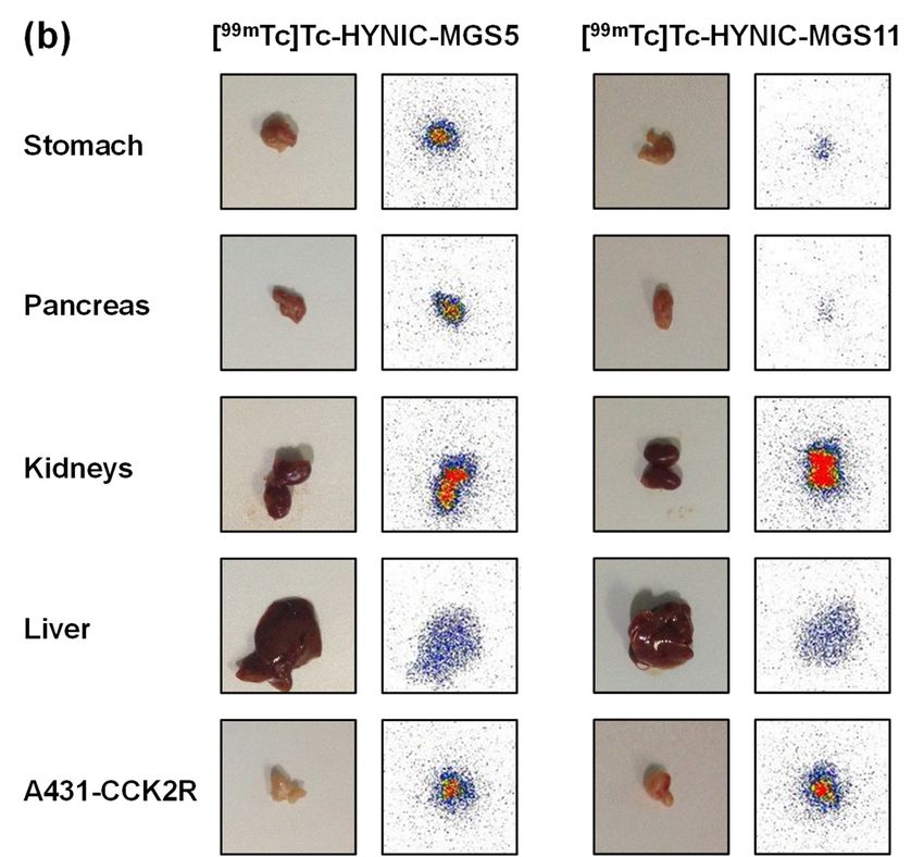
Figure 7.
Figure 7. Biodistribution of [[99m
Biodistribution of 99mTc]Tc‐HYNIC‐MGS5 (red) and [99m
Tc]Tc-HYNIC-MGS5 (red) and [99mTc]Tc‐HYNIC‐MGS11
Tc]Tc-HYNIC-MGS11 (blue)
(blue) in
in the
the
A431-CCK2R/A431-mock xenograft model: (a) tissue distribution and tumor uptake at 1 h and 4 h
A431‐CCK2R/A431‐mock xenograft model: (a) tissue distribution and tumor uptake at 1 h and
expressed as
p.i. with values expressed as % % IA/g
IA/g (mean ±± SD,
SD,nn ==4);
4);(b)
(b) autoradiography
autoradiography performed
performed for selected
organs at 1 h p.i. (color scale, pixel intensity: min 9 (blue), max 100 (red)).Pharmaceuticals 2019, 12, 13 9 of 15
The structural difference of the two radioligands also had a strong impact on the kidney uptake.
The kidney uptake of [99m Tc]Tc-HYNIC-MGS11 (19.90 ± 2.09% IA/g at 1 h and 17.17 ± 2.93% IA/g
at 4 h p.i.) was two times higher in comparison with [99m Tc]Tc-HYNIC-MGS5 (10.56 ± 1.15% IA/g
at 1 h and 7.80 ± 1.47% IA/g at 4 h p.i.). Despite CCK2R-related uptake, reabsorption and retention
of radiolabeled peptides in kidneys is mainly driven by multiple transport mechanisms, involving
megalin and cubulin [34]. In megalin-deficient mice a significantly reduced renal reabsorption was
confirmed for a radiolabeled minigastrin analog, with renal uptake values reduced to 37–49% when
compared to wild-type mice [35]. It has been shown for different radiolabeled MG analogs that the
uptake in stomach and CCK2R-expressing tumor-xenografts can be efficiently blocked by co-injection
of a 1000-fold molar excess of unlabeled peptide, whereas no considerable effect occurs in kidneys [36].
In this study no additional blocking studies were performed in vivo to confirm the receptor-specific
uptake in the different organs.
The tumor targeting profile of [99m Tc]Tc-HYNIC-MGS5 well compares with previous results
obtained with [111 In]In-DOTA-MGS5, showing a similar tumor uptake of 19.53 ± 5.42% IA/g and
23.49 ± 1.25% IA/g at 1 h and 4 h p.i., respectively [15]. This high and persistent tumor uptake
is clearly superior when compared to other 99m Tc-labeled MG analogs previously studied [37].
For [99m Tc]Tc-Demogastrin-2 evaluated in A431-CCK2R xenografted SCID mice at a similar
injected peptide amount of 10 pmol a tumor uptake of 12.89 ± 4.69% ID/g at 4 h p.i. was
reported, which was however, connected with a very high kidney uptake (58.62 ± 8.98% ID/g).
The metabolic stability of this compound (60% intact radiopeptide at 5 min p.i.) was comparable
to [99m Tc]Tc-HYNIC-MGS5 [37]. In our previous studies with radiolabeled DOTA-MGS5 we
concluded that a combination of increased protein binding and stabilization against enzymatic
degradation might be responsible for the highly improved targeting profile. The enhanced
protection against metabolic degradation of [99m Tc]Tc-HYNIC-MGS11 led to a further doubling
in tumor uptake. Such an improvement could not be achieved by enzymatic stabilization alone,
as exemplified by the co-injection of protease inhibitors [37]. [99m Tc]Tc-Demogastrin-2 coinjected
with 300 µg phosphoramidon showed a similar enzymatic stability in vivo (85% intact radiopeptide
at 5 min p.i.) as compared to [99m Tc]Tc-HYNIC-MGS11, but the tumor uptake was clearly
inferior (18.21 ± 5.97% ID/g at 4 h p.i.). When comparing the tumor-to-organ activity ratios of
[99m Tc]Tc-HYNIC-MGS5 and [99m Tc]Tc-HYNIC-MGS11, somewhat lower tumor-to-blood ratios were
observed for [99m Tc]Tc-HYNIC-MGS11. Thus, [99m Tc]Tc-HYNIC-MGS11 also showed increased
non-specific uptake in most organs at both investigated time points. Due to the concomitant increase
of the uptake in A431-CCK2R xenografts and kidneys observed for [99m Tc]Tc-HYNIC-MGS11, a similar
tumor-to-kidney ratio was found for both radiopeptides (2.4–3.3 for [99m Tc]Tc-HYNIC-MGS5 and
2.0–2.6 for [99m Tc]Tc-HYNIC-MGS11). The respective tumor-to-organ activity ratios calculated for
blood, kidney, intestine, pancreas and stomach for both radioligands at different time points are
displayed in Table 1.
Table 1. Tumor-to-organ activity ratios for A431-CCK2R tumor-xenografts of [99m Tc]Tc-HYNIC-MGS5
and [99m Tc]Tc-HYNIC-MGS11 (mean ± SD, n = 4).
[99m Tc]Tc-HYNIC-MGS5 [99m Tc]Tc-HYNIC-MGS11
1 h p.i. 4 h p.i. 1 h p.i. 4 h p.i.
Tumor/blood 21.6 ± 5.1 273 ± 151 15.6 ± 4.5 177 ± 55
Tumor/kidney 2.4 ± 0.3 3.3 ± 1.1 2.0 ± 0.6 2.6 ± 0.8
Tumor/stomach 1.5 ± 0.2 2.0 ± 0.4 6.5 ± 1.9 10.7 ± 1.4
Tumor/pancreas 2.4 ± 0.3 4.2 ± 2.0 20.6 ± 11.7 34.8 ± 9.7
Tumor/intestine 14.2 ± 1.7 19.2 ± 7.5 32.3 ± 5.0 61.1 ± 14.3
To our knowledge this is the first report on 99m Tc-labeled CCK2R targeting peptide analogs
showing such an astonishingly improved tumor uptake along with clearly reduced kidney uptake.Pharmaceuticals 2019, 12, 13 10 of 15
[99m Tc]Tc-HYNIC-MGS5 showed a very similar targeting profile when compared to DOTA-MGS5
radiolabeled with different radiometals suitable for SPECT, PET and TRT [15]. The tumor uptake of
[99m Tc]Tc-HYNIC-MGS11 was further improved, however, connected with a concomitant increase in
kidney uptake. Recently, two 99m Tc-labeled non-peptidic radioligands have been described showing
high tumor uptake in a mouse tumor model based on human epithelial cells transfected with CCK2R,
however, tumor-to-kidney ratio was clearly inferior [38,39].
These developments give high promise that in the near future a kit for 99m Tc-labeling will
be available allowing the localization and staging of CCK2R expressing tumors. Gastrin receptor
scintigraphy might show a lower sensitivity when compared to PET imaging with a 68 Ga-labeled
CCK2R targeting MG analog, but additionally allows for radioguided surgery. The concept of surgical
guidance with conventional gamma probes for intraoperative identification and removal of metastatic
lesions has already been successfully introduced into clinical practice by the use of 99m Tc-labeled
somatostatin analogs in patients with neuroendocrine tumors [40] as well as 99m Tc-labeled ligands
targeting prostate specific membrane antigen in patients with prostate cancer [41]. Due to its high tumor
uptake and tumor retention combined with high clearance from other tissues [99m Tc]Tc-HYNIC-MGS11
might be favorable for imaging and radioguided surgery. [99m Tc]Tc-HYNIC-MGS5 well compares
with DOTA-MGS5 and shows the advantage of using the same peptide for different applications,
[68 Ga]Ga-DOTA-MGS5 for PET, [99m Tc]Tc-HYNIC-MGS5 for SPECT and radioguided surgery, as well
as [177 Lu]Lu-DOTA-MGS5 for TRT.
3. Materials and Methods
3.1. Materials
All commercially obtained chemicals were of analytical grade and used without further
purification. Na[99m Tc]TcO4 was obtained from a commercial 99 Mo/99m Tc-generator (Ultratechnekow,
Mallinckrodt, Petten, The Netherlands) eluted with physiological saline. The A431 human epidermoid
carcinoma cell line stably transfected with the plasmid pCR3.1 containing the full coding sequence
for the human CCK2R (A431-CCK2R) as well as the same cell line transfected with the empty vector
alone (A431-mock) were kindly provided by Dr. Luigi Aloj [42]. Both cell lines were cultured in
Dulbecco’s Modified Eagle Medium (DMEM) supplemented with 10% (v/v) fetal bovine serum and
5 mL of a 100× penicillin-streptomycin-glutamine mixture at 37 ◦ C in a humidified 95% air/5% CO2
atmosphere. Media and supplements were purchased from Invitrogen Corporation (Lofer, Austria).
3.2. Peptide Synthesis
HYNIC-dGlu-Ala-Tyr-Gly-Trp-(N-Me)Nle-Asp-1-Nal-NH2 (HYNIC-MGS5) and HYNIC-dGlu-
Ala-Tyr-Gly-Trp-(N-Me)Nle-Asp-(N-Me)1-Nal-NH2 (HYNIC-MGS11) were synthesized using
9-fluorenylmethoxycarbonyl (Fmoc) chemistry. The peptides were assembled on 60 mg Rink Amide
MBHA resin with capacity 0.5 mmol/g resin (Novabiochem, Hohenbrunn, Germany). The reactive side
chains of the amino acids were masked with the following protection groups: tert-butyl ester for Asp
and dGlu, tert-butyl ether for Tyr, and tertbutyloxycarbonyl (BOC) for Trp. All coupling reactions were
performed using a 5-fold excess of Fmoc-protected amino acids, 1-hydroxy-7-aza-benzotriazole (HOAt)
and O-(7-Azabenzotriazole-1-yl)-N, N,N’N’-tetramethyluronium hexa-fluorophosphate (HATU) in
N-Methyl-2-pyrrolidone (NMP) pH adjusted to 8-9 with N,N’-diisopropylethylamine. Coupling of
the Fmoc-protected amino acids following (N-Me)Nle or (N-Me)1-Nal was repeated twice. For the
coupling of HYNIC a 3-fold molar excess of BOC-HYNIC, HOAt and HATU was used. The introduction
of an additional methyl group into the peptide bound between Asp and 1-Nal in HYNIC-MGS11
was performed by direct N-methylation of 1-Nal during the peptide synthesis on the solid resin as
described by Chatterjee et al. [43]. Cleavage of the peptides from the resin with concomitant removal
of acid-labile protecting groups was achieved by treatment with a mixture of trifluoroacetic acid (TFA),
triisopropylsilane, and water in a ratio 95/2.5/2.5 v/v/v. The crude peptides were precipitated andPharmaceuticals 2019, 12, 13 11 of 15
washed with ether before HPLC purification and characterized by analytical HPLC and MALDI-TOF
MS. The lyophilized peptide derivatives were stored at −20 ◦ C.
3.3. Analytical Systems and Methods
For preparative HPLC purification a Gilson 322 chromatography system (Gilson International,
Limburg, Germany) with Gilson UV/VIS-155 multi-wavelength detector, equipped with an Eurosil
Bioselect Vertex Plus C18A precolumn (300 Å, 5 µm, 30 × 8 mm) and a Eurosil Bioselect Vertex Plus
C18A column (300 Å 5 µm 300 × 8 mm) (Knauer, Berlin, Germany) was used with a gradient system
starting from 80% solvent A (water containing 0.1% TFA) and increasing concentrations of solvent B
(acetonitrile (ACN) containing 0.1% TFA) with a flow rate 2 mL/min: 0–4 min 20% B, 4–24 min 20–60%
B, 24–26 min 60% B, 26–27 min 60–80% B, 27–28 min 80% B, 28–29 min 80–20% B, 29–37 min 20% B.
Analytical HPLC was performed using an UltiMate 3000 chromatography system (Dionex, Germering,
Germany) consisting of a HPLC pump, a variable UV-detector (UV-VIS at λ = 280 nm), an autosampler,
a radiodetector (GabiStar, Raytest, Straubenhardt, Germany), equipped with a Phenomenex Jupiter
4 µm Proteo C12 90 Å 250 × 4.6 mm column (Phenomenex Ltd., Aschaffenburg, Germany) using a flow
rate of 1 mL/min together with the following gradient system: 0–3 min 10% B, 3–18 min 10–55% B,
18–20 min 80% B, 20–21 min 80–10% B, 21–25 min 10% B.
For Matrix Assisted Laser Desorption Ionization Time-of-Flight Mass Spectrometry (MALDI-TOF
MS) a Bruker microflex benchtop MALDI-TOF MS (Bruker Daltonics, Bremen, Germany) was used
in reflector acquisition mode with a positive ion source and 200 shots per spot. MALDI samples
were prepared on a α-cyano-4-hydroxycinnamic acid (HCCA) matrix using dried droplet procedure.
Flex Analysis 2.4 software was used to analyze the recorded data.
3.4. 99m Tc-Radiolabeling Using the Tricine/EDDA Exchange Method
99m Tc-labeling
was performed using a previously described exchange labeling approach with
tricine and EDDA [27]. For this purpose 10–20 µg of the corresponding HYNIC-conjugated peptide
analog (dissolved in EtOH/H2 O 30/70 v/v at a concentration of 0.5 µg/µL) together with 250 µL of
EDDA solution (20 mg/mL in 0.1 M NaOH), 250 µL tricine solution (40 mg/mL in 0.2 M PBS pH 6),
500 µL of Na[99m Tc]TcO4 (≤750 MBq) and 20 µl of tin(II) chloride solution (20 mg of SnCl2 * 2 H2 O
in 10 mL 0.1 N HCl), were incubated in a sealed glass vial at 100 ◦ C for 15–20 min. Radiochemical
purity of [99m Tc]Tc-HYNIC-MGS5 and [99m Tc]Tc-HYNIC-MGS11 was determined by analytical HPLC.
For in vivo assays the radiolabeled peptides were purified by SPE. For this purpose, the labeling
mixture was passed through a C18-SepPak-Light cartridge (Waters, Milford, MA, USA), followed
by 5 mL saline, and the radiolabeled peptide was eluted with EtOH/H2 O 65/35 v/v and diluted
with PBS.
3.5. Evaluation of the in Vitro Properties
The resistance against degradation of [99m Tc]Tc-HYNIC-MGS5 and [99m Tc]Tc-HYNIC-MGS11
(1000 pmol/mL, n = 2) in human serum was studied for up to 24 h. Furthermore, the radiopeptides
were incubated in PBS (1000 pmol/mL, n = 1). At each time point of 1, 2, 4 and 24 h after incubation
the intact radiopeptide was assessed by analytical HPLC. Serum samples were precipitated with
ACN and centrifuged to collect the supernatant and diluted with water prior to radio-HPLC. For the
determination of the distribution coefficient (log D), 500 µL of the radiopeptide solutions (50 pmol/mL
in PBS) were added to 500 µL octanol (1:1) vigorously vortexed (n = 8) for 15 min and centrifuged
to separate the two phases. From each phase a 75 µL sample was taken, the radioactivity measured
in a 2480 Wizard2 automatic gamma-counter (PerkinElmer Life Sciences and Analytical Sciences,
Wallac Oy, Turku, Finland) and the distribution of the radiopeptides calculated. The protein binding in
human serum (500 pmol/mL, n = 2) was assessed by Sephadex G-50 size-exclusion chromatography
(GE Healthcare Illustra, Little Chalfont, UK) for up to 24 h.Pharmaceuticals 2019, 12, 13 12 of 15
3.6. Receptor Binding and Cell Internalization Studies
The receptor affinity of the radioligands prepared using the above described labeling protocol
was evaluated in saturation studies on A431-CCK2R cells. For the assay, 96-well filter plates
(MultiScreenHTS -FB, Merck Group, Darmstadt, Germany) were pretreated with 10 mM TRIS/139 mM
NaCl buffer, pH 7.4 (TRIS-buffer) (2 × 250 µL) and 400,000 A431-CCK2R cells per well were added in
35 mM HEPES buffer, pH 7.4, containing 10 mM MgCl2 , 14 µM bacitracin, and 0.5% bovine serum
albumin (BSA), a hypotonic solution disturbing the integrity of the cell membranes. Thereafter,
increasing concentrations of the radiolabeled peptide conjugates (0.1–112 nM) were added in triplicate
reaching a total volume of 200 µL. In parallel, non-specific binding was determined by co-incubation
with 1 µM pentagastrin. After 1 h incubation at room temperature, the medium was removed by
filtration followed by two rapid rinses with ice-cold TRIS buffer (200 µL). The filters were collected
and counted in a gamma-counter. The Kd value was calculated fitting the data with Origin software
(Origin 6.1, OriginLab Corporation, Northampton, MA, USA) to a one-site binding model using the
formula y = Bmax × x/(Kd + x).
For internalization experiments, A431-CCK2R and A431-mock cells were seeded at a density of
1.0 × 10⁶ cells per well in 6-well plates and grown to confluence for 48 h. At the day of the experiment,
cells were washed twice with ice-cold internalization medium supplemented with 1% (v/v) fetal
bovine serum and supplied with fresh medium before incubation with [99m Tc]Tc-HYNIC-MGS5 and
[99m Tc]Tc-HYNIC-MGS11 at a final peptide concentration of 0.4 nM in a total volume of 1.5 mL in
triplicates. At different time points for up to 2 h incubation the cell uptake was interrupted by removal
of the medium and rapid rinsing with ice-cold internalization medium (two times). Thereafter, the cells
were incubated twice at ambient temperature in acid wash buffer (50 mM glycine buffer pH 2.8,
0.1 M NaCl) for 5 min, to remove the membrane-bound radioligand. Finally, the cells were lyzed
by treatment in 1 M NaOH and collected (internalized radioligand fraction). All collected fractions
(supernatant, surface wash, lyzed cells) were measured together with a standard in the gamma counter.
The radioactivity of the lyzed cells was expressed as percentage of the total radioactivity added (% of
internalized radioactivity). Non-specific binding was evaluated in A431-mock cells and in additional
blocking studies with 1 µM pentagastrin.
3.7. Evaluation of the in Vivo Stability and Biodistribution
All animal experiments were conducted in compliance with the Austrian animal protection laws
and with the approval of the Austrian Ministry of Science (BMWFW-66.011/0075-WF/V/3b/2016).
3.7.1. Metabolic Stability in BALB/c Mice
Metabolic stability studies in vivo with [99m Tc]Tc-HYNIC-MGS5 and [99m Tc]Tc-HYNIC-MGS11
were performed in 5–6 week-old female BALB/c mice (Charles River, Sulzfeld, Germany; n = 2).
Mice were injected intravenously via a lateral tail vain with 37–74 MBq of the 99m Tc-labeled
HYNIC-analogs (corresponding to 2 nmol total peptide). Ten minutes post injection (p.i.) mice
were euthanized and a sample of blood and urine was collected together with the liver and kidneys.
Liver and kidneys were rapidly homogenized in 0.5 mL of a 20 mM HEPES buffer pH 7.3 with
an Ultra-Turrax T8 homogenator (IKA-Werke, Staufen, Germany) for 1 min at RT. Before determining
the percentage of intact radiopeptide by analytical radio-HPLC samples were precipitated with ACN,
centrifuged and diluted with H2 O (1:1).
3.7.2. Biodistribution in Tumor-xenografted BALB/c Nude Mice
Biodistribution studies evaluating the tumor uptake of [99m Tc]Tc-HYNIC-MGS5 and
[99m Tc]Tc-HYNIC-MGS11 were performed in 7 week-old female athymic BALB/c nude mice
(Charles River, Sulzfeld, Germany). To induce tumor-xenografts, mice were injected subcutaneously
with 2 × 10⁶ A431-CCK2R (right flank) and A431-mock cells (left flank). The tumor-xenografts werePharmaceuticals 2019, 12, 13 13 of 15
allowed to grow for 11–12 days reaching medium tumor weights of ~0.2 g. Mice were randomly
divided into groups of four and injected intravenously via a lateral tail vein with 0.3 MBq of the
99m Tc-labeled HYNIC-analogs (corresponding to ~15 pmol total peptide). The groups of animals
were sacrificed at 1 h and 4 h p.i., tumors and other tissues (blood, lung, heart, muscle, bone, spleen,
intestine, liver, kidney, stomach and pancreas) were removed, weighed, and the radioactivity measured
together with a standard in the gamma counter. Results were expressed as percentage of injected
activity per gram tissue (% IA/g) and tumor-to-organ activity ratios were calculated for selected
tissues. Statistical analysis was performed using independent two population t-test (significance level
p = 0.05) with Origin software. The radioactivity in selected organs (stomach, pancreas, liver, kidneys,
A431-CCK2R and A431-mock xenograft) was additionally visualized by autoradiography using
a phosphorimager (Cyclone Plus, PerkinElmer Life Sciences and Analytical Sciences, Downers Grove,
Il, USA). After dissection, the organs were exposed to a multisensitive storage phosphor screen for
40 min and image analysis was performed using OptiQuantTM software (OptiQuant 5.0, PerkinElmer
Life Sciences and Analytical Sciences, Downers Grove, Il, USA).
Supplementary Materials: The following are available online at http://www.mdpi.com/1424-8247/12/1/13/
s1, Table S1: Biodistribution of [99m Tc]Tc-HYNIC-MGS5 and [99m Tc]Tc-HYNIC-MGS11 in BALB/c nude mice
tumor-xenografted with A431-CCK2R and A431-mock tumors (mean ± SD, n = 4).
Author Contributions: Conceptualization, Supervision, Project Administration and Funding Acquisition, E.v.G.;
Methodology and Investigation, M.K., D.S., P.K., E.v.G., C.D. and C.R.; Writing-Original Draft Preparation: M.K.,
D.S.; Writing-Review & Editing, E.v.G., C.R. and C.D.
Funding: This study was funded by the Austrian Science Foundation (FWF) grant P 27844.
Acknowledgments: Technical assistance of Sedigheh Rezaeianpour in stability assays and of Joachim Pfister in
biodistribution studies is greatly acknowledged.
Conflicts of Interest: The authors declare no conflict of interest. The funders had no role in the design of the
study; in the collection, analyses, or interpretation of data; in the writing of the manuscript, and in the decision to
publish the results.
References
1. Laverman, P.; Sosabowski, J.K.; Boerman, O.C.; Oyen, W.J.G. Radiolabelled peptides for oncological diagnosis.
Eur. J. Nucl. Med. Mol. Imaging 2012, 39, 78–92. [CrossRef] [PubMed]
2. Fani, M.; Maecke, H.R. Radiopharmaceutical development of radiolabelled peptides. Eur. J. Nucl. Med.
Mol. Imaging 2012, 39, S11–S30. [CrossRef] [PubMed]
3. Fani, M.; Maecke, H.R.; Okarvi, S.M. Radiolabeled peptides: Valuable tools for the detection and treatment
of cancer. Theranostics 2012, 2, 481–501. [CrossRef] [PubMed]
4. Koopmans, K.P.; Glaudemans, A.W.J.M. Rationale for the use of radiolabelled peptides in diagnosis and
therapy. Eur. J. Nucl. Med. Mol. Imaging 2012, 39, 4–10. [CrossRef] [PubMed]
5. Reubi, J.C.; Schaer, J.C.; Waser, B. Cholecystokinin(CCK)-A and CCK-B/gastrin receptors in human tumors.
Cancer Res. 1997, 57, 1377–1386. [PubMed]
6. Reubi, J.C. Targeting CCK receptors in human cancers. Curr. Top. Med. Chem. 2007, 7, 1239–1242. [CrossRef]
[PubMed]
7. Kaloudi, A.; Nock, B.A.; Krenning, E.P.; Maina, T.; De Jong, M. Radiolabeled gastrin/CCK analogs in tumor
diagnosis: Towards higher stability and improved tumor targeting. Q. J. Nucl. Med. Mol. Imaging 2015,
59, 287–302. [PubMed]
8. Roosenburg, S.; Laverman, P.; van Delft, F.L.; Boerman, O.C. Radiolabeled CCK/gastrin peptides for imaging
and therapy of CCK2 receptor-expressing tumors. Amino Acids 2011, 41, 1049–1058. [CrossRef]
9. Fani, M.; Peitl, P.; Velikyan, I. Current status of radiopharmaceuticals for the theranostics of neuroendocrine
neoplasms. Pharmaceuticals 2017, 10, 30. [CrossRef] [PubMed]
10. Krošelj, M.; Mansi, R.; Reubi, J.C.; Maecke, H.R.; Kolenc Peitl, P. Comparison of DOTA-coupled minigastrin
analogues and corresponding Nle congeners. Eur. J. Nuclear Med. Mol. Imaging 2012, 39, S533–S534.
[CrossRef]You can also read