Novel neutralizing human monoclonal antibodies against tetanus neurotoxin
←
→
Page content transcription
If your browser does not render page correctly, please read the page content below

www.nature.com/scientificreports
OPEN Novel neutralizing human
monoclonal antibodies
against tetanus neurotoxin
Takeharu Minamitani1,2, Karin Kiyose1,2, Ryota Otsubo3, Toshihiro Ito4, Hiroki Akiba5,
Rika A. Furuta6, Tsuyoshi Inoue7, Kouhei Tsumoto8,9,10, Masahiro Satake6 &
Teruhito Yasui1,2,3*
Tetanus is a fatal disease caused by tetanus neurotoxin (TeNT). TeNT is composed of a light chain (Lc)
and a heavy chain, the latter of which is classified into two domains, N-terminus Hn and C-terminus
Hc. Several TeNT-neutralizing antibodies have been reported, but it remains unclear which TeNT
domains are involved in neutralization. To further understand the mechanism of these antibodies,
we isolated TeNT-reactive human antibody clones from peripheral blood mononuclear cells. We then
analyzed the reactivity of the isolated antibody clones to each protein domain and their inhibition
of Hc-ganglioside GT1b binding, which is critical for TeNT toxicity. We also investigated the TeNT-
neutralizing ability of isolated antibody clones and showed that an Hn-reactive clone protected
strongly against TeNT toxicity in mice. Furthermore, combination treatment of Hn-reactive antibody
clones with both Hc-reactive and TeNT mix (the mixture of Hc, Hn, and Lc proteins)–reactive antibody
clones enhanced the neutralizing effect. These results indicated that antibody clones targeting Hn
effectively neutralized TeNT. In addition, the use of a cocktail composed of Hc-, Hn-, and TeNT mix–
reactive antibodies provided enhanced protection compared to the use of each antibody alone.
Tetanus is a common, fatal disease caused by infection with Clostridium tetani, a Gram-positive obligate anaer-
obe. The bacterium enters wounds in the form of spores that germinate to produce tetanus neurotoxin (TeNT).
The toxin blocks the neuronal release of inhibitory neurotransmitters, including acetylcholine, resulting in
characteristic symptoms such as spastic paralysis1. TeNT is synthesized as a single 150-kDa polypeptide and
is subsequently cleaved to generate an active toxin. The active form of TeNT consists of a light chain (Lc, also
called fragment A, 50 kDa) and a heavy chain (100 kDa) linked by a disulfide bond, and the heavy chain is
composed of two domains, Hn (also called fragment B, 50 kDa) and Hc (also called fragment C, 50 kDa)2. Lc
works as a protease to cleave molecules such as vesicle-associated membrane protein-2 (VAMP-2), which is a
soluble neuronal N-ethylmaleimide-sensitive attachment receptor (SNARE) protein, resulting in the blockade
of inhibitory neurotransmitter r elease3,4. Hn plays a functional role in the translocation of Lc into the cytosol of
neuronal cells5. Hc is indispensable for TeNT binding to target cells through ganglioside receptors such as G T1b6.
Human tetanus immunoglobulin (TIG), which is derived from the plasma of vaccine-administrated donors,
is an effective tetanus t herapy7. However, due to the lack of source plasma and the risk of contamination with
1
Laboratory of Infectious Diseases and Immunity, National Institutes of Biomedical Innovation, Health and
Nutrition (NIBIOHN), 7‑6‑8 Saito‑Asagi, Ibaraki City, Osaka 567‑0085, Japan. 2Laboratory of Immunobiologics
Evaluation, Center for Vaccine and Adjuvant Research (CVAR), National Institutes of Biomedical Innovation, Health
and Nutrition (NIBIOHN), 7‑6‑8 Saito‑Asagi, Ibaraki City, Osaka 567‑0085, Japan. 3Laboratory of Pharmaceutical
Integrated Omics, Department of Pharmaceutical Engineering, Facility of Engineering, Toyama Prefectural
University, 5180 Kurokawa, Imizu, Toyama 939‑0398, Japan. 4Laboratory of Proteome Research, National Institutes
of Biomedical Innovation, Health and Nutrition (NIBIOHN), 7‑6‑8 Saito‑Asagi, Ibaraki City, Osaka 567‑0085,
Japan. 5Laboratory of Advanced Biopharmaceuticals, National Institutes of Biomedical Innovation, Health and
Nutrition (NIBIOHN), 7‑6‑8 Saito‑Asagi, Ibaraki City, Osaka 567‑0085, Japan. 6Japanese Red Cross Central Blood
Institute, 1‑67 Tatsumi, Koto‑ku, Tokyo 135‑8521, Japan. 7Division of Advance Pharmaco‑Science, Graduate
School of Pharmaceutical Science, Osaka University, 1‑6 Yamada‑oka, Suita, Osaka 565‑0871, Japan. 8Center for
Drug Discovery Research (CDDR), National Institutes of Biomedical Innovation, Health and Nutrition (NIBIOHN),
7‑6‑8 Saito‑Asagi, Ibaraki City, Osaka 567‑0085, Japan. 9Medical Proteomics Laboratory, The Institute of Medical
Science, The University of Tokyo, 4‑6‑1 Shirokanedai, Minato‑ku, Tokyo 108‑8639, Japan. 10Department of
Bioengineering, School of Engineering, The University of Tokyo, 7‑3‑1 Hongo, Bunkyo‑Ku, Tokyo 113‑8656,
Japan. *email: tyasui@nibiohn.go.jp
Scientific Reports | (2021) 11:12134 | https://doi.org/10.1038/s41598-021-91597-2 1
Vol.:(0123456789)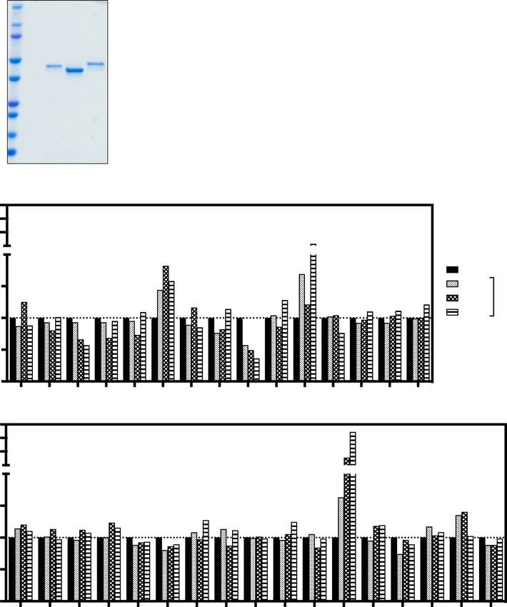
www.nature.com/scientificreports/
infectious materials, the TIG should be replaced with a recombinant antibody (rAb) product7,8. Compared to
mouse antibodies, human antibodies have an advantage as a therapeutic agent because they do not require a
difficult humanization process. Several TeNT-neutralizing human antibodies have been r eported7–11, but it is still
relatively unclear which targeted TeNT domains are required for these antibodies’ neutralization activity. In addi-
tion, it has been reported that combinations of different antibodies, such as TIG, lead to enhanced neutralizing
activity9, but it is not well understood which antibodies in such mixtures contribute to neutralization. To address
these questions we isolated TeNT-reactive human antibody clones and assessed their TeNT inhibitory activity.
Results
Isolation and characterization of TeNT‑reactive lymphoblastoid cell lines (LCLs). To isolate
monoclonal antibodies reactive to TeNT, we isolated peripheral blood mononuclear cells (PBMCs) from the
peripheral blood of healthy volunteers, and infected these cells with Epstein-Barr virus (EBV), and established
LCLs. Two weeks after EBV infection, we analyzed the reactivity of antibodies in LCL supernatants against the
mixture of Lc, Hn, and Hc proteins (the ratio of Lc:Hn:Hc = 1:1:1, defined as the TeNT mix) (Fig. 1A) using an
enzyme-linked immunosorbent assay (ELISA). We performed the screening experiments twice and obtained
18 and 35 TeNT mix–reactive LCL supernatants, respectively (Table 1). We further determined the reactivity of
isolated LCL supernatants against Lc, Hn, or Hc protein by ELISA, and obtained a total of four LCL supernatants
bound to the TeNT mix but not to Hc, Hn, or Lc proteins individually (TeNT mix–reactive supernatants) in
addition to the supernatants bound to each domain (Table 1).
LCL supernatants, 8A7 and 17F7, inhibit the binding of Hc to ganglioside GT1b. Binding of
the TeNT Hc domain to ganglioside GT1b plays a key role in TeNT toxicity, and inhibiting Hc-GT1b binding
interfered with TeNT toxicity. To isolate the TeNT neutralizing antibodies, we performed an ELISA-based Hc-
GT1b binding assay and evaluate the ability of Hc-reactive antibodies in LCL supernatants to block Hc bind-
ing to ganglioside GT1b. We analyzed 31 Hc-reactive LCL supernatants obtained in two rounds of screening
experiments (Table 1), and found that two LCL supernatants, 8A7 and 17F7, inhibited Hc binding to GT1b in a
dilution-dependent manner (Fig. 1B). In contrast, two LCL supernatants, 8H2 and 25H5, enhanced Hc-GT1b
binding (Fig. 1B).
Hn‑ or TeNT mix–reactive antibodies in LCL supernatants protect against TeNT toxicity in
mice. To determine the target domain responsible for the neutralizing ability of antibodies, we performed an
in vivo TeNT neutralization assay using LCL supernatants isolated in the second screening experiment, in which
LCL supernatants reactive to all domains individually were obtained (Table 1). The mixture of all LCL super-
natants (35Ab mix) completely neutralized TeNT (Fig. 2A). Mixtures of three Hn- or three TeNT mix–reactive
supernatants (Hn Abs or TeNT mix Abs, respectively) significantly improved the survival of TeNT-treated mice
compared to negative control (NC). (Log-rank test, p = 0.0082 or p = 0.040, respectively) (Fig. 2A). In addition,
we examined the protective effect enhancement by the combination treatment with Hn- and TeNT mix–reactive
supernatants and found the significantly increased protective activity compared to that of each Hn- or TeNT
mix–reactive mixture alone (Log-rank test, p = 0.029 or p = 0.040, respectively) (Fig. 2B).
Immunoglobulin gene usage analysis and characterization of recombinant antibodies. To
characterize the isolated antibodies, we cloned the immunoglobulin heavy and light chain genes and analyzed
their immunoglobulin gene usage and complementarity determining region (CDR) 3 sequence. As Hc-reactive
antibodies, we cloned 8A7 and 17F7, which inhibited Hc-GT1b binding (Fig. 1B). As Hn-reactive and TeNT
mix–reactive antibodies which had a neutralizing ability (Fig. 2A), we randomly selected 8D8 and 16E8, respec-
tively and analyzed them. As a result, the V (D) J gene combinations and CDR3 amino acid sequences were
differed between each clone, indicating that 8A7, 17F7, 8D8, and 16E8 had different origins (Table 2). We next
generated and purified recombinant antibodies (rAbs) of 8A7, 17F7, 8D8, and 16E8 (Fig. 3A), and assessed their
reactivity by ELISA (Fig. 3B–D). 8A7 and 17F7 reacted with Hc, but not Hn, and Lc, and 8D8 was reactive to Hn
but not Hc, and Lc (Fig. 3B–D). All four antibodies bound to TeNT mix and toxoid in a dose-dependent man-
ner (Fig. 3E,F). However, 8D8 and 16E8 showed very weak reactivity to TeNT (Fig. 3G). We also confirmed that
purified recombinant 8A7 and 17F7 dose-dependently inhibited Hc-GT1b binding, and their inhibitory activity
was significantly increased at the concentration of 1.25 and 10 µg/mL, compared to negative control antibody
(NC) (two-way analysis of variance (ANOVA), p < 0.0001 for each) (Fig. 3H).
The mixture of several antibodies reactive to different domains exerts strong neutralization
activity in mice. We assessed the TeNT-neutralization ability of purified rAbs in mice. For each antibody,
3.75 ng (TeNT:rAb ratio 1:37.5) significantly exerted protective ability against 1 LD50 TeNT compared to nega-
tive control (NC) in mice (Log-rank test, 8A7, p = 0.011; 17F7, p = 0.010; 8D8, p = 0.021; and 16E8, p = 0.010),
and 8D8 showed the strongest protective activity in the four rAbs (Fig. 4A). In all cases, 3,750 ng of each anti-
body (TeNT:rAb ratio 1:37,500) completely protected the mice from 1 L D50 TeNT (Fig. 4B). We next prepared
two antibody cocktails containing Hn-reactive 8D8, TeNT mix–reactive 16E8, and either Hc-reactive 8A7 or
17F7 to assess the neutralizing ability of combined treatment of antibodies. Both antibody cocktails targeting
Hc + Hn + TeNT mix completely neutralized 1 LD50 TeNT toxicity in mice beginning at 1.25 ng each (3.75 ng in
total, TeNT:rAb ratio 1:37.5) (Fig. 4C,D). The results in TeNT:rAb ratio 1:37.5 of Fig. 4A,C,D suggested that the
neutralizing ability of each antibody was enhanced in combination with other antibodies.
Scientific Reports | (2021) 11:12134 | https://doi.org/10.1038/s41598-021-91597-2 2
Vol:.(1234567890)www.nature.com/scientificreports/
A.
r
ke
ar
kDa
n
c
Lc
M
H
H
150
100
75
50
37
25
20
15
10
B.
350
300
250
Percent binding
200
200
Medium
150 1/24 Dilution
1/12 of
100
1/6 Supernatant
50
0
NC 7D4 8A7 8F12 8G10 8H2 9D6 4H7 17F7 23B10 23D1 29G11 2C6 4D2 4D7
350
300
250
Percent binding
200
200
150
100
50
0
3D2 3D8 11H10 15E1 17D5 18C2 20E6 21H6 23C7 23D10 24F1 25H5 26B10 26H9 28F10 30C6 30D1
Figure 1. The 8A7 and 17F7 antibodies inhibit the binding of Hc to ganglioside GT1b. (A) The purified
recombinant Lc, Hc, and Hn proteins were separated by 4–20% SDS-PAGE and stained with Coomassie Brilliant
Blue (CBB). (B) The inhibition of Hc-GT1b by antibodies was analyzed by an ELISA-based Hc-GT1b binding
assay. The supernatants of 31 Hc-reactive clones were serially diluted to 1/6, 1/12, and 1/24. Percent binding of
Hc to GT1b is indicated.
The neutralizing antibody 8D8 binds to the hydrophobic region of Hn. As shown in Fig. 4A,
Hn-reactive antibody 8D8 remarkably neutralized TeNT toxicity in mice at relatively low dose. To deeply under-
stand the molecular mechanism, we determined the binding site of 8D8 in Hn by ELISA. We first tested dele-
tion mutants of Hn. Hn (458–828) showed no binding activity to 8D8, while this activity was retained by Hn
(790–864) (Fig. 5A). We also prepared several additional mutants by replacing every three amino acids from 829
to 864 aa of the Hn (700–864) mutant with alanine and then examined the antibody’s binding ability (Fig. 5B).
Scientific Reports | (2021) 11:12134 | https://doi.org/10.1038/s41598-021-91597-2 3
Vol.:(0123456789)www.nature.com/scientificreports/
Reactive LCLs
Experiment ID Screened PBMCs Hc Hn Lc TeNT mixa Total
7
1 4.7 × 10 14 0 3 1 18
2 2.8 × 10⁷ 17 3 12 3 35
Total 7.5 × 10⁷ 31 3 15 4 53
Table 1. Number of screened PBMCs and isolated LCLs. a TeNT mix is composed of Hc, Hn, and Lc proteins.
The number of TeNT mix–reactive supernatants indicates refers to clones that were not reactive to Hc, Hn, or
Lc individually but were reactive to the mixture of these three antigens in ELISA.
A. (n = 4)
100 NC
Percent survival
35 Ab mix
Hc Abs
Lc Abs
50 Hn Abs
TeNT mix Abs
0
0 48 54 60 66
Time (hr)
B. (n = 4)
100 NC
35 Ab mix
Percent survival
Hn Abs
TeNT mix Abs
50 TeNT mix Abs
+ Hn Abs
0
0 48 60 72 84 96
Time (hr)
Figure 2. Hn- or TeNT mix–reactive antibodies in LCL supernatants neutralizes TeNT in mice. The protective
effect of isolated antibody clones was analyzed by an in vivo TeNT-neutralization assay. We combined 0.5 ng
human IgG-containing LCL supernatants from all 35 supernatants (35 Ab mix), 17 Hc-reactive supernatants
(Hc Abs), 12 Lc-reactive supernatants (Lc Abs), 3 Hn-reactive supernatants (Hn Abs), or 3 TeNT mix–reactive
supernatants (TeNT-mix Abs) prepared in a second screening experiment (Table 1). Then we mixed the
supernatant mixture with 1 LD50 (25 pg) of TeNT and administrated it to ddY mice. Mouse survival rates after
administration are shown (n = 4). NC negative control (medium). Statistical analysis was performed using Log-
rank test.
ID VH DH JH CDR3 Amino acid mutation (aa)
IgH
8A7 IGHV3-33*03 IGHD6-19*01 IGHJ4*02 ARDKGYINGWYVPFFDY 15
17F7 IGHV3-33*01 IGHD6-13*01 IGHJ3*02 ARESGYASSWYFNGDAFDI 13
8D8 IGHV4-39*01 IGHD3-3*01 IGHJ5*02 ARLGVKKITLFGEVIPRSSWFAP 15
16E8 IGHV1-46*04 IGHD6-13*01 IGHJ4*02 ARDRRQQLVFDS 22
ID Vk Jk CDR3 Amino acid mutation (aa)
IgL
8A7 IGKV3-15*01 IGKJ2*01 QQYDNWPPVT 4
17F7 IGKV4-1*01 IGKJ4*01 QQYSSTPLT 5
8D8 IGKV1-33*01 IGKJ5*01 QQYDTLSIT 3
16E8 IGKV4-1*01 IGKJ3*01 QQYYSLSRGLT 9
Table 2. Gene usage and CDR3 sequence of cloned immunoglobulins. CDR complementarity determining
region, aa amino acids.
Scientific Reports | (2021) 11:12134 | https://doi.org/10.1038/s41598-021-91597-2 4
Vol:.(1234567890)www.nature.com/scientificreports/
Hn (700–864) AAA.3, 4, 5, and 7 mutants lost binding to 8D8 (Fig. 5B), indicating that the regions from 835 to
843 aa and from 847 to 849 aa of Hn are important for 8D8 to recognition of Hn.
For more comprehensive assessment for these observations, we applied a computational approach to predict
the variable domain structure of 8D8 and its recognition domain in TeNT. The computational simulation of
antibody-antigen docking revealed that the mode of 8D8-Hn binding was similar to that from the analysis of
the ELISA-based binding activity (Figs. 5 and 6).
Discussion
TIG is widely used to treat tetanus and is known to be effective. This indicates that vaccinated individuals
have TeNT-neutralizing antibodies in their peripheral blood. We hypothesized that we would be able to isolate
neutralizing antibody clones from the PBMCs of such individuals. In this study, we modified a standard EBV
infection method and established LCLs to isolate antibody clones. Then we cloned immunoglobulin heavy and
light chain genes from the RNA of the LCLs. Using this strategy, we successfully isolated 53 TeNT mix–reactive
LCL supernatants and identified four TeNT-neutralizing human antibodies, namely 8A7, 17F7, 8D8, and 16E8,
from the PBMCs of a healthy individual. The healthy volunteer belongs to a presumed population that has been
vaccinated against tetanus, suggesting that the isolated antibodies are derived from affinity-matured B cells. All
four isolated antibodies had TeNT neutralizing activity, and completely protected mice from TeNT-induced
death at high dose. In addition, the results of experiments treated with each antibody or combination of the four
antibodies suggested that treatment with mixture of antibodies reactive to different domains of TeNT enhanced
the neutralizing ability.
In the present study, we isolated a total of three Hn-reactive LCL supernatants from 53 TeNT mix–reactive
supernatants. Compared to Hc- or Lc-reactive supernatants, fewer Hn-reactive supernatants were isolated. It
has been reported that the Hn domain is hidden by the other domains due to the three-dimensional structure of
TeNT12. Interestingly, Hn-reactive recombinant 8D8 did not bind to TeNT in our study. Botulinum neurotoxin
(BoNT) is another highly potent toxin and is composed of three domains responsible for receptor binding (Hc),
toxin translocation into cells (Hn), and proteolytic cleavage of host cell proteins (Lc), respectively13. It has been
reported that most BoNT-neutralizing antibodies recognize H c14,15. In the study, a small number of Lc-reactive
protective antibodies were also isolated, but few antibodies bound to Hn d omain14,15. In the case of diphtheria
toxin (DT) which is consist of three domains, catalytic (C) domain, transmembrane (T) domain, and receptor
binding (R) domain, Wenzel et al. recently reported that all three domains are targets for neutralizing antibodies
and the T-domain-reactive antibodies showed the lowest neutralization p otency16. Nevertheless, in this study
we found that 8D8 bound to Hn via both Hn (835–843), in which hydrophobic amino acids form a cluster,
and Hn (847–849), leading to TeNT-neutralizing activity in mice. Considering the reported three-dimensional
(3-D) structure of TeNT, the region from 835 to 849 aa of TeNT could be relatively antigenic as it is externally
exposed12. These points suggest that this region of Hn is a good target for neutralizing antibodies despite the low
immunogenicity of Hn in humans. The computational simulation results of antibody-antigen docking in this
study supports these agreements, because the simulation results gave a closer match to the experimental results
of ELISA-based binding assays.
On the other hand, we isolated a total of four TeNT mix–reactive LCL supernatants, including 16E8, that did
not bind with each Hn, Hc, or Lc in ELISA, suggesting that these antibodies recognize TeNT in conformation-
dependent manner or the epitopes existing across the three domains. In fact, recombinant 16E8 bound to the
TeNT mix and to toxoid, but not TeNT, native toxin, suggesting that the conformation of TeNT differs from
that of TeNT mix and toxoid, and TeNT mix could form a structure similar to toxoid in our ELISA conditions.
However, recombinant 16E8 antibody protected mice from TeNT toxicity as strongly as recombinant 8D8. Volk
et al. also reported a protective mouse monoclonal antibody derived from tetanus toxoid-immunized mice
that reacted with toxoid but not TeNT in E LISA17. Moreover, it has been reported that the structure of TeNT
is changed by p H12, and thus it may be different in an ELISA assay than in vivo. These findings indicate that to
select neutralizing antibody candidates in vitro, it is important to use ELISA to analyze the reactivity of antibody
clones to the mixtures of each domain protein and to toxoid, as well as to each domain protein individually.
We also isolated a total of 31 Hc-reactive LCL supernatants, and two of these, 8A7 and 17F7, were the only
clones that inhibited the binding of Hc to ganglioside GT1b in vitro. On the other hand, we observed enhance-
ment of Hc-GT1b binding both by 8H2 and 25H5. Fitzsimmons et al. also reported an antibody that promoted
Hc binding to GT1b18. This binding enhancement might be due to a conformational change in Hc. Recombinant
8A7 and 17F7 reacted with toxiod and TeNT in ELISA and had TeNT-neutralizing activity in vivo. Because the
interaction of the TeNT Hc domain with GT1b is a critical to TeNT toxicity, blocking this interaction is thought
to be very important for neutralization. To isolate TeNT-neutralizing antibody clones, therefore, it would be use-
ful to examine whether antibody clones inhibit the binding of Hc to GT1b in vitro. In addition, Felix, L. Y. et al.
has been reported the synaptic vesicle protein 2 (SV2) as a neuron receptor of T eNT19. It would be worthwhile
to analyze the antibodies that inhibit the binding of TeNT to this receptor.
The combined supernatants of 35 TeNT mix–reactive LCL from our second screening experiment containing
0.5 ng of each antibody completely protected mice from 1 L D50 TeNT. Of the various mixtures of domain-reactive
LCL supernatants, the Hn or TeNT mix–reactive mixture conferred the protection. The addition of Hn-reactive
supernatants to the TeNT mix–reactive supernatants enhanced the latter’s protective ability, but unlike the
35Ab mix, it did not provide complete protection. These results suggest that in addition to the Hn- and TeNT
mix–reactive antibody mixtures, other domain-reactive antibody clones are required for complete protection
in our assay. Aliprandini et al. recently reported that the combination of Lc-, Hn- or full-length TeNT-reactive
human antibodies conferred complete protection against TeNT in mice20. In fact, mixture of Hc-reactive 8A7
Scientific Reports | (2021) 11:12134 | https://doi.org/10.1038/s41598-021-91597-2 5
Vol.:(0123456789)www.nature.com/scientificreports/
Figure 3. Reactivity analysis of recombinant antibodies by ELISA. The purified rAbs (8A7, 17F7, 8D8, and ▸
16E8) were separated by 4–20% SDS-PAGE and stained with CBB (A). The reactivity of the rAbs against Hc (B),
Hn (C), Lc (D), TeNT mix (E), tetanus toxoid (Toxoid; F) and native tetanus neurotoxin (TeNT; G) was analyzed
by ELISA in triplicate. The concentration of antibodies was serially diluted from 10 to 0.001 µg/mL (from 64
to 0.0064 nM). Human IgG purified from human sera was used as a negative control (NC). (H) The inhibitory
activity of Hc-GT1b by recombinant 8A7 and 17F7 was analyzed by an ELISA-based Hc-GT1b binding assay in
triplicate. The concentrations of antibodies used for the assay were 0, 0.01, 0.156, 1.25, and 10 µg/mL (0, 0.064,
0.998, 8, and 64 nM). The percent binding of Hc to GT1b is indicated. Human IgG purified from human sera
was used as a negative control (NC). The symbol or bar in each graph indicates the average. Error bars show the
means ± s.d. Statistical analysis was performed using two-way ANOVA.
or 17F7, Hn-reactive 8D8, and TeNT mix–reactive 16E8 completely neutralized 1 L D50 of TeNT in our mouse
experiments.
Finally, we isolated TeNT-neutralizing antibody clones from the PBMCs of a healthy individual using a
modified EBV infection method. An in vivo TeNT neutralization assay revealed that the Hn-reactive and TeNT
mix–reactive antibody clones had a protective ability and the combination of rAbs reactive to Hn, Hc, and TeNT
mix enhanced TeNT-neutralizing activity. Collectively, although it is required for further experiments, such as
treatment of antibodies after TeNT administration, and comparison of their neutralizing ability with TIG and
previously reported human antibodies, the cocktail of the recombinant neutralizing antibodies identified in this
study could be of help to the therapy for TeNT-induced infectious diseases instead of TIG.
Methods
Cell culture and reagents. B95-8-ZHT cells which are derived from B95-8 marmoset cells and stably
express EBV BZLF1 in a 4-hydroxytamoxifen-dependent manner21, were cultured in RPMI1640 (Nacalai Tesque,
Kyoto, Japan) supplemented with 10% fetal bovine serum (FBS; Merck, Darmstadt, Germany), streptomycin/
penicillin, and 2-mercaptoethanol (Nacalai Tesque). LCLs were cultured in RPMI1640 (Nacalai Tesque) supple-
mented with 20% FBS (Merck), streptomycin/penicillin, 2-mercaptoethanol (Nacalai Tesque), K3 (Ajinomoto
Bio-Pharma Services, Osaka, Japan), Ciclosporin (Novartis Pharma, Basel, Switzerland), IL-6, and BAFF (R & D
systems, Minneapolis, MN, USA) (LCL medium).
EBV preparation. To prepare EBV (B95-8 strain), B95-8-ZHT cells were treated with 400 nM 4-hydroxy-
tamoxifen (Merck) for 5 days and then collected the cultured conditioned medium as EBV stock.
Human blood samples. The blood donor was a healthy volunteer belonging to a presumed population that
has been vaccinated against tetanus. Ethical approval for the study was obtained from the Institutional Review
Board (IRB) of National Institutes of Biomedical Innovation, Health and Nutrition (NIBIOHN) (approval num-
ber 198), and informed consent was obtained from all participants. The study was performed in accordance with
the guidelines of the Declaration of Helsinki.
Mice. Four-week-old ddY mice were purchased from SLC Japan, Inc (Izu, Japan). The mice were used 1 week
after purchase. All mice were maintained in a specific pathogen–free animal facility in accordance with the
Osaka University guidelines for animal experimentation.
EBV infection and LCL establishment. PBMCs were separated from whole blood by gradient centrifu-
gation method using Lymphoprep reagent (Abbott Diagnostics Technologies AS, Oslo, Norway). Then I gM+ B
cells were depleted from the PBMCs using Magnetic Cell Sorting-based anti-human IgM Microbeads accord-
ing to the manufacturer’s protocol (Miltenyi Biotec, Bergisch Gladbach, Germany). The IgM+ B cell-depleted
PBMCs were suspended in EBV stock (107 cells/mL) and rotated at 37 °C for 1 h. The cells were then suspended
in LCL medium (5 × 10⁴ cells/mL) and seeded at 200 µL/well in round-bottomed 96-well plates (Thermo Fisher
Scientific, Waltham, MA, USA). After 2-week culture, the supernatant and the cells were collected, and ELISA
and RNA isolation were performed.
Purification of recombinant TeNT subunit and domain proteins. Plasmid DNAs encoding TeNT
Lc (1–424 aa), Hn (458–864 aa, C467S), and Hc (865–1315 aa) were purchased from Eurofins Genomics (Lux-
embourg), and the protein-encoding regions were each inserted into a pCold GST expression vector (Takara,
Shiga, Japan) by homologous recombination using the In-Fusion system (Takara) (pCold GST Lc, pCold GST
Hn and pCold GST Hc, respectively). BL21 Star (DE3) (Merck) was transformed with each expression plasmid
and cultured in Luria Broth medium supplemented with 200 μg/mL ampicillin at 37 °C. Expression was induced
by the addition of isopropyl β-D-1 thiogalactopyranoside to a final concentration of 0.5 mM at an OD600 of ~ 0.5
with cooling to 15 °C. After expression for 24 h, cells were harvested at 4 °C by centrifugation for 20 min at
12,000×g and were stored at − 80 °C.
Frozen cell pellets were resuspended in a 1:5 (w/v) ratio of lysis buffer (50 mM Tris–HCl, pH 8.0, 200 mM
NaCl, EDTA-free complete protease inhibitor cocktail tablets [Roche, Basel, Switzerland]), and lysed using an
EmulsiFlex-C3 homogenizer (Avestin Inc., Ottawa, Canada), and centrifuged for 30 min at 20,000×g and 4 °C.
Then the supernatant was filtered using a 0.45-μm syringe filter (Sartorius, Gottingen, Germany) and applied
onto two 5-mL His Trap TALON columns (Cytiva, Marlborough, MA, USA). The proteins were eluted by 20 mL
Scientific Reports | (2021) 11:12134 | https://doi.org/10.1038/s41598-021-91597-2 6
Vol:.(1234567890)www.nature.com/scientificreports/
A.
8A ker
E8
F7
7
8
ar
8D
17
16
M
kDa
250
150
100
75
50 Heavy
chain
37
Light
25 chain
20
15
10
B.1.5 Hc E.1.5 TeNT mix
Arbitary unit
Arbitary unit
1.0 1.0
0.5 0.5
0 0
0.001 0.01 0.1 1 10 0.001 0.01 0.1 1 10
Ab concentration (µg/mL) Ab concentration (µg/mL)
C. Hn F. Toxoid
1.5 1.5
Arbitary unit
Arbitary unit
1.0 1.0
0.5 0.5
0 0
0.001 0.01 0.1 1 10 0.001 0.01 0.1 1 10
Ab concentration (µg/mL) Ab concentration (µg/mL)
D.1.5 Lc G.1.0 TeNT
Arbitary unit
0.8
Arbitary unit
1.0 0.6
0.4
0.5
0.2
0 0
0.001 0.01 0.1 1 10 0.001 0.01 0.1 1 10
Ab concentration (µg/mL) Ab concentration (µg/mL)
8A7 17F7 8D8
16E8 NC
H.
120
Percent binding
100
80
60
40
20
0
8A7 17F7 NC
Antibody (µg/mL)
0 0.01 0.156 1.25 10
Scientific Reports | (2021) 11:12134 | https://doi.org/10.1038/s41598-021-91597-2 7
Vol.:(0123456789)www.nature.com/scientificreports/
A. 1 : 37.5 (n = 4)
100 NC
8A7
Percent survival
17F7
8D8
16E8
50
0
0 36 48 60 72 84 96 108 120 132 144
Time (hr)
B. 1 : 37500
100 (n = 4)
NC
Percent survival
8A7
17F7
8D8
50 16E8
0
0 36 48 60 72 84 96 108 120 132 144
Time (hr)
C. 8A7 + 8D8 + 16E8 (n = 4)
100 1 : 0 (NC)
1 : 0.75
Percent survival
1 : 3.75
1 : 37.5
1 : 37500
50
0
0 48 60 72 84 96 108 120
Time (hr)
D. 17F7 + 8D8 + 16E8
100 (n = 4)
1 : 0 (NC)
Percent survival
1 : 0.75
1 : 3.75
1 : 37.5
1 : 37500
50
0
0 48 60 72 84 96 108 120
Time (hr)
Figure 4. Mixing antibodies reactive to different domains completely protect the mice from TeNT. The TeNT-
neutralization ability of purified recombinant 8A7, 17F7, 8D8, and 16E8 antibody clones were analyzed by an
in vivo TeNT-neutralization assay. 1 LD50 (25 pg) of TeNT was used for the assay. (A,B) 3.75 ng (TeNT:rAb
ratio 1:37.5) (A) or 3750 ng (TeNT:rAb ratio 1:37,500) (B) of each antibody was assessed. (C,D) The protective
ability of the combination of 8A7, 8D8, and 16E8 antibodies (C) or 17F7, 8D8, and 16E8 antibodies (D) was
analyzed. TeNT:rAb ratios of 1:0.75, 1:3.75, 1:37.5, and 1:37,500 were analyzed. Mouse survival rates following
administration are shown (n = 4). NC negative control (PBS). Statistical analysis was performed using Log-rank
test.
of an elution buffer (20 mM Tris–HCl pH 8.0, 200 mM imidazole, 200 mM NaCl). The hexa-histidine and glu-
tathione S-transferase (GST) tags were removed by adding HRV3C protease directly to the eluent, which was
Scientific Reports | (2021) 11:12134 | https://doi.org/10.1038/s41598-021-91597-2 8
Vol:.(1234567890)www.nature.com/scientificreports/
A.
TeNT
458 864 aa
Hn GST TeNT Hn
700 828
Hn (458-828) GST
Hn (700-864) GST
790
Hn (790-864) GST
852
Hn (852-864) GST
GST GST
0 0.5 1.0 1.5 2.0 2.5
Arbitary unit
B. 3
2
Arbitary unit
1
0
Hn 1 2 3 4 5 6 7 8 9 10 11 12 GST
(700-864) MQYIKANSKFIGITELKKLESKINKVFSTPIPFSYS
829 835 843 847 849 852 864 aa (TeNT)
AAA mutants
Figure 5. Epitope mapping of Hn-reactive 8D8. The reactivity of 8D8 to truncated Hn mutants (A) or Hn
(700–864) mutants with triple alanine replacement (B) was analyzed by ELISA in quadruplicate. The amino acid
sequence of Hn (829–864) is indicated and hydrophobic amino acids are shown in bold (B). The bar in each
graph indicates the average. Error bars show the means ± s.d.
then dialyzed overnight against phosphate-buffered saline (PBS). The protease and cleaved product were removed
by a GSTrap HP column (Cytiva) in PBS containing 5 mM 1,4-dithiothreitol (DTT). The unbound fraction was
dialyzed twice against 10 mM Tris–HCl pH 8.0 for 3 h, and purified by a HiLoad 16/60 Superdex 75 prep-grade
column (Cytiva) equilibrated with size-exclusion buffer (10 mM HEPES pH 7.4, 100 mM NaCl). The purity of
the purified proteins was confirmed by sodium dodecyl-sulfate polyacrylamide gel electrophoresis (SDS-PAGE).
Expression of recombinant TeNT Hn mutant proteins. Expression plasmid vectors for TeNT Hn
mutants were constructed using a KOD-Plus-Mutagenesis Kit according to the manufacturer’s protocol (Toyobo,
Osaka, Japan). The primers used for PCR are shown in Table 3. The method for expressing proteins in E. coli. is
described above. The proteins were extracted using 1% sodium deoxycholate, 5 mM EDTA pH8.0, and PBS as
lysates. The cell lysates were used for capture, and ELISA was performed to determine the 8D8 antibody binding
region for the Hn protein.
ELISA. The reactivity of rAbs and antibodies in LCL supernatant was assessed against each antigen by ELISA.
To detect reactivity against TeNT, 1 µg/mL of Lc, Hn, Hc, TeNT (gifted from Dr. Masaaki Iwaki at the National
Institute of Infectious Diseases), TeNT mix (a mixture of Lc, Hn, and Hc proteins), and tetanus toxoid were
used for capture, and ELISA was performed as previously d escribed22. To measure the concentration of human
Scientific Reports | (2021) 11:12134 | https://doi.org/10.1038/s41598-021-91597-2 9
Vol.:(0123456789)www.nature.com/scientificreports/
Figure 6. Docking model of Hn-reactive 8D8 with full-length TeNT. The docking model of Hn-reactive 8D8
with full-length TeNT (PDB ID: 5N0B) was obtained by using SnugDock software contained in Rosetta software
suite (version 2020.37) (http://www.rosettacommons.org/). The 3-D structure of the variable domain of 8D8
was predicted by using RosettaAntibody software. A whole structure view and a close-up view of 8D8 and TeNT
are shown by ribbon models, and experimentally determined binding sites (NSKFIGITE and LES) are shown
by stick models with 3-letter amino acid codes and numbers. CDR loops in V L and VH of 8D8 are indicated
with green and blue, respectively. Lc, Hn, and Hc domains in TeNT are indicated with cyan, orange, and red,
respectively. Hydrogen bonds are indicated with cyan lines. Antibody-antigen interaction sites within a 5 Å
distance are indicated with black dashes.
Clone name Primer 1 Primer 2
Hn (458–828) TAACTCGAGGGATCCGAATTC CAGGATGTTTTTTGACTGGG
Hn (700–864) CATATGTCCCGGGCCCTGGAACAG ATTATCAAAACTATCGACAACTTC
Hn (790–864) CATATGTCCCGGGCCCTGGAACAG ATGATTAACATCAACATCTTCATG
Hn (852–864) CATATGTCCCGGGCCCTGGAACAG AACAAAGTATTCAGTACCCCC
AAA.1 GCAGCAGCAATTAAAGCGAACTCCAAATTC CAGGATGTTTTTTGACTGGG
AAA.2 GCAGCAGCAAACTCCAAATTCATTGGGATC GTACTGCATCAGGATGTTTTTTG
AAA.3 GCAGCAGCATTCATTGGGATCACCGAACTC CGCTTTAATGTACTGCATCAGG
AAA.4 GCGGCCGCGATCACCGAACTCAAGAAACTG TTTGGAGTTCGCTTTAATGTAC
AAA.5 GCGGCCGCGCTCAAGAAACTGGAAAGCAAG CCCAATGAATTTGGAGTTCGC
AAA.6 GCGGCCGCGCTGGAAAGCAAGATCAACAAAG TTCGGTGATCCCAATGAATTTGG
AAA.7 GCGGCCGCGAAGATCAACAAAGTATTCAG TTTCTTGAGTTCGGTGATCCCAATG
AAA.8 GCGGCCGCGAAAGTATTCAGTACCCCCATAC GCTTTCCAGTTTCTTGAGTTC
AAA.9 GCGGCCGCGAGTACCCCCATACCCTTTTCG GTTGATCTTGCTTTCCAGTTTC
AAA.10 GCGGCCGCGATACCCTTTTCGTATAGCTAAC GAATACTTTGTTGATCTTGC
AAA.11 GCGGCCGCGTCGTATAGCTAACTCGAGGGATC GGGGGTACTGAATACTTTGTTG
AAA.12 GCGGCCGCGTAACTCGAGGGATCCGAATTC AAAGGGTATGGGGGTACTG
Table 3. Primers used to construct Hn mutant expression vectors.
immunoglobulin in LCL supernatant, 10 µg/mL anti-human IgG (SouthernBiotech, Birmingham, AL, USA) was
used for capture and ELISA was performed as previously described23. To determine the binding region of 8D8,
each Hn mutant protein lysate was used for capture, along with 1 µg/mL of 8D8.
Hc‑GT1b binding assay. The inhibitory effect of antibodies on Hc-ganglioside GT1b binding was ana-
lyzed in an Hc-GT1b binding assay. Fifty microliters of 1 µg/mL ganglioside GT1b (Adipogen Life Sciences,
San Diego, CA, USA) were dispensed into 96-well microtiter plates (Thermo Fisher Scientific) and the solvent
methanol was evaporated for 8 h at room temperature. After washing three times with PBS-0.1% Tween 20
(PBS-T), the plates were blocked with 2% bovine serum albumin and PBS for 1 h at room temperature. Fifty
microliters of 5 µg/mL Hc were mixed with serially diluted 50 µL of LCL supernatant or medium and incubated
Scientific Reports | (2021) 11:12134 | https://doi.org/10.1038/s41598-021-91597-2 10
Vol:.(1234567890)www.nature.com/scientificreports/
for 1 h at 37 °C. The plate was washed once with PBS-T, and each preincubated Hc/antibody mixture was added
to the wells and incubated for 1 h at room temperature. The plates were then washed three times and incubated
with mouse anti-Hc (produced in our lab) for 2 h at room temperature. Alkaline phosphatase (AP)-conjugated
goat anti-mouse IgG (SouthernBiotech) was used to detect Hc binding to GT1b. Two-way analysis of variance
(ANOVA) for statistical analysis were performed using Prism software (GraphPad Software, San Diego, CA,
USA). A p value less than 0.05 was considered statistically significant.
In vivo TeNT neutralization assay. The TeNT-neutralizing ability of all antibodies was analyzed in vivo.
To set the survival time with toxin administration to 48 h, we selected the TeNT dose at 1 LD50 (25 pg). TeNT
was mixed with LCL supernatant containing 0.5 ng of antibody, or with 0.025, 0.125, 1.25, or 1250 ng of purified
rAbs and the volume was brought to 400 µL with 2% gelatin, and PBS. After 1 h incubation at room temperature,
each antibody was subcutaneously injected into the left femur of four ddY mice (5 weeks of age) and the mice
were monitored for symptoms of paralysis. The probability curves for survival were calculated according to
the Kaplan–Meier method and compared by the Log-rank test using Prism software (GraphPad Software). A p
value less than 0.05 was considered statistically significant. The protocols of animal experiments were approved
by the Animal Experimentation Committee of the Research Institute for Microbial Diseases, Osaka University
(approval number H30-16–0). The authors complied with the ARRIVE guidelines.
Construct of Ig expression vector. To construct the expression vector for monoclonal antibodies, total
RNA was first isolated from LCLs using a miRNeasy Micro Kit (Qiagen, Hilden, Germany) according to the
manufacturer’s protocol. Then nested RT-PCR was performed using a SMART cDNA Library Construction Kit
according to the manufacturer’s protocol (Takara). The following primers were used for the nested RT-PCR: IgH
(IgG1), 1st forward primer (SMART 1st): 5’-AAGCAGTGGTATCAACGCAGAGT-3’ and 1st reverse primer:
5’-CGGGGAGCGGGGGCTTGCCGGCCGTCGCAC-3’; 2nd forward primer: 5’-GGGGCGGCCGCAGAG
TGGCCATTACGGCCGGG-3’ (SMART 2nd) and 2nd reverse primer: 5’-GGGGAATTCTCATTTACCCGG
AGACAGGG-3’: IgL (Igk), 1st forward primer: SMART 1st and 1st reverse primer: 5’- ACTGAGGAGCAG
GTGGGGGCACTTCTCCCT-3’; 2nd forward primer: SMART 2nd and 2nd reverse primer: 5’-GGGGAATTC
CTAACACTCTCCCCTGTTG-3’. The PCR-amplified fragment was digested with Not I and EcoR I and cloned
into a human EF1α promoter-containing pQEFIP vector or pQEFIN vetctor that was derived from pQCXIP or
pQCXIN vector (Takara), resplectively. The cloned immunoglobulin genes of 8A7, 17F7, 8D8, and 16E8 were
confirmed to be composed of a combination of IgG1 and Igκ by DNA sequencing.
Expression and purification of recombinant antibodies. Transient expression of recombinant Ig was
performed using the Expi293 Expression system (Thermo Fisher Scientific). Expi293F cells were cotransfected
with a mixture of IgH and IgL expression vectors according to the manufacturer’s protocol. Following culture
of the transfected cells for 7 days, the culture supernatants were loaded onto a HiTrap Protein G HP Columns
(Cytiva). Sample preparation on the column continued according to manufacturer-suggested protocols, result-
ing in the solubilized preparation of recombinant monoclonal antibodies. The purity of the purified recombinant
monoclonal antibodies was confirmed by SDS-PAGE.
Analysis of immunoglobulin genes. Analysis of the immunoglobulin gene usage, CDR3 amino acid
sequences, and the number of amino acid mutations was performed using IMGT/V-QUEST at the web site of
the International Immunogenetics Information System (http://www.imgt.org/IMGT_vquest/vquest).
Antibody structure prediction and antibody‑antigen docking. The 3-D structure of the variable
domain of an 8D8 antibody clone was predicted using locally installed Rosetta software (version 2020.37) (http://
www.rosettacommons.org/)24. Briefly, amino acid sequences of light (VL) and heavy (VH) chain variable domains
of the antibody clones were used for a BLASTp search against the Protein Data Bank (PDB) database, in order
to generate CDR-grafted antibody models as templates. Two-hundred runs of antibody structure modeling were
performed using RosettaAntibody software with the generated templates, and the most suitable antibody tem-
plate was selected according to the modeling results. For antibody-antigen docking simulation, the 3-D structure
of full-length TeNT (PDB ID: 5N0B) composed of Lc, Hn, and Hc domains was downloaded from the PDB web-
site (https://www.rcsb.org/)12. Two-hundred runs of antibody-antigen docking simulation were performed using
SnugDock software with the obtained antibody and TeNT structures, and the most suitable docking model was
selected according to the results of the docking simulation and the ELISA-based epitope mapping. The selected
docking model was visualized and analyzed using UCSF Chimera with default s ettings25.
Data availability
The authors confirm that the data supporting the findings of this study are available within the article.
Received: 2 March 2021; Accepted: 21 May 2021
References
1. Yen, L. M. & Thwaites, C. L. Tetanus. Lancet 393, 1657–1668 (2019).
2. Rossetto, O., Scorzeto, M., Megighian, A. & Montecucco, C. Tetanus neurotoxin. Toxicon 66, 59–63 (2013).
3. Schiavo, G. et al. Tetanus and botulinum-B neurotoxins block neurotransmitter release by proteolytic cleavage of synaptobrevin.
Nature 359, 832–835 (1992).
Scientific Reports | (2021) 11:12134 | https://doi.org/10.1038/s41598-021-91597-2 11
Vol.:(0123456789)www.nature.com/scientificreports/
4. Schiavo, G., Rossetto, O., Benfenati, F., Poulain, B. & Montecucco, C. Tetanus and botulinum neurotoxins are zinc proteases specific
for components of the neuroexocytosis apparatus. Ann. N. Y. Acad. Sci. 710, 65–75 (1994).
5. Scott, N., Qazi, O., Wright, M. J., Fairweather, N. F. & Deonarain, M. P. Characterisation of a panel of anti-tetanus toxin single-
chain Fvs reveals cooperative binding. Mol. Immunol. 47, 1931–1941 (2010).
6. Binz, T. & Rummel, A. Cell entry strategy of clostridial neurotoxins. J. Neurochem. 109, 1584–1595 (2009).
7. Lang, A. B., Cryz, S. J., Schürch, U., Ganss, M. T. & Bruderer, U. Immunotherapy with human monoclonal antibodies. Fragment A
specificity of polyclonal and monoclonal antibodies is crucial for full protection against tetanus toxin. J. Immunol. 151, 466–472
(1993).
8. Kamei, M., Hashizume, S., Sugimoto, N., Ozutsumi, K. & Mtsuda, M. Establishment of stable mouse/human-human hybrid cell
lines producing large amounts of anti-tetanus human monoclonal antibodies with high neutralizing activity. Eur. J. Epidemiol. 6,
386–397 (1990).
9. Ghotloo, S., Golsaz-Shirazi, F., Amiri, M. M., Jeddi-Tehrani, M. & Shokri, F. Epitope mapping of tetanus toxin by monoclonal
antibodies: Implication for immunotherapy and vaccine design. Neurotox. Res. 37, 239–249 (2020).
10. Gustafsson, B., Whitmore, E. & Tiru, M. Neutralization of tetanus toxin by human monoclonal antibodies directed against tetanus
toxin fragment C. Hybridoma 12, 699–708 (1993).
11. Arunachalam, B., Ghosh, B., Talwar, G. P. & Raghupathy, R. A single human monoclonal antibody that confers total protection
from tetanus. Hybridoma 11, 165–179 (1992).
12. Masuyer, G., Conrad, J. & Stenmark, P. The structure of the tetanus toxin reveals pH-mediated domain dynamics. EMBO Rep. 18,
1306–1317 (2017).
13. Dong, M., Masuyer, G. & Stenmark, P. Botulinum and tetanus neurotoxins. Annu. Rev. Biochem. 88, 811–837 (2019).
14. Atassi, M. Z. & Dolimbek, B. Z. Mapping of the antibody-binding regions on the HN-domain (residues 449–859) of botulinum
neurotoxin A with antitoxin antibodies from four host species. Full profile of the continuous antigenic regions of the H-chain of
botulinum neurotoxin A. Protein J. 23, 39–52 (2004).
15. Rasetti-Escargueil, C. & Popoff, M. R. Antibodies and vaccines against botulinum toxins: Available measures and novel approaches.
Toxins 11, 528 (2019).
16. Wenzel, E. V. et al. Human antibodies neutralizing diphtheria toxin in vitro and in vivo. Sci. Rep. 10, 571 (2020).
17. Volk, W. A., Bizzini, B., Snyder, R. M., Bernhard, E. & Wagner, R. R. Neutralization of tetanus toxin by distinct monoclonal anti-
bodies binding to multiple epitopes on the toxin molecule. Infect. Immunity 45, 604–609 (1984).
18. Fitzsimmons, S. P., Clark, K. C., Wilkerson, R. & Shapiro, M. A. Inhibition of tetanus toxin fragment C binding to ganglioside
G(T1b) by monoclonal antibodies recognizing different epitopes. Vaccine 19, 114–121 (2000).
19. Felix, L. Y. et al. SV2 mediates entry of tetanus neurotoxin into central neurons. PLoS Pathog. 6, e1001207 (2010).
20. Aliprandini, E. et al. An oligoclonal combination of human monoclonal antibodies able to neutralize tetanus toxin in vivo. Toxicon
2, 100006 (2019).
21. Johannsen, E. et al. Proteins of purified Epstein-Barr virus. Proc. Natl. Acad. Sci. USA 101, 16286–16291 (2004).
22. Uchida, J. et al. Mimicry of CD40 signals by Epstein-Barr virus LMP1 in B lymphocyte responses. Science 286, 300–303 (1999).
23. Yasui, T. et al. Protein kinase N1, a cell inhibitor of Akt kinase, has a central role in quality control of germinal center formation.
Proc. Natl. Acad. Sci. USA 109, 21022–21027 (2012).
24. Weitzner, B. D. et al. Modeling and docking of antibody structures with Rosetta. Nat. Protoc. 12, 401–416 (2017).
25. Pettersen, E. F. et al. UCSF Chimera–a visualization system for exploratory research and analysis. J. Comput. Chem. 25, 1605–1612
(2004).
Acknowledgements
We thank members of the Yasui laboratory; N. Kitagaki for technical assistance, and E. Watanabe for secretarial
support. This study was supported by Grants-in-Aid for Scientific Research from the Japan Society for the
Promotion of Science (JSPS) 16K14650 (to T.Y.) and the Japan Agency for Medical Research and Development
(AMED) 16mk0101048h0002 and 18mk0101115h0001 (to H. A., R. A. F, M. S. and T.Y.).
Author contributions
T.Y. and T.M. designed the study. T.M. K.K. and R.O. performed experiments. T.I. predicted 3-D structure of
proteins using a computer. H. A. and K. T. contributed to immunoglobulin gene cloning and expression. T.I.
assisted protein expression and purification. R.A.F. and M.S. supervised the study. T.M. and T.Y. wrote and all
authors discussed the manuscript.
Competing interests
The authors declare no competing interests.
Additional information
Correspondence and requests for materials should be addressed to T.Y.
Reprints and permissions information is available at www.nature.com/reprints.
Publisher’s note Springer Nature remains neutral with regard to jurisdictional claims in published maps and
institutional affiliations.
Open Access This article is licensed under a Creative Commons Attribution 4.0 International
License, which permits use, sharing, adaptation, distribution and reproduction in any medium or
format, as long as you give appropriate credit to the original author(s) and the source, provide a link to the
Creative Commons licence, and indicate if changes were made. The images or other third party material in this
article are included in the article’s Creative Commons licence, unless indicated otherwise in a credit line to the
material. If material is not included in the article’s Creative Commons licence and your intended use is not
permitted by statutory regulation or exceeds the permitted use, you will need to obtain permission directly from
the copyright holder. To view a copy of this licence, visit http://creativecommons.org/licenses/by/4.0/.
© The Author(s) 2021
Scientific Reports | (2021) 11:12134 | https://doi.org/10.1038/s41598-021-91597-2 12
Vol:.(1234567890)You can also read