The Hippo pathway: key interaction and catalytic domains in organ growth control, stem cell self-renewal and tissue regeneration
←
→
Page content transcription
If your browser does not render page correctly, please read the page content below
9
© The Authors Journal compilation © 2012 Biochemical Society
Essays Biochem. (2012) 53, 111–127: doi: 10.1042/BSE0530111

The Hippo pathway: key
interaction and catalytic
domains in organ growth
control, stem cell self-
renewal and tissue
regeneration

Claire Cherrett, Makoto Furutani-Seiki and
Stefan Bagby1
Department of Biology and Biochemistry, University of Bath, Bath
BA2 7AY, U.K.

Abstract
The Hippo pathway is a conserved pathway that interconnects with several
other pathways to regulate organ growth, tissue homoeostasis and regeneration,
and stem cell self-renewal. This pathway is unique in its capacity to orches-
trate multiple processes, from sensing to execution, necessary for organ expan-
sion. Activation of the Hippo pathway core kinase cassette leads to cytoplasmic
sequestration of the nuclear effectors YAP (Yes-associated protein) and TAZ
(transcriptional coactivator with PDZ-binding motif), consequently disabling
their transcriptional co-activation function. Components upstream of the core
kinase cassette have not been well understood, especially in vertebrates, but are
gradually being elucidated and include cell polarity and cell adhesion proteins.

1To   whom correspondence should be addressed (email s.bagby@bath.ac.uk).

                                                     111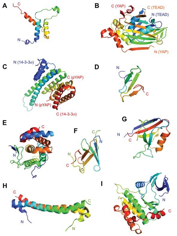
112                                                            Essays in Biochemistry volume 53 2012

Introduction

Like many signalling proteins, Hippo pathway proteins are modular and use
various interaction and catalytic domains to transmit signals and regulate tran-
scription of target genes, often in a context-dependent fashion. In the present
chapter we outline the major protein components, and focus on the structure
and function of some of the key Hippo pathway domains in vertebrates.

The Hippo pathway and its role in organ growth control
The Hippo pathway has emerged over the last decade as a key player in organ
size regulation during development, tissue homoeostasis throughout adult life,
tissue regeneration and stem cell self-renewal [1–8]. Perhaps unsurprisingly, the
pathway also plays a role in tumour suppression. Hippo pathway components
were first identified through loss-of-function genetic screens in Drosophila mel-
anogaster; the Hpo gene, after which the pathway has been called, was named for
the mutant overgrown head phenotype that resembled hippopotamus hide [9].

Hippo pathway components
The core elements of the Hippo pathway are well known, but additional com-
ponents constituting an extended network continue to be identified. Core com-
ponents of the vertebrate pathway (Figure 1) include the MST [mammalian Ste
(sterile) 20-like] 1/2 kinases, each of which autophosphorylates its activation
loop, and then phosphorylates and forms an active complex with Sav1. MST1/2
can then phosphorylate the LATS (large tumour suppressor) 1/2 kinases and
their co-activator MOB1 (Mps one binder). LATS1 or LATS2 subsequently
phosphorylate the most downstream targets of the Hippo pathway, YAP (Yes-
associated protein) and TAZ (transcriptional coactivator with PDZ-binding
motif), enabling 14-3-3 proteins to bind and sequester YAP/TAZ in the cyto-
plasm (Figure 1). The Hippo pathway is less complex in Drosophila than in
vertebrates; where two orthologues exist in vertebrates, for example, only
one exists in Drosophila. The core components in Drosophila, with vertebrate
homologues in parentheses, are Hippo (MST1/2), Sav (Sav1), Warts (LATS1/2),
MATS (MOB1) and Yorkie (YAP/TAZ).
     When the Hippo pathway is inactivated, YAP and TAZ translocate to the
nucleus where they behave as co-activators for various transcription factors.
YAP and TAZ lack a DBD (DNA-binding domain) and so influence transcrip-
tion by interacting with DNA-binding proteins. Organ growth is promoted by
the interaction of YAP and TAZ with TEAD family transcription factors which
up-regulate transcription of genes that promote cell proliferation, survival, dif-
ferentiation and morphogenesis [10]. TEAD transcription factors are ubiqui-
tously expressed, although each TEAD protein (TEAD1–TEAD4) occupies a
slightly different niche with respect to tissue expression and developmental stage.

© The Authors Journal compilation © 2012 Biochemical Society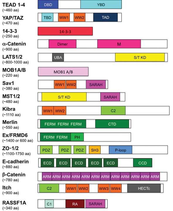
C. Cherrett, M. Furutani-Seiki and S. Bagby 113
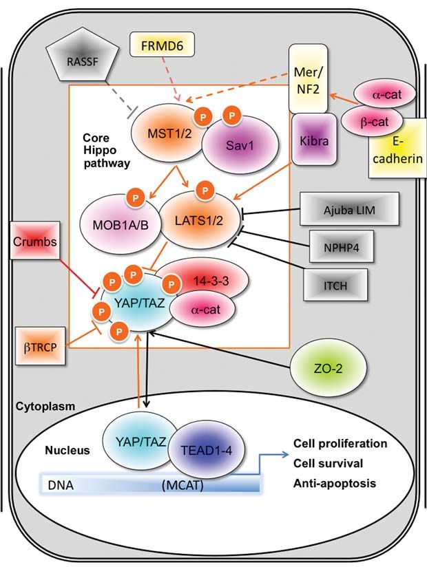
Figure 1. Mammalian Hippo pathway
The core Hippo pathway components within the orange-bordered box (MST1/2 kinases, scaffolding
protein Sav1, LATS1/2 kinases and their co-factors MOB1A/B) target transcriptional co-activator
proteins YAP and TAZ for phosphorylation. Phosphorylated YAP and TAZ are subsequently
anchored in the cytoplasm by 14-3-3 proteins and the interaction is stabilized by α-catenin.
Molecules coloured in grey antagonize the Hippo pathway by inhibiting the core kinases.
Inactivation of the Hippo pathway allows YAP and TAZ to translocate into the nucleus where they
contribute to the up-regulation of target genes through interactions with transcription factors.
Knowledge of upstream signals that regulate organ size and morphology is
key to understanding and modulating the Hippo pathway. Recent studies have
highlighted the influence of cell contacts on regulation of Hippo and associated
pathways. The Crb (Crumbs) complex, which is associated with tight junctions
in the sub-apical region of the cell membrane, regulates the Hippo pathway in
© The Authors Journal compilation © 2012 Biochemical Society114 Essays in Biochemistry volume 53 2012
Drosophila through interactions with the upstream Hippo regulator Ex (Expanded)
[11]. It is unclear whether the Crb–Ex mechanism is conserved in mammals but,
in response to high cell density, Crb can directly inhibit nuclear translocation of
YAP (Figure 1), in addition to contributing to Hippo pathway activation through
an unidentified mechanism [12]. E-cadherin, a transmembrane protein that forms
the intercellular epithelial junction complex, was recently found to be an upstream
regulator of the Hippo pathway. E-cadherin induces cell contact inhibition, a phe-
nomenon that stops cellular proliferation upon confluence. E-cadherin recruits
β-catenin to the cell membrane, and β-catenin then activates the Hippo pathway
through interactions with Merlin/NF2 (neurofibromin 2) [13].
Organ growth control and tumorigenesis
Hippo signalling orchestrates organ growth through its co-ordination of cell pro-
liferation, survival, differentiation and polarity. Numerous examples have illus-
trated that deregulation of the pathway leads to significantly increased organ size.
Up-regulation of YAP, the target protein that is inhibited by the Hippo pathway,
increased mouse liver size from approximately 5% of body weight to approxi-
mately 25% in 4 weeks [14]. Similarly, when core Hippo pathway proteins
MST1/2 and Sav1 were knocked out in mouse livers, the organs were significantly
larger than those of wild-type mice [15–18]. An enlarged heart phenotype has also
been observed in Sav1-knockout mice [19]. In all cases, organ structure was pre-
served, as observed previously for Drosophila Hippo pathway mutants which also
display enlarged organs (heads, imaginal discs) with normal tissue patterns [20].
Constitutive YAP overexpression or liver-specific deletion of MST1/2 or
Sav1 induces multi-focal tumorigenesis, highlighting the role of the pathway
in tumour suppression [14,16–18]. YAP and TAZ can induce anchorage-inde-
pendent growth and EMT (epithelial–mesenchymal transition) of immortalized
mammary and pancreatic epithelial cells [12]. EMT is important in normal mor-
phological processes, but when deregulated in cancers is involved in metasta-
sis, tumour recurrence and therapeutic resistance. Upstream Hippo pathway
components function as tumour suppressors. Hippo pathway mutations have
been observed in a range of human cancers, including breast cancers, soft tissue
sarcomas, melanomas, colorectal cancers, ovarian carcinomas, retinoblastomas,
astrocytomas and neurofibromatosis type 2 [1,5]. There has been speculation
about the possible involvement of Hippo signalling in cancer stem cells due to
the link of the pathway to stem cell self-renewal and cancer; recently the Hippo
pathway, via TAZ, was identified as a molecular link between EMT, cell polarity
and cancer stem cells in breast cancer [21].
Integration of multi-pathway signalling
The Hippo pathway interacts with numerous other signalling pathways (Table 1),
some of which contribute to organ growth regulation. This cross-talk occurs
© The Authors Journal compilation © 2012 Biochemical SocietyC. Cherrett, M. Furutani-Seiki and S. Bagby 115
in both cytoplasm and nucleus, and is probably important for the tight control
of cell proliferation, growth, polarity and differentiation required for forma-
tion and maintenance of functional proportionate organs without crossing the
boundary into tumorigenesis. Some of the inter-connections with the Hippo
pathway are described below [2,7,8,22].
Canonical Wnt pathway
Wnt signalling activates membrane-bound Dvl (Dishevelled) which inhib-
its the axin–GSK (glycogen synthase kinase) 3–APC (adenomatous polyposis
coli) β-catenin destruction complex (β-catenin is the nuclear effector of Wnt
Table 1. Cross-talk between Hippo pathway proteins and other signalling
pathways
EGFR, epidermal growth factor receptor; FOXO, forkhead box O; JAK, Janus kinase;
PI3K, phosphoinositide 3-kinase; Rb, retinoblastoma; Shh, Sonic hedgehog; STAT,
signal transducer and activator of transcription.
Pathway cross-talk Hippo pathway protein interaction
The Hippo pathway inhibits Direct interaction of TAZ with Dvl2 inhibits phosphorylation
Wnt/β-catenin signalling [23] of Dvl2 by CK1δ/ε thereby preventing formation of the
β-catenin destruction complex. This results in β-catenin
nuclear localization and expression of Wnt target genes.
The Hippo pathway inhibits Direct interaction of YAP with phospho-Smad1 and YAP/
BMP/TGFβ signalling [12,44] TAZ with phospho-Smad2/3 retains Smads in the nucleus
and leads to transcription of TGFβ/Smad target genes.
The Hippo pathway inhibits Nuclear Yki induces transcription of cytokines that promote
JAK/STAT signalling [45] JAK/STAT signalling in response to injury.
The Hippo pathway inhibits Nuclear Yki inhibits the Notch ligand Delta which leads to
Notch signalling [46] Notch activation.
Shh signalling inhibits the Hippo Shh signalling up-regulates expression of YAP1 mRNA, and
pathway [47] stabilizes IRS1 (insulin receptor substrate 1) which acts
as a nuclear retention factor for YAP. Shh signalling also
results in decreased levels of phospho-LATS.
The Hippo pathway promotes MST1 phosphorylates FOXO proteins leading to nuclear
FOXO signalling [48] localization and transcription of FOXO target genes.
PI3K/Akt signalling inhibits the Akt phosphorylates MST1 (at Thr120), thereby preventing the
Hippo pathway [49] kinase activity of MST1.
Rb [50,51] The transcription factor E2F is negatively regulated by Rb.
E2F interacts with the Yki–Sd complex and therefore Rb
inhibits E2F–Yki–Sd mediated transcription. In humans,
LATS phosphorylates DYRK (dual-specificity tyrosine
phosphorylation-regulated kinase) which leads to
activation of the ‘DREAM’ complex and inhibition of E2F.
EGFR signalling [52] YAP mediates transcription of the EGFR ligand amphiregulin,
leading to proliferation and migration of neighbouring cells.
© The Authors Journal compilation © 2012 Biochemical Society116 Essays in Biochemistry volume 53 2012 signalling). When membrane-localized β-catenin dissociates from E-cadherin and α-catenin, it translocates to the nucleus where it interacts with Lef (lym- phoid enhancer factor)/TCF (T-cell factor) transcription factors, causing the activation of target genes that determine stem cell survival and differentiation. The Hippo pathway inhibits the canonical Wnt pathway by enhancing levels of cytoplasmic phosphorylated TAZ, which binds to Dvl [23]. This interac- tion prevents phosphorylation of Dvl, rendering Dvl inactive. In the absence of hyperphosphorylated Dvl, β-catenin does not reach the nucleus and is targeted for degradation. The links between Hippo and Wnt signalling are not limited to the cytoplasmic TAZ–Dvl interaction; the E-cadherin–β-catenin–α-catenin complex binds Merlin and activates the Hippo pathway [13]. Also, upon Hippo pathway abrogation, nuclear non-phosphorylated TEAD-bound YAP forms a complex with Lef/TCF-bound β-catenin. YAP–β-catenin complex formation leads to up-regulation of Sox2 and Snai2 genes, and a consequent increase in heart size [19]. TGFβ (transforming growth factor β) and BMP (bone morphogenetic protein) signalling The Hippo pathway has multiple connections with TGFβ and BMP signal- ling. Upon TGFβ or BMP interaction with their respective membrane-bound receptors, cytoplasmic Smad proteins are activated by phosphorylation in the C-terminal region. Smad proteins contain an N-terminal MH1 domain that binds DNA and a C-terminal MH2 protein–protein interaction domain. Upon activation, Smads translocate to the nucleus where they form transcription fac- tor complexes. When nuclear Smad2/3 forms a complex with nuclear YAP/TAZ, Smad2/3 is prevented from returning to the cytoplasm and genes that promote EMT are up-regulated [12]. Phosphorylation of the Smad inter-MH domain linker by CDK (cyclin-dependent kinase) 8/9 promotes the Smad–YAP/TAZ interaction which up-regulates transcription of target genes when the proteins are nuclear [24]. The Hippo pathway can therefore inhibit TGFβ and BMP sig- nalling by inducing cytoplasmic retention of YAP/TAZ, consequently seques- tering YAP/TAZ-bound Smad2/3 or Smad1/5/8 proteins to the cytoplasm. Mechanotransduction An increase in ECM (extracellular matrix) rigidity, such as in bone, causes Rho GTPase activation and leads to stress fibre formation. When cytoskeletal ten- sion increases in response to stiff ECM, YAP and TAZ are retained within the nucleus and MSCs (mesenchymal stem cells) differentiate into osteoblasts. Conversely, when the MSCs are grown on soft ECM with low intracellular cytoskeletal tension, YAP and TAZ are excluded from the nucleus, and the cells can differentiate into other lineages, such as adipocytes. The Hippo pathway regulator E-cadherin can control Rho activation, but in this case E-cadherin may not be involved as mechanotransduction-mediated control of YAP/TAZ cellular localization occurs independently of the core Hippo pathway [25]. © The Authors Journal compilation © 2012 Biochemical Society
C. Cherrett, M. Furutani-Seiki and S. Bagby                                                          117

Pro-apoptotic and other interactions
In addition to TEADs and Smads, YAP and TAZ interact with other transcrip-
tion factors, for example PPARγ (peroxisome-proliferator-activated receptor γ)
and p73, which can result in repressed transcription or pro-apoptotic effects.
YAP and TAZ interact with a host of other proteins (Table 2), in some cases
without any apparent connection to the canonical Hippo pathway. As YAP and
TAZ are the most downstream targets of the pathway, elucidating the function
of these YAP/TAZ complexes is vital for understanding the control of organo-
genesis and tissue homoeostasis.

Table 2. Binding partners of YAP and TAZ
Binding          Description of binding protein                       YAP/TAZ domain
protein                                                               involved
14-3-3 [53,54]        Dimeric proteins that sequester YAP/            Phosphorylated
                        TAZ to the cytoplasm                           14-3-3-binding motif
AMOT [55]             Angiomotin, part of the Crumbs complex          WW domain(s)
ASPP1/2 (p53BP)       Apoptosis-stimulating protein of p53            WW domain(s)
  [56]                  protein family
c-Yes [57]            Tyrosine kinase                                 SH3-binding motif
Crb [12]              Upstream Hippo pathway complex                  WW domain(s) and PDZ-
                        protein Crumbs                                 binding motif
Dvl2 [23]             Dishevelled polarity protein involved in        WW domain and PDZ-
                        Wnt signalling                                 binding motif
ErbB4 [58]            Receptor tyrosine kinase that contains a        WW domain(s)
                        cleavable cytoplasmic fragment
Ex [59]               Upstream Hippo pathway FERM-domain              WW domain(s)
                        protein
HNRNPU [60]           Heterogenous nuclear ribonuclear protein Proline-rich region at
                        U binds to YAP and p73                          N-terminus of YAP
LATS1/2 [61,62]       Hippo pathway serine/threonine kinases          WW domain(s)
NFE2 (p45) [63]       Haemopoietic transcription factor-2             WW domain(s)
NHERF (EPB50)         Recruits YAP and TAZ to plasma                  PDZ-binding motif
  [53,64]               membrane
p73 [65,66]           Pro-apoptotic transcription factor              WW domain(s)
PEBP2 [31]            Polyoma enhancer binding protein 2              WW domain(s)
                        transcription factor
PPARγ [67]            Adipocyte transcription factor                  WW domain(s)
PRGP2 [68]            Proline-rich membrane Gla protein               WW domain(s)
Runx1/2 [67,69]       Transcription factors with Runt DNA-            WW domain(s)
                        binding domain
Smads [24,44]         Transcription factors regulated by the          WW domain(s)
                        TGFβ BMP signalling pathway
TEAD [70]             Transcription factors                           TBD
WBP1/2 [71]           WW domain-binding proteins                      WW domain(s)

                                              © The Authors Journal compilation © 2012 Biochemical Society
118 Essays in Biochemistry volume 53 2012
Key Hippo pathway domains
Regulation of organ growth, tissue homoeostasis, tissue regeneration and stem
cell self-renewal by Hippo pathway components depends on protein–protein,
protein–nucleic acid and protein–membrane interactions, and in some instances
multi-protein complex formation. The binding properties of a range of domains
or motifs within Hippo pathway proteins promote these interactions. Hippo
pathway proteins generally contain multiple domains or motifs separated by
various lengths of often unstructured polypeptide (Figure 2).
TEAD–YAP/TAZ interaction domains
The major nuclear proteins regulated by the Hippo pathway are the TEAD
transcription factors (Figure 1). TEAD proteins comprise an N-terminal DBD
and a C-terminal YBD (YAP-binding domain), both of which are indispen-
sable because individually YAP and TEAD have no transcriptional activity.
The TEAD DBD can bind to a variety of M-CAT-like DNA sequences (the
M-CAT motif is 5′-TCATTCCT-3′) in a fairly promiscuous manner. The solu-
tion structure of the human TEAD1 (TEF-1) A49S mutant DBD comprises
a three-helix bundle in a homeodomain-like fold. The first two α-helices
are almost anti-parallel with the third α-helix lying across them (Figure 3).
Similar to homeodomain proteins, DNA binding is mediated by the third
α-helix (H3) and the preceding loop (L2); H1 and L1 do not bind directly,
but are necessary for full-strength binding of TEAD to tandem M-CAT
sites [26].
The TBD (TEAD-binding domain) of YAP/TAZ, located in the N-terminal
region, is natively unstructured and binds TEAD with high fidelity – to date no
other protein interactions are known to involve the TBD. The crystal structures
of YAP–TEAD interaction domains involving human TEAD2 [27], human
TEAD1–YAP [28] and mouse TEAD4–YAP [29] show that the TEAD YBDs
adopt an immunoglobulin-like β-sandwich fold with the addition of two helix-
turn-helix motifs (Figure 3). The Y421H mutation in TEAD1 that is present
in human Sveinsson’s chorioretinal atrophy was previously found to abrogate
interactions with YAP and TAZ [30]. Consistent with this, Tyr421 is located in
the TEAD–YAP interface where it forms a hydrogen bond with a serine residue
in YAP (Ser94 in human YAP). Upon interaction with TEAD, the YAP TBD
forms two α-helices that pack into binding grooves and are separated by an
extended loop that wraps around the YBD [28,29].
YAP/TAZ TAD (transcription activation domain)
Both YAP and TAZ contain a C-terminal TAD [31]. Although there have been
no experimental studies of the structure of this domain, secondary structure
predictions indicate that it is largely unstructured.
© The Authors Journal compilation © 2012 Biochemical SocietyC. Cherrett, M. Furutani-Seiki and S. Bagby 119
Figure 2. Schematic representations of Hippo network proteins
aa, amino acid; DBD, DNA-binding domain; YBD, YAP-binding domain; TBD, TEAD-binding
domain; WW, domain containing two signature tryptophan residues; TAD, transactivation
domain; M, vinculin-like domain; UBA, ubiquitin-associated domain; S/T KD, serine/threonine
kinase domain; SARAH, Sav/Rassf/Hippo domain; FERM, 4.1 protein/ezrin/radixin/moesin domain;
CTD, C-terminal domain; PH, pleckstrin homology domain; PDZ, PSD-95 (postsynaptic density
95), Dlg (discs large) and zonula occludens-1 domain; SH3, Src homology 3 domain; P-loop,
NTPase domain; ECD, extracellular cadherin domain; CCD, cytoplasmic cadherin domain; ARM,
armadillo repeat; HECTc, homologous with E6-associated protein C-terminus; RA, Ras-
association domain.
YAP/TAZ cytoplasmic sequestration by 14-3-3
C-terminal to the YAP/TAZ TBD is an HxRxxS motif that becomes a
14-3-3-binding site upon serine phosphorylation (Ser127 in human YAP). A
crystal structure of the homodimeric 14-3-3σ–YAP phospho-peptide complex
(Figure 3) reveals that the YAP peptide binds to each monomer of 14-3-3σ with
© The Authors Journal compilation © 2012 Biochemical Society120 Essays in Biochemistry volume 53 2012 Figure 3. Structures of some Hippo pathway protein domains (A), (D), (F) and (H) are NMR solution structures; (B), (C), (E), (G) and (I) are crystal structures. (A) TEAD1 DBD (PDB ID: 2HZD); (B) TEAD1 YBD–YAP TBD complex (PDB ID: 3KYS); (C) 14-3-3σ–YAP phospho-peptide complex (PDB ID: 3MHR); (D) YAP WW2 domain (PDB ID: 2L4J); (E) S. cerevisiae MOB1 (PDB ID: 2HJN); (F) mouse Sav WW2 domain dimer (PDB ID: 2DWV); (G) ZO1 PDZ1 domain (PDB ID: 2H3M); (H) MST1 SARAH domain (PDB ID: 2JO8); and (I) MST1 kinase domain (PDB ID: 3COM). a 1:1 stoichiometry, so each 14-3-3 dimer can bind two molecules of YAP [32]. 14-3-3σ dimerizes in a W-shape via α-helices 1–4. Helices 3, 5, 7 and 9 on each monomer form the YAP-binding groove. The YAP peptide-bound 14-3-3σ structure is very similar to unbound 14-3-3σ with an overall r.m.s.d. (root mean square deviation) between the structures of 1.00 Å (1 Å = 0.1 nm), suggesting that YAP binding does not induce a large conformational change in 14-3-3. © The Authors Journal compilation © 2012 Biochemical Society
C. Cherrett, M. Furutani-Seiki and S. Bagby                                                          121

WW domains
WW domains are prevalent and important features of the Hippo pathway: YAP,
TAZ, Sav1, Kibra and Itch each contain at least one WW domain [33]. The WW
domain (approximately 40 residues) is the smallest known protein domain and
consists of a twisted three-stranded β-sheet (Figure 3). WW domains are named
after two signature tryptophan residues located on the first and third β-strands.
The first tryptophan residue is required for folding and the second tryptophan
residue is involved in ligand binding [34]. WW domains are central mediators of
protein binding events throughout the extended Hippo network via interactions
with proline-rich motifs. WW domains are categorized into five groups (I–V)
according to their cognate ligand. The main Hippo pathway WW domains fall
into group I, i.e. the WW domains bind to PY motifs (PPxY and, less frequently,
LPxY); such motifs are found, for example, in LATS1/2, most of the Smads, Dvl
and p73.
     Itch contains four WW domains and inhibits the Hippo pathway by bind-
ing to PPxY motifs of LATS1/2, predominantly via its first WW domain, lead-
ing to ubiquitination and degradation of LATS1/2 [35]. Sav1 and Kibra both
contain two WW domains; in each case, WW1 is a group I domain, and WW2 is
atypical in that the second tryptophan residue is replaced by another amino acid
(isoleucine in Kibra, tyrosine in vertebrate Sav1 and arginine in Drosophila Sav).
Mouse Sav1 WW2 is the only WW domain known to date to dimerize (Figure 3)
[36]. Sav1 promotes multi-protein complex formation in the Hippo pathway by
acting as a scaffold protein through SARAH domain multimerization. It is pos-
sible that WW2 homodimerization enhances this scaffolding function, whereas
WW1 engages binding partners.
     The YAP1 and YAP2 isoforms of YAP contain one and two WW domains
respectively. It is not currently clear what specific roles the different isoforms
play. YAP and TAZ have approximately 20 known binding partners (Table 2),
many of which bind via at least one of the WW domains; given the prevalence of
PPxY motifs in proteomes, the number of protein–protein interactions mediated
by YAP and TAZ could be much higher than this. The WW domains of YAP and
TAZ belong to group I [37], but YAP WW1 has also been found to interact with
a phospho-serine motif of Smad1 [24]: phosphorylation by CDK8/9 [as part of
BMP (bone morphogenetic protein) signalling] creates a YAP WW1-binding
site on the Smad1 inter-MH domain linker (see above). This region also contains
a PPxY motif that binds to YAP WW2. CDK8/9 phosphorylation also primes
Smad1 for phosphorylation by GSK3. Secondary phosphorylation of Smad1 by
GSK3 reduces the affinity for YAP WW1 and increases the affinity for Smurf1
WW1; Smurf1 WW2 simultaneously binds the PPxY motif. Interactions with
Smurf1 lead to poly-ubiquitination and subsequent proteasomal degradation of
Smad1, thereby marking the end of a YAP–Smad transcriptional event. Wnt
signalling suppresses GSK3, providing another illustration of the complexity of
interpathway connections.

                                              © The Authors Journal compilation © 2012 Biochemical Society
122 Essays in Biochemistry volume 53 2012 SARAH domains The SARAH coiled-coil domain is present in the C-terminal region of Sav1, Rassf and Hippo (MST1/2). SARAH domains homodimerize and can also mediate heterodimerization of Hippo pathway proteins, for example between MST1/2 and Sav1. The solution structure of the human MST1 SARAH homodi- mer (Figure 3) shows that each monomer comprises a short N-terminal α-helix that is oriented towards the N-terminal helix of the other monomer, and an elongated C-terminal α-helix along which the anti-parallel dimer interface lies [38]. The Rassf5 (Nore1) SARAH domain forms a homotetramer but, in the presence of the MST1 SARAH domain, only dimers are observed. The role of mammalian Rassf proteins in the Hippo pathway is currently unclear; in vitro studies indicate that Rassf1 and Rassf5 inhibit the Hippo pathway, as is the case for Drosophila Rassf. Conversely, in some cases the Hippo pathway seems to be activated by Rassf proteins in vivo [1]. Serine/threonine kinase domains Central to the Hippo pathway are the serine/threonine kinase domains of MST1/2 and LATS1/2 that propagate phosphorylation events to retain YAP/ TAZ in the cytoplasm. MST1/2 belong to the Ste group of kinases, whereas LATS kinases are similar to the PKC (protein kinase C) family. In a crystal structure of activated MST1 (Figure 3), the auto-activation loop is di-phospho- rylated. There are currently no published structures of the LATS1 and LATS2 kinase domains. MOB1 MOB1 is part of the MOB family of co-activator proteins [39]. Human MOB1 binding to LATS1/2 triggers LATS1/2 auto-phosphorylation on the activa- tion segment. The MOB1–LATS1/2 complex phosphorylates YAP/TAZ. The C-terminal core domain adopts an α-helical fold common to all MOB proteins, whereas the N-terminal region is less conserved but seems to be functionally important. In Saccharomyces cerevisiae MOB1, this N-terminal region includes structural elements that mediate homodimerization in vitro [40]. One side of the MOB1 surface is mostly acidic and the opposite side is basic. Bioinformatic and experimental analyses indicate that the interaction between MOB1 and LATS1/2 is mediated by the acidic face of the former and the basic region of the N-terminal regulatory domain of the latter. PDZ-binding motifs YAP and TAZ contain a C-terminal PDZ-binding motif (LTWL) that allows interaction with several proteins involved in organ size regulation. PDZ domains typically comprise 80–100 residues forming six β-strands and two α-helices of differing lengths (Figure 3). The binding groove is generally located between the longer α-helix and the second β-strand [41]. Nuclear localization of YAP/TAZ is promoted by interactions with the first PDZ domain of the © The Authors Journal compilation © 2012 Biochemical Society
C. Cherrett, M. Furutani-Seiki and S. Bagby                                                          123

tight junction-associated proteins ZO (zonula occludens)-1 and -2 [42,43].
Interactions involving PDZ-binding motifs and PDZ domains, and WW
domains and PPxY motifs, are important for Hippo pathway cross-talk with
TGFβ signalling {YAP/TAZ interaction with Crumbs components PALS1,
AMOT (angiomotin), PATJ (PALS1-associated tight junction protein) and
LIN7 [12]} and with the Wnt pathway (TAZ interaction with Dvl [23]).

Other domains within Hippo pathway proteins
Remaining protein–protein interaction domains include those involved in self-
association, such as the dimerization domain of α-catenin and those that lead to
proteasomal degradation, e.g. the UBA (ubiquitin-associated) domain of LATS1/2.
Domains involved in membrane interaction and localization include the FERM
domains in Merlin and FRMD6, and the C2 domains in Kibra and Itch.

Conclusions
The Hippo pathway interconnects with numerous other pathways in order to
orchestrate organ growth or tissue regeneration, and might therefore be more
appropriately termed the Hippo network. Substantial knowledge of Hippo net-
work operation has rapidly emerged, but many questions remain. In terms of
protein domains, for example, how are the multiple possible WW, SARAH and
PDZ domain interactions co-ordinated? What are the structural and functional
relationships between multiple domains/motifs within and between proteins?
How do post-translational modifications, predominantly phosphorylation,
modulate domain structures and interactions? Detailed structural, biochemical,
biophysical and computational analyses, including isolation or reconstitution
of multimolecular complexes, are needed to answer questions such as these. In
combination with cellular and organismal studies, one long-term goal of these
molecular level studies is systems level comprehension of Hippo signalling
towards understanding and prediction of responses to particular developmental
and environmental cues, and towards controlled modulation for research and
clinical applications.

Summary

•    The Hippo pathway is a central conserved pathway that interconnects
     with several other pathways to regulate organ growth, tissue homoeo-
     stasis and regeneration, and stem cell self-renewal.
•    The Hippo pathway is unique in its capacity to orchestrate multiple pro-
     cesses, from sensing to execution, necessary for organ expansion.
•    The mechanisms and effects of Hippo pathway cross-talk with other
     pathways such as Wnt and TGFβ growth factor pathways will undoubt-
     edly turn out to be highly complex, but are gradually being elucidated.

                                              © The Authors Journal compilation © 2012 Biochemical Society
124 Essays in Biochemistry volume 53 2012
• The Hippo pathway includes protein domains involved in catalysis,
protein–membrane interactions, protein–protein interactions and pro-
tein–nucleic acid interactions.
References
1. Bao, Y., Hata, Y., Ikeda, M. and Withanage, K. (2011) Mammalian Hippo pathway: from
development to cancer and beyond. J. Biochem. 149, 361–379
2. Mauviel, A., Nallet-Staub, F. and Varelas, X. (2012) Integrating developmental signals: a Hippo in
the (path)way. Oncogene 31, 1743–1756
3. Halder, G. and Johnson, R. L. (2011) Hippo signaling: growth control and beyond. Development
138, 9–22
4. Zhao, B., Tumaneng, K. and Guan, K. L. (2011) The Hippo pathway in organ size control, tissue
regeneration and stem cell self-renewal. Nat. Cell Biol. 13, 877–883
5. Zhao, B., Li, L., Lei, Q. and Guan, K. L. (2010) The Hippo-YAP pathway in organ size control and
tumorigenesis: an updated version. Genes Dev. 24, 862–874
6. Staley, B.K. and Irvine, K.D. (2012) Hippo signaling in Drosophila: recent advances and insights.
Dev. Dyn. 241, 3–15
7. Varelas, X. and Wrana, J.L. (2012) Coordinating developmental signaling: novel roles for the
Hippo pathway. Trends Cell Biol. 22, 88–96
8. Sudol, M. and Harvey, K.F. (2010) Modularity in the Hippo signalling pathway. Trends Biochem.
Sci. 35, 627–633
9. Udan, R.S., Kango-Singh, M., Nolo, R., Tao, C.Y. and Halder, G. (2003) Hippo promotes
proliferation arrest and apoptosis in the Salvador/Warts pathway. Nat. Cell Biol. 5, 914–920
10. Chen, L., Loh, P.G. and Song, H. (2010) Structural and functional insights into the TEAD-YAP
complex in the Hippo signaling pathway. Prot. Cell 1, 1073–1083
11. Parsons, L.M., Grzeschik, N.A., Allott, M.L. and Richardson, H.E. (2010) Lgl/aPKC and Crb
regulate the Salvador/Warts/Hippo pathway. Fly 4, 288–293
12. Varelas, X., Samavarchi-Tehrani, P., Narimatsu, M., Weiss, A., Cockburn, K., Larsen, B.G.,
Rossant, J. and Wrana, J.L. (2010) The Crumbs complex couples cell density sensing to Hippo-
dependent control of the TGF-β-SMAD pathway. Dev. Cell 19, 831–844
13. Kim, N.G., Koh, E., Chen, X. and Gumbiner, B.M. (2011) E-cadherin mediates contact inhibition
of proliferation through Hippo signaling-pathway components. Proc. Natl. Acad. Sci. U.S.A. 108,
11930–11935
14. Dong, J.X., Feldmann, G., Huang, J.B., Wu, S., Zhang, N.L., Comerford, S.A., Gayyed, M.F., Anders,
R.A., Maitra, A. and Pan, D.J. (2007) Elucidation of a universal size-control mechanism in
Drosophila and mammals. Cell 130, 1120–1133
15. Zhou, D., Conrad, C., Xia, F., Park, J.S., Payer, B., Yin, Y., Lauwers, G.Y., Thasler, W., Lee, J.T.,
Avruch, J. and Bardeesy, N. (2009) Mst1 and Mst2 maintain hepatocyte quiescence and suppress
hepatocellular carcinoma development through inactivation of the Yap1 oncogene. Cancer Cell
16, 425–438
16. Lu, L., Li, Y., Kim, S.M., Bossuyt, W., Liu, P., Qiu, Q., Wang, Y., Halder, G., Finegold, M.J., Lee, J.S.
and Johnson, R.L. (2010) Hippo signaling is a potent in vivo growth and tumor suppressor pathway
in the mammalian liver. Proc. Natl. Acad. Sci. U.S.A. 107, 1437–1442
17. Song, H., Mak, K.K., Topol, L., Yun, K., Hu, J., Garrett, L., Chen, Y., Park, O., Chang, J., Simpson, R.M.
et al. (2010) Mammalian Mst1 and Mst2 kinases play essential roles in organ size control and
tumor suppression. Proc. Natl. Acad. Sci. U.S.A. 107, 1431–1436
18. Lee, K.P., Lee, J.H., Kim, T.S., Kim, T.H., Park, H.D., Byun, J.S., Kim, M.C., Jeong, W.I.,
Calvisi, D.F., Kim, J.M. and Lim, D.S. (2010) The Hippo-Salvador pathway restrains hepatic
oval cell proliferation, liver size, and liver tumorigenesis. Proc. Natl. Acad. Sci. U.S.A. 107,
8248–8253
© The Authors Journal compilation © 2012 Biochemical SocietyC. Cherrett, M. Furutani-Seiki and S. Bagby 125
19. Heallen, T., Zhang, M., Wang, J., Bonilla-Claudio, M., Klysik, E., Johnson, R.L. and Martin, J.F.
(2011) Hippo pathway inhibits Wnt signaling to restrain cardiomyocyte proliferation and heart
size. Science 332, 458–461
20. Kango-Singh, M. and Singh, A. (2009) Regulation of organ size: insights from the Drosophila Hippo
signaling pathway. Dev. Dyn. 238, 1627–1637
21. Cordenonsi, M., Zanconato, F., Azzolin, L., Forcato, M., Rosato, A., Frasson, C., Inui, M.,
Montagner, M., Parenti, A.R., Poletti, A. et al. (2011) The Hippo transducer TAZ confers cancer
stem cell-related traits on breast cancer cells. Cell 147, 759–772
22. McNeill, H. and Woodgett, J.R. (2010) When pathways collide: collaboration and connivance
among signalling proteins in development. Nat. Rev. Mol. Cell Biol. 11, 404–413
23. Varelas, X., Miller, B.W., Sopko, R., Song, S.Y., Gregorieff, A., Fellouse, F.A., Sakuma, R., Pawson,
T., Hunziker, W., McNeill, H., Wrana, J.L. and Attisano, L. (2010) The Hippo pathway regulates
Wnt/β-Catenin signaling. Dev. Cell 18, 579–591
24. Aragon, E., Goerner, N., Zaromytidou, A.I., Xi, Q., Escobedo, A., Massagué, J. and Macias, M.J.
(2011) A Smad action turnover switch operated by WW domain readers of a phosphoserine
code. Genes Dev. 25, 1275–1288
25. Dupont, S., Morsut, L., Aragona, M., Enzo, E., Giulitti, S., Cordenonsi, M., Zanconato, F., Le
Digabel, J., Forcato, M., Bicciato, S. et al. (2011) Role of YAP/TAZ in mechanotransduction.
Nature 474, 179–183
26. Anbanandam, A., Albarado, D.C., Nguyen, C.T., Halder, G., Gao, X.L. and Veeraraghavan, S.
(2006) Insights into transcription enhancer factor 1 (TEF-1) activity from the solution structure of
the TEA domain. Proc. Natl. Acad. Sci. U.S.A. 103, 17225–17230
27. Tian, W., Yu, J., Tomchick, D.R., Pan, D. and Luo, X. (2010) Structural and functional analysis of
the YAP-binding domain of human TEAD2. Proc. Natl. Acad. Sci. U.S.A. 107, 7293–7298
28. Li, Z., Zhao, B., Wang, P., Chen, F., Dong, Z.H., Yang, H.R., Guan, K.L. and Xu, Y.H. (2010)
Structural insights into the YAP and TEAD complex. Genes Dev. 24, 235–240
29. Chen, L.M., Chan, S.W., Zhang, X.Q., Walsh, M., Lim, C.J., Hong, W.J. and Song, H.W. (2010)
Structural basis of YAP recognition by TEAD4 in the Hippo pathway. Genes Dev. 24, 290–300
30. Kitagawa, M. (2007) A Sveinsson’s chorioretinal atrophy-associated missense mutation in mouse
Tead1 affects its interaction with the co-factors YAP and TAZ. Biochem. Biophys. Res. Commun.
361, 1022–1026
31. Yagi, R., Chen, L.F., Shigesada, K., Murakami, Y. and Ito, Y. (1999) A WW domain-containing
Yes-associated protein (YAP) is a novel transcriptional co-activator. EMBO J. 18, 2551–2562
32. Schumacher, B., Skwarczynska, M., Rose, R. and Ottmann, C. (2010) Structure of a 14-3-3σ-YAP
phosphopeptide complex at 1.15 Å resolution. Acta Crystallogr. Sect. F Struct. Biol. Crystal.
Commun. 66, 978–984
33. Salah, Z. and Aqeilan, R.I. (2011) WW domain interactions regulate the Hippo tumor suppressor
pathway. Cell Death Dis. 2, e172
34. Koepf, E.K., Petrassi, H.M., Ratnaswamy, G., Huff, M.E., Sudol, M. and Kelly, J.W. (1999)
Characterization of the structure and function of W-to-F WW domain variants: identification of a
natively unfolded protein that folds upon ligand binding. Biochemistry 38, 14338–14351
35. Salah, Z., Melino, G. and Aqeilan, R.I. (2011) Negative regulation of the Hippo pathway by E3
ubiquitin ligase ITCH is sufficient to promote tumorigenicity. Cancer Res. 71, 2010–2020
36. Ohnishi, S., Guntert, P., Koshiba, S., Tomizawa, T., Akasaka, R., Tochio, N., Sato, M., Inoue, M.,
Harada, T., Watanabe, S. et al. (2007) Solution structure of an atypical WW domain in a novel
β-clam-like dimeric form. FEBS Lett. 581, 462–468
37. Webb, C., Upadhyay, A., Giuntini, F., Eggleston, I., Furutani-Seiki, M., Ishima, R. and Bagby, S.
(2011) Structural features and ligand binding properties of tandem WW domains from YAP and
TAZ, nuclear effectors of the Hippo pathway. Biochemistry 50, 3300–3309
38. Hwang, E., Ryu, K.S., Paakkonen, K., Guntert, P., Cheong, H.K., Lim, D.S., Lee, J.O., Jeon, Y.H. and
Cheong, C. (2007) Structural insight into dimeric interaction of the SARAH domains from Mst1
and RASSF family proteins in the apoptosis pathway. Proc. Natl. Acad. Sci. U.S.A. 104, 9236–9241
© The Authors Journal compilation © 2012 Biochemical Society126 Essays in Biochemistry volume 53 2012
39. Hergovich, A. (2011) MOB control: reviewing a conserved family of kinase regulators. Cell.
Signalling 23, 1433–1440
40. Mrkobrada, S., Boucher, L., Ceccarelli, D.F., Tyers, M. and Sicheri, F. (2006) Structural and
functional analysis of Saccharomyces cerevisiae Mob1. J. Mol. Biol. 362, 430–440
41. Lee, H.J. and Zheng, J.J. (2010) PDZ domains and their binding partners: structure, specificity, and
modification. Cell Commun. Signal. 8, 8
42. Remue, E., Meerschaert, K., Oka, T., Boucherie, C., Vandekerckhove, J., Sudol, M. and Gettemans,
J. (2010) TAZ interacts with zonula occludens-1 and -2 proteins in a PDZ-1 dependent manner.
FEBS Lett. 584, 4175–4180
43. Oka, T., Remue, E., Meerschaert, K., Vanloo, B., Boucherie, C., Gfeller, D., Bader, G.D., Sidhu,
S.S., Vandekerckhove, J., Gettemans, J. and Sudol, M. (2010) Functional complexes between YAP2
and ZO-2 are PDZ domain-dependent, and regulate YAP2 nuclear localization and signalling.
Biochem. J. 432, 461–472
44. Alarcón, C., Zaromytidou, A.I., Xi, Q.R., Gao, S., Yu, J.Z., Fujisawa, S., Barlas, A., Miller, A.N.,
Manova-Todorova, K., Macias, M.J. et al. (2009) Nuclear CDKs drive Smad transcriptional
activation and turnover in BMP and TGFβ pathways. Cell 139, 757–769
45. Karpowicz, P., Perez, J. and Perrimon, N. (2010) The Hippo tumor suppressor pathway regulates
intestinal stem cell regeneration. Development 137, 4135–4145
46. Reddy, B.V.V.G., Rauskolb, C. and Irvine, K.D. (2010) Influence of Fat-Hippo and Notch signaling
on the proliferation and differentiation of Drosophila optic neuroepithelia. Development 137,
2397–2408
47. Fernandez, A., Northcott, P.A., Dalton, J., Fraga, C., Ellison, D., Angers, S., Taylor, M.D. and
Kenney, A.M. (2009) YAP1 is amplified and up-regulated in hedgehog-associated
medulloblastomas and mediates Sonic hedgehog-driven neural precursor proliferation. Genes
Dev. 23, 2729–2741
48. Choi, J., Oh, S., Lee D., Oh, H.J., Park, J.Y., Lee, S.B. and Lim, D.S. (2009) Mst1-FoxO signaling
protects naïve T lymphocytes from cellular oxidative stress in mice. PloS ONE 4, e8011
49. Yuan, Z., Kim, D., Shu, S., Wu, J., Guo, J., Xiao, L., Kaneko, S., Coppola, D. and Cheng, J.Q. (2010)
Phosphoinositide 3-kinase/Akt inhibits MST1-mediated pro-apoptotic signaling through
phosphorylation of threonine 120. J. Biol. Chem. 285, 3815–3824
50. Nicolay, B.N., Bayarmagnai, B., Islam, A., Lopez-Bigas, N. and Frolov, M.V. (2011) Cooperation
between dE2F1 and Yki/Sd defines a distinct transcriptional program necessary to bypass cell
cycle exit. Genes Dev. 25, 323–335
51. Tschöp, K., Conery, A.R., Litovchick, L., Decaprio, J.A., Settleman, J., Harlow, E. and Dyson, N.
(2011) A kinase shRNA screen links LATS2 and the pRB tumor suppressor. Genes Dev. 25,
814–830
52. Zhang, J.M., Ji, J.Y., Yu, M., Overholtzer, M., Smolen, G.A., Wang, R., Brugge, J.S., Dyson, N.J. and
Haber, D.A. (2009) YAP-dependent induction of amphiregulin identifies a non-cell-autonomous
component of the Hippo pathway. Nature Cell Biol. 11, 1444–1450
53. Kanai, F., Marignani, P.A., Sarbassova, D., Yagi, R., Hall, R.A., Donowitz, M., Hisaminato, A.,
Fujiwara, T., Ito, Y., Cantley, L.C. and Yaffe, M.B. (2000) TAZ: a novel transcriptional co-activator
regulated by interactions with 14-3-3 and PDZ domain proteins. EMBO J. 19, 6778–6791
54. Basu, S., Totty, N.F., Irwin, M.S., Sudol, M. and Downward, J. (2003) Akt phosphorylates the
Yes-associated protein, YAP, to induce interaction with 14-3-3 and attenuation of p73-mediated
apoptosis. Mol. Cell 11, 11–23
55. Chan, S.W., Lim, C.J., Chong, Y.F., Pobbati, A.V., Huang, C.X. and Hong, W.J. (2011) Hippo
pathway-independent restriction of TAZ and YAP by angiomotin. J. Biol. Chem. 286, 7018–7026
56. Espanel, X. and Sudol, M. (2001) Yes-associated protein and p53-binding protein-2 interact
through their WW and SH3 domains. J. Biol. Chem. 276, 14514–14523
57. Sudol, M., Bork, P., Einbond, A., Kastury, K., Druck, T., Negrini, M., Huebner, K. and Lehman, D.
(1995) Characterization of the mammalian Yap (Yes-associated protein) gene and its role in
defining a novel protein module, the WW domain. J. Biol. Chem. 270, 14733–14741
© The Authors Journal compilation © 2012 Biochemical SocietyC. Cherrett, M. Furutani-Seiki and S. Bagby 127
58. Komuro, A., Nagai, M., Navin, N.E. and Sudol, M. (2003) WW domain-containing protein YAP
associates with ErbB-4 and acts as a co-transcriptional activator for the carboxyl-terminal
fragment of ErbB-4 that translocates to the nucleus. J. Biol. Chem. 278, 33334–33341
59. Badouel, C., Gardano, L., Amin, N., Garg, A., Rosenfeld, R., Le Bihan, T. and McNeill, H. (2009)
The FERM-domain protein Expanded regulates Hippo pathway activity via direct interactions with
the transcriptional activator Yorkie. Dev. Cell 16, 411–420
60. Howell, M., Borchers, C. and Milgram, S.L. (2004) Heterogeneous nuclear ribonuclear protein U
associates with YAP and regulates its co-activation of Bax transcription. J. Biol. Chem. 279,
26300–26306
61. Lei, Q.Y., Zhang, H., Zhao, B., Zha, Z.Y., Bai, F., Pei, X.H., Zhao, S., Xiong, Y. and Guan, K.L.
(2008) TAZ promotes cell proliferation and epithelial-mesenchymal transition and is inhibited by
the Hippo pathway. Mol. Cell. Biol. 28, 2426–2436
62. Zhang, J., Smolen, G.A. and Haber, D.A. (2008) Negative regulation of YAP by LATS1
underscores evolutionary conservation of the Drosophila Hippo pathway. Cancer Res. 68,
2789–2794
63. Gavva, N.R., Gavva, R., Ermekova, K., Sudol, M. and Shen, C.K.J. (1997) Interaction of WW
domains with hematopoietic transcription factor p45/NF-E2 and RNA polymerase II. J. Biol.
Chem. 272, 24105–24108
64. Mohler, P.J., Kreda, S.M., Boucher, R.C., Sudol, M., Stutts, M.J. and Milgram, S.L. (1999) Yes-
associated protein 65 localizes p62 (c-Yes) to the apical compartment of airway epithelia by
association with EBP50. J. Cell Biol. 147, 879–890
65. Strano, S., Monti, O., Baccarini, A., Sudol, M., Sacchi, A. and Blandino, G. (2001) Physical
interaction with yes-associated protein enhances p73 transcriptional activity. Eur. J. Cancer 37,
S279
66. Oka, T. and Sudol, M. (2009) Nuclear localization and pro-apoptotic signaling of YAP2 require
intact PDZ-binding motif. Genes Cells 14, 607–615
67. Hong, J.H., Hwang, E.S., McManus, M.T., Amsterdam, A., Tian, Y., Kalmukova, R., Mueller, E.,
Benjamin, T., Spiegelman, B.M., Sharp, P.A. et al. (2005) TAZ, a transcriptional modulator of
mesenchymal stem cell differentiation. Science 309, 1074–1078
68. Kulman, J.D., Harris, J.E., Xie, L. and Davie, E.W. (2007) Proline-rich Gla protein 2 is a cell-surface
vitamin K-dependent protein that binds to the transcriptional coactivator Yes-associated protein.
Proc. Natl. Acad. Sci. U.S.A. 104, 8767–8772
69. Zaidi, S.K., Sullivan, A.J., Medina, R., Ito, Y., van Wijnen, A.J., Stein, J.L., Lian, J.B. and Stein, G.S.
(2004) Tyrosine phosphorylation controls Runx2-mediated subnuclear targeting of YAP to
repress transcription. EMBO J. 23, 790–799
70. Vassilev, A., Kaneko, K.J., Shu, H.J., Zhao, Y.M. and DePamphilis, M.L. (2001) TEAD/TEF
transcription factors utilize the activation domain of YAP65, a Src/Yes-associated protein
localized in the cytoplasm. Genes Dev. 15, 1229–1241
71. Chen, H.I. and Sudol, M. (1995) The WW domain of Yes-associated protein binds a proline-rich
ligand that differs from the consensus established for Src homology 3-binding modules. Proc. Natl.
Acad. Sci. U.S.A. 92, 7819–7823
© The Authors Journal compilation © 2012 Biochemical SocietyYou can also read