Structural Origins of the Functional Divergence of Human Insulin-Like Growth Factor-I and Insulin
←
→
Page content transcription
If your browser does not render page correctly, please read the page content below
Biochemistry 2002, 41, 9389-9397                                                    9389

    Structural Origins of the Functional Divergence of Human Insulin-Like Growth
                                 Factor-I and Insulin†,‡
         Andrzej M. Brzozowski,* Eleanor J. Dodson, Guy G. Dodson, Garib N. Murshudov, Chandra Verma,
                         Johan P. Turkenburg, Frederik M. de Bree,# and Zbigniew Dauter§
    Structural Biology Laboratory, Chemistry Department, UniVersity of York, Heslington, York YO10 5DD, United Kingdom
                                ReceiVed January 28, 2002; ReVised Manuscript ReceiVed April 23, 2002

        ABSTRACT: Human insulin-like growth factors I and II (hIGF-I, hIGF-II) are potent stimulators of cell
        and growth processes. They display high sequence similarity to both the A and B chains of insulin but
        contain an additional connecting C-domain, which reflects their secretion without specific packaging or
        precursor conversion. IGFs also have an extension at the C-terminus known as the D-domain. This paper
        describes four homologous hIGF-1 structures, obtained from crystals grown in the presence of the detergent
        SB12, which reveal additional detail in the C- and D-domains. Two different detergent binding modes
        observed in the crystals may reflect different hIGF-I biological properties such as the interaction with
        IGF binding proteins and self-aggregation. While the helical core of hIGF-I is very similar to that in
        insulin, there are distinct differences in the region of hIGF-I corresponding to the insulin B chain C-terminus,
        residues B25-B30. In hIGF-I, these residues (24-29) and the following C-domain form an extensive
        loop protruding 20 Å from the core, which results in a substantially different conformation for the receptor
        binding epitope in hIGF-I compared to insulin. One notable feature of the structures presented here is
        demonstration of peptide-bond cleavage between Ser35 and Arg36 resulting in an apparent gap between
        residues 35 and 39. The equivalent region of proinsulin is involved in hormone processing demanding a
        reassessment of the structural integrity of hIGF-I in relation to its biological function.

  Human insulin-like growth factor I (hIGF-I) is a 70-amino
acid single chain protein that mediates somatic growth. It
has a high (45-52%) sequence similarity with the B and A
chains of human insulin, and 67% sequence identity with
human insulin-like growth factor-II (IGF-II) (see Figure 1)
(1).
  A number of NMR studies (2-5) and a few recent
crystallographic analyses (6, 7) have revealed the essentially
identical nature of the core region and the organization of
the three helical segments of insulin and hIGF-I. The short
    †
      This work was partially supported by the European Community
(Human Capital and Mobility Program, Contract No. CHRX-CT94-
0556). The infrastructure of the Structural Biology Laboratory at York    FIGURE 1: The sequence and chain organization of hIGF-I, hIGF-
is supported by the BBSRC. We thank the European Union for support        II, and insulin. The IGF-specific C- and D-domains are colored
of the work carried out at EMBL Hamburg outstation through the            grey and pink, respectively; the B and A chains of insulin and their
HCMP access to large installations project (Contract No. CHGE-CT93-       equivalents in hIGF-I/II are highlighted in yellow and blue,
0040).                                                                    respectively. Residues important for the IGF-1R or IR binding are
    ‡ Atomic coordinates of the hIGF-I-esrf, hIGF-I-dares, hIGF-I-hamb-
                                                                          in red, with residues responsible for association with IGFBPs in
RT, hIGF-I-inh-RT crystal structures have been deposited in Protein       green (mutations of the highlighted residues result in a minimum
Data Bank (accession codes: 1GZR, 1H02, 1GZZ, 1GZY).                      90% drop in binding; residues for which substitution results in even
    * Corresponding author. E-mail: marek@ysbl.york.ac.uk; tele-          higher impact on affinities toward receptors and IGFBPs are in
phone: +44-1904-432570; fax: +44-1904-410519.
    #
      Present address: Netherlands Institute for Brain Research, Aca-
                                                                          italic (for details, see refs 8-11).
demic Medical Center, Meibergdreef 33, 1105 AZ Amsterdam, The
Netherlands.                                                              N- and C-terminal extensions in hIGFs are directed away
    §
      Present address: Synchrotron Radiation Research Section, NCI,       from the body of the molecule and are generally mobile.
Brookhaven National Laboratory, Bldg. 725A-X9, Upton, NY 11973,           These regions are not detected by NMR and are only partially
USA.
    1
      Abbreviations: hIGF, insulin-like growth factor; IGFBP, IGF-        visible in the crystallographic analyses. Although the IGF-
binding protein; IGF-1(2)R, IGF-type 1 and type 2 receptor; IR, insulin   specific C-domain is covalently attached to the region that
receptor; ELISA, enzyme-linked immunosorbent assay; TRIS, (Tris-          is the equivalent of the A and B chains of insulin, it is still
[hydroxymethyl]aminomethane); SB12, N-dodecyl-N,N-dimethyl-3-             relatively mobile. The presence of the C-domain does not
ammonio-1-propanesulfonate; big deoxy CHAPS, N,N-bis(3-D-glu-
conoamidopropyl) deoxycholamide; 2K PEG MME, poly(ethylene                affect the conformation of the A chain, which is insulin-
glycol) monomethyl ether of 2000 M.W.                                     like. It is, however, associated with structural differences in
                                 10.1021/bi020084j CCC: $22.00 © 2002 American Chemical Society
                                                    Published on Web 07/04/2002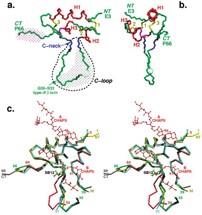
9390 Biochemistry, Vol. 41, No. 30, 2002                                                                               Brzozowski et al.
the conformation of the residues that are equivalent to insulin   Table 1: X-ray Data and Refinement Statistics
residues B25-B30. Both insulin and hIGF-I bind as mono-
                                                                                structure name                           IGF-I-esrf
mers to their receptors and display an ability to bind to each
other’s receptors; this latter phenomenon is considered to                        Data Collection and Processing Statistics
                                                                      data collection site                       ID14-4 ESRF
be physiologically significant (12). By contrast, the two             wavelength (Å)                             0.93
molecules have very different cellular origins; insulin is            space group                                C2221
synthesized in the â-cell of the Islets of Langerhans, while          unit cell dimensions (Å) (a, b, c)         30.78, 69.47, 65.0
hIGF is synthesized mainly in the liver, although other tissues       resolution range (Å) (outer shell)         20-2.0 (2.07-2.0)
                                                                      observations                               17172
are also involved. The two molecules undergo profoundly               unique reflections                         4839
different mechanisms of processing, secretion, and circulation        I/σ(I)                                     18(12)
in the blood. It is the characteristic hexamer structure that         completeness (%)                           98.3(95.8)
                                                                      Rmerge a                                   3.7(20.3)
provides the solution properties and stability needed for
transport, processing, and storage of proinsulin in the â-cell.                              Refinement Statistics
Proinsulin, contrary to IGF, is processed further by specific         resolution range (Å)                           20-2.0
                                                                      reflections used (Rfree set)                   4620(208)
convertases to insulin, whose hexamers rapidly disassemble            Rcryst/Rfreeb                                  23.4/29.5
upon release to the bloodstream into the native monomer               protein/ligand atoms/waters                    475/20/35
that binds to its receptor; this complex is then endocytosed          r.m.s. bonds/angles (Å)c                       0.015/2.6
and degraded. hIGF-I, in a contrasting process, circulates as         r.m.s. main chain ∆B (Å2)d                     1.05
                                                                      mean B-factor (Å2)e                            31.3/33.3/35.5/38.5
an inactive species for lengthy periods protected by the              %A, B, L (a, b, l, p)f                         89.8(10.2)
numerous IGF binding protein (IGFBP) molecules (see for              a
                                                                       Rmerge ) 100∑|I - ⟨I⟩ |/∑⟨I⟩. b Rcryst )∑|Fobs - Fcalc|/∑Fobs; Rfree is
example refs 13-15). hIGF-I exerts its pleiotropic actions        as Rcryst but calculated over 4.5% of data that were excluded from the
by binding and activating a 440-kDa type 1 IGF receptor           refinement process. c Root-mean-square deviations in bond length and
(IGF-1R), an R2â2 tyrosine kinase heterodimer (50% ho-            angle distances from Engh and Huber ideal values. d Root-mean-square
mologous with the insulin receptor (IR)) (15, 16).                deviations between Bfactors for bonded main chain atoms. e Mean
                                                                  temperature factor for whole molecule, main chain, side chain, ligand,
   Here we present four crystal structures of hIGF-1 analyzed     and water atoms, respectively. f Percentage of residues located in the
at 100 K and room temperature, based on crystallographic          most favored (additional) regions of Ramachandran plot as determined
data collected using X-ray sources at home, the SRS               by PROCHECK (17).
(Daresbury), the EMBL (Hamburg), and the ESRF (Greno-
ble). The crystals were grown from media containing a
detergent SB12 that acts to reduce aggregation and, perhaps,
conformational flexibility of the monomer. Although the axial
parameters of the crystals are very similar, the quality of
the data collected at 300 and 100 K varies considerably,
reflecting overall mobility of the hIGF-I. The structures are
generally similar to those reported in the earlier investiga-
tions, but there is more detail detected at the N- and
C-terminal extensions and particularly in the B- and C-
domain loop.
   The corresponding chain numberings in insulin and hIGF-
II are given respectively in [ ] and {} brackets. Comparisons
of hIGF-I with insulin structures and hIGF-II result from
structure alignments using helix Ala8[B9]{Gly11}-Cys18-
[B19]{Cys22}.

RESULTS AND DISCUSSION
   Structural Organization of hIGF-I. The description and
discussion of hIGF-I are based mostly on the 2.0 Å X-ray
data collected at the ESRF (referred to as hIGF-I-esrf), which    FIGURE 2: Comparison of rms deviations (in Å) between CR atoms
has the best data statistics (Table 1). The other three hIGF-I    of hIGF-I-dares (red), hIGF-I-hamb-RT (blue), hIGF-I-inh-RT
structures are referred to a: hIGF-I-dares (data collected at     (green), hIGF-I described by Vajdos et al. (6) (magenta), and hIGF-
the SRS at a temperature of 120 K), hIGF-I-hamb-RT (data          I-esrf as a reference structure.
collected in Hamburg at room temperature), and hIGF-I-inh-
RT (data collected in-house at room temperature); for all         I-esrf molecule, while the more profound structural differ-
data and refinement statistics see Table 1 of the Supporting      ences are located around the C-domain and N- and C-termini
Information.                                                      (Figure 2, Figure 3c).
   The atomic structure of hIGF-I was determined almost              Comparison of the four hIGF-I structures reported here,
completely in the ESRF analysis (hIGF-I-esrf is referred to       the hIGF-I structure described by Vajdos et al. (6), and
here also as hIGF-I); only two N-terminal, four C-terminal,       insulin reveals that the hIGF-I core (consisting of three
and three C-domain residues (Arg36-Arg37-Ala38) have              helices) is very similar to its equivalent in insulin (the 1.5 Å
not been detected satisfactorily (Figure 3c). The core of all     resolution structure described by Baker et al. 1988 (18) has
four structures shows close overall similarity with the hIGF-     been used throughout as a reference structure for insulin).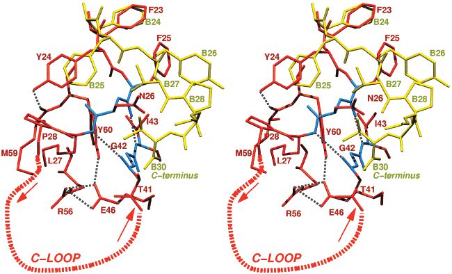
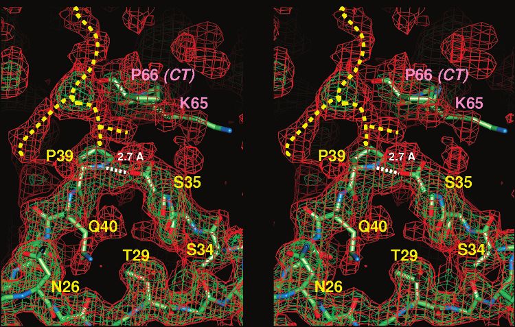
hIGF-I Crystal Structure                                                               Biochemistry, Vol. 41, No. 30, 2002 9391

FIGURE 3: (a) and (b) Two orthogonal views of a backbone representation of the hIGF-1 structure. Helices are in red and are defined as
helix 1 or H1 (Gly7-C18), helix 2 or H2 (Ile43-Cys47), helix 3 or H3 (Leu54-Leu57); the 310 helix (Glu58-Tyr60) is in magenta;
disulfides are in yellow with 1 corresponding to Cys6-Cys47, 2 to Cys46-Cys52, and 3 to Cys18-Cys61. The C-neck (Phe25-Asn26
and Gly42-Ile43) is in blue; NT and CT are the N- and C-termini; gray and magenta dotted surfaces indicate the areas of the C- and
D-domains, respectively. (c) Overall CR chain comparison of the four hIGF-I structures: hIGF-I-esrf (green), hIGF-I-dares (grey), hIGF-
I-hamb-RT (yellow), hIGF-I-inh-RT (blue) and hIGF-I-CHAPS (6) (red), with detergents as ball-and-stick models; numbers indicate the N-
and C-termini and the C-domain gaps observed in these structures. CHAPS A corresponds to its position described in (6); CHAPS B
depicts a symmetry related orientation of CHAPS A (not discussed in ref 6) that is close to the SB12 location reported here (symmetry
equivalent of SB12 in CHAPS A position is not shown for picture clarity) (divergent stereo).

   The most striking departure from the insulin fold is at            motif of three pseudo-â-strands. It is worth noting that a
Tyr24 and Phe25 ([B25&B26]). The Gly22[B23]-Phe25-                    similar hydrogen bond motif also occurs in insulin, where
[B26] â strand does not extend as far as in insulin where it          [A4Glu] is salt bridged to the free amino group of [A1Gly]
finishes at Thr29[B30]. In hIGF-I, it ends with a tight bend          (18).
at Phe25[B26] leading to the IGF-specific C-loop, which                  The detergent SB12 was an additive in our crystallization
includes residues Asn26-Thr29 and the C-domain peptide                of hIGF-I, and one SB12 molecule is found to pack between
that links back to the A-domain. This C-loop extends such             protein molecules in the crystal. Its alkyl chain stretches
as a dagger ∼20 Å away from the core of the molecule,                 along the hydrophobic patch formed by Val11[B12], Phe23-
giving hIGF-I an overall shape of a sharp wedge. Residues             [B24], Phe25[B26] and bends to involve its SO3- group in
Gly30-Tyr31-Gly32-Ser33 of the C-domain form a clas-                  hydrogen bonds with NH main chain atoms of Asn26 and
sical type-II â-turn at the center of the C-loop, directing it        Phe25 (see Figure 4 in Supporting Information). As the
to the A-domain. There is a gap in the electron density for           Val11[B12], Phe23[B24], Phe25[B26] surface corresponds
the C-loop for residues Arg36-Arg37-Ala38 in hIGF-I. The              to the dimer-forming interface of insulin, it seems likely that
C-domain polypeptide chain then returns back to the core              the SB12 detergent prevents nonspecific aggregation of
of IGF-I via Gly42, with a sharp bend of the main chain at            hIGF-I observed during crystallization without detergent (6).
Ile43 [A2]. Two antiparallel hydrogen bonds between the                  The fragments of hIGF-I reported as highly mobile in all
beginning and end of the C-loop (25CO‚‚‚NH43 and 27NH‚                NMR structures, namely, the N- and C-termini and the
‚‚OC41) create two short antiparallel â-strands, referred to          C-domain, occupy cavities in the crystal and are free of
here as the “C-neck” (Figure 3a,b). The definition of this            strong lattice contacts; hence, the conformations observed
new structural feature is reinforced by additional hydrogen           here may be physiologically relevant.
bonds between 42CO‚‚‚HN45 and 42NH‚‚‚45OD1 of Asp45-                     Structural Variation in hIGF-I. The comparison of all
[A4] from the contiguous R-helix 2 (Ile43[A2]-Cys47[A6]).             hIGF-I crystal structures solved during this study reveals the
The involvement of the Asp45[A4] side chain extends the               dynamic nature of some parts of this molecule (Figure 3c).
neck, which consists of two, short â-strands, into a structural       The mobility of the C-loop is visibly increased in both room
9392 Biochemistry, Vol. 41, No. 30, 2002                                                                     Brzozowski et al.

temperature structures (hIGF-I-inh-RT and hIGF-I-hamb-            MALDI-TOF analyses of different hIGF-samples also showed
RT), where the gaps in the electron density are widened from      a significant population of hIGF-I in dimeric form (data in
Tyr31 to Pro39 (in hIGF-I-inh-RT). The inherent flexibility       Supporting Information), while the dissolved crystal samples
of the C-loop is further corroborated by comparison with          behaved differently. In these, the dimer population was
the recent hIGF-I structure (6) obtained, as described above,     significantly diminished. As a MALDI-TOF-characteristic,
from crystals grown in the presence of detergent big CHAPS        artificial sample dimerization occurs fairly frequently, these
(referred to here as IGF-I-CHAPS). Despite different crystal-     observations have to be considered with care. Whether SB12
lization conditions and detergent used, this crystal form         (or CHAPS) interferes with IGF’s insulin-like dimerization,
belongs to the same space group C2221, with a maximum             the interactions with cognate binding proteins or destabiliza-
difference of ∼3% in the unit cell dimensions. Although the       tion of other types of dimers, remains unclear.
crystal packing and overall structure of hIGF-I reported here        It is possible, however, that breaks in the C-domain
(grown in the presence of SB12; the four structures are           electron density in the 31-39 region of hIGF-I result from
collectively referred to in this paragraph as hIGF-I-SB12         cleavage at Arg36-Arg37, although there is no evidence up
when discussing common features) and hIGF-I-CHAPS are             to now as to whether this occurs during or before structural
similar, the inherent mobility of the C-loop and differences      experiments. The short (∼3 Å) Ser35‚‚‚Pro39 separation in
in the chemical nature of the detergents used, result in local    the hIGF-I-esrf and hIGF-I-dares maps and the well-defined
structural variations and partially different detergent-protein   local electron density made it very difficult to close the gap
interactions in these, otherwise relatively isomorphous,          (see Figure 4), suggesting cleavage. Moreover, there is
structures.                                                       continuous electron density between Ser35 and Pro39 that
   The most significant structural divergence between IGF-        allows modeling of the -COOH group at Ser35 to lie adjacent
I-SB12 and IGF-I-CHAPS occurs in the conformation of the          to the main chain of Pro39. The presence of a weak (at 0.5σ
C-loop. In hIGF-I-CHAPS, the weak and disconnected                level on 2Fo-Fc maps) electron density stretching out from
electron density for part of the C-loop is longer than in hIGF-   Pro39 into the intermolecular space may indicate the presence
I-SB12 and includes the Ser35-Gln40 stretch of the C-             of the “missing” Arg36-Arg37-Ala38- residues. The ful-
domain. The conformation at Asn26-Ser34 in the “CHAPS”            filment of the stereochemical criteria for this “alternative”,
crystal form is also significantly different from that in hIGF-   extended conformation of the Arg36-Pro39 chain would
I-SB12. The C-loop starts to deviate from hIGF-I-SB12 at          require repositioning of Pro39 from its current conformation;
Asn27 (0.75 Å between their CR atoms) to reach a separation       this rearrangement would fit the observed electron density.
of ∼9 Å between the CR atoms of Ser34; consequently, the          It should be noted that the high mobility of the C-loop,
side chains of Tyr31, crucial for IGF-1R binding, are ∼8.6        especially its 35-39 region, is correlated in the crystal with
Å apart (see Figure 2c). The structure of the terminal part       the disorder of the C-terminus, which is in close contact with
of the C-loop (around Thr41-Gly42) is very alike in both          some of the residues of the C-domain.
structures. Additionally, these structural differences might         The SDS-PAGE electrophoresis of reduced samples from
also reflect slightly different detergent binding modes in both   crystals used for X-ray data collection showed more than
crystal structures. Although the deoxycholamide headgroup         one major band, implying cleavage, a behavior exhibited also
of the detergent (CHAPS B in Figure 3c, symmetry related          by the other hIGF-I samples used for crystallization (see
to CHAPS A described in ref 6) in hIGF-I-CHAPS occupies           Supporting Information). Additionally, the Electrospray and
the position of the alkyl chain of SB12, its long hydrophilic     MALDI-TOF mass spectrometry showed that the mass of
moieties do not overlap with the short polar group of SB12.       hIGF-I in the crystal was increased by 16 Da in comparison
They would also sterically clash with the distal part of the      with a fresh solution of the hIGF-I sample. This would
C-loop in its hIGF-I-SB12-like conformation. Additionally,        correspond to one oxygen atom taken up by peptide bond
the Br- ion found in hIGF-I-CHAPS mimics the position of          hydrolysis. An alternative explanation for the 16 Da mass
the SO3- group of the SB12 detergent (with Br-S distance          increase could be the oxygenation of Met59; there is,
of 0.66 Å).                                                       however, no extra electron density at this residue. The
   It is perhaps unexpected that despite the different chemical   N-terminal sequencing of the crystal-derived IGF-I sample
natures of the two detergents (nonionic big CHAPS com-            also points to heterogeneity, as both the RA- sequence and
pared to zwitterionic SB12), both compounds target similar        the main GPETL- N-terminal motif are observed. All these
surfaces of hIGF-I. Their perceived impact on the biologi-        data show the presence of hIGF-I cleavage in the crystals
cally relevant behavior of hIGF-I may, however, differ            and that there is a need for careful reexamination and more
depending on whether the interactions represented by              rigorous assessment of the structural integrity of hIGF-I in
CHAPS-A or CHAPS-B are considered (see Figure 3c). The            vitro and in vivo. There is an additional relevance to this
binding mode “A” corresponds roughly to the IGF-I/IGFBP-5         cleavage as the sequence -Arg36-Arg37-Ala38- of the
complex interface (7), and results in the interference with       “missing” region is similar to the proinsulin B chain-C chain
IGFBP-1 and IGFBP-3 association with the hormone (6).             junction processed during its maturation (20) and a single
The hIGF-I displacement from the IGF-I/IGFBP-3 complex            cleavage in the C-domain (before Arg37) in the plasma-
by the nonpeptide, isoquinoline compounds (19) also cor-          derived hIGF-I has already been reported (21).
roborates this hypothesis, as their poly-hydroxy-aromatic            IGF-I and Insulin Structural Relationship. Although the
character is similar to the deoxycholamide functional group       overall architecture of the helices 1, 2, and 3 in hIGF-I is
of CHAPS. In contrast to the binding mode “A”, the main           very like its counterpart in insulin, there are substantial
effect of detergent binding mode “B” would be to diminish         structural dissimilarities between hIGF-I and insulin (Figure
dimerization of hIGF-I, as dimer formation has already been       5). The most striking difference results from the presence
reported from sedimentation equilibrium data (6). Our             of the C-loop in hIGF-I, which alters completely the character
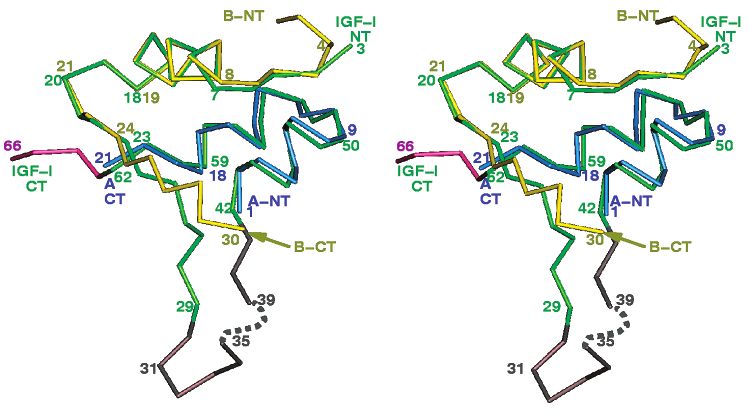
hIGF-I Crystal Structure                                                               Biochemistry, Vol. 41, No. 30, 2002 9393

FIGURE 4: The electron density for the 35-39 gap region of the C-domain in the hIGF-esrf structure; green, 1σ level; red, 0.5σ level.
Yellow dashed lines indicate unassigned low-contour electron density, next to Pro39, that may correspond to the missing Arg36-Arg37-
Ala38 residues. The white dashed line represents the potential hydrogen bond between OH of the carboxyl group of Ser35 and the NH of
Pro39 (the Ser35 carboxyl group is not included in the deposited PDB file as it was modeled only after additional cycles of REFMAC
following completion of the model building and refinement; if the R36-R37-A38 are considered as present, this hydrogen bond could not
be formed by Pro39 in its current conformation as this residue would have to be substantially remodeled to be joined with Ala38); pink
labels are associated with a symmetry related C-terminus of a neighboring hIGF-I molecule (divergent stereo).

FIGURE 5: Comparisons of the CR chains of hIGF-I (hIGF-I-esrf) and human insulin with domain color coding as in Figure 1: B and A
chains of insulin in yellow and blue, respectively; hIGF-I C-domain in gray, D-domain in magenta (remaining parts of hIGF-I in green;
divergent stereo).

of the surface that in insulin is responsible for its dimerization    molecule and has not been observed in proinsulin or in any
and receptor binding. The most visible consequence of this            insulin molecule containing an engineered link between the
characteristic hIGF-I structural feature is a repositioning of        B chain C-terminus and the A chain N-terminus (see for
the B chain C-terminal residues into an integral part of the          example refs 22-26). The main chain bend at residues
C-loop. The neck at the C-loop brings the Gly22[B23]-                 Gly22[B23]-Thr29[B30] (by ca. 105°) in hIGF-I at the CR
Phe25[B26] â-strand much closer to the N-terminus of helix            atom of Phe25 directs these residues outward into the
2 [A1-A2] and to the core of the protein. A ∼25° angle                C-domain (Figure 6). This positioning of the 26-29[B27-
between these strands in hIGF-I and insulin (with the CR’s            B30] polypeptide exposes residues 42-43[A1-A2]) that in
of Phe23[B24] as the reference starting point) results in a           insulin form the main IR binding epitope (the so-called site
∼3.4 Å difference between the corresponding CR atoms of               1 (27)) (Figure 7). In insulin, these residues are buried by
Phe26 and [TyrB27]. This conformation prevents the forma-             the C-terminal part of the B chain, and it has been postulated
tion of an antiparallel â-sheet structure in the hIGF-1               that its dislocation/rearrangement is a prerequisite for receptor
9394 Biochemistry, Vol. 41, No. 30, 2002                                                                               Brzozowski et al.

FIGURE 6: Comparison of the conformation of the hIGF-I 23-29 region (in red) and the corresponding C-terminal part of the insulin B
chain (in yellow); the main chain of the C-neck part of hIGF-I in blue, hydrogen bonds are shown as dashed gray lines. The C-loop is
depicted as a thick, red dashed half-circle with arrows indicating the direction of the polypeptide chain.

FIGURE 7: van der Waals representations of insulin (a) and hIGF-I (b) surfaces. Corresponding regions of the B chains implicated in
interactions with receptors are marked in yellow and gold, with residues crucial for IR or IGF-1R binding are highlighted in gold. Residues
[A1-A4] of the insulin A chain (and their 42-43 equivalents in hIGF-I) that are also important for interactions of insulin with its cognate
receptor are marked in red. They are buried under the B chain in the insulin molecule, and are exposed in hIGF-I due to the presence of
the C-loop and the tight main-chain bend at Phe25 in this growth factor.

binding (e.g., refs 23 and 28). In contrast to insulin, the             of Tyr60[TyrA19], a residue crucial for the hIGF-I, hIGF-
hIGF-I binding surface’s exposure is achieved through the               II, and insulin activity (30). In hIGF-I, the hydroxyl group
well-defined conformation at the C-loop.                                of Tyr60[TyrA19] forms a hydrogen bond with the OE1 atom
   The new “IGF-like” fold of the 22-29[B23-B30] chain                  of Glu46([GlnA5] (a contact preserved in insulin and hIGF-
in the hIGF-I-SB12 crystal structure (typical also for the              II (to {Glu45}) as well). This hydrogen bond seems specific
hIGF-I-CHAPS molecule) is additionally stabilized by a                  to hIGF-I as the carboxyl of Glu46 is replaced in insulin by
hydrogen bond between the hydroxyl group of Tyr24 and                   the amide group of [GlnA19]. Furthermore, the stability of
the carbonyl oxygen of Met59 [AsnA18] (see Figure 6). It                Tyr60 and its neighboring C-neck region may also benefit
is obvious, however, that the hydrogen bond interactions of             from complete burial and protection of this residue under
Tyr24 alone are insufficient for defining the conformation              the side chains of Arg56, Lys27, and Met59. However, the
in the 23-25 region since a hIGF-I mutant with the insulin-             side chains of Lys27 and Met59 in hIGF-I-SB12 are
like sequence Phe23-Phe24-Tyr25 has the same affinity                   stabilized by van der Waals contacts only and may adopt
as the wild-type hormone towards the IGF-1, -2, and insulin             alternative conformations upon receptor binding.
receptors. On the other hand, the aromatic character of the                As a result of the hIGF-I-specific conformation at residues
side chain at position 24 is still required for the full biological     22-29 that mimics the [B23-B30] chain displacement in
activity (29). It is also possible that the hIGF-specific               insulin (e.g., refs 31 and 32), the IGF-1R binding epitopes
positioning of Tyr24 may be a part of a wider network of                (Gly22[B23]-Phe25[B26] and Gly42-Asp45[A1-A4]) on
interactions, leading in consequence to further stabilization           the hIGF-I surface can be involved in binding of this
hIGF-I Crystal Structure Biochemistry, Vol. 41, No. 30, 2002 9395
hormone to both receptors (IGF-1R and IR) without any composed of various ratios of hIGF-I at 7 mg/mL (in H2O)
major structural reorganization within this hormone molecule with reservoir solution consisting of 0.1 M Tris/HCl pH 7.5,
(Figure 7). Thus, the well-defined structure of the Gly22- 12-15% (w/v) PEG 2K MME and 5 mM SB12 detergent.
29[B23-B30] chain and the C-loop neck region in hIGF-I Platelike orthorhombic crystals with unit cell dimensions a
(and in hIGF-II) imply that the postulated rearrangement of ) 30.78 Å, b ) 69.47 Å, c ) 65.0 Å appeared within 2-3
the C-terminus of the B chain in IGF-I and IGF-II upon IGF- weeks, with one molecule of the hormone per asymmetric
1/2R (or IR) binding (33), as proposed for the insulin-IR unit.
association, is unlikely. Data Collection. Initially, a room temperature 2.5 Å data
While the hIGF-I specific conformation of the Gly22- set was collected in-house using an RaxisIIc imaging plate
29[B23-B30] chain will maintain/impose a monomeric state detector mounted on a Rigaku RU200 rotating anode X-ray
of the molecule, other residues that differ from those in generator with MSC/Yale mirrors. Subsequently, the resolu-
insulin (e.g., [HisB10] f Glu9, [TyrB16] f Gln15) can also tion was extended to 2.3 Å at station W7B at DESY (EMBL,
be relevant. For example, the switch of the Pro [B28] and Hamburg) and station 14.2 at SRS (Daresbury). The final
Lys [B29] amino acids in insulin to Lys27-Pro28 is 100 K 2.0 Å data set was collected at station ID14-1 at the
characteristic of IGF-I. As this switch is known to prevent ESRF (Grenoble, France). Prior to freezing, the crystal was
insulin dimer formation and has been successfully used to cryoprotected by sequential, 5% step, soaks in the mother
create a monomeric insulin (34) its presence in hIGF-I is liquor containing glycerol with a final concentration of 30%
likely to contribute to the monomeric state of this hormone. (v/v). Data were recorded in one sweep and 1° oscillations
Importance of the C-Loop. The role of the C-domain for using a MAR Research CCD detector placed at 150 mm from
IGF-1R binding is clearly indicated by the observations that the crystal. All data were integrated and reduced using
its deletion or replacement by short-linkers abolish or DENZO and SCALEPACK (42). X-ray data statistics for
diminish dramatically the affinity of hIGF-I for IGF-1R (33, this data set are summarized in Table 1.
35). The structure at the C loop (residues 25-41) may have Structure Determination and Refinement. The coordinates
a double impact on receptor binding affinities and specifici- of the publicly available hIGF-I NMR structures (2-4) and
ties of hIGF-I and insulin. First, it enforces a particular, IGF- its theoretical model (43) were initially used as search models
specific, conformation of the Gly22-Phe25 â-strand, pre- in AmoRe (44) to solve the hIGF-I structure by molecular
serving at the same time an ‘insulin-like” fold across residues replacement. However, as these trials were unsuccessful
42-43 ([A1-A2]). subsequent molecular replacement trials were carried out with
Second, the C-domain is likely to be directly involved in human insulin (PDB entry: 4INS, 18) as the best search
specific IGF-1R interactions through some of its side chains, model. Careful analysis of the solutions (no single, clear
especially Tyr31, which is one of the key residues in IGF- solution was obtained) allowed determination of the position
1R affinity (30). The positioning of Tyr31 in the center of of the core of the hormone molecule. Initial electron density
the structurally well-defined Gly30‚‚‚Ser33 type-II â-turn maps, calculated after rigid-body refinement in AmoRe,
underlines the importance of this interaction; its probable unambiguously indicated the position of all disulfides
hydrophobic character is emulated by van der Waals contacts although the model building was rather difficult. The
between Tyr31 and the alkyl moiety of a symmetry related refinement with the use of an older version of REFMAC
SB12 detergent molecule in the crystal. The structural role (45) was unsuccessful.
of the C-loop is underlined further by the high IGF-like Refinement of hIGF-I was only possible using translation,
activity of an insulin analogue with its A and B chains linked libration, screw (TLS) (46), and individual atomic parameter
by the C-domain of hIGF-I (36, 37) and very low biological (45) refinement. Each round of TLS refinement was followed
potency of both hormones with C-domain modifications (e.g., by individual atomic (positional and thermal) refinement.
refs 22-26 and 35). The C-domain therefore contributes Because of the high mobility of IGF, TLS refinement was
directly to the hIGF-1 receptor binding, and is at least in crucial for the success of structure determination.
part responsible for the hIGF-I specificity. By contrast, the The successive rounds of refinement using all data between
C-domain of pro-insulin does not even prevent its aggrega- 15 and 2.0 Å (no sigma cutoffs), and manual rebuilding gave
tion (38, 39), and may be considered as a polypeptide whose a final model with an Rcryst of 23.6 and an Rfree of 29.5. The
main role is an assurance of the efficient folding and final model comprises 475 protein atoms, 20 ligand (SB12)
maturation of an active hormone (40, 41). atoms, and 35 water molecules. Poorly defined residues 1-2,
36-38, 67-70 were not included in the final structure, and
In summary, our X-ray studies of the hIGF-I shed light
occupancies of some side chain atoms of Gln40, Asp56, and
on a structural role of the C-loop of this hormone in its
Lys65 are set to zero because of their disorder. All model
functional divergence from insulin and underlined the need
building was carried out using the molecular graphics
for further studies of the chemical integrity of hIGF-I in vitro
package QUANTA (QUANTA98; Accelrys INC, San Diego,
and in vivo. The likelihood of the cleavage occurring at
CA). Dictionaries for the SB12 ligand were derived from
Arg36-Arg37 and the resulting flexibility of the C-loop
REFMAC. A summary of refinement statistics is given in
delay any final conclusions about the hIGF-I “active”
Table 1.
conformation in complex with IGF-1R.
Superpositions of different insulin and IGF models were
MATERIALS AND METHODS carried out in QUANTA; after global superposition, the
overlaps were fine-tuned using the “match closest residue”
Crystallization. Pure hIGF-I was kindly supplied by option in QUANTA. All NMR structures were minimized
Pharmacia-Upjohn (Stockholm, Sweden). The protein was using CHARMM (47; details to be published elsewhere)
crystallized by the hanging drop method, in which drops were prior to analysis. Figures 3c, 4, and 5 were produced using9396 Biochemistry, Vol. 41, No. 30, 2002 Brzozowski et al.
QUANTA, and Figure 3a and 7 were produced with 14. Jones, J. I., and Clemmons, D. R. (1995) Insulin-like growth factors
MOLVIEWER (M. Harsthorn, personal communication) and their binding proteins: biological actions. Endocrine ReV. 16,
3-34.
15. Adams, T. E., Epa, V. C., Garret, T. P. J., and Ward, C. W. (2000)
ACKNOWLEDGMENT Structure and function of type 1 insulin-like growth factor receptor.
Cell. Mol. Life Sci. 57, 1050-1093.
We would like to thank Par Gellerfors (Pharmacia-Upjohn, 16. Roberts, C. T., Lasky, S. R., Lowe, W. L., Seaman, W. T., and
Stockholm) for protein samples and a very friendly col- LeRoith, D. (1987) Molecular cloning of rat insulin-like growth
laboration. The technical assistance of the beam line manag- factor I complementary deoxyribonucleic acids: differential
ers at the ESRF, EMBL Hamburg, and SRS Daresbury messenger ribonucleic acid processing and regulation by growth
hormone in extrahepatic tissues. Mol. Endocrinol. 1, 243-248.
during data collection is gratefully acknowledged. We also 17. Laskowski, R. A., MacArthur, M. W., Moss, D. M., and Thornton,
thank Steven Howell (NIMR, Mill Hill, London) and Arthur J. M. (1993) PROCHECK - a program to check the stereochem-
Moir (Department of Molecular Biology, University of ical quality of protein structures. J. Appl. Crystallogr. 26, 283-
Sheffield) for kind help with mass spectrometry and N- 291.
18. Baker, E. N., Blundell, T. N., Cutfield, J. F., Cutfield, S. M.,
terminus sequencing analyses. Dodson, E. J., Dodson, G. G., Crowfoot Hodgkin, D. M., Hubbard,
R. E., Isaacs, N. W., Reynolds, C. D., Sakabe, K., Sakabe, N.,
SUPPORTING INFORMATION AVAILABLE and Vijayan, N. M. (1988) The structure of 2Zn pig insulin crystals
at 1.5 Å resolution. Philos. Trans. R. Soc. (London) 319, 369-
ES-, MALDI-TOF spectroscopy, and SDS gel electro- 456.
phoresis results; X-ray data and refinement statistics for other 19. Liu, X.-J., Xie, Q., Zhu, Y.-F., Chen, C., and Ling, N. (2001)
than hIGF esrf structures discussed in the text. This material Identification of a nonpeptide ligand that releases bioactive insulin-
like growth factor-I from its Binding Protein complex. J. Biol.
is available free of charge via Internet at http://pubs.acs.org. Chem. 276, 32419-32422.
20. Steiner, D. F., Cunningham, D., Spigelman, L., and Aten, B. (1967)
REFERENCES Insulin biosynthesis: evidence for a precursor. Science 157, 697-
700.
1. Rinderknecht, E., and Humbel, R. E. (1978) The amino acid 21. Jansen, J., Van Buul-Offers, S. C., Hoogerbrugge, C. M., and Van
sequence of human insulin-like growth factor I and its structural Den Brande, J. L. (1990) Effects of a single cleavage in insulin-
homology with pro-insulin. J. Biol. Chem. 253, 2769-2776. like growth factors I and II on binding to receptors, carrier proteins
2. Cooke, R. M., Harvey, T. S., and Campbell, I. D. (1991) Solution and antibodies. Biochem. J. 266, 513-520.
structure of human insulin-like growth factor 1: a nuclear 22. Cutfield, J., Cutfield, S., Dodson, E. J., Dodson, G. G., Hodgkin,
resonance and restrained molecular dynamics study. Biochemistry D., and Reynolds, C. D. (1981) Evidence concerning insulin
30, 5484-5491. activity from the structure of a cross-linked derivative. Hoppe-
3. Sato, A., Nishimura, S., Ohukuba, T., Kyogoku, Y., Koyama, S., Seyler’s Z. Physiol. Chem. 362, 755-761.
Kobayashi, M., Ysuda, T., and Kobayashi, Y. (1993) Three-
23. Derewenda, U., Derwenda, Z., Dodson, E. J., Dodson, G. G., Bing,
dimensional structure of human insulin-like growth factor-1 (IGF-
X., and Markussen, J. (1991) X-ray analysis of the single chain
1) determined by 1H NMR and distance geometry. Int. J. Pept.
B29-A1 peptide-linked insulin molecule. J. Mol. Biol. 220, 425-
Protein Res. 41, 433-440.
433.
4. Torres, A. M., Forbes, B. E., Aplin, S. E., Wallace, J. C., Francis,
G. L., and Norton, R. S. (1995) Solution structure of human 24. Derewenda, U., Derwenda, Z., Dodson, E. J., Dodson, G. G.,
insulin-like growth factor II. Relationship to receptor and binding Reynolds, C. D., Smith, G. D., Sparks, C., and Swenson, D. (1989)
protein interactions. J. Mol. Biol. 248, 385-401. Phenol stabilizes more helix in new symmetrical zinc insulin
hexamer. Nature 338, 594-596.
5. Tersawa, H., Kohda, D., Hatanaka, H., Nagata, K., Higashihashi,
N., Fujiwara, H., Sakano, K., and Inagaki, F. (1994) Solution 25. Whittingham, J. L., Edwards, D. J., Antson, A. A., Clarkson, J.,
structure of human insulin-like growth factor II.; recognition sites and Dodson, G. G. (1998) Interactions of phenol and m-cresol in
for receptors and binding proteins. EMBO J. 13, 5590-5597. the insulin hexamer, and their effects on the association properties
6. Vajdos, F. F., Ultsch, M., Schaffer, M. L., Deshayes, K. D., Liu, of B28Pro f Asp insulin analogues. Biochemistry 37, 11516-
J., Skelton, J., and de Vos A. M. (2001) Crystal structure of human 11523.
insulin-like growth factor-1: detergent binding inhibits Binding 26. Cho, Y. S., Chang, S. G., Choi, K. D., Shin, H. C., Ahn, B. Y.,
Protein interactions. Biochemistry 40, 11022-11029. and Kim, K. S. (2000) Solution structure of an active mini-
7. Żesławski, W., Beisel, H.-G., Kamionka, M., Kalus, W., Engh, proinsulin, M2PI: interchain flexibility is crucial for insulin
R. A., Huber, R., Lang, K., and Holak, T. A. (2001) The activity. J. Biochem. Mol. Biol. 33, 120-125.
interaction of insulin-like growth factor with N-terminal domain 27. Schäffer, L. (1994) A model for insulin binding to the insulin
of IGFBP-5. EMBO J. 20, 3638-3644. receptor, Eur. J. Biochem. 221, 1127-1132.
8. Dubaquie, Y., and Lowman, H. B. (1999) Total alanine-scanning 28. Dodson, E. J., Dodson, G. G., Hubbard, R. E., and Reynolds, C.
mutagenesis of insulin-like growth factor I (IGF-I) identifies D. (1983) Insulin’s structural behaviour and its relation to activity.
differential binding epitopes for IGFBP-1 and IGFBP-3. Bio- Biopolymers 22, 281-291.
chemistry 38, 6386-6396. 29. Cascieri, M. A., Chicchi, G. G., Applebaum, J., Hayes, N. S.,
9. Jansson, M., Uhlen, M., and Nilsson, B. (1997) Structural changes Green, B. G., and Bayne, M. L. (1988) Mutants of human insulin-
in insulin-like growth factor (IGF) I mutant proteins affecting like growth factor I with reduced affinity for the type 1 insulin-
binding kinetic rates to IGF binding protein 1 and IGF-I receptor. like growth factor receptor. Biochemistry 27, 3229-3233.
Biochemistry 36, 4108-4117. 30. Bayne, M. L., Applebaum, J., Chicchi, G. G., Miller, R. E., and
10. Jansson, M., Andersson, Uhlén, M., Nilsson, B., and Kördel, J. Cascieri, M. A. (1990) Role of tyrosines 24, 31 and 60 in the
(1998) The insulin-like growth factor (IGF) binding protein 1 high affinity binding of insulin-like growth factor-1 to the type 1
binding epitope on IGF-I probed by heteronuclear NMR spec- insulin-like growth factor receptor. J. Biol. Chem. 265, 15648-
troscopy and mutational analysis. J. Biol. Chem. 273, 24701- 15652.
24707. 31. Ludwigsen, S., Olsen, H. B., and Kaarlshom, N. C. (1998) A
11. Magee, B. A., Shooter, G. K., Wallace, J. C, and Francis, G. L. structural switch in mutant insulin exposes key residues for
(1999) Insulin-like growth factor I and its binding proteins: a receptor binding. J. Mol. Biol. 279, 1-7.
study of the binding interface using B-domain analogues. Bio- 32. Hua, X. Q., Shoelson, S. E., Kochoyan, M., and Weiss, M. A.
chemistry 38, 15863-15870. (1991) Receptor binding redefined by a structural switch in a
12. Massague, J., and Czech, M. P. (1982) The subunit structures of mutant human insulin. Nature 354, 238-241.
two distinct receptors for insulin-like growth factors I and II and 33. Gill, R., Wallach, B., Verma, C., Ursø, B., De Wolf, E., Grötzinger,
their relationship to the insulin receptor. J. Biol. Chem. 257, 5038- J., Murray-Rust, J., Pitts, J., Wollmer, A., De Meyts, P., and Wood,
5045. S. (1996) Engineering the C-region of human insulin-like growth
13. Collett-Sorberg, P. F., and Cohen, P. (2000) Genetics, chemistry, factor-1: Implications for receptor binding. Protein Eng. 9, 1011-
and function of the IGF/IGFBP system. Endocrine 12, 121-136. 1019.hIGF-I Crystal Structure Biochemistry, Vol. 41, No. 30, 2002 9397
34. Ciszak, E., Beals, J. M., Frank, B. H., Baker, J. C., Carter, N. D., convertases. Biochemistry 38, 890-896.
and Smith, G. D. (1995) Role of C-terminal B chain residues in 41. Qiao, Z.-S., Guo, Z.-Y., and Feng, Y.-M. (2001) Putative disulfide-
insulin assembly: the structure of hexameric LysB28ProB29-human forming pathway of porcine insulin precursor during its refolding
insulin. Structure 3, 615-622. in vitro. Biochemistry 40, 2662-2668.
35. Bayne, M. L., Applebaum, J., Underwood, D., Chicchi, G. G., 42. Otwinowski, Z., and Minor, W. (1997) Processing X-ray diffraction
Green, B. G., Hayes, N. S., and Cascieri, M. A (1989) The C data collected in oscillation mode. Methods Enzymol. A 276, 307-
region of human insulin-like growth factor (IGF) I is required for 326.
high affinity binding to the type 1 IGF receptor. J. Biol. Chem. 43. Blundell, T. L., and Humbel, R. E. (1980) Hormone families:
264, 11004-11008. pancreatic hormones and homologous growth factors. Nature 287,
36. Kristensen, J., Andersen, A. S., Hach, M., Wiberg, F. C., Schäffer, 781-787.
L., and Kjeldsen, T. (1995) The single chain insulin-like growth 44. Collaborative Computational Project No. 4 (1994) The CCP4
factor I/insulin hybdrid binds with high affinity to the insulin suite: programs for protein crystallography. Acta Crystallogr. D50,
receptor. Biochem. J. 305, 981-986. 760-763.
37. Cara, J. F., Mirmira, R. G., Nakagawa, S. H., and Tager, H. S. 45. Murshudov, G. N., Vagin, A. A., and Dodson, E. J. (1997)
(1990) An insulin-like growth factor I/insulin hybrid exhibiting Refinement of macromolecular structures by the maximum
high potency for interactions with type I insulin-like growth factor likelihood method. Acta Crystallogr. D53, 240-255.
and insulin receptors of placental plasma membranes. J. Biol. 46. Winn, M. D., Isupov, M. N, and Murshudov, G. N. (2001) Use of
Chem. 265, 17820-17825. TLS parameters to model anisotropic displacement in macromo-
38. Steiner, D. F. (1973) Cocrystallisation of proinsulin with insulin. lecular refinement. Acta Crystallogr. D57, 122-133.
Nature 243, 528-530. 47. Brooks, B. R., Bruccoleri, R. E., Olafson, B. D., States, D. J.,
39. Pekar, A. H., and Frank, B. H. (1972) Conformation of proinsulin. Swaminathan, S., and Karplus, M. (1983) CHARMM - a program
A comparison of insulin and proinsulin self-association at neutral for macromolecular energy, minimization, and dynamics calcula-
pH. Biochemistry 11, 4013-4016. tions. J. Comput. Chem. 4, 187-217.
40. Lipkind, G., and Steiner, D. F. (1999) Predicted structural
alternations in proinsulin during its interactions with prohormone BI020084JYou can also read