Translational Control of Canonical and Non-Canonical Translation Initiation Factors at the Sea Urchin Egg to Embryo Transition - MDPI
←
→
Page content transcription
If your browser does not render page correctly, please read the page content below
International Journal of
           Molecular Sciences

Article
Translational Control of Canonical and
Non-Canonical Translation Initiation Factors at the
Sea Urchin Egg to Embryo Transition
Héloïse Chassé, Sandrine Boulben, Patrick Cormier and Julia Morales *
 Centre National de la Recherche Scientifique (CNRS), Sorbonne Université, Integrative Biology of Marine
 Models (LBI2M), Station Biologique de Roscoff (SBR), 29680 Roscoff, France; hchasse@sb-roscoff.fr (H.C.);
 boulben@sb-roscoff.fr (S.B.); cormier@sb-roscoff.fr (P.C.)
 * Correspondence: morales@sb-roscoff.fr; Tel.: +33-298-292-369

 Received: 7 December 2018; Accepted: 30 January 2019; Published: 1 February 2019

 Abstract: Sea urchin early development is a powerful model to study translational regulation
 under physiological conditions. Fertilization triggers an activation of the translation machinery
 responsible for the increase of protein synthesis necessary for the completion of the first embryonic
 cell cycles. The cap-binding protein eIF4E, the helicase eIF4A and the large scaffolding protein eIF4G
 are assembled upon fertilization to form an initiation complex on mRNAs involved in cap-dependent
 translation initiation. The presence of these proteins in unfertilized and fertilized eggs has already
 been demonstrated, however data concerning the translational status of translation factors are still
 scarce. Using polysome fractionation, we analyzed the impact of fertilization on the recruitment of
 mRNAs encoding initiation factors. Strikingly, whereas the mRNAs coding eIF4E, eIF4A, and eIF4G
 were not recruited into polysomes at 1 h post-fertilization, mRNAs for eIF4B and for non-canonical
 initiation factors such as DAP5, eIF4E2, eIF4E3, or hnRNP Q, are recruited and are differentially
 sensitive to the activation state of the mechanistic target of rapamycin (mTOR) pathway. We discuss
 our results suggesting alternative translation initiation in the context of the early development of
 sea urchins.

 Keywords: sea urchin fertilization; translational control; initiation complex; eIF4E; mTOR pathway

1. Introduction
      Regulation of protein synthesis is critical for cell growth, development and survival and is
now recognized as a key control of gene expression [1,2]. Translation is predominantly regulated
during the initiation step. The majority of eukaryotic mRNAs are translated using a cap-dependent
mechanism: eukaryotic initiation factors (eIFs) assemble on the 5’ cap structure of the mRNA to
recruit the 43S preinitiation complex which scans mRNA in a 3’ direction to locate the AUG start
codon and facilitate 60S subunit joining, forming a translationally active 80S ribosome [3]. The
initiation complex includes the cap-binding protein (eIF4E), the scaffold protein (eIF4G), and an
RNA helicase (eIF4A) [4]. The translation initiation factor eIF4B potentiates eIF4A RNA helicase
activity in vitro [5]. The poly(A)-binding protein (PABP) bridges the 5’ and 3’ ends of the mRNA by
binding to eIF4G and the poly(A) tail of the mRNA, conferring a closed-loop conformation to the
mRNA, that increases translation efficiency [6]. The small protein 4E-BP competes with eIF4G for the
binding domain (YX4 LΦ) of eIF4E [7] and consequently is able to inhibit translation initiation. The
phosphorylation status of 4E-BP is regulated by the mTOR (mechanistic target of rapamycin) pathway
and controls its association with eIF4E [8]. Cell proliferation is tightly regulated by translational
control and has been shown to involve the eIF4E-dependent translation of proto-oncogenes [9].

Int. J. Mol. Sci. 2019, 20, 626; doi:10.3390/ijms20030626                           www.mdpi.com/journal/ijms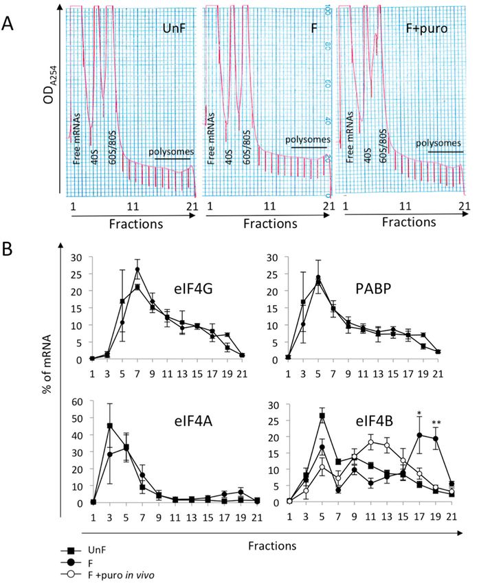
Int. J. Mol. Sci. 2019, 20, 626                                                                      2 of 14

The mRNAs of proto-oncogenes have structured untranslated regions that regulate cap-dependent
translation initiation through helicase-mediated unwinding of RNA structures [10,11]. Furthermore,
overexpression of translation factors leads to proliferation and cellular transformation. The mTOR and
MAPK/ERK (mitogen-activated protein kinase) pathways target several translation factors to regulate
protein synthesis [12]. As a major effector of cell growth and proliferation, mTOR is a favorite target
for therapeutic inhibition. However, mTOR pathway inhibition can lead to a hyperactivation of the
MAPK pathway in mammalian cells [13], both pathways being connected by compensatory feedback
loops [14]. Deciphering mRNA targets of the different pathways will allow a better comprehension of
the molecular circuitry impacting translational control.
      The eIF4E family is composed of three classes represented by eIF4E1, eIF4E2, and eIF4E3, which
share ~30% identity [15,16]; eIF4E1 has been found in all eukaryotes; eIF4E2 in all metazoans; and
eIF4E3 in deuterostomes [15,16], including sea urchins [17]. eIF4E1 is the canonical cap-binding
protein implicated in translation initiation, whereas eIF4E2 and eIF4E3 are involved in mRNA-specific
translation regulation [18–21]. In some specific physiological or physio-pathological cases, alternative
cap- and/or eIF4E1-independent translation can occur, in which non-canonical translation factors take
control [22]. DAP5 is a protein homologous to the C-terminal end of eIF4G that is unable to interact
with eIF4E or PABP but still retains the capacity to bind eIF3 and eIF4A. DAP5 can mediate translation
of its own mRNA by the direct recruitment of ribosomes at the vicinity of the ORF start codon on IRES
(internal ribosome entry site) [23] as well as for the translation of a subset of mRNAs [24–26]. Recently,
DAP5 was shown to also affect translation of mRNAs that do not contain IRES [26]. New members
of the poly(A)-binding family have been characterized recently, among them hnRNP Q (also termed
Syncrip) [27]. This protein, a competitive inhibitor of PABP binding, acts as a translation inhibitor [28]
but also promotes IRES-dependent translation of specific mRNAs [29,30].
      Sea urchin early development is a model in which to study translation and cell cycle regulation.
Indeed, the first cell cycles of the sea urchin embryo depend on the translational up-regulation that
occurs at fertilization [31,32]. The sea urchin genome contains full repertoire of known translation
factors, each one encoded by a unique gene [17,33]. In unfertilized eggs, translation activity is
low. Fertilization triggers an increase in protein synthesis dependent upon the activation of the
translation machinery and the polysomal recruitment of the stored maternal mRNAs, independently
of transcription [34–36]. The increase in protein synthesis triggered by the fertilization of sea urchin
eggs is partly explained by an increase in the activity of the cap-binding complex [37–39]. The
cap-dependent translation inhibitor 4E-BP plays an important role in eIF4E sequestration in unfertilized
eggs. Following fertilization, 4E-BP is rapidly phosphorylated and degraded in an mTOR-dependent
manner, leading to the release of eIF4E [40–42]. eIF4E is available to associate with eIF4G [43].
Therefore, eIF4E is now recognized as a crucial actor for the onset of the first mitotic division following
fertilization, suggesting that cap-dependent translation is highly regulated during the egg-to-embryo
transition in sea urchins [44]. Interestingly, when performed in the presence of the mTOR inhibitor
PP242, a differential recruitment of maternal mRNAs to polysome was observed after fertilization.
While several mRNAs showed sensitivity to the inhibitor, indicative of recruitment via the canonical
cap and eIF4E recognition, other mRNAs are still recruited to the polysomes in the presence of the
inhibitor, suggesting that an alternative mode of translation is also functional at fertilization [45].
Furthermore, PP242 has been identified as an ERK activator in multiple myeloma cells [46], raising the
question of whether a similar relay could operate physiologically in the developing sea urchin embryo.
      Besides the post-translational regulation and protein interactions described above, an additional
potential layer of regulation of the protein machinery is the translational control of mRNAs encoding
the translation factors. The study of the translational status of specific mRNAs by analysis of polysome
localization allows the direct assessment of mRNAs recruited after fertilization [47]. In this report, we
focused on the polysomal recruitment of mRNAs for canonical and non-canonical factors involved in
mRNA recruitment at the initiation step of translation.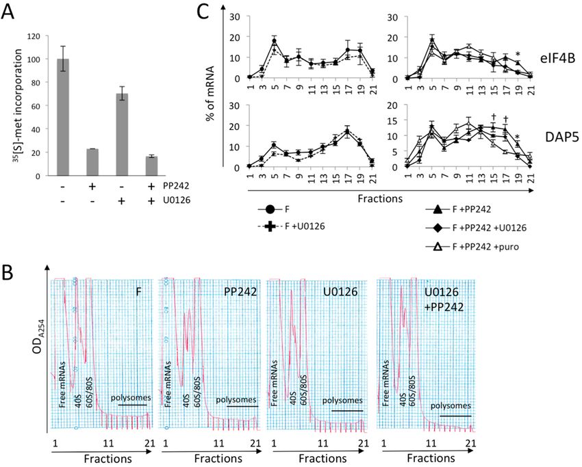
Int. J. Mol. Sci. 2019, 20, 626                                                                                    3 of 14

2. Results

2.1. mRNA Coding Translation Initiation Factors Involved in mRNA Recognition Are Present as Maternal
mRNAs in Sea Urchin Eggs
      An antibody directed to the human full-length protein eIF4E1 cross-reacts with two bands at
24–25 kDa, the lower band being the most prominently associated to an m7 -GTP column, revealing the
existence of cap-binding protein in sea urchin eggs [40]. Whether the upper band revealed by western
blot corresponds to eIF4E1, eIF4E2, or eIF4E3 is currently unknown. Information on mRNA abundance
of the translation factors was derived from transcriptome analysis of maternal mRNAs using FPKM
values as an indicator of mRNA abundance [45]. Based on FPKM values, mRNAs encoding eIF4E1
were present at very low levels (94. FPKM values and the primers
used for PCR amplification are presented in Table 1.

         Table 1. Translation initiation factors in the sea urchin maternal transcriptome (retrieved from [45]).

                                                                                                         Efficiency
      Name             Transcript #       FPKM *            Primer Sequence        Product Length (bp)
                                                                                                            (R2 )
                                                       TGGTCAAGAAGGAAGAAC
 eIF4A               comp73316_c0_seq1     59.53                                          103              0.990
                                                       CGTCTCATACAAGTCACA
                                                       GGAGGAGCAAAGCCTGTAGA
 eIF4B               comp78411_c2_seq1     94.72                                          200              0.992
                                                       ACGCGTTCTGCTTTCTCTTC
                                                       GGTGGAAGGTGGCTCATAGG
 eIF4E1              comp52193_c0_seq1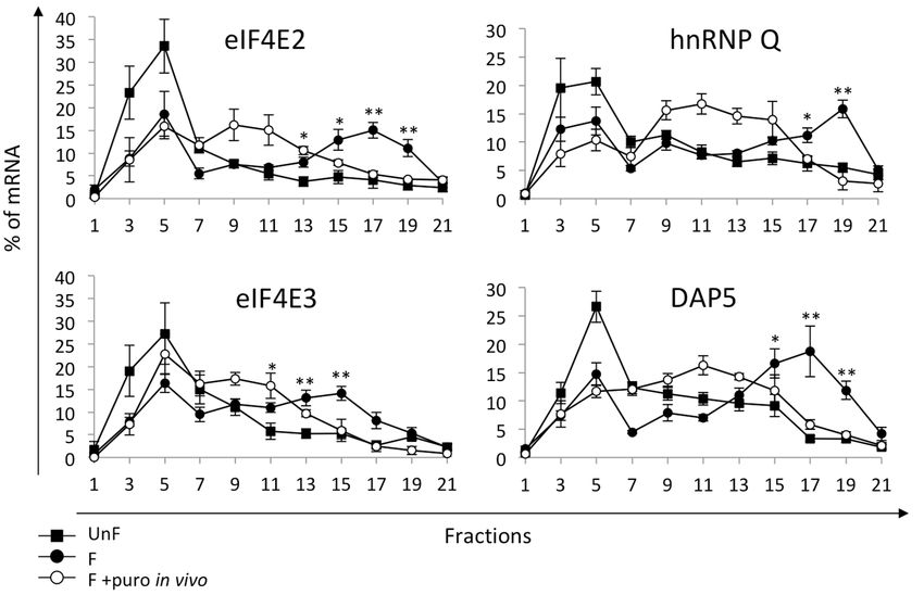
in the light fractions (1–7) of the gradient before fertilization and are associated with the heavy
 fractions (17–21) after fertilization. Upon puromycin treatment, which disrupts only actively
 translating polysomes but not co-migrating mRNPs, the eIF4B mRNAs shift to the middle of the
 gradient
Int.           (fractions
     J. Mol. Sci.             9–13), suggesting that eIF4B is translationally regulated at fertilization. 4These
                  2019, 20, 626                                                                             of 14
 results are in agreement with the translatome data analysis at the egg-to-embryo transition [45].

     Figure 1. (A) Optical density profiles (ODA254 ) of polysome fractionation from unfertilized eggs
     (UnF),
       Figureembryos      at 1 hdensity
                1. (A) Optical   post-fertilization
                                         profiles (OD  (F)A254
                                                           and ) ofinpolysome
                                                                       presencefractionation
                                                                                 of puromycin    (F+puro).
                                                                                               from          Fractions
                                                                                                     unfertilized  eggs 1(UnF),
                                                                                                                          and
     21  correspond
       embryos     at 1 to  top and bottom, (F)
                         h post-fertilization     respectively,
                                                      and in presence of the 15–40%   sucrose(F+puro).
                                                                              of puromycin       gradient;Fractions
                                                                                                            (B) Distribution
                                                                                                                     1 and 21
     incorrespond
         polysomestooftop      and bottom,
                            mRNAs     encoding respectively,
                                                    initiation of      the 15–40%
                                                                   factors           sucrose eIF4B,
                                                                             eIF4G, eIF4A,    gradient;
                                                                                                      and(B)  Distribution in
                                                                                                           poly(A)-binding
     protein (PABP) before fertilization (UnF, square), at 1 h post-fertilization (F, black dot), and protein
       polysomes     of mRNAs     encoding    initiation    factors     eIF4G, eIF4A,  eIF4B,  and  poly(A)-binding     at 1 h
       (PABP) before fertilization
     post-fertilization    in presence(UnF,      square), (F+puro
                                         of puromycin        at 1 h post-fertilization     (F, black
                                                                          in vivo, white dot).   mRNAsdot),were
                                                                                                             and detected
                                                                                                                  at 1 h post-
                                                                                                                            by
       fertilization
     RT-PCR     of RNAin presence
                           purifiedof puromycin
                                    from              (F+puro
                                          each fraction           in vivo,
                                                             of the          whiteAmplified
                                                                        gradient.  dot). mRNAs     were detected
                                                                                              products             by RT-PCR
                                                                                                         were separated     on
       of RNAgel
     agarose     purified   from each
                    and quantified      fraction ofinthe
                                     as described        thegradient.
                                                              MaterialsAmplified      products
                                                                            and Methods   section.were separatedison
                                                                                                   Distribution        agarose
                                                                                                                    shown   as
       gel  and   quantified   as described    in  the  Materials       and  Methods   section.
     a percentage of total mRNA (n = 5; UnF vs. F: * p-value < 0.05, ** p-value < 0.01).         Distribution  is shown    as a
       percentage of total mRNA (n = 5; UnF vs. F: * p-value < 0.05, ** p-value < 0.01).
      In agreement with the low level of mRNAs for eIF4E1 present in the transcriptome, eIF4E1
 2.3.
mRNAs Non-Canonical Initiation Complex
          were not detectable          mRNAsfractions
                                on polysome   Are Present in Unfertilized
                                                      by RT-PCR    of RNA Eggs and unfertilized
                                                                             from  Translated at eggs or
 Fertilization
from embryos at 1 h post-fertilization. As shown in Figure 1B, mRNAs for eIF4A, eIF4G, and PABP
were mostly    present ininitiation
       Each canonical      the light factor
                                      fractions  (1–7) ofinthe
                                             involved           gradient,
                                                              the  mRNA and      their distribution
                                                                             activation  possesses is  not modified
                                                                                                     a non-canonical
after fertilization,
  counterpart         suggesting
                (described         no increase
                            in Table            in act
                                       2) that can their
                                                       asrecruitment.      In contrast,
                                                           a specific translation       eIF4B
                                                                                     factor     mRNAs are
                                                                                            or inhibitor       present
                                                                                                          for selective
intranslation.
    the light fractions   (1–7)  of  the gradient   before    fertilization   and  are associated
               The mRNAs for eIF4E2 and eIF4E3 were present on the transcriptome and detectable     with    the  heavyby
fractions  (17–21) after fertilization. Upon   puromycin     treatment,   which   disrupts
  RT-PCR, in contrast to eIF4E1 mRNAs. The mRNAs coding DAP5, a truncated homolog of eIF4G, only actively  translating
polysomes    but not coding
  and the mRNAs       co-migrating
                              hnRNP   mRNPs,   the eIF4B
                                        Q, a protein  ablemRNAs       shiftwith
                                                             to interact    to the middle were
                                                                                 poly(A),  of thedetected
                                                                                                  gradient also
                                                                                                             (fractions
                                                                                                                  in the
9–13), suggesting    that eIF4B  is translationally  regulated    at fertilization.
  maternal transcriptome in amounts exceeding the canonical counterpart (Table 1).  These  results are in  agreement
with the translatome data analysis at the egg-to-embryo transition [45].
Int. J. Mol. Sci. 2019, 20, 626 5 of 14
2.3. Non-Canonical Initiation Complex mRNAs Are Present in Unfertilized Eggs and Translated
at Fertilization
Each canonical initiation factor involved in the mRNA activation possesses a non-canonical
counterpart (described in Table 2) that can act as a specific translation factor or inhibitor for selective
translation. The mRNAs for eIF4E2 and eIF4E3 were present on the transcriptome and detectable by
RT-PCR, in contrast to eIF4E1 mRNAs. The mRNAs coding DAP5, a truncated homolog of eIF4G,
and the mRNAs coding hnRNP Q, a protein able to interact with poly(A), were detected also in the
maternal transcriptome in amounts exceeding the canonical counterpart (Table 1).
Table 2. Features of canonical and non-canonical eIFs involved in mRNA recruitment.
Interactions
Function Protein Role and References
Cap eIF4G 4E-BP
eIF4E1 +++ +++ +++ Canonical eIF [3,15,48]
Cap-binding
eIF4E2 + / + Selective translation
proteins
eIF4E3 + + / [15,18–21,48]
eIF4E1 PABP eIF3 eIF4A
Scaffolding
protein eIF4G +++ +++ + + Canonical eIF [3]
DAP5 / / + + Selective translation [24–26]
poly(A) eIF4G
Poly(A)-binding
protein PABP +++ +++ Canonical eIF [3]
hnRNP
+ +++ Selective translation [28–30]
Q
In addition, we asked whether these mRNAs were translationally regulated (Figure 2). Before
fertilization, eIF4E2 and eIF4E3 mRNAs sedimented in the light fractions (1–7) of the gradient and after
fertilization shifted towards the heavy fractions (15–21). For eIF4E2, the mRNA was present in fractions
15 to 21 and was displaced to the middle of the gradient when embryos were treated with puromycin.
For eIF4E3, the mRNAs were associated to smaller polysomes, peaking at fraction 15, and were also
displaced by puromycin treatment. These data demonstrate that the two members of the eIF4E family,
namely eIF4E2 and eIF4E3, were actively recruited after fertilization. The mRNAs for eIF4E2 and
eIF4E3 were found in lighter fractions than mRNA for eIF4B, suggesting a lower recruitment rate.
This localization explains why these mRNAs were not detected in the earlier transcriptome analysis,
which focused on recruitment in polysome fractions 18 to 21 at fertilization [45]. In contrast, DAP5
is strongly recruited into heavy polysomes after fertilization and displaced by puromycin treatment
(Figure 2), indicative of active recruitment after fertilization in agreement with the translatome data [45].
Distribution in polysome of the hnRNP Q mRNA showed that it is also recruited into fractions 17–21
of the gradient after fertilization, and it is displaced by puromycin treatment, indicating that hnRNP Q
is actively recruited after fertilization.the translatome data [45]. Distribution in polysome of the hnRNP Q mRNA showed that it is also
recruited into fractions 17–21 of the gradient after fertilization, and it is displaced by puromycin
treatment, indicating that hnRNP Q is actively recruited after fertilization.
Altogether, among the maternally stored mRNAs in sea urchin eggs, we demonstrated that
Int. J. Mol.eIF4E2,
eIF4B, Sci. 2019, eIF4E3,
20, 626 DAP5, and hnRNP-Q mRNAs are recruited into polysomes and translated
6 of 14
after fertilization, whereas mRNAs for eIF4A, eIF4G, and PABP were not.
Figure2.2.Distribution
Figure DistributionofofmRNAs
mRNAsforfor eIF4E2, eIF4E3,
eIF4E2, eIF4E3,hnRNP
hnRNP Q and DAP5
Q and in polysomes,
DAP5 monitored
in polysomes, as
monitored
inasFigure 1 (n1=(n
in Figure 5; =UnF vs. F:
5; UnF p-value
vs.* F: < 0.05,
* p-value ** p-value
< 0.05, < 0.01).
** p-value < 0.01).
Altogether, among the maternally stored mRNAs in sea urchin eggs, we demonstrated that
eIF4B, eIF4E2, eIF4E3, DAP5, and hnRNP-Q mRNAs are recruited into polysomes and translated after
fertilization, whereas mRNAs for eIF4A, eIF4G, and PABP were not.
2.4. mTOR Pathway Differentially Impacts the Polysomal Recruitment of Initiation Factor mRNAs
In order to study the possible involvement of the mTOR pathway on polysomal recruitment, we
investigated the effect of the mTOR active site inhibitor, PP242, on mRNA recruitment in embryos at
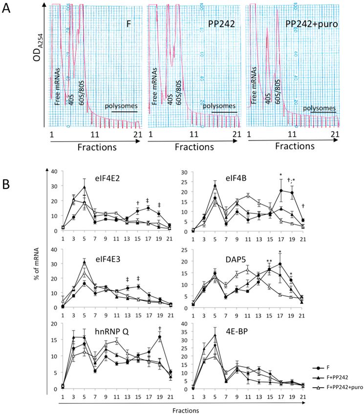
1 h post-fertilization. Upon PP242 treatment, the mRNAs for eIF4E2, eIF4E3, and hnRNP Q completely
shifted from heavy fractions (15–21) of the polysome gradient to the light fractions (Figure 3). The
polysomal distribution was similar in PP242-treated embryos and those treated with both PP242 and
puromycin, indicating that all mRNAs were released from polysomes upon mTOR inhibition. These
results suggested that entry into polysomes at fertilization was completely dependent on the mTOR
pathway for eIF4E2, eIF4E3, and hnRNP Q mRNAs. As expected [45], DAP5 mRNA recruitment was
similar in control and in PP242-treated embryos. Recruitment of eIF4B mRNA was only partially
inhibited by PP242 treatment (Figure 3). The puromycin treatment performed on PP242-treated
embryos shifted the remaining DAP5 and eIF4B mRNAs completely towards the middle of the
gradient, suggesting that the mRNAs remaining in polysome when mTOR is inhibited were still
actively translated.dependent on the mTOR pathway for eIF4E2, eIF4E3, and hnRNP Q mRNAs. As expected [45], DAP5
mRNA recruitment was similar in control and in PP242-treated embryos. Recruitment of eIF4B
mRNA was only partially inhibited by PP242 treatment (Figure 3). The puromycin treatment
performed on PP242-treated embryos shifted the remaining DAP5 and eIF4B mRNAs completely
Int.towards
J. Mol. Sci.the middle
2019, 20, 626 of the gradient, suggesting that the mRNAs remaining in polysome when7 mTOR
of 14
is inhibited were still actively translated.
Figure
Figure 3. (A)
3. (A) Polysome
Polysome profile
profile of fertilized
of fertilized embryos embryos
(F) with(F) with or PP242
or without without PP242and
inhibitor inhibitor and in
in presence
presence of(F+puro).
of puromycin puromycin (B)(F+puro).
mTOR impact(B) mTOR impact on
on polysomal polysomalofrecruitment
recruitment of mRNAs
mRNAs encoding encoding
translation
initiation factors
translation after fertilization.
initiation factors after Localization ofLocalization
fertilization. the targeted mRNAs along the
of the targeted gradient
mRNAs is monitored
along the gradient
as in
is Figure
monitored1. Samples were treated
as in Figure or notwere
1. Samples with treated
PP242 and puromycin
or not (n = 5;
with PP242 andF vs. F+PP242: (n
puromycin † p-value
= 5; F vs.
< 0.05, ‡ p-value
F+PP242: < 0.01;Int. J. Mol. Sci. 2019, 20, 626 8 of 14
inhibition, and both inhibitors were kept in contact with the embryos throughout the experiment.
Protein synthesis was monitored one hour post-fertilization by [35 S]-methionine incorporation. The
Int. J. Mol. Sci. 2019, 20, x 7 of 13
mTOR inhibitor PP242 inhibited strong global translation activity, while the MAPK pathway inhibitor
U0126We inhibited
next asked it moderately.
whether mTORDual treatment
inhibitionof embryos
would weakly
impact increased
4E-BP the inhibitory
translation. The 4E-BPeffect
proteinof
PP242 on protein synthesis (Figure 4A). The distribution on polysome gradients
remains present in PP242-treated embryos whereas it is degraded in control embryos following of eIF4B and DAP5
mRNAs,
fertilizationexhibiting a residual
[49]. 4E-BP mRNA translation
is presentinas
presence
a maternalof PP242,
mRNA was analyzed
(Table 1). Asin shown
three independent
in Figure 3,
experiments. In embryos exposed to U0126 only, no significant
analysis of the mRNA distribution on sucrose gradient showed that 4E-BP mRNA variation in polysomal recruitment
is mostly
after
associated with light fractions (3–5), showing no significant translation. The polysome profile eIF4B
fertilization was observed (Figure 4C left). When both PP242 and U0126 were present, of 4E-
mRNA
BP mRNAs was noinlonger recruited
presence to polysomes
of mTOR inhibitor(Figure
PP2424C right), suggesting
showed no increasethat
of athe
MAPKmRNAactivity
levelrelay
into
ispolysomal
involved in its translation.
fractions. In contrast,ruled
This observation DAP5outmRNA recruitment
a possible was independent
contribution of mTOR activity
of newly translated protein
and
to thewas4E-BP
still partially
pool, in recruited
addition when
to theembryos
already were treated with
demonstrated bothinhibition
PP242 inhibitors,ofsuggesting a MAPK
4E-BP degradation
relay and
rates [41,50].an additional regulatory mechanism, yet undetermined (Figure 4B right).
Figure4.4.(A)
Figure (A)mTOR
mTORinhibition
inhibitionand anddual
dualmTOR/MAPK
mTOR/MAPK inhibition impacts protein synthesis activity. activity.
Protein
Protein synthesis
synthesis activity
activity isis measured
measured by incorporationofof[35
byincorporation [35S]-methionine
S]-methionine in in TCA-precipitated
TCA-precipitated
proteins,
proteins,normalized
normalizedto tothe
thevalues
valuesin infertilized
fertilizedcontrol
controlembryos
embryos(representative
(representativeof oftwo
twoindependent
independent
experiments).
experiments).(B) (B)Polysome
Polysome profile of fertilized
profile embryos
of fertilized (F) with
embryos (F)orwith
without
or PP242
without andPP242
U0126and
inhibitors.
U0126
(C) Redistribution
inhibitors. of mRNAs for
(C) Redistribution eIF4B (top)
of mRNAs and DAP5
for eIF4B (top)(bottom)
and DAP5 on(bottom)
polysome ongradients,
polysomemonitored
gradients,
as in Figureas1,ininFigure
monitored presence
1, inofpresence
U0126 (left) or in(left)
of U0126 combination with PP242
or in combination (right).
with PP242Values
(right).are shown
Values are
as a mean of three biological replicates, error bars represent SEM (F+PP242 vs.
shown as a mean of three biological replicates, error bars represent SEM (F+PP242 vs. F+PP242+U0126: F+PP242+U0126:
**p-value
p-valueInt. J. Mol. Sci. 2019, 20, 626 9 of 14
Q were recruited following fertilization. Unfortunately, no cross-reacting antibodies were available
against the sea urchin proteins, therefore it was not possible to study the presence or accumulation
of the proteins for which we have shown a translational regulation. The stored maternal initiation
factors drive protein synthesis initiation for the first cell cycles; our results suggest that non-canonical
initiation factors that are translated after fertilization may have a specific role later in development.
eIF4E2 and eIF4E3 are implicated in selective translation [18–21] rather than in global translation,
which is driven by eIF4E1. During embryonic development in mice, Drosophila, and Caenorhabditis
elegans, eIF4E2 acts as a selective translational repressor, replacing eIF4E1 [18,52,53]. In metazoan
early development in general, and in sea urchin early development in particular, a redox gradient
is responsible of the oral–aboral axis specification of the embryo [54,55]. A spatial rearrangement of
mitochondria in the embryo confers an asymmetric distribution of oxygen availability and renders
some embryonic regions hypoxic. eIF4E2 is able to drive protein synthesis in hypoxic conditions [56,57],
when it associates to HIF2α and RBM4 to select the appropriate mRNAs to translate [19]. Although
hypoxia induces 4E-BP reappearance in sea urchins [58], eIF4E2 may still mediate translation because
it does not efficiently bind 4E-BP [15,48,59]. Furthermore, an RBM4 homolog mRNA is recruited into
polysomes to a high extent after fertilization [45]. In our experiments, we observed the increased
recruitment of eIF4E2 mRNA, suggesting a role of selective translation in sea urchin early development.
Therefore, taken together, we suggest that newly translated eIF4E2 could be part of a regionalized
non-canonical initiation complex and could drive spatial specific mRNA translation in hypoxic
embryonic cells.
The initiation factor eIF4E3 binds to the cap in an atypical manner and binds less efficiently to
eIF4G than eIF4E1 [15,21]. These features are responsible for the competition between eIF4E3 and
eIF4E1 for eIF4E1 target mRNAs [21]. MNKs, the eIF4E1 kinases [60] regulated by the MAPK pathway,
have a master role in the use of eIF4E1 or eIF4E3 to regulate mRNA recruitment in diffuse large B-cell
lymphoma [20]. Inhibition of MNKs activity leads to an increase of eIF4E3 mRNA translation and to the
preferential use of eIF4E3 as a cap-binding protein in these cells. In early sea urchin embryos, the MAPK
pathway is physiologically inactivated at fertilization and peaks transiently at M-phase [51]. eIF4E3 has
not previously been identified as a developmental eIF4E. Whether physiological MAPK inactivation is
directly involved in the recruitment of eIF4E3 mRNA observed post fertilization and whether newly
synthesized eIF4E3 is engaged into initiation complex formation in sea urchin development remains to
be investigated.
A recent report showed that DAP5, through its interaction with eIF3d, also being a cap-binding
protein [61], was able to drive the alternative cap-dependent translation of 20% of mammalian cell
mRNAs [26]. Moreover, DAP5 was shown to promote IRES-mediated translation during mitosis [25,62].
Since the translation of some mRNAs, including DAP5, is not impacted by the mTOR inhibitor
PP242, we suggest that DAP5 could participate in alternative cap-dependent and/or IRES-dependent
translations in sea urchins. IRES-containing mRNAs have yet to be identified in sea urchins. In
mammalian cells, hnRNP Q regulates the translation of several cellular IRES-containing mRNAs [30];
however, in sea urchins, its own translation is sensitive to the mTOR pathway. It would be interesting
to investigate the hnRNP Q protein level in sea urchin eggs and embryos.
Overall, our results demonstrate that the polysomal recruitment of initiation factor mRNAs differs
depending on their sensitivity to the mTOR pathway, ranging from completely dependent to completely
independent. Our study also demonstrates the conservation of the mTOR and MAPK pathway relays
in sea urchins [13,14] for the translation of specific mRNAs, namely eIF4B and DAP5 mRNAs.
In summary, the non-canonical initiation factors we identified as translationally regulated after
sea urchin fertilization are all global protein synthesis repressors, and enhancers of selective translation.
We suggest that these non-canonical eIFs could establish non-canonical translation initiation complexes
enabling their translation selectivity. An interesting perspective would be to investigate, in parallel, the
dynamics of the different initiation complexes and the maternal mRNA recruitment during the first
cell divisions of early development up to the maternal-to-zygotic transition. These results highlightInt. J. Mol. Sci. 2019, 20, 626 10 of 14
that the translation initiation process might be more complex than previously thought during the
development of the sea urchin embryo.
4. Materials and Methods
4.1. Handling and Treatment of Eggs and Embryos
Paracentrotus lividus sea urchins were collected in the bay of Crozon (Brittany, France) and
maintained in the CRBM facility of the Station Biologique de Roscoff. Gametes were obtained after
intracoelomic injection of 1 mL acetylcholine 0.1 M. Unfertilized eggs were dejellied and rinsed
before resuspension at 5% dilution in filtered sea water (FSW). Diluted sperm was added to the
unfertilized eggs. Experiments were only performed on batches of embryos exhibiting >90% of
fertilization rate. Embryos were collected for polysome analyses at 60 min post-fertilization. Inhibitors
were added to the eggs or embryos at the indicated time points: PP242 [10 µM] at 10 min before
fertilization; U0126 [60 µM], puromycin [0.6 mM], and emetine [0.1 mM] at 5 min, 40 min, and 55 min
post-fertilization respectively.
4.2. Polysome Gradients and RT-PCR Analysis
Polysome gradients and their analysis were performed as described in [47]. Briefly, 250 µL of
pelleted cells were lysed in a Dounce homogenizer with 1 mL polysome lysis buffer (10 mM Tris pH 7.4;
250 mM KCl; 10 mM MgCl2 ; 25 mM EGTA; 0.4% Igepal; 5% sucrose; 1 mM DTT; 10 µg/mL aprotinin;
2 µg/mL leupeptin; 100 µg/mL emetine; and 40 U RNase inhibitor). Lysates were clarified for 10 min
at 13,000 rpm in a tabletop centrifuge. Supernatants were fractionated on a linear 15–40% sucrose
gradient (10 mM Tris pH 7.4; 250 mM KCl; 10 mM MgCl2 ; 25 mM EGTA; and 1 mM DTT) for 2.5 h
at 38,000 rpm in a SW41Ti rotor at 4 ◦ C. Gradients were fractionated into 21 equal fractions. RNAs
were extracted from each fraction using acid phenol–chloroform (v/v), precipitated with 1 volume
isopropanol, washed with 70% ethanol, and resuspended in RNAse-free water. RNA quality was
checked on a 2% agarose/TBE gel electrophoresis. Specific mRNA distribution along the polysome
gradient was analyzed by semi-quantitative RT-PCR using an equal volume of RNA from each fraction
as described [47]. Reverse transcription (RT) was performed using a constant volume of RNAs from
each fraction with reverse transcriptase SuperScript II (Invitrogen, Courtaboeuf, France), following the
manufacturer’s instructions. The resulting cDNA was diluted in RNase-free water (1 vol. RT/300 vol.
H2 O) for the PCR reaction, so that the amplification with each primer pair (see list in Table 1) was in the
linear range for 30 cycles of amplification. Primers’ efficiency was determined using total RNA purified
from eggs (except for eIF4E1 done on blastulae RNA). PCR were carried out with the GoTaq Flexi kit
(Promega, Charbonnières-les-Bains, France) and [5 µM] primers, using the following thermal profile:
95 ◦ C for 2min; followed by 30 cycles of 3 steps: 95 ◦ C for 30 s, 60 ◦ C for 30 s, 72 ◦ C for 1 min; and finally,
72 ◦ C for 5min. For each mRNA tested, a RT-PCR performed without reverse transcriptase was done
in parallel to control for non-specific amplification. PCR products were analyzed on 2% agarose/TBE
gels electrophoresis, scanned on a Typhoon Trio (GE Healthcare Life Sciences, Velizy-Villacoublay,
France), and quantified with ImageJ software (NIH). Statistical analyses were done using a two-tailed
Student’s t test.
4.3. In Vivo Protein Synthesis Analysis
Embryos (5% suspension in seawater) were taken one hour after fertilization and incubated for
15 min in 10 µCi/mL [35 S]-L-methionine. [35 S]-L-methionine incorporation into proteins was measured
on duplicate aliquots after 10% TCA precipitation.
Author Contributions: Conceptualization: H.C., P.C. and J.M.; investigation, validation, and formal analysis:
H.C, S.B. and J.M.; writing: H.C., P.C. and J.M. All authors reviewed and approved the final draft.
Funding: This work was supported by research grants from the French Cancer League (La Ligue contre le Cancer,
comités Finistère, Côtes d’Armor, Morbihan, Deux-Sèvres et Charente), the Brittany Regional Council (RégionInt. J. Mol. Sci. 2019, 20, 626 11 of 14
Bretagne), and the Finistère Departmental Council (CG29). H.C. was supported by the Brittany Regional Council
(Région Bretagne) PhD fellowship.
Acknowledgments: We thank the Marine and Diving facility and the Roscoff Aquarium Service for collecting
and maintaining the sea urchins at the Roscoff Marine Station, respectively. We are grateful to the reviewers for
useful suggestions to improve the manuscript.
Conflicts of Interest: The authors declare no conflict of interest. The funders had no role in the design of the
study; in the collection, analyses, or interpretation of data; in the writing of the manuscript; or in the decision to
publish the results.
References
1. Sonenberg, N.; Hinnebusch, A.G. New modes of translational control in development, behavior, and disease.
Mol. Cell 2007, 28, 721–729. [CrossRef] [PubMed]
2. Schwanhäusser, B.; Busse, D.; Li, N.; Dittmar, G.; Schuchhardt, J.; Wolf, J.; Chen, W.; Selbach, M. Global
quantification of mammalian gene expression control. Nature 2011, 473, 337–342. [CrossRef] [PubMed]
3. Jackson, R.J.; Hellen, C.U.; Pestova, T.V. The mechanism of eukaryotic translation initiation and principles of
its regulation. Nat. Rev. Mol. Cell. Biol. 2010, 11, 113–127. [CrossRef] [PubMed]
4. Merrick, W.C. eIF4F: A retrospective. J. Biol. Chem. 2015, 290, 24091–24099. [CrossRef] [PubMed]
5. Rozen, F.; Edery, I.; Meerovitch, K.; Dever, T.E.; Merrick, W.C.; Sonenberg, N. Bidirectional RNA helicase
activity of eucaryotic translation initiation factors 4A and 4F. Mol. Cell. Biol. 1990, 10, 1134–1144. [CrossRef]
6. Kahvejian, A.; Roy, G.; Sonenberg, N. The mRNA closed-loop model: The function of PABP and
PABP-interacting proteins in mRNA translation. Cold Spring Harb. Symp. Quant. Biol. 2001, 66, 293–300.
[CrossRef]
7. Mader, S.; Lee, H.; Pause, A.; Sonenberg, N. The translation initiation factor eIF-4E binds to a common motif
shared by the translation factor eIF-4 gamma and the translational repressors 4E-binding proteins. Mol. Cell.
Biol. 1995, 15, 4990–4997. [CrossRef]
8. Bah, A.; Vernon, R.M.; Siddiqui, Z.; Krzeminski, M.; Muhandiram, R.; Zhao, C.; Sonenberg, N.; Kay, L.E.;
Forman-Kay, J.D. Folding of an intrinsically disordered protein by phosphorylation as a regulatory switch.
Nature 2015, 519, 106–109. [CrossRef]
9. Mamane, Y.; Petroulakis, E.; Martineau, Y.; Sato, T.A.; Larsson, O.; Rajasekhar, V.K.; Sonenberg, N. Epigenetic
Activation of a Subset of mRNAs by eIF4E Explains Its Effects on Cell Proliferation. PLoS ONE 2007, 2, e242.
[CrossRef]
10. Modelska, A.; Turro, E.; Russell, R.; Beaton, J.; Sbarrato, T.; Spriggs, K.; Miller, J.; Gräf, S.; Provenzano, E.;
Blows, F.; et al. The malignant phenotype in breast cancer is driven by eIF4A1-mediated changes in the
translational landscape. Cell Death Dis. 2015, 6, e1603. [CrossRef]
11. Leppek, K.; Das, R.; Barna, M. Functional 50 UTR mRNA structures in eukaryotic translation regulation and
how to find them. Nat. Rev. Mol. Cell Biol. 2018, 19, 158–174. [CrossRef] [PubMed]
12. Roux, P.P.; Topisirovic, I. Signaling pathways involved in the regulation of mRNA translation. Mol. Cell. Biol.
2018, 38, MCB.00070-18. [CrossRef]
13. Carracedo, A.; Ma, L.; Teruya-Feldstein, J.; Rojo, F.; Salmena, L.; Alimonti, A.; Egia, A.; Sasaki, A.T.;
Thomas, G.; Kozma, S.C.; et al. Inhibition of mTORC1 leads to MAPK pathway activation through a
PI3K-dependent feedback loop in human cancer. J. Clin. Investig. 2008, 118, 3065–3074. [CrossRef] [PubMed]
14. Mendoza, M.C.; Er, E.E.; Blenis, J. The Ras-ERK and PI3K-mTOR pathways: Cross-talk and compensation.
Trends Biochem. Sci. 2011, 36, 320–328. [CrossRef] [PubMed]
15. Joshi, B.; Cameron, A.; Jagus, R. Characterization of mammalian eIF4E-family members. Eur. J. Biochem.
2004, 271, 2189–2203. [CrossRef]
16. Joshi, B.; Lee, K.; Maeder, D.L.; Jagus, R. Phylogenetic analysis of eIF4E-family members. BMC Evol. Biol.
2005, 5, 48. [CrossRef]
17. Morales, J.; Mulner-Lorillon, O.; Cosson, B.; Morin, E.; Bellé, R.; Bradham, C.A.; Beane, W.S.; Cormier, P.
Translational control genes in the sea urchin genome. Dev. Biol. 2006, 300, 293–307. [CrossRef] [PubMed]
18. Morita, M.; Ler, L.W.; Fabian, M.R.; Siddiqui, N.; Mullin, M.; Henderson, V.C.; Alain, T.; Fonseca, B.D.;
Karashchuk, G.; Bennett, C.F.; et al. A novel 4EHP-GIGYF2 translational repressor complex is essential for
mammalian development. Mol. Cell. Biol. 2012, 32, 3585–3593. [CrossRef] [PubMed]Int. J. Mol. Sci. 2019, 20, 626 12 of 14
19. Uniacke, J.; Holterman, C.E.; Lachance, G.; Franovic, A.; Jacob, M.D.; Fabian, M.R.; Payette, J.; Holcik, M.;
Pause, A.; Lee, S. An oxygen-regulated switch in the protein synthesis machinery. Nature 2012, 486, 126–129.
[CrossRef] [PubMed]
20. Landon, A.L.; Muniandy, P.A.; Shetty, A.C.; Lehrmann, E.; Volpon, L.; Houng, S.; Zhang, Y.; Dai, B.;
Peroutka, R.; Mazan-Mamczarz, K.; et al. MNKs act as a regulatory switch for eIF4E1 and eIF4E3 driven
mRNA translation in DLBCL. Nat. Commun. 2014, 5, 5413. [CrossRef] [PubMed]
21. Osborne, M.J.; Volpon, L.; Kornblatt, J.A.; Culjkovic-Kraljacic, B.; Baguet, A.; Borden, K.L.B. eIF4E3 acts as a
tumor suppressor by utilizing an atypical mode of methyl-7-guanosine cap recognition. Proc. Natl. Acad.
Sci. USA 2013, 110, 3877–3882. [CrossRef] [PubMed]
22. Gebauer, F. Versatility of the translational machinery during stress: Changing partners to keep dancing.
Cell Res. 2012, 22, 1634–1636. [CrossRef] [PubMed]
23. Henis-Korenblit, S.; Strumpf, N.L.; Goldstaub, D.; Kimchi, A. A novel form of DAP5 protein accumulates in
apoptotic cells as a result of caspase cleavage and internal ribosome entry site-mediated translation. Mol.
Cell. Biol. 2000, 20, 496–506. [CrossRef] [PubMed]
24. Liberman, N.; Gandin, V.; Svitkin, Y.V.; David, M.; Virgili, G.; Jaramillo, M.; Holcik, M.; Nagar, B.; Kimchi, A.;
Sonenberg, N. DAP5 associates with eIF2 and eIF4AI to promote Internal Ribosome Entry Site driven
translation. Nucl. Acids Res. 2015, 43, 3764–3775. [CrossRef] [PubMed]
25. Marash, L.; Liberman, N.; Henis-Korenblit, S.; Sivan, G.; Reem, E.; Elroy-Stein, O.; Kimchi, A. DAP5 promotes
cap-independent translation of Bcl-2 and CDK1 to facilitate cell survival during mitosis. Mol. Cell 2008, 30,
447–459. [CrossRef] [PubMed]
26. de la Parra, C.; Ernlund, A.; Alard, A.; Ruggles, K.; Ueberheide, B.; Schneider, R.J. A widespread alternate
form of cap-dependent mRNA translation initiation. Nat. Commun. 2018, 9, 3068. [CrossRef] [PubMed]
27. Wigington, C.P.; Williams, K.R.; Meers, M.P.; Bassell, G.J.; Corbett, A.H. Poly(A) RNA-binding proteins
and polyadenosine RNA: New members and novel functions. Wiley Interdiscip. Rev. RNA 2014, 5, 601–622.
[CrossRef]
28. Svitkin, Y.V.; Yanagiya, A.; Karetnikov, A.E.; Alain, T.; Fabian, M.R.; Khoutorsky, A.; Perreault, S.;
Topisirovic, I.; Sonenberg, N. Control of translation and miRNA-dependent repression by a novel poly(A)
binding protein, hnRNP-Q. PLoS Biol. 2013, 11, e1001564. [CrossRef]
29. Lee, K.-H.; Woo, K.-C.; Kim, D.-Y.; Kim, T.-D.; Shin, J.; Park, S.M.; Jang, S.K.; Kim, K.-T. Rhythmic interaction
between Period1 mRNA and hnRNP Q leads to circadian time-dependent translation. Mol. Cell. Biol. 2012,
32, 717–728. [CrossRef]
30. Kim, D.-Y.; Kim, W.; Lee, K.-H.; Kim, S.-H.; Lee, H.-R.; Kim, H.-J.; Jung, Y.; Choi, J.-H.; Kim, K.-T. hnRNP Q
regulates translation of p53 in normal and stress conditions. Cell Death Differ. 2012, 20, 226–234. [CrossRef]
31. Epel, D. Protein synthesis in sea urchin eggs: A “late” response to fertilization. Proc. Natl. Acad. Sci. USA
1967, 57, 899–906. [CrossRef] [PubMed]
32. Wagenaar, E.B. The timing of synthesis of proteins required for mitosis in the cell cycle of the sea urchin
embryo. Exp. Cell Res. 1983, 144, 393–403. [CrossRef]
33. Sodergren, E.; Weinstock, G.M.; Davidson, E.H.; Cameron, R.A.; Gibbs, R.A.; Angerer, R.C.; Angerer, L.M.;
Arnone, M.I.; Burgess, D.R.; Burke, R.D.; et al. The genome of the sea urchin Strongylocentrotus purpuratus.
Science 2006, 314, 941–952. [CrossRef] [PubMed]
34. Malkin, L.I.; Gross, P.R.; Romanoff, P. Polyribosomal protein synthesis in fertilized sea urchin eggs: The
effect of Actinomycin treatment. Dev. Biol. 1964, 10, 378–394. [CrossRef]
35. Davidson, E.H. The nature and function of maternal transcripts. In Gene Activity in Early Development;
Academic Press: Cambridge, MA, USA, 1986; pp. 46–125.
36. Winkler, M.M.; Nelson, E.M.; Lashbrook, C.; Hershey, J.W. Multiple levels of regulation of protein synthesis
at fertilization in sea urchin eggs. Dev. Biol. 1985, 107, 290–300. [CrossRef]
37. Jagus, R.; Huang, W.I.; Hansen, L.J.; Wilson, M.A. Changes in rates of protein synthesis and eukaryotic
initiation factor-4 inhibitory activity in cell-free translation systems of sea urchin eggs and early cleavage
stage embryos. J. Biol. Chem. 1992, 267, 15530–15536. [PubMed]
38. Lopo, A.; Lashbrook, C.; Hershey, J. Characterization of translation systems in vitro from three developmental
stages of Strongylocentrotus purpuratus. Biochem. J. 1989, 258, 553–561. [CrossRef]
39. Huang, W.I.; Hansen, L.J.; Merrick, W.C.; Jagus, R. Inhibitor of eukaryotic initiation factor 4F activity in
unfertilized sea urchin eggs. Proc. Natl. Acad. Sci. USA 1987, 84, 6359–6363. [CrossRef]Int. J. Mol. Sci. 2019, 20, 626 13 of 14
40. Cormier, P.; Pyronnet, S.; Morales, J.; Mulner-Lorillon, O.; Sonenberg, N.; Belle, R. eIF4E association with
4E-BP decreases rapidly following fertilization in sea urchin. Dev. Biol. 2001, 232, 275–283. [CrossRef]
41. Salaun, P.; Pyronnet, S.; Morales, J.; Mulner-Lorillon, O.; Bellé, R.; Sonenberg, N.; Cormier, P. eIF4E/4E-BP
dissociation and 4E-BP degradation in the first mitotic division of the sea urchin embryo. Dev. Biol. 2003,
255, 428–439. [CrossRef]
42. Oulhen, N.; Boulben, S.; Bidinosti, M.; Morales, J.; Cormier, P.; Cosson, B. A variant mimicking
hyperphosphorylated 4E-BP inhibits protein synthesis in a sea urchin cell-free, cap-dependent translation
system. PLoS ONE 2009, 4, e5070. [CrossRef]
43. Oulhen, N.; Salaun, P.; Cosson, B.; Cormier, P.; Morales, J. After fertilization of sea urchin eggs, eIF4G
is post-translationally modified and associated with the cap-binding protein eIF4E. J. Cell Sci. 2007, 120,
425–434. [CrossRef]
44. Cormier, P.; Chassé, H.; Cosson, B.; Mulner-Lorillon, O.; Morales, J. Translational control in echinoderms:
The calm before the storm. In Evolution of the Protein Synthesis Machinery and Its Regulation;
Hernández, G., Jagus, R., Eds.; Springer International Publishing: Cham, Switzerland, 2016; pp. 413–432.
ISBN 978-3-319-39468-8.
45. Chassé, H.; Aubert, J.; Boulben, S.; Le Corguillé, G.; Corre, E.; Cormier, P.; Morales, J. Translatome analysis at
the egg-to-embryo transition in sea urchin. Nucl. Acids Res. 2018, 46, 4607–4621. [CrossRef] [PubMed]
46. Hoang, B.; Benavides, A.; Shi, Y.; Yang, Y.; Frost, P.; Gera, J.; Lichtenstein, A. The PP242 mammalian target
of rapamycin (mTOR) inhibitor activates extracellular signal-regulated kinase (ERK) in multiple myeloma
cells via a target of rapamycin complex 1 (TORC1)/eukaryotic translation initiation factor 4E (eIF-4E)/RAF
pathway. J. Biol. Chem. 2012, 287, 21796–21805. [CrossRef]
47. Chassé, H.; Boulben, S.; Costache, V.; Cormier, P.; Morales, J. Analysis of translation using polysome profiling.
Nucl. Acids Res. 2017, 45, e15. [CrossRef] [PubMed]
48. Robalino, J.; Joshi, B.; Fahrenkrug, S.C.; Jagus, R. Two zebrafish eIF4E family members are differentially
expressed and functionally divergent. J. Biol. Chem. 2004, 279, 10532–10541. [CrossRef] [PubMed]
49. Chassé, H.; Mulner-Lorillon, O.; Boulben, S.; Glippa, V.; Morales, J.; Cormier, P. Cyclin B translation depends
on mTOR activity after fertilization in sea urchin embryos. PLoS ONE 2016, 11, e0150318. [CrossRef]
50. Laurent, S.; Richard, A.; Mulner-Lorillon, O.; Morales, J.; Flament, D.; Glippa, V.; Bourdon, J.; Gosselin, P.;
Siegel, A.; Cormier, P.; et al. Modelization of the regulation of protein synthesis following fertilization
in sea urchin shows requirement of two processes: A destabilization of eIF4E:4E-BP complex and a great
stimulation of the 4E-BP-degradation mechanism, both rapamycin-sensitive. Front. Genet. 2014, 5. [CrossRef]
51. Mulner-Lorillon, O.; Chassé, H.; Morales, J.; Bellé, R.; Cormier, P. MAPK/ERK activity is required for the
successful progression of mitosis in sea urchin embryos. Dev. Biol. 2017, 421, 194–203. [CrossRef]
52. Dinkova, T.D.; Keiper, B.D.; Korneeva, N.L.; Aamodt, E.J.; Rhoads, R.E. Translation of a Small Subset of
Caenorhabditis elegans mRNAs Is Dependent on a Specific Eukaryotic Translation Initiation Factor 4E
Isoform. Mol. Cell. Biol. 2005, 25, 100–113. [CrossRef]
53. Cho, P.F.; Gamberi, C.; Cho-Park, Y.A.; Cho-Park, I.B.; Lasko, P.; Sonenberg, N. Cap-dependent translational
inhibition establishes two opposing morphogen gradients in Drosophila embryos. Curr. Biol. 2006, 16,
2035–2041. [CrossRef] [PubMed]
54. Coffman, J.A.; McCarthy, J.J.; Dickey-Sims, C.; Robertson, A.J. Oral-aboral axis specification in the sea
urchin embryo: II. Mitochondrial distribution and redox state contribute to establishing polarity in
Strongylocentrotus purpuratus. Dev. Biol. 2004, 273, 160–171. [CrossRef] [PubMed]
55. Coffman, J.A.; Denegre, J.M. Mitochondria, redox signaling and axis specification in metazoan embryos.
Dev. Biol. 2007, 308, 266–280. [CrossRef] [PubMed]
56. Ho, J.J.D.; Wang, M.; Audas, T.E.; Kwon, D.; Carlsson, S.K.; Timpano, S.; Evagelou, S.L.; Brothers, S.;
Gonzalgo, M.L.; Krieger, J.R.; et al. Systemic reprogramming of translation efficiencies on oxygen stimulus.
Cell Rep. 2016, 14, 1293–1300. [CrossRef] [PubMed]
57. Kelly, N.J.; Varga, J.F.A.; Specker, E.J.; Romeo, C.M.; Coomber, B.L.; Uniacke, J. Hypoxia activates cadherin-22
synthesis via eIF4E2 to drive cancer cell migration, invasion and adhesion. Oncogene 2018, 37, 651–662.
[CrossRef] [PubMed]
58. Le Bouffant, R.; Cormier, P.; Mulner-lorillon, O.; Belle, R. Hypoxia and DNA-damaging agent bleomycin
both increase the cellular level of the protein 4E-BP. J. Cell. Biochem. 2006, 99, 126–132. [CrossRef] [PubMed]Int. J. Mol. Sci. 2019, 20, 626 14 of 14
59. Rom, E.; Kim, H.C.; Gingras, A.C.; Marcotrigiano, J.; Favre, D.; Olsen, H.; Burley, S.K.; Sonenberg, N. Cloning
and characterization of 4EHP, a novel mammalian eIF4E-related cap-binding protein. J. Biol. Chem. 1998, 273,
13104–13109. [CrossRef]
60. Pyronnet, S.; Imataka, H.; Gingras, A.C.; Fukunaga, R.; Hunter, T.; Sonenberg, N. Human eukaryotic
translation initiation factor 4G (eIF4G) recruits MNK1 to phosphorylate eIF4E. EMBO J. 1999, 18, 270–279.
[CrossRef]
61. Lee, A.S.Y.; Kranzusch, P.J.; Doudna, J.A.; Cate, J.H.D. eIF3d is an mRNA cap-binding protein required for
specialized translation initiation. Nature 2016, 536, 96–99. [CrossRef]
62. Liberman, N.; Marash, L.; Kimchi, A. The translation initiation factor DAP5 is a regulator of cell survival
during mitosis. Cell Cycle 2009, 8, 204–209. [CrossRef]
© 2019 by the authors. Licensee MDPI, Basel, Switzerland. This article is an open access
article distributed under the terms and conditions of the Creative Commons Attribution
(CC BY) license (http://creativecommons.org/licenses/by/4.0/).You can also read


