Astrocyte-Derived TGF-b1 Accelerates Disease Progression in ALS Mice by Interfering with the Neuroprotective Functions of Microglia and T Cells
←
→
Page content transcription
If your browser does not render page correctly, please read the page content below
Article

Astrocyte-Derived TGF-b1 Accelerates Disease
Progression in ALS Mice by Interfering with the
Neuroprotective Functions of Microglia and T Cells
Graphical Abstract                                             Authors
                                                               Fumito Endo, Okiru Komine, ...,
                                                               Tony Wyss-Coray, Koji Yamanaka

                                                               Correspondence
                                                               kojiyama@riem.nagoya-u.ac.jp

                                                               In Brief
                                                               Endo et al. show that TGF-b1,
                                                               upregulated in astrocytes of ALS patients
                                                               and mice, negatively regulates
                                                               neuroprotective inflammatory responses.
                                                               Astrocyte-derived TGF-b1 accelerates
                                                               disease progression in ALS mice, which is
                                                               ameliorated by a TGF-b signaling
                                                               inhibitor. These findings indicate that
                                                               targeting glial TGF-b signaling may
                                                               represent a therapeutic approach for
                                                               ALS.

Highlights
d   Astrocyte-derived TGF-b1 accelerates disease progression
    in ALS mice

d   TGF-b1 inhibits neuroprotective inflammatory response by
    microglia and T cells

d   TGF-b signaling inhibitor slows disease progression and
    extends survival of ALS mice

d   Cell-type-specific dysregulation of TGF-b signaling is a
    therapeutic target for ALS

            Endo et al., 2015, Cell Reports 11, 592–604
            April 28, 2015 ª2015 The Authors
            http://dx.doi.org/10.1016/j.celrep.2015.03.053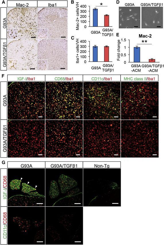
Cell Reports

                                                                                                                 Article

Astrocyte-Derived TGF-b1 Accelerates Disease
Progression in ALS Mice by Interfering with the
Neuroprotective Functions of Microglia and T Cells
Fumito Endo,1,2,3 Okiru Komine,1 Noriko Fujimori-Tonou,2 Masahisa Katsuno,4 Shijie Jin,1 Seiji Watanabe,1 Gen Sobue,4,5
Mari Dezawa,3 Tony Wyss-Coray,6 and Koji Yamanaka1,2,5,*
1Department of Neuroscience and Pathobiology, Research Institute of Environmental Medicine, Nagoya University, Nagoya, Aichi 4648601,

Japan
2Laboratory for Motor Neuron Disease, RIKEN Brain Science Institute, Wako, Saitama 3510198, Japan
3Department of Stem Cell Biology and Histology, Tohoku University Graduate School of Medicine, Sendai, Miyagi 9808575, Japan
4Department of Neurology, Nagoya University Graduate School of Medicine, Nagoya, Aichi 4668550, Japan
5CREST, Japan Science and Technology Agency, Saitama 3320012, Japan
6Department of Neurology and Neurological Sciences, Stanford University School of Medicine, Stanford, CA 94305, USA

*Correspondence: kojiyama@riem.nagoya-u.ac.jp
 http://dx.doi.org/10.1016/j.celrep.2015.03.053
This is an open access article under the CC BY-NC-ND license (http://creativecommons.org/licenses/by-nc-nd/4.0/).

SUMMARY                                                               with ALS-linked mutations recapitulate both the clinical and
                                                                      pathological characteristics of ALS.
Neuroinflammation, which includes both neuropro-                         Neuroinflammation, characterized by activated astrocytes,
tective and neurotoxic reactions by activated glial                   microglia, infiltrated T cells, and the subsequent overproduc-
cells and infiltrated immune cells, is involved in the                tion of proinflammatory cytokines and other neurotoxic or pro-
pathomechanism of amyotrophic lateral sclerosis                       tective molecules, is a pathological hallmark not only of mutant
(ALS). However, the cytokines that regulate the                       SOD1 mice but also of ALS patients (Engelhardt et al., 1993).
                                                                      Along with other researchers, we have demonstrated previously
neuroprotective inflammatory response in ALS are
                                                                      that the selective reduction of mutant SOD1 expression in
not clear. Here, we identify transforming growth fac-
                                                                      microglia (Beers et al., 2006; Boillée et al., 2006; Wang et al.,
tor-b1 (TGF-b1), which is upregulated in astrocytes                   2009) or astrocytes (Wang et al., 2011; Yamanaka et al.,
of murine and human ALS, as a negative regulator                      2008) significantly extends the survival time of ALS mice. More-
of neuroprotective inflammatory response. We                          over, elimination of functional T cells from mutant SOD1 mice
demonstrate that astrocyte-specific overproduction                    shortens the survival time (Beers et al., 2008; Chiu et al.,
of TGF-b1 in SOD1G93A mice accelerates disease                        2008). Thus, non-neuronal cells, such as astrocytes, microglia,
progression in a non-cell-autonomous manner, with                     and T cells, are able to modify the course of disease through a
reduced IGF-I production in deactivated microglia                     non-cell-autonomous mechanism within the CNS (Ilieva et al.,
and fewer T cells with an IFN-g-dominant milieu.                      2009; Lasiene and Yamanaka, 2011). On the other hand, the
Moreover, expression levels of endogenous TGF-b1                      targeting of single proinflammatory cytokines has been tested
                                                                      as a treatment for ALS mice; however, the effectiveness of
in SOD1G93A mice negatively correlate with lifespan.
                                                                      this strategy remains inconclusive. For example, some studies
Furthermore, pharmacological administration of a
                                                                      showed that deletion of TNF-a or IL-1b has marginal effects
TGF-b signaling inhibitor after disease onset extends                 on the survival time of mutant SOD1 mice, while one study
survival time of SOD1G93A mice. These findings                        showed extended survival of IL-1b-deficient SOD1G93A mice
indicate that astrocytic TGF-b1 determines disease                    (Gowing et al., 2006; Meissner et al., 2010; Nguyen et al.,
progression and is critical to the pathomechanism                     2001). In contrast, overexpression of neurotrophic factors,
of ALS.                                                               such as IGF-I and GDNF, extends the survival time of mutant
                                                                      SOD1 mice (Dodge et al., 2008; Henderson et al., 1994; Kaspar
                                                                      et al., 2003; Wang et al., 2002). Targeting a single proinflamma-
INTRODUCTION                                                          tory cytokine may not be sufficient to modify disease course,
                                                                      but instead inducing a neuroprotective environment composed
Amyotrophic lateral sclerosis (ALS) is an adult-onset, fatal neuro-   of glial cells and T cells may be more effective. However, it
degenerative disease characterized by the selective loss of           has not been elucidated what regulates the neuroprotective
motor neurons. While most cases of ALS are of sporadic etiol-         glia/immune response.
ogy, 10% of cases are familial ALS among which dominant                  Transforming growth factor bs (TGF-bs) are pleiotropic cyto-
mutations in the gene for Cu/Zn superoxide dismutase (SOD1)           kines that have key roles in immune homeostasis (Li et al.,
are the frequent causes. Mice overexpressing the SOD1 gene            2006), neurotrophic response (Katsuno et al., 2011), and

592 Cell Reports 11, 592–604, April 28, 2015 ª2015 The Authors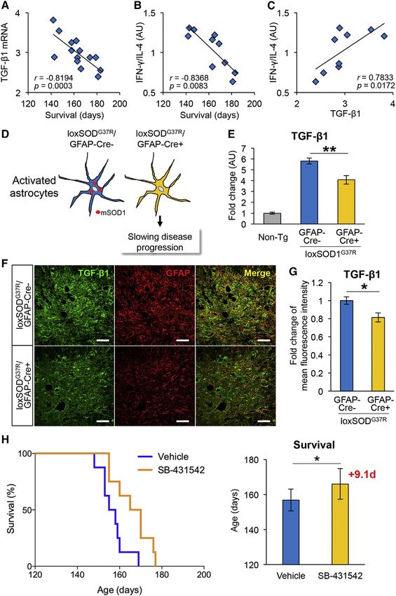
microglial development (Butovsky et al., 2014). In mouse astrocytes during disease progression of ALS mice and sporadic
models of Alzheimer’s disease (AD) in which the amyloid pre- ALS patients.
cursor gene is overexpressed, increasing astrocytic TGF-b1
levels or blockade of TGF-b signaling in peripheral macro- Astrocyte-Specific Overproduction of TGF-b1
phages is shown to reduce senile plaque formation in the brain Accelerates Disease Progression in SOD1G93A Mice
parenchyma (Wyss-Coray et al., 1995, 2001; Town et al., We next crossed SOD1G93A mice with mice modestly expressing
2008). In ALS patients, TGF-b1 protein levels are elevated in the bioactive form of porcine TGF-b1 under the control of a glial
the serum, plasma, and cerebrospinal fluid (CSF) (Houi et al., fibrillary acidic protein (GFAP) promoter (GFAP-TGF-b1 mice)
2002; I1zecka et al., 2002), and recent studies indicate that (Wyss-Coray et al., 1995) and monitored body weight, rotarod
TGF-b signaling is implicated in ALS (Iida et al., 2011; Katsuno performance, and survival time. Astrocytic TGF-b1 expression
et al., 2011; Phatnani et al., 2013). However, the exact roles of in GFAP-TGF-b1 mice was confirmed by immunoblots, and total
TGF-b1 in the pathomechanism of ALS have not been TGF-b1 protein level was elevated in end-stage SOD1G93A/TGF-
elucidated. b1 mice compared with SOD1G93A mice (Figures 2A and 2B).
In this study, we found TGF-b1 upregulation in the spinal Although the time of disease onset remained unchanged in
cord astrocytes of sporadic ALS patients and ALS mice. By SOD1G93A/TGF-b1 double-transgenic mice (Figure 2C), the
using SOD1G93A mice with overexpression of TGF-b1 in astro- mean survival time was unexpectedly shortened by about
cytes and SOD1G37R mice with astrocyte-specific deletion of 10 days (SOD1G93A/TGF-b1: 151.7 ± 10.4 days; SOD1G93A:
mutant gene, we identified astrocytic TGF-b1 negatively regu- 161.9 ± 9.5 days) (Figure 2D). SOD1G93A/TGF-b1 mice showed
lates neuroprotective inflammatory responses through micro- accelerated disease progression in the late stage (Figure 2E) of
glia and T cells and accelerates disease progression of ALS ALS, with faster weight loss (Figure 2F) and a decreased perfor-
mice. Moreover, pharmacological administration of TGF-b mance on the rotarod task (Figure 2G). On the other hand, het-
signaling inhibitor after disease onset extends the survival of erozygous GFAP-TGF-b1 mice showed neither motor deficit
ALS mice. On the other hand, a defect in canonical TGF-b nor weight loss with a normal lifespan (Figures 2D, 2F, and
signaling in motor neurons was unaffected by exogenous 2G). Additionally, analysis of motor axons in cross-sections of
TGF-b1. These findings indicate that cell-type-specific dysre- L5 ventral root revealed a trend toward more progressive axonal
gulation of TGF-b signaling is critical to the pathomechanism degeneration in SOD1G93A/TGF-b1 mice compared to SOD1G93A
underlying ALS. mice, while L5 motor axons in GFAP-TGF-b1 were not affected
(Figures 2H and 2I). Together, these results suggest that astro-
cyte-specific overproduction of TGF-b1 plays a detrimental
RESULTS role in the disease progression of SOD1G93A mice.
TGF-b1 Is Upregulated in the Spinal Cord Astrocytes of Dysregulated TGF-b Signaling in Spinal Motor Neurons
SOD1G93A Mice and Sporadic ALS Patients of SOD1G93A Mice
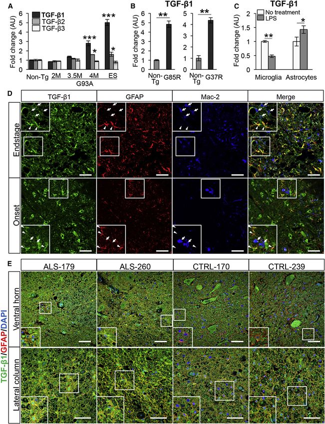
To explore the role of TGF-b1 in ALS pathogenesis, we examined To determine whether TGF-b signaling is altered during disease
expression levels of TGF-b1 mRNA in the spinal cord of mutant progression of SOD1G93A and SOD1G93A/TGF-b1 mice, we ex-
SOD1 mice. We found that, among its different isoforms, TGF- amined Smad2, which is phosphorylated following stimulation
b1 mRNA level was highly upregulated during disease progres- of TGF-b signaling and transported to the nucleus (Shi and Mas-
sion of SOD1G93A mice (Figure 1A) as well as the mice expressing sagué, 2003). High levels of phosphorylated Smad2 (pSmad2)
other mutant SOD1 genes, SOD1G85R and SOD1G37R (Figure 1B). were observed in motor neuron nuclei of non-transgenic mice;
Immunoblotting also confirmed that TGF-b1 protein level was however, pSmad2 levels in SOD1G93A mice were significantly
upregulated during disease progression of SOD1G93A mice (Fig- decreased from pre-symptomatic stage and rarely observed in
ure 2B). To further examine the cell types that produce TGF-b1 end stage (Figures S1A and S1B). Moreover, the loss of pSmad2
under an acute inflammatory condition, we examined expression in motor neuron nuclei was not recovered by overexpression of
levels of TGF-b1 in SOD1G93A-bearing primary astrocytes and TGF-b1 in SOD1G93A/TGF-b1 mice (Figures S1A and S1B).
microglia induced by lipopolysaccharide (LPS) stimulation, since Considering that the expressions of TGF-b receptor type 2
these glial cells were implicated to produce TGF-b1 under (TGFbR2) and type 1 (TGFbR1) and the level of cytoplasmic
various neurological diseases (Flanders et al., 1998). We found pSmad2 in the lumbar motor neurons of SOD1G93A mice were
a relatively elevated level of TGF-b1 mRNA in SOD1G93A-primary preserved (Figures S1A and S1C), nuclear transport of pSmad2
astrocytes, but reduced TGF-b1 expression in SOD1G93A-pri- is likely to be defective in spinal motor neurons of SOD1G93A
mary microglia upon LPS stimulation (Figure 1C). Immunofluo- mice. In contrast, glial nuclear pSmad2 expressions were pre-
rescence analysis revealed that TGF-b1 protein expressions in served in end-stage SOD1G93A and SOD1G93A/TGF-b1 mice
astrocytes were elevated at end stage compared with onset (Figures S2A and S2B).
and that they were markedly observed compared with ones in
microglia of end-stage SOD1G93A mice (Figure 1D). More impor- Astrocyte-Specific Overproduction of TGF-b1 Reduces
tantly, astrocytic TGF-b1 expression also was elevated in the the Levels of T Cell-Related Cytokines and Neurotrophic
spinal ventral horn and lateral column of sporadic ALS patients Factors in SOD1G93A Mice
compared with control (Figure 1E). These results indicate that To identify the molecules linked to the rapid disease progres-
expression levels of endogenous TGF-b1 are upregulated in sion in SOD1G93A/TGF-b1 mice, we performed qRT-PCR
Cell Reports 11, 592–604, April 28, 2015 ª2015 The Authors 593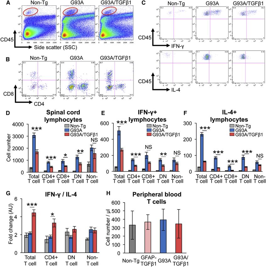
Figure 1. TGF-b1 Is Upregulated in Astrocytes in the Spinal Cords of Mutant SOD1 Mice and Sporadic ALS Patients
(A) Mean mRNA levels of TGF-b isoforms in the lumbar spinal cord of SOD1G93A (2M, 2-month-old; 3.5M, 3.5-month-old; 4M, 4-month-old; and ES, end-stage)
mice relative to the ones in 6-month-old non-transgenic (Non-Tg) mice are shown.
(B) Mean TGF-b1 mRNA levels in the lumbar spinal cord of end-stage SOD1G85R (G85R) and SOD1G37R (G37R) mice relative to the ones in age-matched Non-Tg
mice are shown.
(C) Mean mRNA levels of TGF-b1 in SOD1G93A-primary microglia and astrocytes with or without LPS treatment (1 mg/ml) are shown. (A–C) *p < 0.05, **p < 0.01,
***p < 0.001. Error bars denote SEM (n = 3 in A and C and n = 3–5 in B).

                                                                                                                         (legend continued on next page)

594 Cell Reports 11, 592–604, April 28, 2015 ª2015 The Authors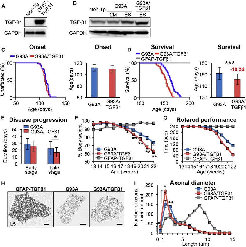
analyses of the lumbar spinal cord of end-stage SOD1G93A and                       lower CD68 mRNA levels observed in SOD1G93A/TGF-b1 spinal
SOD1G93A/TGF-b1 mice. We observed significantly decreased                          cords (Figure 3E).
mRNA levels of T cell-related cytokines, IFN-g, and IL-4, in                          Moreover, to test whether TGF-b1 directly reduces microglial
end-stage SOD1G93A/TGF-b1 mice compared with SOD1G93A                              activation, morphology and Mac-2 mRNA levels were evaluated
mice (Figures 3A and 3B). Further, mRNA levels of the T helper                     in SOD1G93A-primary microglia cultured in astrocyte-condi-
2 (Th2) cell-specific transcription factor GATA-3 tended to be                     tioned media (ACM) from SOD1G93A or SOD1G93A/TGF-b1
reduced in SOD1G93A/TGF-b1 mice, while ones of the Th1 tran-                       mice. We found that the sizes of SOD1G93A-primary microglia
scription factor T-bet were unaffected (Table S2). The mRNA                        cultured with ACM from SOD1G93A/TGF-b1 mice were smaller
levels of the neurotrophic factors IGF-I and GDNF were                             than those cultured with ACM from SOD1G93A mice (Figure 4D).
decreased significantly (Figures 3C and 3D) in SOD1G93A/                           Mac-2 mRNA levels were noticeably reduced in SOD1G93A-pri-
TGF-b1 mice. We also observed lower mRNA levels of micro-                          mary microglia with ACM from SOD1G93A/TGF-b1 mice (Fig-
glia activation marker CD68 in SOD1G93A/TGF-b1 mice                                ure 4E). These findings indicate that TGF-b1 directly inhibits
(Figure 3E), while the levels of Iba-1, classically activated (M1)                 microglial activation. Furthermore, flow cytometric analysis of
microglia markers (iNOS, CD86, TNF-a, and IL-1b), and alterna-                     spinal microglia from end-stage SOD1G93A and SOD1G93A/
tively activated (M2) microglia markers (Arginase1 and CD206)                      TGF-b1 mice revealed no differences in cell number, cellular
were unaffected with TGF-b1 overproduction (Figures 3F and                         size, granularity (Figures S4A and S4B), surface expression of
3G; Table S2). Overexpression of TGF-b1 itself reduced expres-                     CD86 and CD206, and intracellular expression of TNF-a (Fig-
sion levels of IFN-g, IL-4, and GDNF in GFAP-TGF-b1 mice                           ure S4C) between the two genotypes, indicating no significant
compared to non-transgenic mice (Figures 3A, 3B, and 3D),                          changes in M1 (CD86 and TNF-a) or M2 markers (CD206) in
while the levels of others were unaffected. Intriguingly, expres-                  SOD1G93A microglia by the in vivo expression of TGF-b1.
sion levels of astrocytic marker GFAP measured by qRT-PCR                             Having observed a decrease in the mRNA levels of CD68
(Figure S3A) and immunohistochemistry (Figure S3B) were un-                        and IGF-I in the lumbar spinal cord of SOD1G93A/TGF-b1
changed during the disease progression between SOD1G93A                            mice (Figures 3C and 3E), we next evaluated their expression
and SOD1G93A/TGF-b1. With regard to astrocytic functions,                          levels using immunofluorescence. We found that the expres-
mRNA levels of S100b, glutamate transporter (GLT-1), and neu-                      sion levels of IGF-I, CD68, CD11c, a dendritic cell marker,
rocan were not significantly affected (Table S2). The levels of                    and major histocompatibility complex (MHC) class II were
other glia- or immune-related molecules determined by qRT-                         significantly decreased in microglia of the ventral horn of
PCR analyses are summarized in Table S2. In summary,                               SOD1G93A/TGF-b1 mice compared with SOD1G93A mice (Fig-
T cell-related cytokines and neurotrophic factors were signifi-                    ure 4F). Moreover, expression of IGF-I in macrophages of the
cantly affected by astrocyte-specific overproduction of TGF-                       lumbar ventral root, which was markedly elevated in SOD1G93A
b1 in SOD1G93A mice, while the molecules linked to astrocytic                      mice, was substantially decreased in SOD1G93A/TGF-b1 mice
function were relatively unaffected. Therefore, our next focus                     (Figure 4G). Expression levels of both CD11c and CD68 in
was on the roles of TGF-b1 in microglia and lymphocytes of                         macrophages of the ventral root were also decreased in
SOD1G93A/TGF-b1 mice.                                                              SOD1G93A/TGF-b1 mice compared with SOD1G93A mice (Fig-
                                                                                   ure 4G). In the intact ventral root of non-transgenic mice,
Astrocyte-Specific Overproduction of TGF-b1 Inhibits                               IGF-I- and CD68-positive macrophages were both not detect-
Microglial Activation and Reduces the Neuroprotective                              able (Figure 4G). These findings indicate that astrocyte-spe-
Properties of Microglia/Macrophages of SOD1G93A Mice                               cific overproduction of TGF-b1 inhibits activation and neuro-
We first investigated the effect of astrocyte-secreted TGF-b1 on                   protective properties of microglia/macrophages in the lumbar
microglial activation in the lumbar spinal cord by immunohisto-                    spinal cord.
chemistry. We observed that the expression of Mac-2, a marker
for microglial activation, was markedly decreased in the lumbar                    Astrocyte-Specific Overproduction of TGF-b1 Induces
ventral horn of SOD1G93A/TGF-b1 compared with SOD1G93A                             an IFN-g-Dominant Milieu with Reduced Number of
(Figure 4A, left) mice. Additionally, the number of Mac-2-positive                 Infiltrated T Cells in SOD1G93A Mice
cells in the ventral horn was significantly decreased in                           Our observation of decreased mRNA levels for IFN-g and IL-4 in
SOD1G93A/TGF-b1 mice (Figure 4B), although there were no dif-                      the lumbar spinal cord of SOD1G93A/TGF-b1 mice (Figures 3A
ferences in expression of the microglial marker Iba-1 (Figure 4A,                  and 3B) led us to evaluate infiltrated CD45hi mononuclear cells
right) and the number of Iba-1-positive cells (Figure 4C) between                  in the spinal cord by flow cytometry. We found that total
the two genotypes. These data were in accordance with the                          numbers of CD4+, CD8+, and CD4 CD8 double-negative

(D) Representative images show the ventral horn of the lumbar spinal cord of onset and end-stage SOD1G93A mice stained for TGF-b1 (green), GFAP (red), and
Mac-2 (blue), along with the merged image. Magnified images of the outlined areas (white square) also are shown. TGF-b1 expression was elevated in astrocytes
(arrowheads) compared with microglia (arrows). Scale bars, 50 mm.
(E) Representative images show the ventral horn and the lateral column of the cervical spinal cord in sporadic ALS and control patients stained for TGF-b1 (green),
GFAP (red), and DAPI (blue). Note that the signals double-positive for GFAP and TGF-b1 were more prominent in ALS spinal cords than controls. Magnified
images of the outlined areas (white square) also are shown. Scale bars, 50 mm.
See also Table S1.

                                                                                    Cell Reports 11, 592–604, April 28, 2015 ª2015 The Authors 595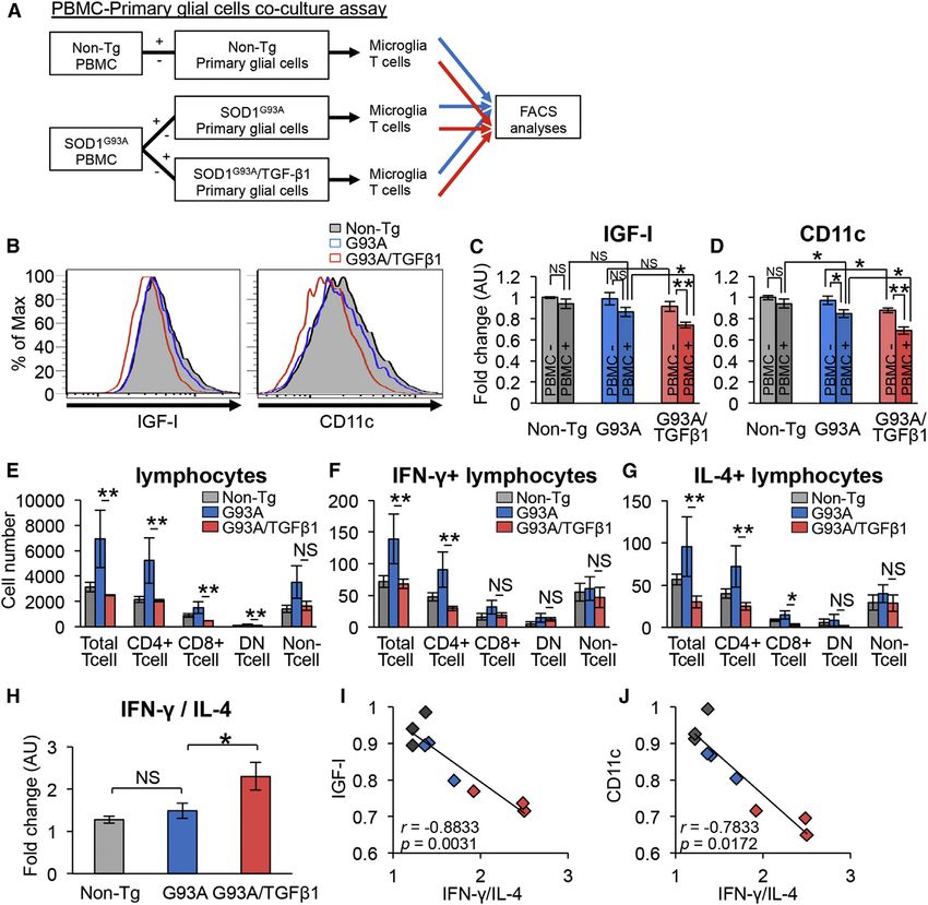
Figure 2. Astrocyte-Specific Overproduction of TGF-b1 Accelerates Disease Progression in SOD1G93A Mice
(A and B) Immunoblot analysis for TGF-b1 in (A) primary astrocytes isolated from Non-Tg and GFAP-TGF-b1 mice and in (B) the lumbar spinal cords of Non-Tg,
2-month-old (2M), and end-stage (ES) SOD1G93A (G93A) and SOD1G93A/TGF-b1 (G93A/TGF-b1) mice. GAPDH was used as the internal loading control.
(C and D) Kaplan-Meier curves for (C) onset and plotted mean onset and (D) survival time and plotted mean survival time for G93A (n = 36), G93A/TGF-b1 (n = 33),
and GFAP-TGF-b1 (n = 5) mice are shown.
(E) Plotted durations of early (from onset to 10% weight loss) and late (from 10% weight loss to end stage) stages for G93A and G93A/TGF-b1 mice are shown.
(F) Weekly mean body weight was plotted for GFAP-TGF-b1 (n = 5), G93A (n = 36), and G93A/TGF-b1 (n = 33) mice. (C–F) *p < 0.05, **p < 0.01, ***p < 0.001.
Mean ± SD were plotted.
(G) Rotarod performances were evaluated weekly for GFAP-TGF-b1 (n = 5), G93A (n = 19), and G93A/TGF-b1 (n = 13) mice. Mean holding times on the rotating rod
at indicated ages were plotted.
(H) Images show toluidine blue-stained axial sections of the L5 ventral root of GFAP-TGF-b1 mice and end-stage G93A and G93A/TGF-b1 mice. Scale bars,
50 mm.
(I) The distribution of motor axonal diameters in cross-sections of L5 ventral root of GFAP-TGF-b1 (n = 2), G93A (n = 6), and G93A/TGF-b1 (n = 6) mice were
measured and plotted. *p < 0.05, **p < 0.01. Error bars denote SEM.
See also Figures S1 and S2.

(DN) T cells were decreased in SOD1G93A/TGF-b1 mice                              the two genotypes (Figures 5A, 5B, and 5D). We also evaluated
compared to SOD1G93A mice, while no significant difference                       intracellular expression of IFN-g and IL-4 in lymphocytes, and
was observed in the numbers of CD45hiCD3 cells between                           found that the numbers of IFN-g/IL-4 producing total, CD4+,

596 Cell Reports 11, 592–604, April 28, 2015 ª2015 The Authors
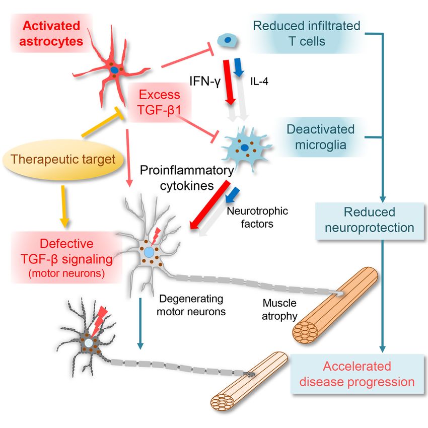
Figure 3. Quantitative RT-PCR Analyses of Glia/Immune System-Related Molecules in the Lumbar Spinal Cord of SOD1G93A and SOD1G93A/
TGF-b1 Mice
(A–I) Mean mRNA levels of indicated glia/immune system-related molecules in the lumbar spinal cord of end-stage SOD1G93A (G93A) and SOD1G93A/TGF-b1
(G93A/TGF-b1) mice along with GFAP-TGF-b1 mice relative to the ones from Non-Tg littermates were plotted. Each result was normalized to b-actin. *p < 0.05,
**p < 0.01, ***p < 0.001. Error bars denote SEM (n = 3–5).
See also Table S2 and Figure S3.

and DN T cells were significantly decreased in SOD1G93A/TGF-                   and decreased number of infiltrated T cells with higher ratios
b1 mice (Figures 5C, 5E, and 5F). Moreover, the ratio of IFN-g/                of IFN-g/IL-4-producing T cells in SOD1G93A/TGF-b1 mice,
IL-4 in total and CD4+ T cells was significantly increased in                  we performed primary glial and peripheral blood mononuclear
SOD1G93A/TGF-b1 mice (Figure 5G), indicating that overpro-                     cell (PBMC) co-culture assays using primary glial cells
duction of TGF-b1 induces an IFN-g-dominant environment                        obtained from non-transgenic, SOD1G93A, and SOD1G93A/
rather than IL-4. On the other hand, there were no significant                 TGF-b1 newborn mice and PBMCs from non-transgenic
differences in the numbers of T cells in the peripheral blood                  and end-stage SOD1G93A mice (Figure 6A). In this assay, we
among all genotypes (Figure 5H). These findings indicate that                  examined intracellular expression of IGF-I and surface ex-
astrocyte-specific overproduction of TGF-b1 reduces the num-                   pressions of CD11c in microglia as well as intracellular expres-
ber of infiltrated T cells and induces an IFN-g-dominant milieu in             sions of both IFN-g and IL-4 in T cells using flow cytometric
the spinal cord of SOD1G93A mice.                                              analysis. We found that microglial IGF-I expression was
                                                                               significantly decreased in the SOD1G93A/TGF-b1-primary
Astrocyte-Derived TGF-b1 Reduces Neuroprotective                               glial cells when cultured with SOD1G93A-PBMC (Figures 6B
Microglial IGF-I Expression and Dendritic Cell-like                            and 6C), while IGF-I levels were unaffected without PBMC
Activation via a Dominant IFN-g-Producing T Cell                               (Figure 6C). Additionally, TGF-b1 itself marginally affected
Environment                                                                    expression levels of IGF-I in microglia (Figures 6C and S5A).
To uncover the mechanism through which astrocyte-derived                       The numbers of T cells as well as IFN-g/IL-4-producing
TGF-b1 results in a decreased IGF-I expression in microglia                    T cells were significantly reduced when co-cultured with

                                                                                Cell Reports 11, 592–604, April 28, 2015 ª2015 The Authors 597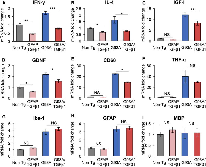
Figure 4. Decreased IGF-I and CD11c Ex-
                                                                                         pressions of Deactivated Microglia/Macro-
                                                                                         phages of SOD1G93A/TGF-b1 Mice
                                                                                         (A) Images show the lumbar spinal cord of end-
                                                                                         stage SOD1G93A (G93A) and SOD1G93A/TGF-b1
                                                                                         (G93A/TGF-b1) mice stained for Mac-2 (left) and
                                                                                         Iba-1 (right). Scale bars, 250 mm.
                                                                                         (B and C) Numbers of (B) Mac-2- and (C) Iba-1-
                                                                                         positive microglia within the ventral horn area from
                                                                                         the lumbar spinal cord of end-stage G93A (blue)
                                                                                         and G93A/TGF-b1 (red) mice are shown. *p < 0.05.
                                                                                         Mean ± SEM were plotted (n = 3).
                                                                                         (D) Images show SOD1G93A primary microglia
                                                                                         cultured with ACM derived from G93A or G93A/
                                                                                         TGF-b1 mice. Scale bars, 50 mm.
                                                                                         (E) Mac-2 mRNA levels of SOD1G93A primary mi-
                                                                                         croglia cultured in the indicated conditions are
                                                                                         shown. **p < 0.01. Mean ± SEM were plotted
                                                                                         (n = 4).
                                                                                         (F) Images show the ventral horn of the lumbar
                                                                                         spinal cord of G93A (top) and G93A/TGF-b1
                                                                                         (bottom) mice stained for IGF-I, CD68, CD11c, and
                                                                                         MHC class II (green) together with Iba-1 (red).
                                                                                         Scale bars, 100 mm.
                                                                                         (G) Images show the lumbar ventral root of end-
                                                                                         stage G93A (left), G93A/TGF-b1 (middle), and
                                                                                         Non-Tg (right) mice stained for IGF-I or CD11c
                                                                                         (green) and CD68 (red). CD68-positive cells in the
                                                                                         ventral root show IGF-I expression (arrowheads).
                                                                                         Scale bars, 100 mm.
                                                                                         See also Figure S4.

                                                                                        Higher Level of TGF-b1 Is a
                                                                                        Negative Prognostic Factor by
                                                                                        Determining the IFN-g/IL-4 Ratio in
                                                                                        SOD1G93A Mice
                                                                                        We examined the mean mRNA levels
                                                                                        determined by qRT-PCR in the lumbar
                                                                                        spinal cord to evaluate the correlation
                                                                                        among survival time, IFN-g/IL-4 ratio,
                                                                                        and endogenous TGF-b1 levels in
                                                                                        SOD1G93A mice. We found that the sur-
                                                                                        vival time of SOD1G93A mice negatively
                                                                                        correlated not only with TGF-b1 mRNA
                                                                                        level (Figure 7A), but also with the
                                                                                        ratio of IFN-g/IL-4 mRNA (Figure 7B).
                                                                                        Moreover, TGF-b1 levels significantly
                                                                                        correlated with the ratio of IFN-g/IL-4
SOD1G93A/TGF-b1-primary glial cells (Figures 6E–6G). The ra-       (Figure 7C). In summary, these findings suggest that TGF-b1
tio of IFN-g/IL-4-producing T cells was significantly increased    is a negative prognostic factor in ALS mice likely through its
in the presence of SOD1G93A/TGF-b1-primary glial cells (Fig-       determination of IFN-g/IL-4 ratio.
ure 6H). Furthermore, microglial IGF-I expression was nega-
tively correlated with the ratio of IFN-g/IL-4-producing cells     Astrocytic TGF-b1 Is Downregulated in Slowly
(Figure 6I), and expression of IGF-I in BV-2 microglia was         Progressive SOD1G37R Mice with Astrocyte-Specific
altered by the IFN-g/IL-4 ratio (Figure S5B). Lastly, microglial   Deletion of Mutant SOD1
CD11c expression behaved like IGF-I (Figures 6B, 6D, and           We previously demonstrated that the selective reduction of
6J). These results suggest that astrocyte-derived TGF-b1 re-       mutant SOD1 in astrocytes significantly slowed the disease pro-
duces the neuroprotective properties of microglia, such as         gression of loxSOD1G37R mice using Cre-loxP system (Figure 7D;
IGF-I expression and dendritic cell-like phenotype, alongside      Yamanaka et al., 2008). However, astrocyte-derived molecules
an IFN-g-dominant milieu of T cells.                               that are linked to neurotoxicity have not been fully elucidated.

598 Cell Reports 11, 592–604, April 28, 2015 ª2015 The Authors
Figure 5. Decreased Number of Infiltrated T Cells and Increased Ratio of IFN-g/IL-4-Producing T Cells in SOD1G93A/TGF-b1 Mice
Flow cytometric analysis of spinal cord lymphocytes from non-transgenic (Non-Tg), end-stage SOD1G93A (G93A), and SOD1G93A/TGF-b1 (G93A/TGF-b1) mice.
(A–C) Flow cytometric analyses of (A) CD45hi cells (cells in red-circled areas), (B) CD4/CD8 expression for T cells, and (C) cytokine staining of IL-4 or IFN-g in
T cells are shown.
(D) Numbers of each subset of lymphocytes in the spinal cord from each mouse line are shown.
(E and F) Numbers of each subset of (E) IFN-g- or (F) IL-4-producing lymphocytes in the spinal cord from each mouse line are shown.
(G) Mean ratios of IFN-g/IL-4-producing cells are shown.
(H) Cell numbers per microliter of CD3+ T cells in the peripheral blood of Non-Tg, GFAP-TGF-b1, end-stage G93A, and G93A/TGF-b1 mice are shown. (D–H)
*p < 0.05, **p < 0.01, ***p < 0.001. NS, not significant. Mean ± SEM were plotted (n = 6–7 in D–G and n = 8 in H).
We found that TGF-b1 mRNA level was significantly reduced in Pharmacological Inhibition of TGF-b Signaling Slows
loxSOD1G37R/GFAP-Cre+ mice compared with loxSOD1G37R/ Disease Progression and Extends Survival of SOD1G93A
GFAP-Cre mice (Figure 7E). Furthermore, immunofluorescence Mice
analysis showed that TGF-b1 as well as GFAP expressions were To explore the therapeutic potential of modifying glial TGF-b
reduced in the spinal cord astrocytes of loxSOD1G37R/GFAP- signaling and to validate an adverse role of excess TGF-b in
Cre+ mice compared with loxSOD1G37R/GFAP-Cre mice (Fig- neuroinflammation, we administered TGF-b signaling inhibitor
ures 7F and 7G). In summary, astrocytic TGF-b1 is one of the SB-431542 intraperitoneally to symptomatic SOD1G93A mice.
key factors induced by mutant SOD1 that is linked to accelerated SB-431542 slowed disease progression and extended survival
disease progression in ALS mice. time of SOD1G93A mice (Figure 7H). We also examined the
Cell Reports 11, 592–604, April 28, 2015 ª2015 The Authors 599Figure 6. Correlation between Decreased Microglial IGF-I/CD11c Expressions and Decreased Number of T Cells with Increased IFN-g/IL-4 Ratio in the Presence of TGF-b1 Overproduction in Astrocytes Flow cytometric analyses of primary microglia from non-transgenic (Non-Tg), SOD1G93A (G93A), and SOD1G93A/TGF-b1 (G93A/TGF-b1) mice and T cells in PBMC from Non-Tg and end-stage G93A mice in the primary glial and PBMC co-culture assay. (A) Experimental design for the primary glial and PBMC co-culture assay and flow cytometry is shown. (B) Flow cytometric analysis shows IGF-I and CD11c for primary microglia cultured with PBMC (black, Non-Tg; blue, G93A; red, G93A/TGF-b1). (C and D) Plotted relative expression levels of (C) IGF-I and (D) CD11c in primary microglia. Each datum was normalized to that of Non-Tg primary microglia cultured without PBMC. (E) Plotted numbers of each subset of T cells and non-T cells in PBMC from Non-Tg, G93A mice cultured with primary glial cells from each mouse line are shown. (F and G) Plotted numbers of each subset of (F) IFN-g- and (G) IL-4-producing lymphocyte in PBMC from each mouse line are shown. (H) Plotted mean ratios of IFN-g/IL-4-producing T cells in PBMC from each mouse line are shown. (C–H) *p < 0.05, **p < 0.01. NS, not significant. Error bars denote SEM (n = 3). (I and J) Plotted Spearman correlations between expression level of (I) IGF-I and (J) CD11c in primary microglia and the ratio of IFN-g/IL-4-producing T cells in PBMC (n = 9, r = 0.8833, and p = 0.0031 in I and n = 9, r = 0.7833, and p = 0.0172 in J) are shown. See also Figure S5. effect of SB-431542 on the very late symptomatic SOD1G93A DISCUSSION mice (140 days old), revealing a trend toward extension of their survival times (Figure S6). These results indicate that inhibiting In this study, we have demonstrated that astrocyte-specific TGF-b signaling after disease onset ameliorates disease in ALS overproduction of TGF-b1 in mutant SOD1 mice results in accel- mice. erated disease progression in a non-cell-autonomous manner, 600 Cell Reports 11, 592–604, April 28, 2015 ª2015 The Authors
Figure 7. Astrocytic TGF-b1 Is a Determinant
                                                                                         for Disease Progression and Survival for ALS
                                                                                         Mice, and TGF-b Signaling Inhibitor Extends
                                                                                         the Survival Time of ALS Mice
                                                                                         (A–C) Correlations between survival time, IFN-g/IL-
                                                                                         4 ratio, and TGF-b1 level in SOD1G93A mice. (A)
                                                                                         Plotted Spearman correlations (A) between TGF-b1
                                                                                         mRNA and the survival time of SOD1G93A mice (n =
                                                                                         14, r = 0.8194, and p = 0.0003), (B) between ratio
                                                                                         of IFN-g/IL-4 mRNA and the survival time (n = 10, r =
                                                                                           0.8368, and p = 0.0083), and (C) between TGF-b1
                                                                                         mRNA and ratio of IFN-g/IL-4 mRNA (n = 10, r =
                                                                                         0.7833, and p = 0.0172) are shown.
                                                                                         (D–G) Astrocytic TGF-b1 is downregulated in lox-
                                                                                         SOD1G37R mice with astrocyte-specific deletion of
                                                                                         mutant SOD1. (D) A schematic drawing shows
                                                                                         astrocyte-specific deletion of mutant SOD1 in lox-
                                                                                         SOD1G37R mice using Cre-loxP system, which
                                                                                         leads to slowing the disease progression.
                                                                                         (E) TGF-b1 mRNA levels in the lumbar spinal
                                                                                         cord of end-stage loxSOD1G37R/GFAP-Cre– and
                                                                                         loxSOD1G37R/GFAP-Cre+ mice relative to age-
                                                                                         matched Non-Tg mice are shown. **p < 0.01. Mean
                                                                                         ± SEM were plotted (n = 6).
                                                                                         (F) Representative images show the ventral horn of
                                                                                         the lumbar spinal cord of end-stage loxSOD1G37R/
                                                                                         GFAP-Cre– and loxSOD1G37R/GFAP-Cre+ mice
                                                                                         stained for TGF-b1 (green) and GFAP (red), along
                                                                                         with the merged image. Scale bars, 50 mm.
                                                                                         (G) Mean fluorescent intensities of TGF-b1 in the
                                                                                         gray matter of lumbar ventral horn of loxSOD1G37R/
                                                                                         GFAP-Cre+ mice relative to loxSOD1G37R/GFAP-
                                                                                         Cre– mice. *p < 0.05. Error bars denote SEM (n = 3).
                                                                                         (H) Peripheral administration of TGF-b inhibitor
                                                                                         extended the survival time of SOD1G93A mice.
                                                                                         SOD1G93A mice were intraperitoneally injected with
                                                                                         SB-431542 (10 mg/kg, n = 8) or vehicle (DMSO/
                                                                                         PBS, n = 8) five times per week from 16 weeks old
                                                                                         (after disease onset). Kaplan-Meier analysis for the
                                                                                         survival time and plotted mean survival showed that
                                                                                         SB-431542 extended the survival of SOD1G93A
                                                                                         mice (SB-431542: 166.0 ± 8.7 days, vehicle: 156.9
                                                                                         ± 6.3 days, log-rank test: p = 0.0168). *p < 0.05.
                                                                                         Error bars denote SD (n = 8, four males and four
                                                                                         females in each group).
                                                                                         See also Figure S6.

                                                                                         implicated in the pathogenesis of ALS
                                                                                         (Iida et al., 2011; Phatnani et al., 2013),
                                                                                         the detailed mechanisms are yet to be
with reduced neurotrophic factor production in deactivated mi-     elucidated (Katsuno et al., 2011). We demonstrated that
croglia/macrophages and an IFN-g-dominant environment of in-       decreased expression of pSmad2 in motor neuron nuclei
filtrated T cells. Moreover, the lower level of TGF-b1 was         occurred at the pre-symptomatic stage and was exacerbated
achieved by astrocyte-specific deletion of mutant SOD1 from        during disease progression in ALS mice. Moreover, exogenous
ALS mice, which slows disease progression. Furthermore, phar-      expression of TGF-b1 in SOD1G93A mice did not improve
macological inhibition of TGF-b signaling in symptomatic ALS       pSmad2 level within motor neuron nuclei, the motor function,
mice extended survival time. These results provide compelling      or disease course of mice. In spinobulbar muscular atrophy
evidence that astrocytic TGF-b1 inhibits the neuroprotective in-   (SBMA), an inherited motor neuron disease caused by mutant
flammatory responses coordinated by microglia/macrophages          androgen receptor, we previously reported a defect of TGF-b
and T cells.                                                       signaling in motor neurons (Katsuno et al., 2010). Intriguingly,
    Our study demonstrated elevated TGF-b1 levels in astrocytes    while downregulation of TGFbR2 was observed in SBMA motor
of ALS patients and mice. Although TGF-b signaling has been        neurons, a defect in nuclear transport of pSmad2 was observed

                                                                    Cell Reports 11, 592–604, April 28, 2015 ª2015 The Authors 601
in SOD1-ALS motor neurons rather than dysregulation of TGF-b the survival of motor and cortical neurons (Ozdinler and Macklis, receptors. In addition, dysregulated pSmad2/3 expression has 2006; Ueno et al., 2013). Moreover, IGF-I administration pro- been observed in motor neuron nuclei of patients with sporadic longs the survival time of SOD1G93A mice (Dodge et al., 2008; ALS (Nakamura et al., 2008). Our results along with these reports Kaspar et al., 2003). Thus, in the current study, the marked indicate that the dysfunction of TGF-b signaling, especially de- reduction of IGF-I in the lumbar spinal cord of SOD1G93A/TGF- fects downstream of the TGF-b receptor in motor neurons, is b1 mice might have contributed to an accelerated disease pro- involved in neurodegeneration in both familial and sporadic ALS. gression. Although TGF-b1 itself slightly reduces expression Previous studies have shown that elimination of functional levels of IGF-I, we observed that IFN-g, and not TGF-b1, showed T cells in SOD1G93A mice shortens their survival time (Beers a strong antagonizing effect on the expression of IGF-I in micro- et al., 2008; Chiu et al., 2008). However, what regulates the neu- glia induced by IL-4 in vitro. Furthermore, IGF-I expression in roprotective immune response in ALS mice remains unclear. In microglia was regulated by the IFN-g/IL-4 balance in vitro. We this study, we found that TGF-b1, known to inhibit T cell prolifer- found that the level of GDNF, a potent survival factor for motor ation and differentiation (Li et al., 2006), regulates the number neurons that prolongs the survival of SOD1G93A mice (Henderson and IFN-g/IL-4 balance in T cells both in vivo and in vitro and et al., 1994; Kaspar et al., 2003; Wang et al., 2002), also was that microglia-related molecules were misregulated partly reduced in SOD1G93A/TGF-b1 mice, suggesting that the through altered IFN-g/IL-4 balance. This finding suggests that neurodegenerative mechanism, similar to IGF-I reduction, TGF-b1 is likely to be one of the regulators responsible for con- might involve a low level of GDNF in SOD1G93A/TGF-b1 mice. trolling neuroprotective immune responses. Together, decreased levels of these neurotrophic factors On the other hand, TGF-b1 has been reported to deactivate through enhanced expression level of TGF-b1 seem to have microglia (Lodge and Sriram, 1996; Suzumura et al., 1993) important roles in the accelerated disease progression in and to regulate antigen-presentation function of microglia SOD1G93A/TGF-b1 mice. in vitro (Abutbul et al., 2012; Suzumura et al., 1993). The influ- We demonstrated that expression levels of endogenous TGF- ence of TGF-b1 on microglia, however, is unclear in the context b1 mRNA at the end stage negatively correlates with the survival of neurodegeneration (Flanders et al., 1998). Our results show time of SOD1G93A mice and positively correlates with the IFN-g/ that astrocyte-specific overproduction of TGF-b1 deactivates IL-4 ratio. These findings suggest a functional relationship be- microglia/macrophages with reduced expression of Mac-2, tween astrocytes producing TGF-b1 and T cells producing CD68, CD11c, MHC class II, and IGF-I both in vivo and IFN-g/IL-4 in the disease progression, not only in SOD1G93A/ in vitro. Of note, T cell activation requires expression of MHC TGF-b1 mice but also in SOD1G93A mice. Moreover, the negative class II in antigen-presenting cells, including microglia and correlation between TGF-b1 level and survival time of ALS mice macrophages. Furthermore, IGF-I+ CD11c+ microglia have is consistent with our observation that astrocyte-specific dele- been reported to exert beneficial effects over neurodegenera- tion of mutant SOD1 extended survival time with a lower level tion (Butovsky et al., 2006; Chiu et al., 2008). Thus, the current of astrocytic TGF-b1. Our results indicate that astrocytic TGF- study suggests that astrocytic TGF-b1 inhibits the neuropro- b1 is a determinant of disease progression in ALS mice, and tective properties of microglia not only indirectly by regulating TGF-b1 shall be evaluated as a candidate biomarker to predict the number and balance of IFN-g/IL-4 in T cells, but also disease progression of ALS. directly through the deactivation of microglial functions, Finally, pharmacological administration of TGF-b signaling in- including antigen presentation. Previous studies have estab- hibitor SB-431542 after disease onset extended the survival time lished that infiltration and activation of a large number of mac- of SOD1G93A mice. Though peripherally administered, SB- rophages occurs in the peripheral nerves of SOD1G93A mice 431542 was presumably effective in the diseased spinal cord, (Chiu et al., 2009; Kano et al., 2012). In the current study, since the blood-spinal cord barrier was damaged in the symp- reduced expression of IGF-I, CD11c, and CD68 in macro- tomatic mutant SOD1 mice (Zhong et al., 2008). In addition, an phages was observed in the lumbar ventral root of SOD1G93A/ adverse effect on motor neurons by inhibiting TGF-b signaling TGF-b1 mice, indicating that the TGF-b1-induced deactivation is likely to be minimal, since TGF-b signaling in motor neurons of macrophages in the ventral root also contributes to acceler- is already defective at the late symptomatic stage. The effect ated disease progression. of a TGF-b1 inhibitor on extension of the survival time might be In addition, TGF-b1 is critical to the development of micro- more robust if TGF-b signaling in motor neurons would be glia (Butovsky et al., 2014). Although nuclear pSmad2 was simultaneously protected. Nevertheless, our data validated an preserved in both microglia and astrocytes of SOD1G93A adverse role of excess glial TGF-b1 in neuroinflammation and mice, expressions of microglia-related molecules such as uncovered the therapeutic potential of modifying glial TGF-b CD68 were significantly reduced compared with those related signaling in ALS. to astrocytes in SOD1G93A/TGF-b1 mice. These results impli- In conclusion, our study provides evidence that astrocytic cate that TGF-b1 exhibits more robust effects on microglia TGF-b1 plays a key role in the neuroprotective inflammatory than on astrocytes, likely because expressions of TGF-b re- response in ALS mice by regulating microglial activation, T cell ceptors in microglia are highly dependent on TGF-b1 (Butov- number, and IFN-g/IL-4 balance. Our findings suggest that tar- sky et al., 2014). geting TGF-b signaling in a cell-type-specific manner, such as IGF-I has been found to exhibit neuroprotective properties in restoration of TGF-b signaling in motor neurons and suppressing motor neurons. For example, IGF-I enhances axonal outgrowth excess TGF-b1 in astrocytes, may represent a therapeutic of motor neurons, and microglia-derived IGF-I is required for approach for the treatment of motor neuron diseases. 602 Cell Reports 11, 592–604, April 28, 2015 ª2015 The Authors
EXPERIMENTAL PROCEDURES                                                                Administration of TGF-b Signaling Inhibitor In Vivo
                                                                                       SOD1G93A mice were intraperitoneally injected with TGF-b signaling inhibitor
Detailed information is available in the Supplemental Experimental                     SB-431542 (Sigma, 5 or 10 mg/kg) or vehicle (DMSO/PBS) five times per
Procedures.                                                                            week either at 16 weeks old (early symptomatic phase) or 20 weeks old (late
                                                                                       symptomatic phase).

Postmortem Human Tissues
                                                                                       Statistical Analysis
Specimens of spinal cords from two patients with sporadic ALS and two
                                                                                       Statistical analyses of survival time and disease duration were performed with
other neurological disease patients as controls were obtained by autopsy
                                                                                       a log-rank test and unpaired t test, respectively. Statistical analysis of the fold
with informed consent (Table S1). The diagnosis of ALS was confirmed by
                                                                                       change of relative mRNA levels determined by qRT-PCR was performed with a
El Escorial diagnostic criteria as defined by the World Federation of
                                                                                       one-way ANOVA followed by Tukey’s test or paired or unpaired t test. All an-
Neurology. The collection of tissues and their use in this study were
                                                                                       alyses were carried out using GraphPad Prism.
approved by the ethics committee of Nagoya University. For immunofluo-
rescence analysis, sections were prepared from formalin-fixed and
                                                                                       SUPPLEMENTAL INFORMATION
paraffin-embedded tissues, deparaffinized, and boiled for 30 min in
50 mM citrate buffer (pH 6.0).
                                                                                       Supplemental Information includes Supplemental Experimental Procedures,
                                                                                       six figures, and two tables and can be found with this article online at http://
Mice                                                                                   dx.doi.org/10.1016/j.celrep.2015.03.053.
Transgenic mice expressing familial ALS-linked SOD1 mutations, SOD1G93A
(B6.Cg-Tg (SOD1*G93A)1Gur/J, Jackson Laboratory), LoxSOD1G37R (Boillée                AUTHOR CONTRIBUTIONS
et al., 2006), and SOD1G85R (Bruijn et al., 1997), were described previously.
GFAP-TGF-b1 mice (a low-expressing line [line T64] on a C57BL/6 genetic                F.E., O.K., and K.Y. designed the experiments and analyzed the data. F.E. per-
background) (Wyss-Coray et al., 1995) and GFAP-Cre mice (Bajenaru et al.,              formed the research with support from O.K., N.F-T., S.J., and S.W. M.K., G.S.,
2002) were described previously. To generate mice heterozygous for both                M.D., and T.W.-C. contributed critical analytic tools and input. F.E. and K.Y.
SOD1G93A and GFAP-TGF-b1 transgenes, SOD1G93A males were bred with                     wrote the paper.
GFAP-TGF-b1 females. To generate loxSOD1G37R mice with astrocyte-spe-
cific deletion of mutant SOD1, loxSOD1G37R males were bred with GFAP-                  ACKNOWLEDGMENTS
Cre females. The animal study was approved by the Animal Care and Use
Committee of Nagoya University and RIKEN.                                              The authors thank the Support Unit for Animal Resource Development and
                                                                                       Biomaterial Analysis in RIKEN BSI Research Resources Center for supporting
                                                                                       animal experiments and fluorescence-activated cell sorting (FACS) analysis,
Analysis of Disease Progression, Survival, and Motor Function of
                                                                                       and BSI-Olympus Collaboration Center for supporting morphological ana-
Mice
                                                                                       lyses. This work was supported by Grants-in-Aid for Scientific Research on
SOD1G93A mice always were compared with their SOD1G93A/TGF-b1 litter-
                                                                                       Innovative Areas (23111006) and Scientific Research (B) (26293208) from the
mates. Disease onset was determined as the time when mice reached
                                                                                       Ministry of Education, Culture, Sports, Science and Technology of Japan;
maximum body weight, and the end stage was determined by the inability of
                                                                                       Grant-in-Aid for Research on rare and intractable diseases, the Research
an animal to right itself within 20 s when placed on its side. Definitions for early
                                                                                       Committee on Establishment of Novel Treatments for Amyotrophic Lateral
and late disease were described previously (Boillée et al., 2006). The motor
                                                                                       Sclerosis, from the Ministry of Health, Labour and Welfare of Japan; Japan
function of mice was tested by an accelerated rotarod task with a rotarod de-
                                                                                       Intractable Disease Research Foundation; Daiko Foundation; and ‘‘Inochi-
vice (MK-610A, Muromachi Kikai).
                                                                                       no-Iro’’ ALS research grant (K.Y.).
   See the Supplemental Experimental Procedures for details.
                                                                                       Received: October 6, 2013
Primary Glial Cell and PBMC Co-culture Assay                                           Revised: February 20, 2015
To isolate PBMCs, blood was collected from the central tail artery of                  Accepted: March 24, 2015
mice. PBMCs were isolated using Lymphosepar II (IBL) by centrifugation at              Published: April 16, 2015
400 3 g for 30 min. Then, 5 3 105 PBMCs were cultured in 10% fetal bovine
serum (FBS) RPMI-1640 medium with primary glial cells in 12-well plates for            REFERENCES
4 days. For flow cytometric analyses, PBMCs were obtained from the culture
media of this assay, and primary microglia were obtained from adhesive glial           Abutbul, S., Shapiro, J., Szaingurten-Solodkin, I., Levy, N., Carmy, Y., Baron,
cells after treatment with trypsin-EDTA (Gibco).                                       R., Jung, S., and Monsonego, A. (2012). TGF-b signaling through SMAD2/3 in-
                                                                                       duces the quiescent microglial phenotype within the CNS environment. Glia
                                                                                       60, 1160–1171.
Flow Cytometry Analyses of Spinal Cord Immune Cells, PBMCs, and                        Bajenaru, M.L., Zhu, Y., Hedrick, N.M., Donahoe, J., Parada, L.F., and Gutmann,
Primary Microglia                                                                      D.H. (2002). Astrocyte-specific inactivation of the neurofibromatosis 1 gene
Mice were transcardially perfused with PBS. Spinal cords were dissected,               (NF1) is insufficient for astrocytoma formation. Mol. Cell. Biol. 22, 5100–5113.
minced into 1-mm3 pieces in collagenase working solution (1 mg/ml Colla-
                                                                                       Beers, D.R., Henkel, J.S., Xiao, Q., Zhao, W., Wang, J., Yen, A.A., Siklos, L.,
genase IV [Worthington] and 0.4 mg/ml DNase I [Roche]), and incubated
                                                                                       McKercher, S.R., and Appel, S.H. (2006). Wild-type microglia extend survival
at 37 C for 15 min. For isolation of immune cells and microglia, cells were
                                                                                       in PU.1 knockout mice with familial amyotrophic lateral sclerosis. Proc. Natl.
re-suspended in 37% Percoll (GE Healthcare) and centrifuged at 780 3 g
                                                                                       Acad. Sci. USA 103, 16021–16026.
for 20 min. After centrifugation, myelin debris was removed and cell pellets
were collected. Cells were then washed twice with PBS and suspended in                 Beers, D.R., Henkel, J.S., Zhao, W., Wang, J., and Appel, S.H. (2008). CD4+
10% FBS RPMI-1640 medium or DMEM. Intracellular staining was per-                      T cells support glial neuroprotection, slow disease progression, and modify
formed as previously described (Komine et al., 2003) with modifications.               glial morphology in an animal model of inherited ALS. Proc. Natl. Acad. Sci.
Flow cytometry analyses were performed on FACS Aria and FACS Verse                     USA 105, 15558–15563.
flow cytometer (Becton Dickinson). The data were analyzed by using FlowJo              Boillée, S., Yamanaka, K., Lobsiger, C.S., Copeland, N.G., Jenkins, N.A., Kassio-
Software (Tree Star). Details are described in the Supplemental Experi-                tis, G., Kollias, G., and Cleveland, D.W. (2006). Onset and progression in inherited
mental Procedures.                                                                     ALS determined by motor neurons and microglia. Science 312, 1389–1392.

                                                                                        Cell Reports 11, 592–604, April 28, 2015 ª2015 The Authors 603
Bruijn, L.I., Becher, M.W., Lee, M.K., Anderson, K.L., Jenkins, N.A., Copeland, tion factor inhibits the differentiation of naive CD4+ T cells into the Th2 lineage
N.G., Sisodia, S.S., Rothstein, J.D., Borchelt, D.R., Price, D.L., and Cleveland, by repressing GATA3 expression. J. Exp. Med. 198, 51–61.
D.W. (1997). ALS-linked SOD1 mutant G85R mediates damage to astrocytes Lasiene, J., and Yamanaka, K. (2011). Glial cells in amyotrophic lateral scle-
and promotes rapidly progressive disease with SOD1-containing inclusions. rosis. Neurol. Res. Int. 2011, 718987.
Neuron 18, 327–338.
Li, M.O., Wan, Y.Y., Sanjabi, S., Robertson, A.K., and Flavell, R.A. (2006).
Butovsky, O., Koronyo-Hamaoui, M., Kunis, G., Ophir, E., Landa, G., Cohen, Transforming growth factor-beta regulation of immune responses. Annu.
H., and Schwartz, M. (2006). Glatiramer acetate fights against Alzheimer’s dis- Rev. Immunol. 24, 99–146.
ease by inducing dendritic-like microglia expressing insulin-like growth factor
Lodge, P.A., and Sriram, S. (1996). Regulation of microglial activation by TGF-
1. Proc. Natl. Acad. Sci. USA 103, 11784–11789.
beta, IL-10, and CSF-1. J. Leukoc. Biol. 60, 502–508.
Butovsky, O., Jedrychowski, M.P., Moore, C.S., Cialic, R., Lanser, A.J., Ga-
Meissner, F., Molawi, K., and Zychlinsky, A. (2010). Mutant superoxide dismut-
briely, G., Koeglsperger, T., Dake, B., Wu, P.M., Doykan, C.E., et al. (2014).
ase 1-induced IL-1beta accelerates ALS pathogenesis. Proc. Natl. Acad. Sci.
Identification of a unique TGF-b-dependent molecular and functional signature
USA 107, 13046–13050.
in microglia. Nat. Neurosci. 17, 131–143.
Nakamura, M., Ito, H., Wate, R., Nakano, S., Hirano, A., and Kusaka, H. (2008).
Chiu, I.M., Chen, A., Zheng, Y., Kosaras, B., Tsiftsoglou, S.A., Vartanian, T.K.,
Phosphorylated Smad2/3 immunoreactivity in sporadic and familial amyotro-
Brown, R.H., Jr., and Carroll, M.C. (2008). T lymphocytes potentiate endoge-
phic lateral sclerosis and its mouse model. Acta Neuropathol. 115, 327–334.
nous neuroprotective inflammation in a mouse model of ALS. Proc. Natl. Acad.
Nguyen, M.D., Julien, J.P., and Rivest, S. (2001). Induction of proinflammatory
Sci. USA 105, 17913–17918.
molecules in mice with amyotrophic lateral sclerosis: no requirement for pro-
Chiu, I.M., Phatnani, H., Kuligowski, M., Tapia, J.C., Carrasco, M.A., Zhang,
apoptotic interleukin-1beta in neurodegeneration. Ann. Neurol. 50, 630–639.
M., Maniatis, T., and Carroll, M.C. (2009). Activation of innate and humoral im-
Ozdinler, P.H., and Macklis, J.D. (2006). IGF-I specifically enhances axon
munity in the peripheral nervous system of ALS transgenic mice. Proc. Natl.
outgrowth of corticospinal motor neurons. Nat. Neurosci. 9, 1371–1381.
Acad. Sci. USA 106, 20960–20965.
Phatnani, H.P., Guarnieri, P., Friedman, B.A., Carrasco, M.A., Muratet, M.,
Dodge, J.C., Haidet, A.M., Yang, W., Passini, M.A., Hester, M., Clarke, J., Ros-
O’Keeffe, S., Nwakeze, C., Pauli-Behn, F., Newberry, K.M., Meadows, S.K.,
kelley, E.M., Treleaven, C.M., Rizo, L., Martin, H., et al. (2008). Delivery of
et al. (2013). Intricate interplay between astrocytes and motor neurons in
AAV-IGF-1 to the CNS extends survival in ALS mice through modification of
ALS. Proc. Natl. Acad. Sci. USA 110, E756–E765.
aberrant glial cell activity. Mol. Ther. 16, 1056–1064.
Shi, Y., and Massagué, J. (2003). Mechanisms of TGF-beta signaling from cell
Engelhardt, J.I., Tajti, J., and Appel, S.H. (1993). Lymphocytic infiltrates in the
membrane to the nucleus. Cell 113, 685–700.
spinal cord in amyotrophic lateral sclerosis. Arch. Neurol. 50, 30–36.
Suzumura, A., Sawada, M., Yamamoto, H., and Marunouchi, T. (1993). Trans-
Flanders, K.C., Ren, R.F., and Lippa, C.F. (1998). Transforming growth factor-
forming growth factor-beta suppresses activation and proliferation of micro-
betas in neurodegenerative disease. Prog. Neurobiol. 54, 71–85.
glia in vitro. J. Immunol. 151, 2150–2158.
Gowing, G., Dequen, F., Soucy, G., and Julien, J.P. (2006). Absence of tumor
Town, T., Laouar, Y., Pittenger, C., Mori, T., Szekely, C.A., Tan, J., Duman,
necrosis factor-alpha does not affect motor neuron disease caused by super-
R.S., and Flavell, R.A. (2008). Blocking TGF-beta-Smad2/3 innate immune
oxide dismutase 1 mutations. J. Neurosci. 26, 11397–11402.
signaling mitigates Alzheimer-like pathology. Nat. Med. 14, 681–687.
Henderson, C.E., Phillips, H.S., Pollock, R.A., Davies, A.M., Lemeulle, C., Ar-
Ueno, M., Fujita, Y., Tanaka, T., Nakamura, Y., Kikuta, J., Ishii, M., and Yama-
manini, M., Simmons, L., Moffet, B., Vandlen, R.A., Simpson LC corrected to
shita, T. (2013). Layer V cortical neurons require microglial support for survival
Simmons, L., et al. (1994). GDNF: a potent survival factor for motoneurons pre-
during postnatal development. Nat. Neurosci. 16, 543–551.
sent in peripheral nerve and muscle. Science 266, 1062–1064.
Wang, L.J., Lu, Y.Y., Muramatsu, S., Ikeguchi, K., Fujimoto, K., Okada, T., Miz-
Houi, K., Kobayashi, T., Kato, S., Mochio, S., and Inoue, K. (2002). Increased
ukami, H., Matsushita, T., Hanazono, Y., Kume, A., et al. (2002). Neuroprotec-
plasma TGF-beta1 in patients with amyotrophic lateral sclerosis. Acta Neurol.
tive effects of glial cell line-derived neurotrophic factor mediated by an
Scand. 106, 299–301.
adeno-associated virus vector in a transgenic animal model of amyotrophic
Iida, A., Takahashi, A., Kubo, M., Saito, S., Hosono, N., Ohnishi, Y., Kiyotani, lateral sclerosis. J. Neurosci. 22, 6920–6928.
K., Mushiroda, T., Nakajima, M., Ozaki, K., et al. (2011). A functional variant
Wang, L., Sharma, K., Grisotti, G., and Roos, R.P. (2009). The effect of mutant
in ZNF512B is associated with susceptibility to amyotrophic lateral sclerosis
SOD1 dismutase activity on non-cell autonomous degeneration in familial
in Japanese. Hum. Mol. Genet. 20, 3684–3692.
amyotrophic lateral sclerosis. Neurobiol. Dis. 35, 234–240.
Ilieva, H., Polymenidou, M., and Cleveland, D.W. (2009). Non-cell autonomous
Wang, L., Gutmann, D.H., and Roos, R.P. (2011). Astrocyte loss of mutant
toxicity in neurodegenerative disorders: ALS and beyond. J. Cell Biol. 187,
SOD1 delays ALS disease onset and progression in G85R transgenic mice.
761–772.
Hum. Mol. Genet. 20, 286–293.
I1zecka, J., Stelmasiak, Z., and Dobosz, B. (2002). Transforming growth factor-
Wyss-Coray, T., Feng, L., Masliah, E., Ruppe, M.D., Lee, H.S., Toggas, S.M.,
Beta 1 (tgf-Beta 1) in patients with amyotrophic lateral sclerosis. Cytokine 20,
Rockenstein, E.M., and Mucke, L. (1995). Increased central nervous system
239–243.
production of extracellular matrix components and development of hydro-
Kano, O., Beers, D.R., Henkel, J.S., and Appel, S.H. (2012). Peripheral nerve cephalus in transgenic mice overexpressing transforming growth factor-beta
inflammation in ALS mice: cause or consequence. Neurology 78, 833–835. 1. Am. J. Pathol. 147, 53–67.
Kaspar, B.K., Lladó, J., Sherkat, N., Rothstein, J.D., and Gage, F.H. (2003). Wyss-Coray, T., Lin, C., Yan, F., Yu, G.Q., Rohde, M., McConlogue, L., Mas-
Retrograde viral delivery of IGF-1 prolongs survival in a mouse ALS model. Sci- liah, E., and Mucke, L. (2001). TGF-beta1 promotes microglial amyloid-beta
ence 301, 839–842. clearance and reduces plaque burden in transgenic mice. Nat. Med. 7,
Katsuno, M., Adachi, H., Minamiyama, M., Waza, M., Doi, H., Kondo, N., Miz- 612–618.
oguchi, H., Nitta, A., Yamada, K., Banno, H., et al. (2010). Disrupted transform- Yamanaka, K., Chun, S.J., Boillee, S., Fujimori-Tonou, N., Yamashita, H., Gut-
ing growth factor-beta signaling in spinal and bulbar muscular atrophy. mann, D.H., Takahashi, R., Misawa, H., and Cleveland, D.W. (2008). Astro-
J. Neurosci. 30, 5702–5712. cytes as determinants of disease progression in inherited amyotrophic lateral
Katsuno, M., Adachi, H., Banno, H., Suzuki, K., Tanaka, F., and Sobue, G. sclerosis. Nat. Neurosci. 11, 251–253.
(2011). Transforming growth factor-b signaling in motor neuron diseases. Zhong, Z., Deane, R., Ali, Z., Parisi, M., Shapovalov, Y., O’Banion, M.K., Sto-
Curr. Mol. Med. 11, 48–56. janovic, K., Sagare, A., Boillee, S., Cleveland, D.W., and Zlokovic, B.V. (2008).
Komine, O., Hayashi, K., Natsume, W., Watanabe, T., Seki, Y., Seki, N., Yagi, ALS-causing SOD1 mutants generate vascular changes prior to motor neuron
R., Sukzuki, W., Tamauchi, H., Hozumi, K., et al. (2003). The Runx1 transcrip- degeneration. Nat. Neurosci. 11, 420–422.
604 Cell Reports 11, 592–604, April 28, 2015 ª2015 The AuthorsYou can also read