Hippocampal MicroRNAs Respond to Administration of Antidepressant Fluoxetine in Adult Mice - MDPI
←
→
Page content transcription
If your browser does not render page correctly, please read the page content below
International Journal of
           Molecular Sciences

Article
Hippocampal MicroRNAs Respond to Administration
of Antidepressant Fluoxetine in Adult Mice
Nan Miao 1      ID
                     , Junghee Jin 2 , Seung-Nam Kim 2,3    ID
                                                                 and Tao Sun 1,2, *
 1    Center for Precision Medicine, School of Medicine and School of Biomedical Sciences, Huaqiao University,
      668 Jimei Road, Xiamen 361021, China; miaonan@hqu.edu.cn
 2    Department of Cell and Developmental Biology, Cornell University Weill Medical College,
      1300 York Avenue, Box 60, New York, NY 10065, USA; jjin@rockefeller.edu (J.J.);
      snkim@dongguk.edu (S.-N.K.)
 3    College of Korean Medicine, Dongguk University, Ilsandonggu, Goyangsi 10326, Gyeonggido, Korea
 *    Correspondence: taosun@hqu.edu.cn

 Received: 25 January 2018; Accepted: 24 February 2018; Published: 27 February 2018

 Abstract: Current antidepressant treatments to anxiety and depression remain inadequate, burdened
 by a significant percentage of misuse and drug side-effects, due to unclear mechanisms of actions of
 antidepressants. To better understand the regulatory roles of antidepressant fluoxetine-related drug
 reactions, we here investigate changes of expression levels of hippocampal microRNAs (miRNAs)
 after administration of fluoxetine in normal adult mice. We find that 64 miRNAs showed significant
 changes between fluoxetine treatment and control groups by analyzing 626 mouse miRNAs. Many
 miRNAs in response to fluoxetine are involved in neural-related signaling pathways by analyzing
 miRNA-target gene pairs using the Kyoto encyclopedia of genes and genomes (KEGG) and Gene
 Ontology (GO). Moreover, miRNAs with altered expression are mainly associated with the repression
 of the dopaminergic synapse signals, which may affect hippocampal function after fluoxetine
 treatment. Our results demonstrate that a number of miRNAs respond to antidepressants even
 in normal mice and may affect target gene expression, which supports the safety consideration of
 inappropriate treatment and off-label use of antidepressant drugs.

 Keywords: miRNAs; fluoxetine; antidepressant; depression; hippocampus

1. Introduction
     Depression is a major mood disorder characterized by chronic low mood, disturbance of sleep
and appetite, as well as feelings of inferiority, despair, and suicide [1–3]. Recent studies on the
mechanisms of depression are mainly concentrated on the genetic, neural and biochemical factors,
neuroendocrine function, electroencephalogram (EEG) dynamics, neuroimaging and psychosocial
problems [4]. Despite unremitting efforts of a few decades, the molecular and cellular mechanisms
associated with depression remain unclear. No objective biomarkers are available for accurate
evaluation in the therapeutic effect of antidepressant treatment.
     Antidepressant drugs, including monoamine oxidase inhibitors (MAOIs) and tricyclic
antidepressants (TCAs), were first used in the 1950s. Fluvoxamine was launched in the European
market in 1983 and in the US market in 1994 [5]. However, due to their nonspecific interactions
with multiple receptors in the central nervous system (CNS), they exhibit unwanted side effects that
limit their use in the clinics [6–9]. Selective serotonin reuptake inhibitors (SSRIs), the well-developed
third-generation antidepressants, have been proved to be more effective [10–12]. Among them,
fluoxetine has the longest half-life and well absorption after oral administration [12–15]. Since
then, antidepressants have been increasingly used, more than two dozen antidepressants have been
approved to treat mood and anxiety disorders, and one in 10 people takes them [16,17]. Side effects of

Int. J. Mol. Sci. 2018, 19, 671; doi:10.3390/ijms19030671                             www.mdpi.com/journal/ijms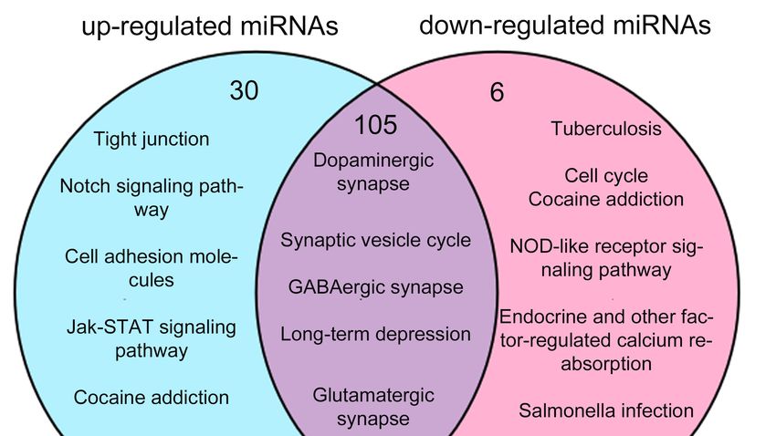
Int. J. Mol. Sci. 2018, 19, 671                                                                  2 of 15

antidepressants are usually underreported in clinical trials, and a small but growing literature on the
inappropriate use of antidepressants largely consists of case reports. Furthermore, individuals without
professional diagnosis may be vulnerable to misuse or abuse of medications [18].
     MicroRNAs (miRNAs) are a family of noncoding RNA molecules, and regulate different biological
pathways by negatively modulating target gene expression [19]. Since one miRNA may regulate up to
hundreds of genes, and collectively 50–60% of total transcriptomes, miRNAs could have pleiotropic
biological effects [20,21]. In mammals, miRNAs have been shown as one of the key regulators
that control development of the CNS, including neurogenesis, neuronal proliferation and synaptic
plasticity [22–25]. Disruptions to these processes have been linked to development of depression [26].
The involvement of miRNAs in the response to fluoxetine is only beginning to be explored,
for example, miR-16 mediated the action of fluoxetine by acting as a micromanager of hippocampal
neurogenesis [27–29]. The therapeutic action of fluoxetine in shocked mice was associated with a
significant reduction of miR-1971 in the cortex [30]. Moreover, studies have shown that miRNAs may
function as pre-diagnosis markers in different diseases via post-transcriptional regulation, such as
cardiovascular disorders, meiosis-associated cancer and reproductive dysfunction [31–35].
     In this study, we show that 39 miRNAs are up-regulated (signal > 500) and 25 miRNAs are
down-regulated (signal > 500) after the antidepressant fluoxetine administration of normal mice
by using an Affymetrix microarray analysis. We also demonstrate potential roles of up- and
down-regulated miRNAs in hippocampus by analyzing miRNA predicted targets using Kyoto
encyclopedia of genes and genomes (KEGG) and Gene Ontology (GO). Finally, we find that changes of
miRNAs are associated with the repression of the dopaminergic synapse signal, which could cause
fluoxetine-related dopaminergic abnormal inhibition.

2. Results

2.1. Differential Expression of miRNAs upon Fluoxetine Treatment in Adult Mice
     Our previous study has shown that miRNAs are associated with anxiety- and depression-like
behaviors in mice [36]. To test whether antidepressant itself has an effect on expression of
miRNAs in hippocampus in wild type mice, adult C57BL/6 mice were directly administrated daily
fluoxetine or vehicle for 2 weeks. Hippocampus was dissected for extracting RNA and then miRNA
microarray analyses.
     1892 miRNA probes were tested in the microarray analysis (Tables S1 and S2). 623 known mature
miRNAs have been mapped randomly in the mouse chromosomes through the mouse miRBase
(Figure 1A and Figure S1, and Table S3). To screen differentially expressed miRNAs, the transcriptional
level of miRNAs was compared between hippocampi of the antidepressant treated and control
groups (Figure 1B and Table S3). Altogether, while 103 and 74 miRNAs were highly expressed in the
antidepressant treated and control groups (p < 0.1), respectively, 446 of 623 miRNAs did not show
differential expression (Figure 1B and Table S3). When the signal of differentially expressed miRNAs
was set larger than 500 and the two fold-change cutoff was adjusted p-value as p < 0.1, 64 miRNAs (39
up-regulated miRNAs and 25 down-regulated miRNAs) were found to show differential expression in
the antidepressant treated and control groups (Figures 1C and 2, and Table 1).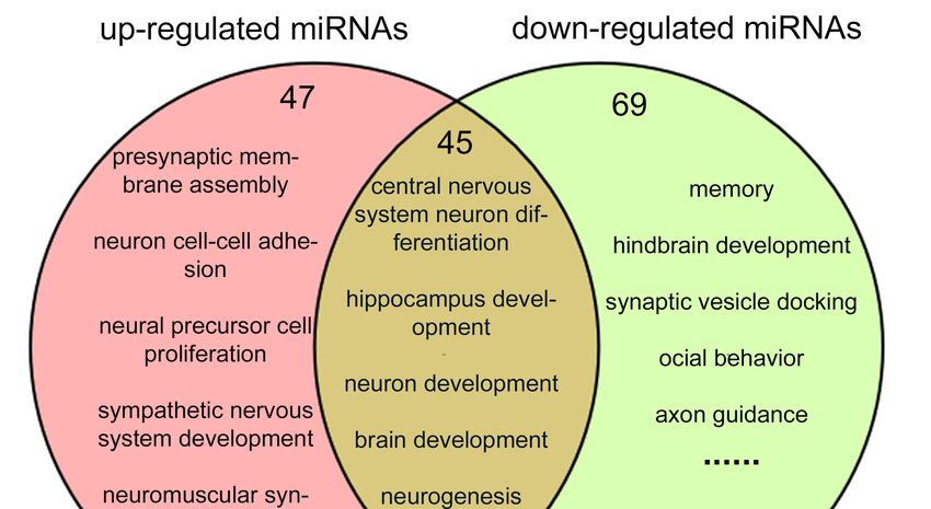
Int. J. Mol. Sci. 2018, 19, 671                                                                                          3 of 15
 Int. J. Mol. Sci. 2018, 19, x                                                                                           3 of 15

      Figure 1. MiRNA expression in hippocampi of antidepressant administration and control mice.
       Figure
      (A)  The 1. MiRNAofexpression
               numbers       miRNAs ininmousehippocampi     of antidepressant
                                                    chromosome                   administration
                                                                   loci in Affymetrix             and control
                                                                                       array analysis;          mice.plot
                                                                                                        (B) Volcano    (A)
       The  numbers    of miRNAs     in mouse   chromosome      loci in Affymetrix  array  analysis; (B) Volcano
      of miRNA expression levels in antidepressant administration and control group. Each point represents        plot  of
       miRNA    expression    levels  in antidepressant    administration   and  control group.
      a miRNA, “red” dot means hyper, “green” dot means hypo expression and “black” dot means noneEach  point represents
       a miRNA, “red”
      significance         dot means hyper,
                     (antidepressant            “green”(C)
                                         vs. control);    dotVenn
                                                               means    hypo expression
                                                                     diagram   of miRNAand     “black”
                                                                                            array       dot The
                                                                                                  results.  means    none
                                                                                                                 ellipses
       significance
      in             (antidepressant
          different color                vs. control);
                            (blue, yellow,    red and(C)    Vennrepresent
                                                         green)    diagram miRNAs
                                                                              of miRNAinarray
                                                                                           four results.
                                                                                                 groups:The   ellipses
                                                                                                           signal  > 500in
       different  color  (blue, yellow,   red  and   green)   represent   miRNAs    in four  groups:
      (175 miRNAs), p < 0.1 (177 miRNAs), up-regulated (319 miRNAs) and down-regulated (296 miRNAs),  signal  > 500   (175
       miRNAs), p The
      respectively.   < 0.1overlapping
                            (177 miRNAs), partsup-regulated     (319 miRNAs)
                                                 of ellipses represent           and down-regulated
                                                                         the intersection                (296 miRNAs),
                                                                                           of different groups,  such as
      103 and 74 miRNAs were in up-regulated and down-regulated groups (p < 0.1), respectively. such as
       respectively.  The  overlapping     parts of  ellipses represent   the intersection of different  groups,
       103 and 74 miRNAs were in up-regulated and down-regulated groups (p < 0.1), respectively.
Int. J. Mol. Sci. 2018, 19, 671 4 of 15
Int. J. Mol. Sci. 2018, 19, x 5 of 15
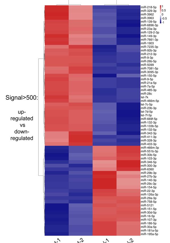
Figure 2. The heatmap of normalized expression of miRNAs in hippocampi of antidepressant
Figure 2. The heatmap
administration of normalized
and control expression
mice to distinguish theofdifference
miRNAs of in expression.
hippocampi The
of antidepressant
heatmap was
administration and controlexpression
drawn with normalized mice to distinguish the difference
((x − min)/(max of expression.
− min)) The heatmap
of each miRNA. The was
“x” drawn
means
with normalized expression ((x − min)/(max − min)) of each miRNA. The “x” means the
the expression of each miRNA, “min” means the lowest expressed miRNA and “max” means the expression of
each miRNA, “min” means the lowest expressed miRNA and “max” means the highest expressed
highest expressed miRNA. Blue, white and red indicate relatively low, middle and high expression of
miRNA.
miRNAs,Blue, white and red indicate relatively low, middle and high expression of miRNAs,
respectively.
respectively.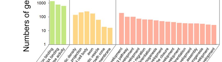
Int. J. Mol. Sci. 2018, 19, 671                                                                        5 of 15

           Table 1. Up-regulated and down-regulated hippocampal miRNAs upon fluoxetine treatment.

                                              Up-Regulated miRNAs
                            miRNA Name (Fold-Change, Fold-Change = Treatment/Control)
        miR-7081-5p (22.83)          miR-5099 (1.71)       miR-485-3p (1.40)     miR-3095-3p (1.23)
          miR-3962 (3.43)              let-7k (1.64)         let-7f-5p (1.39)      let-7c-5p (1.23)
        miR-133b-5p (2.27)          miR-92b-3p (1.61)       let-7d-5p (1.39)     miR-466m-5p (1.22)
         miR-21a-5p (2.14)           miR-1903 (1.53)       miR-411-3p (1.39)     miR-23b-3p (1.20)
          miR-3963 (2.01)           miR-150-5p (1.50)      miR-23a-3p (1.37)     miR-149-3p (1.20)
         miR-26b-5p (1.80)          miR-328-3p (1.50)       miR-7a-5p (1.34)      miR-342-3p (1.19)
         miR-132-3p (1.79)          miR-6896-3p (1.49)     miR-7235-3p (1.30)      miR-9-5p (1.16)
         miR-212-3p (1.75)          miR-132-5p (1.44)      miR-433-3p (1.30)     miR-341-5p (1.15)
         miR-329-3p (1.75)          miR-129-2-3p (1.43)    miR-139-5p (1.27)     miR-7661-3p (1.13)
         miR-218-5p (1.74)            miR-28c (1.40)       miR-669f-5p (1.27)
                                            Down-Regulated miRNAs
                            miRNA Name (Fold-Change, Fold-Change = Control/Treatment)
        miR-466m-3p (1.09)           miR-22-3p (1.23)      miR-30d-5p (1.39)      miR-103-3p (1.22)
         miR-151-5p (1.09)           miR-107-3p (1.23)     miR-195a-5p (1.42)     miR-30b-5p (1.32)
         miR-300-3p(1.1)             miR-27b-3p (1.24)      miR-5121 (1.43)       miR-186-5p (1.55)
        miR-126a-3p (1.16)           miR-346-5p (1.24)     miR-758-5p (1.44)      miR-551b-3p (1.23)
         miR-140-3p (1.18)           miR-16-5p (1.25)      miR-29c-3p (1.50)
         miR-154-5p (1.22)           miR-29a-3p (1.27)     miR-29b-3p (1.50)
        miR-181a-5p (1.22)           miR-30a-5p (1.38)      miR-6360 (2.26)

     Among 64 miRNAs (signal > 500, p < 0.1), 39 miRNAs exhibited higher expression levels in
the antidepressant treated group than the control (termed up-regulated miRNA group or hyper
miRNA group) (Figure 1B,C and Figure 2, Table 1). The well-studied miRNAs within this group
included let-7 family (let-7c/d/f/k), miR-212 cluster (miR-212-3p and miR-132-3p/5p), miR-23a/b,
miR-9-3p/5p, miR-411 clusters (miR-299a and miR-329) and miR-466 clusters (miR-466m-5p and
miR-669f-5p) (Figure 2 and Table 1). On the other hand, 25 miRNAs were expressed at a lower level
in the antidepressant treated group than the control (termed down-regulated miRNA group or hypo
miRNA group). This group included miR-30 family (miR-30a/b/d) and miR-29 family (miR-29a/b/c).
Overall, identification of 64 miRNAs with significant changes after the fluoxetine administration
in normal adult mice suggests that a considerable number of miRNAs respond to antidepressants
in hippocampus.

2.2. Prediction of miRNA Target Genes and Pathway Analysis
     Because miRNAs normally function through silencing their target genes, we next searched
targets of differentially expressed miRNAs. Using TargetScan (http://www.targetscan.org/vert_71/)
and miRDB (http://www.mirdb.org/), 7478 genes were predicted as targets of the 39 up-regulated
miRNAs, and 3404 genes as targets of 25 down-regulated miRNAs (Table S4).
     To comprehensively study the interaction between miRNAs and their predicted targets, analyses
of KEGG pathway for the miRNA-target pairs were performed in miRNAs of up-regulated and
down-regulated groups (Tables S5 and S6). We found that some enriched pathways are associated with
neural development and function, for example pathways in the endocrine system development and
neurotrophin signaling (Figure 3 and Figure S2). Pathways related to hippocampal neurogenesis
for the predicted targets of up-regulated miRNAs included genes involved in axon guidance,
dopaminergic synapse, long-term depression and GABAergic synapse (Figure 3). Predicted targets for
the down-regulated miRNAs were associated with the wingless-type MMTV integration site (Wnt)
signaling pathway and cholinergic synapse (Figure S2). These results suggest that many miRNAs in
response to fluoxetine may participate in neural-related signaling pathways.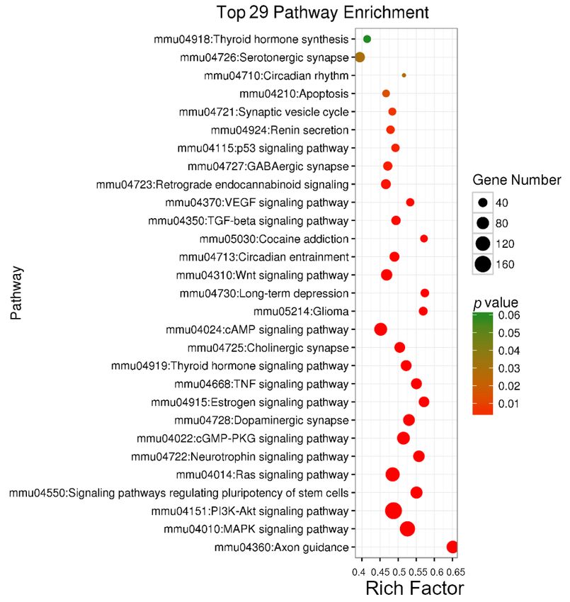
Int. J. Mol. Sci. 2018, 19, 671 6 of 15
Int. J. Mol. Sci. 2018, 19, x 6 of 15
Figure3.
Figure 3. The
The bubble
bubble chart
chart of
of up-regulated
up-regulated miRNAs
miRNAs in in KEGG
KEGG analysis.
analysis. Y-axis
Y-axis represents
represents the
the pathway
pathway
name, X-axis
name, X-axis represents
represents the
the rich
rich factor,
factor, the
the size
size of
of bubble
bubble represents
represents the
the number
number of of genes,
genes, and
and the
the
color of
color of bubble
bubble represents
representsthe
thep-value.
p-value. VEGF:
VEGF: vascular
vascular endothelial
endothelial growth
growthfactor;
factor;TGF:
TGF:transforming
transforming
growth factor;
growth factor; Wnt:
Wnt: wingless-type
wingless-type MMTV
MMTV integration
integration site;
site; cAMP:
cAMP: cathelicidin
cathelicidin antimicrobial
antimicrobial peptide;
peptide;
TNF:
TNF:tumor
tumornecrosis
necrosisfactor;
factor;cGMP-PKG:
cGMP-PKG: cyclic cyclic GDP-mannose
GDP-mannose pyrophosphorylase
pyrophosphorylase dependent
dependent protein
protein
kinase;
kinase; Ras:
Ras: rat
rat sarcoma
sarcoma viral
viral oncogene
oncogene homolog;
homolog; PI3K:
PI3K:phosphatidylinositol
phosphatidylinositol 3-kinase;
3-kinase; Akt:
Akt: RAC
RAC
serine/threonine-protein
serine/threonine-protein kinase; MAPK: mitogen-activated protein kinase.
2.3. GO
2.3. GO Analysis
Analysis of
of Predicted
Predicted Targets
Targetsfor
formiRNAs
miRNAs
Tomake
To makeaabetter
betterunderstanding
understanding of ofbiological
biological processes
processes ofof predicted
predicted targets
targets for
for miRNAs,
miRNAs, GeneGene
Ontology(GO)
Ontology (GO)analyses
analyseswerewereperformed
performed inin up-regulated
up-regulated andand down-regulated
down-regulated miRNA
miRNA targets
targets that that
are
are associated with biological process (BP), cellular components (CC) and molecular
associated with biological process (BP), cellular components (CC) and molecular function (MF). We function (MF).
We found
found thatGO
that 923 923items
GO items
(567 BP(567 BP 155
items, items,CC155
itemsCCand
items
201and 201 MFwere
MF items) items) were in
enriched enriched in up-
up-regulated
regulated(Tables
miRNAs miRNAs (Tables
S7–S9), andS7–S9),
614 GOanditems
614 GO (395items (395 BP114
BP items, items,
CC 114 CCand
items items105and
MF105 MF items)
items) were
were abundant in down-regulated miRNAs (Tables S10–S12). Among GO items
abundant in down-regulated miRNAs (Tables S10–S12). Among GO items that are associated with that are associated
with neurogenesis
neurogenesis (Tables
(Tables S13 and
S13 and S14),S14),
some some
itemsitems of BP
of BP in in up-regulatedmiRNAs
up-regulated miRNAswere wereinvolved
involvedinin
functions regulating
functions regulating postsynaptic
postsynaptic density,
density,neuronal
neuronalprojection
projectionandandsynaptic
synaptic vesicle,
vesicle, and
and some
some items
items
of CC
of CC were
were involved
involved in in regulating
regulating neuronal
neuronal projection,
projection, migration
migration and differentiation (Figure 4).
Moreover,the
Moreover, themost
mostsignificant
significantGOGOterms
termsin indown-regulated
down-regulated miRNAs
miRNAs were were related
related to
tosynapse,
synapse, axon
axon
and synaptic membrane (Figure S3). These results indicate that many up and down-regulated
miRNAs are associated with hippocampal neurogenesis.
Int. J. Mol. Sci. 2018, 19, 671                                                                                   7 of 15
        Int. J. Mol. Sci. 2018, 19, x                                                                        7 of 15

Int. J.synaptic
and     Mol. Sci. 2018, 19, x
                   membrane                                                                          7 of 15
                              (Figure S3). These results indicate that many up and down-regulated miRNAs
are associated with hippocampal neurogenesis.

                           Figure 4. The histogram of target genes for up-regulated miRNAs in GO analysis.

        2.4. Functional Comparison of Predicted Targets for Up-Regulated and Down-Regulated miRNAs
            To study functional correlations of predicted targets between up-regulated and down-regulated
       miRNAs, we validated the overlapping KEGG and GO items among these targets (Figure 5).
                Figure 4.
                Figure  4. The
                           The histogram
                               histogram ofof target
                                               target genes
                                                      genesfor
                                                             forup-regulated
                                                                  up-regulatedmiRNAs
                                                                                 miRNAsin inGO
                                                                                             GOanalysis.
                                                                                                analysis.
            In the KEGG analysis, most of the pathways were overlapped between two groups (105 out of
       135 in up-regulated miRNAs and 105 out of 111 in down-regulated miRNAs) (Figure 5A and Table
2.4. Functional
2.4. Functional
       S16). Many
                  Comparison
                 Comparison     of Predicted
                               of
                    of them were
                                   Predicted  Targets
                                    involvedTargets
                                                        fordevelopment,
                                                       for
                                               in neural
                                                            Up-Regulatedfor
                                                           Up-Regulated     and
                                                                           and    Down-Regulated
                                                                                 Down-Regulated
                                                                               instance
                                                                                                   miRNAs
                                                                                                  miRNAs
                                                                                        the dopaminergic  synapse,
       synaptic
      Tostudy    vesicle
         studyfunctional cycle,  GABAergic
                 functionalcorrelations
                              correlationsof   synapse
                                             ofpredicted and   long-term
                                                  predictedtargets         depression.
                                                              targetsbetween            30
                                                                        betweenup-regulatedpathways
                                                                                  up-regulatedand     were specific
                                                                                                 anddown-regulated
                                                                                                      down-regulated
      To
       abundant
miRNAs,we         in up-regulated
            wevalidated
                validatedthe        miRNAs,
                             theoverlapping
                                 overlappingKEGGfor example
                                                  KEGGand    the
                                                           andGO  tight
                                                                 GOitemsjunction,
                                                                     itemsamong   notch
                                                                            amongthese  signaling
                                                                                     thesetargets pathway
                                                                                            targets(Figure and
                                                                                                            5). cell
                                                                                                    (Figure5).
miRNAs,
       adhesion molecules, while only 6 pathways were enriched in down-regulated miRNAs.
     In the KEGG analysis, most of the pathways were overlapped between two groups (105 out of
135 in up-regulated miRNAs and 105 out of 111 in down-regulated miRNAs) (Figure 5A and Table
S16). Many of them were involved in neural development, for instance the dopaminergic synapse,
synaptic vesicle cycle, GABAergic synapse and long-term depression. 30 pathways were specific
abundant in up-regulated miRNAs, for example the tight junction, notch signaling pathway and cell
adhesion molecules, while only 6 pathways were enriched in down-regulated miRNAs.

                                                         Figure 5. Cont.
Int. J. Mol. Sci. 2018, 19, 671                                                                                  8 of 15

        Int. J. Mol. Sci. 2018, 19, x                                                                        8 of 15

               Figure 5. Venn diagrams of the comparison analysis of target genes for up- and down-regulated
      Figure 5. Venn diagrams of the comparison analysis of target genes for up- and down-regulated
            miRNAs. (A) Venn picture of GO analysis; (B) Venn picture of KEGG analysis. Jak-STAT: Janus
      miRNAs. (A) Venn picture of GO analysis; (B) Venn picture of KEGG analysis. Jak-STAT:
            kinase/signal transducers and activators of transcription; NOD: nucleotide binding oligomerization
      Janus domain.
             kinase/signal transducers and activators of transcription; NOD: nucleotide binding
      oligomerization domain.
            In the GO analyses of BP, 92 items in up-regulated miRNAs and 114 items in down-regulated
       miRNAs
     In the KEGG were  found tomost
                     analysis,   be associated with neural
                                     of the pathways    weredevelopment
                                                             overlapped(Figure 5B,two
                                                                         between   Tables S13–S15).
                                                                                      groups   (105 47
                                                                                                    outand
                                                                                                        of 135
       69 items were specifically abundant in up-regulated and down-regulated miRNAs, respectively. 45
in up-regulated miRNAs and 105 out of 111 in down-regulated miRNAs) (Figure 5A and Table S16).
       GO items were found overlapping in up- and down-regulated miRNAs, for example neuronal
Many of them were involved in neural development, for instance the dopaminergic synapse, synaptic
       differentiation and hippocampal development.
vesicle cycle,  GABAergic
            These  functional synapse   and long-term
                               analyses indicate         depression.
                                                 that most            30 pathways
                                                            GO and KEGG             were in
                                                                          are overlapping  specific abundant
                                                                                             the predicted
in up-regulated    miRNAs, for
       targets of up-regulated    example
                                and          the tightmiRNAs.
                                     down-regulated    junction, notch signaling pathway and cell adhesion
molecules, while only 6 pathways were enriched in down-regulated miRNAs.
     In2.5.
        themiRNA    Interaction
            GO analyses       ofNetwork  for Elements
                                 BP, 92 items          Involved in Dopaminergic
                                                in up-regulated     miRNAs and  Synapse
                                                                                    114 Signals
                                                                                          items in down-regulated
miRNAs were      found     to  be  associated   with    neural  development     (Figure
            Previous studies have shown that antidepressant targeted Na /Cl -coupled+   −  5B, Tables  S13–S15). 47
                                                                                                   dopaminergic
and 69neurotransmitter
        items were specifically      abundant    in up-regulated     and down-regulated      miRNAs,
                            uptake defines a key therapeutic strategy to treat clinical depression       respectively.
                                                                                                              and
45 GOneuropathic
        items werepainfound    overlapping
                            [37–40].  We found in that
                                                  up- 71andpredicted
                                                             down-regulated
                                                                      targets of miRNAs,
                                                                                 up-regulatedfor miRNAs
                                                                                                 exampleand neuronal
                                                                                                               39
       predictedand
differentiation   targets  of down-regulated
                      hippocampal                 miRNAs are involved in the dopaminergic synapse signals
                                       development.
       (Figure
     These     3 and Figure
             functional        S4, andindicate
                           analyses    Table S17).  Overall,
                                                 that  most GO26 predicted
                                                                   and KEGGtargets
                                                                                arewere  overlapped
                                                                                    overlapping    ininthe
                                                                                                        these two
                                                                                                           predicted
       groups, 45  and  13 predicted  targets  were
targets of up-regulated and down-regulated miRNAs.  specifically  abundant  in up-regulated  and down-regulated
       miRNAs that are involved in regulating dopaminergic synapse, respectively (Figure S4 and Table
       S17). Interestingly,
2.5. miRNA                  we found
              Interaction Network   forthat many miRNAs
                                         Elements  Involved(18
                                                             inof 21) negativelySynapse
                                                                Dopaminergic       regulateSignals
                                                                                             dopaminergic synapse
       pathways by inhibiting the positive genes (Figure 6). Moreover, some sub-pathways in hippocampal
     Previous    studies   have shown       that antidepressant     targeted    Na+ /Cl   − -coupled dopaminergic
       neurons might    be modulated    by up-regulated  miRNAs, such     as synaptic  vesicle cycle, glutamatergic
neurotransmitter    uptake defines protein
       synapse, mitogen-activated    a key therapeutic
                                              kinase (MAPK)strategy  toand
                                                               signal,  treat
                                                                            RACclinical depression and neuropathic
                                                                                   serine/threonine-protein   kinase
       (Akt) signal.
pain [37–40].         Some that
                We found    miRNAs     may altertargets
                                 71 predicted     the positive  regulators that
                                                         of up-regulated          are associated
                                                                             miRNAs               with Parkinson’s
                                                                                         and 39 predicted     targets of
       disease.
down-regulated miRNAs are involved in the dopaminergic synapse signals (Figure 3 and Figure S4,
            TheseOverall,
and Table S17).     results 26
                            suggest  that targets
                               predicted    miRNAswereaffected  by the in
                                                          overlapped    antidepressant     fluoxetine
                                                                            these two groups,    45 andin the  adult
                                                                                                          13 predicted
       hippocampus largely regulate target genes controlling dopaminergic synapse.
targets were specifically abundant in up-regulated and down-regulated miRNAs that are involved
in regulating dopaminergic synapse, respectively (Figure S4 and Table S17). Interestingly, we found
that many miRNAs (18 of 21) negatively regulate dopaminergic synapse pathways by inhibiting the
positive genes (Figure 6). Moreover, some sub-pathways in hippocampal neurons might be modulated
by up-regulated miRNAs, such as synaptic vesicle cycle, glutamatergic synapse, mitogen-activated
protein kinase (MAPK) signal, and RAC serine/threonine-protein kinase (Akt) signal. Some miRNAs
may alter the positive regulators that are associated with Parkinson’s disease.
Int. J. Mol. Sci. 2018, 19, 671 9 of 15
Int. J. Mol. Sci. 2018, 19, x 9 of 15
Figure6.6.Network
Figure NetworkofofmiRNA-target
miRNA-targetpathways
pathwaysinvolved
involvedinindopaminergic
dopaminergicsynapse
synapseforforup-regulated
up-regulated
miRNAs.The
miRNAs. The rhombus
rhombus represents
represents the genes,
the genes, hexagonhexagon represents
represents the miRNAsthe and
miRNAs and represents
the square the square
represents
the the main
main pathways in pathways
dopaminergic in dopaminergic
synapse. Thesynapse. The
solid lines solid
with linesrepresent
arrows with arrows represent the
the relationship
between genesbetween
relationship and pathways,
genes andandpathways,
the dot lines
andrepresent the relationship
the dot lines represent thebetween genes between
relationship and miRNAs.
genes
The
andsolid lines without
miRNAs. The solid arrows represent
lines without the sub-pathways.
arrows represent the The double blueThe
sub-pathways. solid line represent
double blue solidthe
line
cell structure
represent theofcell
neuron (presynaptic
structure of neuronterminal and postsynaptic
(presynaptic terminal and cell) and glia cell.
postsynaptic cell) and glia cell.
3. Discussion
These results suggest that miRNAs affected by the antidepressant fluoxetine in the adult
hippocampus
Current largely regulate treatments
antidepressant target genesremain
controlling dopaminergic
inadequate, synapse.
burdened by a significant percentage of
misuse and side-effects. The fluoxetine may be deleterious to immature or undifferentiated neural
3. Discussion
cells, but the mechanisms are unclear [41–43]. Our study shows the diverse changes of miRNAs in
hippocampus after the fluoxetine
Current antidepressant administration
treatments of normalburdened
remain inadequate, mice. Some by aup-regulated miRNAs may
significant percentage of
negatively
misuse regulate the The
and side-effects. dopaminergic
fluoxetine synapse pathway. Our
may be deleterious results suggest
to immature that miRNAs respond
or undifferentiated neural
to antidepressants
cells, but the mechanisms in hippocampus of normal
are unclear [41–43]. Ourmice
study byshows
inhibiting a widechanges
the diverse range of target genes
of miRNAs in
involved in many
hippocampus after aspects of neural
the fluoxetine developmentofand
administration functions.
normal mice. Some up-regulated miRNAs may
Studies
negatively have the
regulate shown that miRNA
dopaminergic is onepathway.
synapse of the most Ourimportant prognosis
results suggest markers respond
that miRNAs in differentto
clinical scenario.
antidepressants miR-181a-5p/miR-21a-5p
in hippocampus of normal mice ratios were useda wide
by inhibiting to detect
range mycotrophic
of target genes lateral sclerosis
involved in
with aspects
many 90 and 85% sensitivity
of neural in cerebrospinal
development fluid [44]. miRNAs have been found to be involved in
and functions.
cardiac
Studiesresynchronization
have shown that therapy
miRNA through
is one association
of the most with cardiac
important angiogenesis
prognosis (miR-30,
markers miR-92
in different
and miR-145),
clinical scenario.cardiac apoptosis (miR-30) and
miR-181a-5p/miR-21a-5p cardiac
ratios werefibrosis
used to(miR-29) [35]. Recently,
detect mycotrophic several
lateral studies
sclerosis
have90reported
with and 85%that dysregulation
sensitivity of miRNAs
in cerebrospinal may
fluid precipitate
[44]. miRNAsorhave aggravate anxiety
been found to and depression.
be involved in
For example,
cardiac miR-17-92 knockout
resynchronization mice show
therapy through anxiety- and
association withdepression-like behaviors,
cardiac angiogenesis whereas
(miR-30, miR-92miR-
17-92
and overexpressing
miR-145), mice exhibit
cardiac apoptosis anxiolytic
(miR-30) and and antidepressant-like
cardiac fibrosis (miR-29)behaviors [36]. Higher
[35]. Recently, several or lower
studies
levels
have of miR-135
reported that can alter anxiety-
dysregulation and depression-like
of miRNAs may precipitatebehaviors in mice anxiety
or aggravate via regulating activity of
and depression.
serotonergic
For (5HT) neurons,
example, miR-17-92 knockout 5HTmice levels
show in anxiety-
blood and brain, and behavioral
and depression-like behaviors, response
whereasto
antidepressant
miR-17-92 treatment [45].
overexpressing miceFluoxetine is a widely
exhibit anxiolytic andused antidepressant inbehaviors
antidepressant-like treatment[36].
of mood
Higher and
oranxiety
lower disorders via regulating
levels of miR-135 miR-29
can alter family
anxiety- andand genes such asbehaviors
depression-like brain derived neurotrophic
in mice via regulating factor
(BDNF),
activity of response
serotonergicregulator
(5HT) in two-component
neurons, 5HT levelsregulatory
in blood and system with
brain, andCreC (CREB),
behavioral and HUS-
response to
associated diffuse
antidepressant adherence
treatment [45].(HDACs)
Fluoxetine[46–49].
is a However,
widely used the involvement
antidepressant of miRNAs
in treatmentin theofresponse
mood
to antidepressants
and anxiety disorders in via
adult hippocampus
regulating miR-29is family
only beginning
and genestosuchbe explored. Previousneurotrophic
as brain derived studies have
shown
factor that miR-17-92
(BDNF), response mediates
regulator antidepressant-regulated
in two-component regulatory adult hippocampal
system with neurogenesis,
CreC (CREB), whichandis
verified by quantitative reverse transcription PCR (qRT-PCR) and behavioral tests [36]. We identified
64 miRNAs with significant changes after the fluoxetine administration in normal adult mice,Int. J. Mol. Sci. 2018, 19, 671 10 of 15
HUS-associated diffuse adherence (HDACs) [46–49]. However, the involvement of miRNAs in the
response to antidepressants in adult hippocampus is only beginning to be explored. Previous studies
have shown that miR-17-92 mediates antidepressant-regulated adult hippocampal neurogenesis,
which is verified by quantitative reverse transcription PCR (qRT-PCR) and behavioral tests [36]. We
identified 64 miRNAs with significant changes after the fluoxetine administration in normal adult
mice, suggesting that a large number of miRNAs respond to antidepressants in hippocampus. Even
though expression of some of these miRNAs have been validated by qRT-PCR, a thorough verification
will draw a comprehensive profile of miRNA expression in the future.
Among altered miRNAs, some of them have been shown to play roles in brain functions.
For example, the fluoxetine induced up-regulation of let-7 family (let-7c/d/f/k) and miR-23a/b have
been shown to have high risk of temporal lobe epilepsy [50]. Abnormal ascending of miR-132/212
alters memory and learning by modulating dendritic growth and arborization of newborn neurons
via attenuating hippocampal transcriptome [51–53]. In addition, reduced expression of miR-29 family
causes several neurodegenerative disorders, including Alzheimer’s disease, Huntington’s disease,
and spinocerebellar ataxias, by targeting voltage-dependent anion channel 1 (VDAC1), β-secretase 1
(BACE1) and neuron navigator 3 (NAV3) [54–57]. Interestingly, these miRNAs were also found up- or
down-regulated after fluoxetine treatment in the hippocampus.
Since miRNAs function through silencing target genes, KEGG and GO analyses of targets for
altered miRNAs after fluoxetine treatment, have identified some pathways important for neural
development and function. We have found that miR-16 is down-regulated, and its predicted
targets, including BDNF, apoptosis regulator Bcl-2 (BCL-2), Serotonin transporter (SERT), and
Wnt2, have been shown to regulate hippocampal response to serotonin reuptake inhibitor (SRI)
antidepressants [27,58–60]. Even though the functional relationship between altered miRNAs and
their targets needs to be further verified, the KEGG and GO analyses have provided a reference for
future identification of miRNA-target interactions responding to antidepressants. Because the false
positive of miRNA target prediction is inevitable, the future work will be to validate miRNA-target
regulation and to examine the biological meaning of these interactions.
Moreover, we have found that many pathways of predicted targets are overlapped between up-
and down-regulated miRNAs, which suggests a counter-balanced functional output. Interestingly,
the dopaminergic synapse signal appears to be suppressed in hippocampal neurons by miRNAs
after fluoxetine treatment, suggesting that fluoxetine may be deleterious to normal dopaminergic
neurons by abnormal inhibition [61,62]. Some miRNA-target pairs in dopaminergic synapse
signal have been reported. For example, miR-9-CREB negative feedback minicircuitry plays a
critical role in the determination of proliferation and migration in glioma cells [63,64]; miR-433-3p
suppresses cell growth and enhances chemosensitivity by targeting CREB in human glioma [65];
miR-26a/Kruppel like factor 4 (KLF4) and cAMP responsive element binding protein CCAAT/enhancer
binding protein (CREB-C/EBPβ) signaling pathways regulate survival of mycobacterium tuberculosis
in macrophages [66]. Since CRE-mediated transcription promotes neuronal plasticity, some
up-regulated miRNAs (miR-9, miR-433-3p and miR-26a) may play essential roles in fluoxetine-induced
dopaminergic synapse suppression by targeting CREB [67–69]. We have found that fluoxetine treatment
in hippocampus of wild type mice causes up-regulation of miRNAs that normally suppress the
dopaminergic synapse signal, which might in turn regulate hippocampal function related to anxiety
and depression.
In summary, this study has identified many up- and down-regulated hippocampal miRNAs after
the fluoxetine administration in wild type mice, suggesting that a large number of miRNAs respond to
antidepressants even under normal conditions. Subsequently, a broad range of target genes are affected
by altered miRNAs. This data further suggests safety considerations related to antidepressant usage.
Further analyses of these altered miRNAs and related pathways identified in this study will help to
promote the understanding of molecular mechanisms involved in inappropriate use of antidepressants
in the future.Int. J. Mol. Sci. 2018, 19, 671 11 of 15
4. Materials and Methods
4.1. Antidepressant Treatment
The C57BL/6 mice from the same parents was fed in the same condition (temperature, humidity,
fodder and etc.). The adult mice were administrated daily fluoxetine (20 mg/kg, Sigma-Aldrich,
St. Louis, MO, USA) or vehicle for 2 weeks (13-week-old) until the day before the experiment [36].
Fluoxetine was dissolved in dimethylsulfoxide (DMSO) and then diluted in saline. The drug was finally
diluted in 100 µL of 0.9% saline for administration [36,70]. Animal use was overseen by the Animal
Facility at Weill Cornell Medical College (#2011-0062, 25 July 2011), and was performed according to
the institutional ethical guidelines for animal experiments.
4.2. RNA Preparation
The adult hippocampi of the 13-week-old mice were dissected from control group and fluoxetine
administration group, each of them from a different litter. To avoid contamination of cortical tissues,
the adult hippocampi including the Cornu Ammonis 1 (CA1), CA2, CA3, CA4 areas and the dentate
gyrus (DG) were collected through fine dissection from the cerebral cortex. All tissue samples were
washed briefly with phosphate buffered solution (PBS), and total RNA from each brain was isolated
by using Trizol reagent (Invitrogen, Carlsbad, CA, USA). The quality and quantity of these RNA
samples were determined by NanoDrop spectrophotometer (ND-2000C, Thermo Fisher Scientific Inc.,
Wilmington, DE, USA). Agilent RNA 6000 Nano assay (Agilent Technologies, Inc., Santa Clara, CA,
USA) and agarose gel electrophoresis were used to check purity of the RNA samples.
4.3. miRNA Microarray Assay
A commercial Affymetrix array service (LC Sciences, LLC, Houston, TX, USA) was used for
miRNA array analyses. The labeled RNA was hybridized to the Affymetrix GeneChip miRNA1.0 array
(Affymetrix, Santa Clara, CA, USA) containing 1917 miRNA probes, and the array was later scanned
by Axon 4000B (Axon Instruments, Union City, CA, USA) from Molecular Devices (San Jose, CA, USA).
The RNA labeling, miRNA array hybridization, and quantification were performed via Affymetrix
GeneChip system instruments and protocols. The sequence for the array result was selected from the
miRBase database 21 (http://www.mirbase.org).
4.4. Microarray Data Analysis
The data analysis of miRNAs expression profiling (data filtering, normalization and statistical
calculations) was processed by R (R-Foundation for Statistical Computing, Vienna, Austria; version
2.12.1). Between 2 antidepressant treatments and 2 control tissues, the significant difference of miRNAs
expression was selected by fold-change and a p-value with the following criteria: fold-change > 1 and
p-value < 0.1 [71]. The heatmap was performed with normalized expression ((x − min)/(max − min)) of
each miRNA by pheatmap package (https://cran.r-project.org/web/packages/pheatmap/index.html)
of R language. The heat map was drawn. Blue, white and red indicate relatively low, middle and high
expression of miRNAs, respectively.
4.5. miRNA Target Prediction
Target prediction of miRNAs was performed using a combination of 2 prediction programs
including miRDB and TargetScan [72]. The significantly changed miRNAs were analyzed with their
chromosome location and p-value using miRBase [73].
4.6. miRNA Target Functional Analysis
Predicted target genes were imported into the Database for Annotation, Visualization and
Integrated Discovery (DAVID) v6.8 (https://david.ncifcrf.gov/), an online functional annotationInt. J. Mol. Sci. 2018, 19, 671 12 of 15
tools for investigators to understand biological meaning. We performed Gene Ontology (GO) and
KEGG analysis of the miRNAs-targets with differential expression. In this study, modules in the
gene-miRNA-KEGG were mined with Cytoscape 3.6 (http://www.cytoscape.org/) and then functional
enrichment analysis was applied to the miRNAs in the modules.
4.7. Statistical Analysis
The p-value to verify significant difference is set toInt. J. Mol. Sci. 2018, 19, 671 13 of 15
15. Akbari Hasanjani, H.R.; Sohrabi, M.R. Artificial Neural Networks (ANN) for the Simultaneous
Spectrophotometric Determination of Fluoxetine and Sertraline in Pharmaceutical Formulations and
Biological Fluid. Iran. J. Pharm. Res. 2017, 16, 478–489. [PubMed]
16. Abbing-Karahagopian, V.; Huerta, C.; Souverein, P.C.; de Abajo, F.; Leufkens, H.G.; Slattery, J.; Alvarez, Y.;
Miret, M.; Gil, M.; Oliva, B.; et al. Antidepressant prescribing in five European countries: Application of
common definitions to assess the prevalence, clinical observations, and methodological implications. Eur. J.
Clin. Pharmacol. 2014, 70, 849–857. [CrossRef] [PubMed]
17. Sultana, J.; Italiano, D.; Spina, E.; Cricelli, C.; Lapi, F.; Pecchioli, S.; Gambassi, G.; Trifiro, G. Changes in the
prescribing pattern of antidepressant drugs in elderly patients: An Italian, nationwide, population-based
study. Eur. J. Clin. Pharmacol. 2014, 70, 469–478. [CrossRef] [PubMed]
18. Evans, E.A.; Sullivan, M.A. Abuse and misuse of antidepressants. Subst. Abuse Rehabil. 2014, 5, 107–120. [PubMed]
19. Huntzinger, E.; Izaurralde, E. Gene silencing by microRNAs: Contributions of translational repression and
mRNA decay. Nat. Rev. Genet. 2011, 12, 99–110. [CrossRef] [PubMed]
20. Baek, D.; Villen, J.; Shin, C.; Camargo, F.D.; Gygi, S.P.; Bartel, D.P. The impact of microRNAs on protein
output. Nature 2008, 455, 64–71. [CrossRef] [PubMed]
21. Krol, J.; Loedige, I.; Filipowicz, W. The widespread regulation of microRNA biogenesis, function and decay.
Nat. Rev. Genet. 2010, 11, 597–610. [CrossRef] [PubMed]
22. Liu, C.; Teng, Z.Q.; Santistevan, N.J.; Szulwach, K.E.; Guo, W.; Jin, P.; Zhao, X. Epigenetic regulation of
miR-184 by MBD1 governs neural stem cell proliferation and differentiation. Cell Stem Cell 2010, 6, 433–444.
[CrossRef] [PubMed]
23. Bian, S.; Hong, J.; Li, Q.; Schebelle, L.; Pollock, A.; Knauss, J.L.; Garg, V.; Sun, T. MicroRNA cluster miR-17-92
regulates neural stem cell expansion and transition to intermediate progenitors in the developing mouse
neocortex. Cell Rep. 2013, 3, 1398–1406. [CrossRef] [PubMed]
24. Hou, Q.; Ruan, H.; Gilbert, J.; Wang, G.; Ma, Q.; Yao, W.D.; Man, H.Y. MicroRNA miR124 is required for the
expression of homeostatic synaptic plasticity. Nat. Commun. 2015, 6, 10045. [CrossRef] [PubMed]
25. Cohen, J.E.; Lee, P.R.; Chen, S.; Li, W.; Fields, R.D. MicroRNA regulation of homeostatic synaptic plasticity.
Proc. Natl. Acad. Sci. USA 2011, 108, 11650–11655. [CrossRef] [PubMed]
26. Manji, H.K.; Quiroz, J.A.; Sporn, J.; Payne, J.L.; Denicoff, K.; Gray, N.A.; Zarate, C.A., Jr.; Charney, D.S.
Enhancing neuronal plasticity and cellular resilience to develop novel, improved therapeutics for
difficult-to-treat depression. Biol. Psychiatry 2003, 53, 707–742. [CrossRef]
27. Launay, J.M.; Mouillet-Richard, S.; Baudry, A.; Pietri, M.; Kellermann, O. Raphe-mediated signals control the
hippocampal response to SRI antidepressants via miR-16. Transl. Psychiatry 2011, 1, e56. [CrossRef] [PubMed]
28. Baudry, A.; Mouillet-Richard, S.; Schneider, B.; Launay, J.M.; Kellermann, O. miR-16—A key for adaptive
responses of neurons to fluoxetine. Med. Sci. 2011, 27, 128–131.
29. Baudry, A.; Mouillet-Richard, S.; Schneider, B.; Launay, J.M.; Kellermann, O. miR-16 targets the serotonin
transporter: A new facet for adaptive responses to antidepressants. Science 2010, 329, 1537–1541. [CrossRef]
[PubMed]
30. Schmidt, U.; Herrmann, L.; Hagl, K.; Novak, B.; Huber, C.; Holsboer, F.; Wotjak, C.T.; Buell, D.R. Therapeutic
Action of Fluoxetine is Associated with a Reduction in Prefrontal Cortical miR-1971 Expression Levels in a
Mouse Model of Posttraumatic Stress Disorder. Front. Psychiatry 2013, 4, 66. [CrossRef] [PubMed]
31. Li, Y.; Fang, Y.; Liu, Y.; Yang, X. MicroRNAs in ovarian function and disorders. J. Ovarian Res. 2015, 8, 51.
[CrossRef] [PubMed]
32. Lu, L.; Mao, X.; Shi, P.; He, B.; Xu, K.; Zhang, S.; Wang, J. MicroRNAs in the prognosis of triple-negative
breast cancer: A systematic review and meta-analysis. Medicine 2017, 96, e7085. [CrossRef] [PubMed]
33. Santamaria, X.; Taylor, H. MicroRNA and gynecological reproductive diseases. Fertil. Steril. 2014, 101,
1545–1551. [CrossRef] [PubMed]
34. Santulli, G. MicroRNAs and Endothelial (Dys) Function. J. Cell. Physiol. 2016, 231, 1638–1644. [CrossRef]
[PubMed]
35. Sardu, C.; Marfella, R.; Santulli, G.; Paolisso, G. Functional role of miRNA in cardiac resynchronization
therapy. Pharmacogenomics 2014, 15, 1159–1168. [CrossRef] [PubMed]
36. Jin, J.; Kim, S.N.; Liu, X.; Zhang, H.; Zhang, C.; Seo, J.S.; Kim, Y.; Sun, T. miR-17-92 Cluster Regulates Adult
Hippocampal Neurogenesis, Anxiety, and Depression. Cell Rep. 2016, 16, 1653–1663. [CrossRef] [PubMed]Int. J. Mol. Sci. 2018, 19, 671 14 of 15
37. Jessell, T.M.; Kandel, E.R. Synaptic transmission: A bidirectional and self-modifiable form of cell-cell
communication. Cell 1993, 72, 1–30. [CrossRef]
38. Masson, J.; Sagne, C.; Hamon, M.; El Mestikawy, S. Neurotransmitter transporters in the central nervous
system. Pharmacol. Rev. 1999, 51, 439–464. [PubMed]
39. Kristensen, A.S.; Andersen, J.; Jorgensen, T.N.; Sorensen, L.; Eriksen, J.; Loland, C.J.; Stromgaard, K.;
Gether, U. SLC6 neurotransmitter transporters: Structure, function, and regulation. Pharmacol. Rev. 2011, 63,
585–640. [CrossRef] [PubMed]
40. Penmatsa, A.; Wang, K.H.; Gouaux, E. X-ray structure of dopamine transporter elucidates antidepressant
mechanism. Nature 2013, 503, 85–90. [CrossRef] [PubMed]
41. Man, K.K.; Tong, H.H.; Wong, L.Y.; Chan, E.W.; Simonoff, E.; Wong, I.C. Exposure to selective serotonin
reuptake inhibitors during pregnancy and risk of autism spectrum disorder in children: A systematic review
and meta-analysis of observational studies. Neurosci. Biobehav. Rev. 2015, 49, 82–89. [CrossRef] [PubMed]
42. Kawada, T. Selective serotonin reuptake inhibitors exposure during pregnancy and neonatal outcomes.
J. Clin. Psychopharmacol. 2014, 34, 751. [CrossRef] [PubMed]
43. Viktorin, A.; Uher, R.; Reichenberg, A.; Levine, S.Z.; Sandin, S. Autism risk following antidepressant
medication during pregnancy. Psychol. Med. 2017, 47, 2787–2796. [CrossRef] [PubMed]
44. Benigni, M.; Ricci, C.; Jones, A.R.; Giannini, F.; Al-Chalabi, A.; Battistini, S. Identification of miRNAs as
Potential Biomarkers in Cerebrospinal Fluid from Amyotrophic Lateral Sclerosis Patients. Neuromol. Med.
2016, 18, 551–560. [CrossRef] [PubMed]
45. Issler, O.; Haramati, S.; Paul, E.D.; Maeno, H.; Navon, I.; Zwang, R.; Gil, S.; Mayberg, H.S.; Dunlop, B.W.;
Menke, A.; et al. MicroRNA 135 is essential for chronic stress resiliency, antidepressant efficacy, and intact
serotonergic activity. Neuron 2014, 83, 344–360. [CrossRef] [PubMed]
46. Issler, O.; Carter, R.N.; Paul, E.D.; Kelly, P.A.; Olverman, H.J.; Neufeld-Cohen, A.; Kuperman, Y.; Lowry, C.A.;
Seckl, J.R.; Chen, A.; et al. Increased anxiety in corticotropin-releasing factor type 2 receptor-null mice
requires recent acute stress exposure and is associated with dysregulated serotonergic activity in limbic
brain areas. Biol. Mood Anxiety Disord. 2014, 4, 1. [CrossRef] [PubMed]
47. Krishnan, V.; Nestler, E.J. The molecular neurobiology of depression. Nature 2008, 455, 894–902. [CrossRef]
[PubMed]
48. Wang, H.; Goehring, A.; Wang, K.H.; Penmatsa, A.; Ressler, R.; Gouaux, E. Structural basis for action by
diverse antidepressants on biogenic amine transporters. Nature 2013, 503, 141–145. [CrossRef] [PubMed]
49. Albert, P.R.; Francois, B.L. Modifying 5-HT1A Receptor Gene Expression as a New Target for Antidepressant
Therapy. Front. Neurosci. 2010, 4, 35. [CrossRef] [PubMed]
50. Song, Y.J.; Tian, X.B.; Zhang, S.; Zhang, Y.X.; Li, X.; Li, D.; Cheng, Y.; Zhang, J.N.; Kang, C.S.; Zhao, W.
Temporal lobe epilepsy induces differential expression of hippocampal miRNAs including let-7e and
miR-23a/b. Brain Res. 2011, 1387, 134–140. [CrossRef] [PubMed]
51. Hansen, K.F.; Sakamoto, K.; Aten, S.; Snider, K.H.; Loeser, J.; Hesse, A.M.; Page, C.E.; Pelz, C.; Arthur, J.S.;
Impey, S.; et al. Targeted deletion of miR-132/-212 impairs memory and alters the hippocampal transcriptome.
Learn. Mem. 2016, 23, 61–71. [CrossRef] [PubMed]
52. Magill, S.T.; Cambronne, X.A.; Luikart, B.W.; Lioy, D.T.; Leighton, B.H.; Westbrook, G.L.; Mandel, G.;
Goodman, R.H. microRNA-132 regulates dendritic growth and arborization of newborn neurons in the adult
hippocampus. Proc. Natl. Acad. Sci. USA 2010, 107, 20382–20387. [CrossRef] [PubMed]
53. Aten, S.; Hansen, K.F.; Hoyt, K.R.; Obrietan, K. The miR-132/212 locus: A complex regulator of neuronal
plasticity, gene expression and cognition. RNA Dis. 2016, 3, e1375. [PubMed]
54. Yang, G.; Song, Y.; Zhou, X.; Deng, Y.; Liu, T.; Weng, G.; Yu, D.; Pan, S. DNA methyltransferase 3, a
target of microRNA-29c, contributes to neuronal proliferation by regulating the expression of brain-derived
neurotrophic factor. Mol. Med. Rep. 2015, 12, 1435–1442. [CrossRef] [PubMed]
55. Yang, G.; Song, Y.; Zhou, X.; Deng, Y.; Liu, T.; Weng, G.; Yu, D.; Pan, S. MicroRNA-29c targets beta-site amyloid
precursor protein-cleaving enzyme 1 and has a neuroprotective role in vitro and in vivo. Mol. Med. Rep.
2015, 12, 3081–3088. [CrossRef] [PubMed]
56. Zong, Y.; Yu, P.; Cheng, H.; Wang, H.; Wang, X.; Liang, C.; Zhu, H.; Qin, Y.; Qin, C. miR-29c regulates
NAV3 protein expression in a transgenic mouse model of Alzheimer’s disease. Brain Res. 2015, 1624, 95–102.
[CrossRef] [PubMed]Int. J. Mol. Sci. 2018, 19, 671 15 of 15
57. Roshan, R.; Shridhar, S.; Sarangdhar, M.A.; Banik, A.; Chawla, M.; Garg, M.; Singh, V.P.; Pillai, B.
Brain-specific knockdown of miR-29 results in neuronal cell death and ataxia in mice. RNA 2014, 20,
1287–1297. [CrossRef] [PubMed]
58. Yang, Y.; Hu, Z.; Du, X.; Davies, H.; Huo, X.; Fang, M. miR-16 and Fluoxetine Both Reverse Autophagic
and Apoptotic Change in Chronic Unpredictable Mild Stress Model Rats. Front. Neurosci. 2017, 11, 428.
[CrossRef] [PubMed]
59. Zhang, B.; Chen, C.F.; Wang, A.H.; Lin, Q.F. MiR-16 regulates cell death in Alzheimer’s disease by targeting
amyloid precursor protein. Eur. Rev. Med. Pharmacol. Sci. 2015, 19, 4020–4027. [PubMed]
60. Bai, M.; Zhu, X.; Zhang, Y.; Zhang, S.; Zhang, L.; Xue, L.; Yi, J.; Yao, S.; Zhang, X. Abnormal hippocampal
BDNF and miR-16 expression is associated with depression-like behaviors induced by stress during early
life. PLoS ONE 2012, 7, e46921. [CrossRef] [PubMed]
61. Sekine, Y.; Suzuki, K.; Ramachandran, P.V.; Blackburn, T.P.; Ashby, C.R., Jr. Acute and repeated administration
of fluoxetine, citalopram, and paroxetine significantly alters the activity of midbrain dopamine neurons in
rats: An in vivo electrophysiological study. Synapse 2007, 61, 72–77. [CrossRef] [PubMed]
62. Suzuki, K.; Okada, K.; Wakuda, T.; Shinmura, C.; Kameno, Y.; Iwata, K.; Takahashi, T.; Suda, S.; Matsuzaki, H.;
Iwata, Y.; et al. Destruction of dopaminergic neurons in the midbrain by 6-hydroxydopamine decreases
hippocampal cell proliferation in rats: Reversal by fluoxetine. PLoS ONE 2010, 5, e9260. [CrossRef] [PubMed]
63. Laneve, P.; Gioia, U.; Andriotto, A.; Moretti, F.; Bozzoni, I.; Caffarelli, E. A minicircuitry involving REST
and CREB controls miR-9-2 expression during human neuronal differentiation. Nucleic Acids Res. 2010, 38,
6895–6905. [CrossRef] [PubMed]
64. Tan, X.; Wang, S.; Yang, B.; Zhu, L.; Yin, B.; Chao, T.; Zhao, J.; Yuan, J.; Qiang, B.; Peng, X. The CREB-miR-9
negative feedback minicircuitry coordinates the migration and proliferation of glioma cells. PLoS ONE 2012,
7, e49570. [CrossRef] [PubMed]
65. Sun, S.; Wang, X.; Xu, X.; Di, H.; Du, J.; Xu, B.; Wang, Q.; Wang, J. MiR-433-3p suppresses cell growth and enhances
chemosensitivity by targeting CREB in human glioma. Oncotarget 2017, 8, 5057–5068. [CrossRef] [PubMed]
66. Sahu, S.K.; Kumar, M.; Chakraborty, S.; Banerjee, S.K.; Kumar, R.; Gupta, P.; Jana, K.; Gupta, U.D.; Ghosh, Z.;
Kundu, M.; et al. MicroRNA 26a (miR-26a)/KLF4 and CREB-C/EBPβ regulate innate immune signaling,
the polarization of macrophages and the trafficking of Mycobacterium tuberculosis to lysosomes during
infection. PLoS Pathog. 2017, 13, e1006410. [CrossRef] [PubMed]
67. Wood, M.A.; Kaplan, M.P.; Park, A.; Blanchard, E.J.; Oliveira, A.M.; Lombardi, T.L.; Abel, T. Transgenic
mice expressing a truncated form of CREB-binding protein (CBP) exhibit deficits in hippocampal synaptic
plasticity and memory storage. Learn. Mem. 2005, 12, 111–119. [CrossRef] [PubMed]
68. Honjo, K.; Furukubo-Tokunaga, K. Distinctive neuronal networks and biochemical pathways for appetitive
and aversive memory in Drosophila larvae. J. Neurosci. 2009, 29, 852–862. [CrossRef] [PubMed]
69. Shaw-Lutchman, T.Z.; Impey, S.; Storm, D.; Nestler, E.J. Regulation of CRE-mediated transcription in mouse
brain by amphetamine. Synapse 2003, 48, 10–17. [CrossRef] [PubMed]
70. Oh, Y.S.; Gao, P.; Lee, K.W.; Ceglia, I.; Seo, J.S.; Zhang, X.; Ahn, J.H.; Chait, B.T.; Patel, D.J.; Kim, Y.; et al.
SMARCA3, a chromatin-remodeling factor, is required for p11-dependent antidepressant action. Cell 2013,
152, 831–843. [CrossRef] [PubMed]
71. Martinez-Ramos, R.; Garcia-Lozano, J.R.; Lucena, J.M.; Castillo-Palma, M.J.; Garcia-Hernandez, F.;
Rodriguez, M.C.; Nunez-Roldan, A.; Gonzalez-Escribano, M.F. Differential expression pattern of microRNAs
in CD4+ and CD19+ cells from asymptomatic patients with systemic lupus erythematosus. Lupus 2014, 23,
353–359. [CrossRef] [PubMed]
72. Dweep, H.; Sticht, C.; Pandey, P.; Gretz, N. miRWalk—Database: Prediction of possible miRNA binding sites
by “walking” the genes of three genomes. J. Biomed. Inform. 2011, 44, 839–847. [CrossRef] [PubMed]
73. Kozomara, A.; Griffiths-Jones, S. miRBase: Integrating microRNA annotation and deep-sequencing data.
Nucleic Acids Res. 2011, 39, D152–D157. [CrossRef] [PubMed]
© 2018 by the authors. Licensee MDPI, Basel, Switzerland. This article is an open access
article distributed under the terms and conditions of the Creative Commons Attribution
(CC BY) license (http://creativecommons.org/licenses/by/4.0/).You can also read