Mucosal Barrier Functions of Fish under Changing Environmental Conditions - MDPI
←
→
Page content transcription
If your browser does not render page correctly, please read the page content below
fishes
Review
Mucosal Barrier Functions of Fish under Changing
Environmental Conditions
Nikko Alvin R. Cabillon 1, * and Carlo C. Lazado 2
 1    Scottish Association for Marine Science—University of Highlands and Islands, Oban PA37 1QA, UK
 2    Nofima, Norwegian Institute of Food, Fisheries and Aquaculture Research, 1430 Ås, Norway;
      carlo.lazado@nofima.no
 *    Correspondence: nikkoalvin@yahoo.com; Tel.: +63-907-420-5685

 Received: 27 November 2018; Accepted: 7 January 2019; Published: 10 January 2019

 Abstract: The skin, gills, and gut are the most extensively studied mucosal organs in fish.
 These mucosal structures provide the intimate interface between the internal and external milieus
 and serve as the indispensable first line of defense. They have highly diverse physiological functions.
 Their role in defense can be highlighted in three shared similarities: their microanatomical structures
 that serve as the physical barrier and hold the immune cells and the effector molecules; the mucus
 layer, also a physical barrier, contains an array of potent bioactive molecules; and the resident
 microbiota. Mucosal surfaces are responsive and plastic to the different changes in the aquatic
 environment. The direct interaction of the mucosa with the environment offers some important
 information on both the physiological status of the host and the conditions of the aquatic environment.
 Increasing attention has been directed to these features in the last year, particularly on how to improve
 the overall health of the fish through manipulation of mucosal functions and on how the changes in
 the mucosa, in response to varying environmental factors, can be harnessed to improve husbandry.
 In this short review, we highlight the current knowledge on how mucosal surfaces respond to various
 environmental factors relevant to aquaculture and how they may be exploited in fostering sustainable
 fish farming practices, especially in controlled aquaculture environments.

 Keywords: aquaculture; changing environment; mucosal barrier; mucosal immunity

1. Introduction
      Mucosal surfaces (i.e., skin, gills, gut, and olfactory organ) provide fish the crucial first line of defense
against the threats present in the immediate environment [1,2]. Besides their role in defense, mucosal
structures have other physiological functions—for example, skin in osmotic balance and sensory reception,
gills in osmotic, ionic, and acid-base regulation as well as excretion of nitrogenous wastes, and the gut in
catabolism and nutrient uptake [3–5]. The mucosal structures in fish have provided immense knowledge
on the evolution of barrier functionality in vertebrates, particularly in cases where the organism and its
environment are in constant interactions. The mucosal interface allow these highly organized structures
to respond remarkably to external manipulations and perturbations. In recent years, extensive studies
have been directed at understanding the role of mucosa to the overall health and welfare of fish and at
securing optimum conditions in controlled aquaculture environments [6,7]. The current knowledge on
the fundamental structures of the mucosa and the associated defense factors and mechanisms highlight
both the complexity and peculiarity of mucosal surfaces in fish.
      The immune system of teleost fish consists of primary lymphoid organs (i.e., thymus and head
kidney) and the secondary lymphoid organs, comprising the spleen, the kidney, and mucosal-associated
lymphoid tissues (MALT) present in peripheral immune tissues. Mucosal structures play a crucial role in
immunity and MALT can be sub-categorized further into four main lymphoid tissues: skin-associated

Fishes 2019, 4, 2; doi:10.3390/fishes4010002                                         www.mdpi.com/journal/fishes
Fishes 2019, 4, 2 2 of 10
lymphoid tissue (SALT), gill-associated lymphoid tissue (GIALT), gut-associated lymphoid tissue (GALT),
and nasal-associated lymphoid tissue (NALT) [2]. The first three MALTs are the most intensively
characterized in fish and the majority of the current knowledge on teleost mucosal immunity is based
on these tissues. Nasal-associated lymphoid tissue is a recently discovered and characterized MALT in
fish and our current understanding is mainly on the fundamental aspects of its anatomy and physiology.
Since there are limited information on how environmental changes in aquaculture affect their functions,
they will not be discussed in this mini-review. For in depth discussion on mucosal immunity in teleost fish,
Fishes 2018, 3, x FOR PEER REVIEW 2 of 10
kindly refer to the following highly relevant articles: Rombout et al., (2010), Sunyer (2013), Lazado and
Caipang (2014), aandcrucial role in immunity and MALT can be sub-categorized further into four main lymphoid tissues:
Salinas (2015) [1,2,8,9].
skin-associated lymphoid tissue (SALT), gill-associated lymphoid tissue (GIALT), gut-associated
An emblematic
lymphoid feature of mucosal
tissue (GALT), surfaces islymphoid
and nasal-associated the presence of a[2].
tissue (NALT) mucus
The firstlayer. This slimy polymer
three MALTs
are the most intensively characterized in fish and the majority of the
secreted by mucous and goblet cells contains inhibitory activity against pathogens, with potent molecules current knowledge on teleost
mucosal immunity is based on these tissues. Nasal-associated lymphoid tissue is a recently
present in the matrix
discoveredincluding mucins,MALT
and characterized lysozymes,in fish andcomplement proteins, lectins,
our current understanding is mainly antimicrobial
on the peptides,
and immunoglobulins,
fundamental among aspectsmany others and
of its anatomy [10,11]. Mucosal
physiology. Since surfaces are also
there are limited uniqueonmicroenvironments
information how
environmental changes in aquaculture affect their functions, they will not be discussed in this mini-
for non-pathogenic and co-habitant microorganisms and they likewise provide another layer of defense
review. For in depth discussion on mucosal immunity in teleost fish, kindly refer to the following
by antagonizinghighly
pathogens through
relevant articles: the production
Rombout of bacteriocins,
et al., (2010), Sunyer (2013), Lazado and H2Caipang
O2 , and antimicrobial
(2014), and Salinas peptides and
(2015) [1,2,8,9].
other inhibitory compounds. Disruption of microbial homeostasis may lead to increased susceptibility to
An emblematic feature of mucosal surfaces is the presence of a mucus layer. This slimy polymer
infectious agentssecreted
and eventually
by mucous and facilitates
goblet cellsthe development
contains of disease
inhibitory activity against [12]. There
pathogens, withispotent
mounting evidence
showing that a molecules
balanced present
mucosalin the microbiota
matrix including hasmucins, lysozymes,impact
a significant complement proteins, functionality
on barrier lectins, and in
antimicrobial peptides, and immunoglobulins, among many others [10,11]. Mucosal surfaces are also
totality, the health of the
unique organism. for non-pathogenic and co-habitant microorganisms and they likewise
microenvironments
The general mechanism
provide another layerof immune
of defense responsepathogens
by antagonizing in thethroughmucosal surfaces
the production involves the concerted
of bacteriocins,
H2O2, and antimicrobial peptides and other inhibitory compounds. Disruption of microbial
action of physical barriers, i.e., mucus, scales, and epithelium, which orchestrate the first line of
homeostasis may lead to increased susceptibility to infectious agents and eventually facilitates the
defense by trapping and by
development of direct elimination
disease [12]. of pathogens
There is mounting evidence (Figure
showing 1). that Once the pathogenic
a balanced mucosal organism
microbiota has a significant impact on barrier functionality and in totality, the health of the organism.
manages to infiltrate the physical barriers, pattern recognition receptors (PRR) of immune cells detect
The general mechanism of immune response in the mucosal surfaces involves the concerted
pathogenic agents through
action of physical their pathogen-associated
barriers, i.e., mucus, scales, and molecular
epithelium, which patterns, triggering
orchestrate theofinnate immune
the first line
system. Antigen defense
uptake by trapping
can haveand bydifferent
direct elimination of pathogens
pathways: (a)(Figure
start1).ofOncethetheinflammatory
pathogenic organism process brought
manages to infiltrate the physical barriers, pattern recognition receptors (PRR) of immune cells detect
about by the release of cytokine mediators and attractants corresponding
pathogenic agents through their pathogen-associated molecular patterns, triggering the innate to the type of cell and
(b) presentation of the antigen that activates the action of antigen-specific lymphocytes that contain
immune system. Antigen uptake can have different pathways: (a) start of the inflammatory process
brought about by the release of cytokine mediators and attractants corresponding to the type of cell
receptors whichand recognize specific molecules, characteristic of each pathogen. This elicits secondary
(b) presentation of the antigen that activates the action of antigen-specific lymphocytes that
responses, including processes
contain receptors whichinvolved in adaptive
recognize specific molecules, immunity
characteristic[6].of each pathogen. This elicits
secondary responses, including processes involved in adaptive immunity [6].
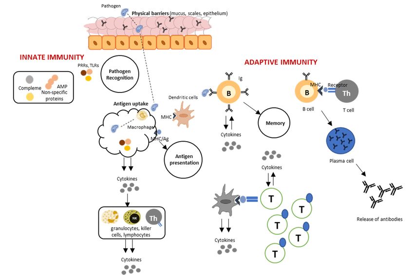
Figure 1. Immune response at the fish mucosa. When pathogens succeed to infiltrate the physical
barriers, the innate immune system is triggered by the action of pathogen recognition receptors (PRRs).
Antigen uptake results in the release of cytokines that activates different types of cells involved in
inflammatory processes, and antigen presentation facilitated by lymphocytes containing receptors,
which leads to subsequent responses as well as memory. This diagram is the simplified version from
Beck and Peatman (2015) with Elsevier License number 4477021492352 [6].Fishes 2019, 4, 2 3 of 10
2. Variability of Environmental Parameters in Aquaculture
Adaptations to varying environmetal pressures are often advantageous, since they can be
crucial to the survival of an organism [13,14]. Phenotypic plasticity, the phenomenon of producing
different phenotypes in response to change in environmental conditions, is a ubiquitous aspect of
the organism’s evolutionary adaptation [15,16]. Fish are highly vulnerable to environmental changes
because of the constant and intimate contact with the aquatic environment. In the wild, fish encounter
fluctuating environmental factors throughout their lifetime. Some of these environmental variations
are short-lived, while others may last for a considerable period, which may leave both phenotypic and
physiological imprints in the organism. Many organisms exhibit an impressive repertoire of adaptive
mechanisms to thrive in various environmental conditions, hence facilitating survival.
Fish domestication relies heavily on the control of environmental parameters. Successful and
sustainable husbandry is fostering a rearing environment for fish to grow and survive under optimum
controlled conditions. Environmental control in aquaculture often involves the manipulations
of physico-chemical factors that support the physiological functions in fish. The changes in the
environmental conditions in the production system may also be driven by the excretion products,
both from fish and the microbial populations present in the rearing environment or the lack of
equilibrium among the physico-chemical factors that support fish in the system. Some of the
key environmental parameters that are critical in supporting the physiological processes in fish,
especially in farming conditions, include dissolved oxygen, temperature, salinity, and photoperiod,
among many others.
Moreover, these changes have pervasive impacts on the overall welfare status of fish.
These environmental parameters can influence the survival and resistance of fish against diseases,
stressful conditions, and more. The mucosal immune system, in particular, has inherent importance
in the ability of fish to respond and adapt to these changes because of their direct connection with
the environment. In a number of studies, it has been documented that mucosal surfaces respond
exceptionally to these environmental changes—for example, phenotypic alterations (e.g., increase
in mucus cell size and numbers), molecular responses (e.g., transcriptional and proteomic changes),
and diversification (e.g., microbiota profile).
This short review discusses how different environmental factors relevant to aquaculture influence
the basic physiological functions of the mucosal barriers and how the adaptive changes contribute to the
plasticity of mucosal functionality. To the best of our knowledge, no published paper has synthesized
the responses of fish to environmental changes, particularly within the context of mucosal barriers.
Previous papers discussed several aspects of this theme separately. Studies that are enumerated in this
short review highlight both the current knowledge and the gaps about this timely and highly relevant
topic in aquaculture.
3. Dissolved Oxygen
Dissolved oxygen (DO) is the main limiting factor in an aquaculture system [17]. Oxygen is
important in respiration and thus controls all cellular functions. Low DO (hypoxia) is known to alter
the structure of the gills to accommodate more oxygen. Hypoxia-induced morphological adaptations
in the gills were highly documented in cold-water carp species, which are known for their tolerance
in low DO. Crucian carp (Carassius carassius) exposed to 0.75 mg L−1 DO display protruding gill
lamellae accompanied by an increased interlamellar cell mass that resulted to a 7-fold surface area
increase in at least 24 h of exposure [18]. A similar gill remodeling strategy was also observed in
goldfish (Carassius auratus) [19]. In Qinghai scaleless carp (Gymnocypris przewalskii), increase in gill
surface area occurred 8 h post-exposure to 0.3 mg L−1 DO and a shift into the small “shallow-basin”
type of mitochondria rich-cells (MRC) from wavy-convex-type was observed 12 h post-exposure [20].
These changes induce gas transfer during hypoxia and were found to be reversible after exposure to
normoxic conditions.Fishes 2019, 4, 2 4 of 10
The sensitivity of the gut to hypoxic conditions has been observed in Atlantic salmon (Salmo salar).
Exposure to low DO levels (50% saturation) caused chronic inflammation in the gut but the effect
varied depending on the temperature. At 8 ◦ C, mucosal neutrophil infiltration increased, together with
a down-regulation in the expression of inhibitor of nuclear factor kB (ikB) gene, while at 16 ◦ C, expression
of the pro-inflammatory cytokine interleukin 1β (il-1 β) decreased, while expression of interleukin 10
(il-10) increased. Differential expression of immune factors was more striking at 16 ◦ C, which shows
that temperature intensifies the effects of hypoxia in this species [21].
The effects of high DO levels (hyperoxia) have been studied in some species. In Crucian carp and
goldfish, high temperature and high DO (300% saturation) resulted in gill remodelling (i.e., increase
in interlamellar cell mass) [22]. The exposure of Beluga sturgeon (Huso huso) to 115% saturation for
8 weeks resulted in changes in the gill structure, such as impairment of the secondary lamellae [23].
At the molecular level, hyperoxia of up to 200% saturation caused a decrease of expression of
cyclooxygenase-2 (cox2), a gene important for pro-inflammatory pathways, in the gills of Senegalese sole
(Solea senegalensis) after 24 h [24].
Suboptimal DO levels put the fish in a challenging condition that would result in the perturbations
of mucosal barrier functions. These have been shown in the histological and molecular changes in the
mucosal surfaces in response to variable DO levels. Disturbances in the functions of mucosal surfaces
brought about by the highly demanding conditions at low DO may increase their permeability to
pathogens. Thus, it may likely result in the increase of susceptibility to infection. Many fish pathogens
are opportunistic; they cause infection when the environmental conditions are sub-optimal and host
barriers are compromised.
4. Water pH
Next to DO, water pH is one of the secondary limiting factors in an aquaculture system [17].
Water pH is influenced by carbon dioxide (CO2 ) through the bicarbonate-carbonate balance, as well as
alkalinity, which serves as a buffer system that prevents sudden changes in pH [25]. Non-CO2 change
in water pH is driven by organic acids, inorganic substances, and anthropogenic factors (e.g., mining,
industrial effluents) [26].
Changes in water pH as a result of pollution are a recurring problem, especially in freshwater
aquaculture which is a major industry in areas near water basins. Responses of freshwater species
to water pH beyond the normal physiological range have been studied, with considerable changes
observed in mucosal organs. In brond snout (Chondrostoma regium), acute exposure (i.e., 24 h) to high
pH (9.8–9.9) caused gill lamella hyperplasia and fusion of secondary lamella, while exposure to low pH
(4.4) resulted in club-shaped cells and hypertrophy of the epithelium [27]. In zebrafish (Danio rerio) gills,
pH 4.0 increased the density of H+ -ATPase-rich cells, involved in acid secretion and Na+ secretion. [28].
These morphological changes allow the fish to adapt their respiratory structures to the physiological
demands of variable water pH. Tight junctions, which limit the passage of molecules in spaces between
epithelial cells [29], are also affected by water pH. In rainbow trout (Oncorhynchus mykiss), length of
tight junctions between pavement epithelial cells and chloride cells decreased by 25% at pH 4.0 [30].
Claudins, a large class of tight junction proteins, are also regulated in acidic environments, as shown by
the increase in expression of claudin-b (cldnb), linked to the tight junction between lamellar epithelium
cells [31] in zebrafish gills after exposure to pH 3.8–4.0 [32]. The antioxidant defense at the mucosa
is also responsive to changes in water pH. Zebrafish exposed to the same pH showed differential
regulation of gene coding for superoxide dismutase (sod), catalase (cat), and glutathione peroxidase (gpx)
in the gills. This reveals that the antioxidant system is not only important in combating oxidative
damage in the presence of high oxygen radicals, but may also have key roles in the responses of the
gills in pH variability [33].
In tambaqui (Colossoma macropomum), a freshwater fish found in the Amazon river, skin microbiota
is affected by lower water pH (for example, pH 4.0) [34]. It was further shown that unlike the skin
microbiota, the diversity and structure of the gut microbiota of tambaqui was more robust under lowerFishes 2019, 4, 2 5 of 10
pH, indicating that the microbial community in the gut is quite resistant to suboptimal pH levels.
These alterations in mucosal features not only have implications on their functionality but can also
serve as determinants of the extent of pollution in freshwater environments.
5. Carbon Dioxide
Carbon dioxide (CO2 ), which exists in the water as bicarbonate, carbonate and carbonic acid
forms, is an environmental factor in an aquaculture system that has a striking impact on the mucosal
functionality in fish [25]. CO2 can both directly impact the physiological functions of fish and indirectly
affect the host by changing the water pH [17]. Carbonate concentration is important in maintaining
the buffer capacity of the rearing water against pH fluctuations and can affect the toxicity of some
components, such as ammonia and hydrogen sulfide.
The gills, being involved in acid-base regulation, are especially susceptible to high CO2 levels
(hypercarbia). Mitochondria-rich cells (MRC) that are involved in acid-base regulation [35] have
been studied to determine the effects of hypercarbia in the gills. The results varied across species.
For instance, in channel catfish (Ictalurus punctatus) exposed to 8% CO2 and in olive flounder
(Paralichthys olivaceus) exposed to 3–5% CO2 , the apical opening surface area of MRCs increased
to accommodate the rise in CO2 concentrations and ease the conversion to HCO3− and CO3 2− [36,37].
In contrast, a decrease in the MRC apical opening surface area was observed in brown bullhead
(Ictalurus nebulosus) exposed to 2% CO2 , in white sturgeon (Acipenser transmontanus) exposed to 1.53%
CO2 , and slightly in rainbow trout exposed to 1% CO2 [38–40]. It is apparent that changes in MRC are
more likely to occur at higher concentrations of CO2, and interspecies differences are quite remarkable
in the observed changes. A good acid-base regulation would result in healthy gills, which allows a
fully functional mucosal barrier.
The interaction of CO2 with other environmental variables, such as temperature and heavy metals,
was also assessed in several studies. Hypercarbia at 10 ◦ C resulted in an increase in gill tissue mass of
Atlantic cod (Gadus morhua), but not at 18 ◦ C [41]. The increase in size can be attributed to the response
of fish to rapidly eliminate high CO2 concentrations [42]. Co-occurring acidification (CO2 + Hg)
decreased the accumulation of Hg in Argyrosomus regius and decreased the activity of oxidative stress
markers i.e. catalase, superoxide dismutase (SOD), and glutathione S-transferase activities in the
gills [43]. These findings may give insights into the adaptive mechanisms of the gills to pollution in
relation to projected ocean acidification, and consequently, on the future of marine cage culture.
Given the low water change that results in high CO2 accumulation, hypercarbia is a foreseen
problem in recirculating aquaculture system (RAS). In a recent study, Mota et al. (2019) observed thinner
epidermis in Atlantic salmon exposed to a CO2 concentration greater than 19 ppm, which renders the
host more susceptible to pathogen infiltration [44]. This information shall give rise to more studies
on the effect of high CO2 concentrations on the mucosal barriers of other aquaculture species reared
in RAS.
6. Temperature
Temperature is a widely studied environmental cue in fish, not only in reproduction and animal
behavior but also in immune response and the progression of infectious diseases [45,46]. Temperature
can also intensify the effects of other physicochemical parameters (e.g., DO levels) in the rearing
water [20].
Eurythermal species are a suitable model for determining the phenotypic changes in response to
temperature variations within the physiological range. In Crucian carp and goldfish with temperature
tolerances of 2–22 ◦ C and 0–41 ◦ C, respectively, hypertrophy of interlamellar cells was observed after a
30-day exposure to 7.5 ◦ C [47]. In fathead minnow (Pimephales promela) with temperature tolerance of
0–33 ◦ C acclimated to 5 ◦ C, no changes occurred in interlamellar cells, although mucus cells in the gills
were observed to be fewer but larger after acclimation [48].Fishes 2019, 4, 2 6 of 10
Cellular responses varied when fish were exposed to the upper or lower temperature limits of
their physiological tolerance. Lysozyme activity, hepcidin, and immunoglobulin M (IgM) increased in
the skin of turbot (Scophthalmus maximus), with a temperature tolerance of 16–20 ◦ C after exposure
to 27 ◦ C [49]. Genes coding for proteins that block viral replication in the early phase, macrophage
activation, mucus secretion, and pro-inflammatory responses were upregulated while the genes coding
for antigen presentation and immunoglobulins were downregulated in fathead minnow acclimated
to 5 ◦ C [48]. In channel catfish, the adaptive immune response to T-dependent antigens in the gills is
inhibited by low temperature [50].
The antioxidant systems of mucosal surfaces are also responsive to thermal variability. The SOD
levels in the skin mucus of turbot increased significantly when temperature was elevated from
16 ◦ C to 20 ◦ C. While it is within the normal range for turbot, it is apparent that in the upper
temperature limits, the markers for oxidative stress were remarkably affected. The same tendency
of increased SOD levels at a temperature higher than the normal rearing temperature (21–25 ◦ C)
was also documented in the gills of Asian stinging catfish (Heteropneustes fossilis) reared at 32 ◦ C [51].
Reduced gill glutathione peroxidase (GPX) activity was observed in Antarctic fishes, Notothenia coriiceps,
and N. rossi after exposure to 4 ◦ C for 1 day (long-term) in contrast to 2 ◦ C for 6 days (short-term) [52].
The temperature at 20 ◦ C can also activate a protective antioxidant defense response in gills of discus
fish (Symphysodon aequifasciatus), as evidenced by the increase in SOD and GPX activities, compared to
a temperature of 28 ◦ C [53].
Thermal plasticity of mucosal surfaces has a key role in the robustness of fish under variable
temperatures. Thermal manipulations at early life stages have been observed to influence robustness
at a later stage of fish, however, little is known on its role in mucosal immunomodulation.
7. Salinity
Salinity is important in the osmoregulatory functions of fish. The interesting overlap between
osmoregulation and immunity purported some studies to determine the effect of changes in the
salinity on the immune responses of fish. Acclimation to different salinity levels in euryhaline species,
such as Atlantic salmon, an anadromous species, increases susceptibility to diseases during transitional
stages. In fact, mortalities of up to 16% were observed after seawater transfer of post-smolts [54].
However, Karlsen et al. (2018) saw a progressive improvement in the protective functions in the skin
of Atlantic salmon post-smolts, such as the increased mucus cell number and thickness observed
4 months after seawater transfer [55]. Furthermore, assessment of the mucus lysozyme activity of
salmonids reared in either freshwater or seawater shows that Atlantic salmon had the least activity
compared with coho salmon (Oncorhynchus kisutch) and rainbow trout when reared in seawater, while
in freshwater, Atlantic salmon and coho salmon had the highest activity [56]. The variability in the
results gives insights into the diversity of salmonid cutaneous defense system in response to various
salinity conditions.
Mucosal barriers are influenced by salinity at the molecular level. For instance, freshwater
acclimation reduced the expression of genes coding for tight junction proteins claudin-3 (cldn3)
and claudin-4 (cldn4) in the gills of southern flounder (Paralichthys lethostigma) [57]. In green-spotted
pufferfish (Tetraodon nigroviridis), claudin-10d (cldn10d) and cldn10e increased in the gills of fish
acclimated to seawater versus freshwater, while claudin-6 (cldn6) decreased in the gills of fish acclimated
to seawater [58]. Seawater acclimation of Atlantic salmon resulted in differential regulation of claudins
(i.e., increase in the expression of claudin-10e (cldn10e) and decrease in the expression of claudin-27a and
-30 (cldn27a, cldn30) in the gills) [59]. Moreover, an increase in the expression of genes encoding antigen
presentation, complement, immunoglobulins, acute phase proteins, and lymphocytes, to mention a
few, was observed in the skin Atlantic salmon 4 months after seawater transfer [55]. On the other hand,
acclimating in seawater significantly reduced the expression of immune-related genes, i.e., C-reactive
protein (crp), toll-like receptor 2 (tlr2), and interleukin-1 receptor type 2 (il1-1r2), in the gills of Japanese eel
(Anguilla japonica), a catadromous species [60].Fishes 2019, 4, 2 7 of 10
Acclimation to different salinities also impacts the microbiota at the mucosa. Transfer of Atlantic
salmon from freshwater to seawater resulted in the destabilization of skin microbiota. The abundance
of Proteobacteria, for instance, increased from 45% in freshwater to 89% in seawater. This drastic
change reveals that microbiota of mucosal surfaces undergoes restructuring as a form of adaptation to
salinity changes [61].
8. Photoperiod
Photoperiod, or the light-dark cycle, is an environmental cue that controls many biological
activities in fish, including their defense mechanisms. Photoperiod manipulation is a practice to
regulate sexual maturation and spawning. Though the use of artificial photoperiod has beneficial
effects on regulating the reproduction cycles of farmed fish, it has been observed to cause morphological
changes, such as skin lesions and ulcerative-type necrosis in rainbow trout, accompanied by mortalities
of up to 36% in LD 14:10 and 25% in LD 24:0 compared to the control setup (7%; LD10:14) [62].
Flavobacterium psychrophilum was identified as the causative agent of the mortalities, though the
isolation of Aeromonas, Pseudomonas, and Saprolegnia in the diseased fish implies an increased
susceptibility of fish to opportunistic diseases.
The current understanding proposes that partitioning of the immune system characterizes the
circadian impact to immune functions into a state of anticipation and enhanced immune activity, and a
state of repair and regeneration [63]. Understanding the circadian rhythm of mucosal defenses in
fish can be exploited to design photoperiod strategies that do not hamper normal barrier functions,
and at the same time, can be used to deliver time-dependent strategies that aim at modulating fish
immunity. The impact of daily rhythm to key defense factors in the mucus of permit (Trachinotus falcatus)
reared under a 12L:12D cycle has been described by Lazado et al. (2015) [64]. Daily rhythmicity of
defense enzymes in the mucus was observed in alkaline phosphatase (ALP) and GPX, with the latter
having a dark-biased activity. The significant daily rhythmic pattern and similarity in acrophase of
ALP in both serum and mucus suggests that this defense enzyme may be one of the vital components
in the circadian-dependent immune defenses in permit.
9. Conclusions
The studies enumerated in this short review highlighted the remarkable influence of different
environmental factors to fish mucosal barrier functionality. The changes in these factors not only
impact the mucosal structural phenotypes and molecular response but also influence the mucus and
microbiota that line the mucosal surfaces. It is quite apparent, however, that our current understanding
of how environmental parameters in aquaculture systems impact mucosal barrier functions is quite
limited and fragmentary. Future studies should be directed not only on how mucosal surfaces respond
to environmental changes but also their mechanisms on adaption and the long-term impacts of the
variability of environmental parameters. A thorough understanding of this interaction is particularly
important in modern fish husbandry, in which production technologies are addressing strict control of
aquaculture environments to foster heightened biosecurity and more sustainable production systems.
Author Contributions: Conceptualization, N.A.R.C.; writing—original draft preparation, N.A.R.C., C.C.L.;
writing—review and editing, C.C.L., N.A.R.C.; visualization, N.A.R.C.
Funding: This research received no external funding.
Acknowledgments: The writing of this short review is made possible through SFI CtrlAQUA—Centre for Closed
Containment Aquaculture funded by the Norwegian Research Council (ref. 237856).
Conflicts of Interest: The authors declare no conflict of interest.
References
1. Lazado, C.C.; Caipang, C.M.A. Mucosal immunity and probiotics in fish. Fish Shellfish Immunol. 2014,
39, 78–89. [CrossRef] [PubMed]Fishes 2019, 4, 2 8 of 10
2. Salinas, I. The Mucosal Immune System of Teleost Fish. Biology 2015, 4, 525–539. [CrossRef] [PubMed]
3. Smith, H.W. The absorption and excretion of water and salts by marine teleosts. Am. J. Physiol. Legacy Content
1930, 93, 480–505. [CrossRef]
4. Evans, D.H.; Piermarini, P.M.; Choe, K.P. The multifunctional fish gill: Dominant site of gas exchange,
osmoregulation, acid-base regulation, and excretion of nitrogenous waste. Physiol. Rev. 2005, 85, 97–177.
[CrossRef] [PubMed]
5. Grossel, M.; Farrell, A.; Brauner, C. Fish Physiology: The Multifunctional Gut of Fish, 1st ed.; Academic Press:
Cambridge, MA, USA, 2010; Volume 30.
6. Beck, B.H.; Peatman, E. Mucosal Health in Aquaculture, 1st ed.; Academic Press: Cambridge, MA, USA, 2015.
7. Lazado, C.C.; Caipang, C.M.A.; Estante, E.G. Prospects of host-associated microorganisms in fish and
penaeids as probiotics with immunomodulatory functions. Fish Shellfish Immunol. 2015, 45, 2–12. [CrossRef]
[PubMed]
8. Rombout, J.H.W.M.; Yang, G.; Kiron, V. Adaptive immune responses at mucosal surfaces of teleost fish.
Fish Shellfish Immunol. 2014, 40, 634–643. [CrossRef]
9. Sunyer, J.O. Fishing for mammalian paradigms in the teleost immune system. Nature Immunol. 2013, 14, 320.
[CrossRef] [PubMed]
10. Nigam, A.K.; Kumari, U.; Mittal, A.K. Comparative analysis of innate immune parameters of the skin mucous
secretions from certain freshwater teleosts, inhabiting different ecological niches. Fish Physiol. Biochem. 2012,
38, 1245–1256. [CrossRef]
11. Ellis, A.E. Innate host defense mechanisms of fish against viruses and bacteria. Dev. Comp. Immunol. 2001,
25, 827–839. [CrossRef]
12. Boutin, S.; Bernatchez, L.; Audet, C.; Derôme, N. Network analysis highlights complex interactions between
pathogen, host and commensal microbiota. PLoS ONE 2013, 8, e84772. [CrossRef]
13. Dudley, S.A.; Schmitt, J. Testing the Adaptive Plasticity Hypothesis: Density-Dependent Selection on
Manipulated Stem Length in Impatiens capensis. Am. Nat. 1996, 147, 445–465. [CrossRef]
14. Meyers, L.A.; Bull, J.J. Fighting change with change: Adaptive variation in an uncertain world.
Trends Ecol. Evol. 2002, 17, 551–557. [CrossRef]
15. Travis, J. Evaluating the adaptive role of morphological plasticity. In Ecological Morphology: Integrative
Organismal Biology; University of Chicago Press: Chicago, IL, USA, 1994; pp. 99–122.
16. West-Eberhard, M.J. Developmental Plasticity and Evolution; Oxford University Press: Cary, NC, USA, 2003;
ISBN 978-0-19-512235-0.
17. Fivelstad, S. Long-term carbon dioxide experiments with salmonids. Aqua. Eng. 2013, 53, 40–48. [CrossRef]
18. Sollid, J.; De Angelis, P.; Gundersen, K.; Nilsson, G.E. Hypoxia induces adaptive and reversible gross
morphological changes in crucian carp gills. J. Exp. Biol. 2003, 206, 3667–3673. [CrossRef] [PubMed]
19. Sollid, J.; Nilsson, G.E. Plasticity of respiratory structures—Adaptive remodeling of fish gills induced by
ambient oxygen and temperature. Respir. Physiol. Neurobiol. 2006, 154, 241–251. [CrossRef] [PubMed]
20. Matey, V.; Richards, J.G.; Wang, Y.; Wood, C.M.; Rogers, J.; Davies, R.; Murray, B.W.; Chen, X.-Q.; Du, J.;
Brauner, C.J. The effect of hypoxia on gill morphology and ionoregulatory status in the Lake Qinghai
scaleless carp, Gymnocypris przewalskii. J. Exp. Biol. 2008, 211, 1063–1074. [CrossRef]
21. Niklasson, L.; Sundh, H.; Fridell, F.; Taranger, G.L.; Sundell, K. Disturbance of the intestinal mucosal
immune system of farmed Atlantic salmon (Salmo salar), in response to long-term hypoxic conditions.
Fish Shellfish Immunol. 2011, 31, 1072–1080. [CrossRef]
22. Tzaneva, V.; Bailey, S.; Perry, S.F. The interactive effects of hypoxemia, hyperoxia, and temperature on
the gill morphology of goldfish (Carassius auratus). Am. J. Physiol. Regul. Integr. Comp. Physiol. 2011,
300, R1344–R1351. [CrossRef] [PubMed]
23. Bagherzadeh Lakani, F.; Sattari, M.; Falahatkar, B. Effect of different oxygen levels on growth performance,
stress response and oxygen consumption in two weight groups of great sturgeon Huso huso. Iran. J. Fish. Sci.
2013, 12, 533–549.
24. Machado, M.; Malheiro, D.; Couto, A.; Wilson, J.M.; Guerreiro, M.; Azeredo, R.; Svendsen, J.C.; Afonso, A.;
Serradeiro, R.; Costas, B. Acute hyperoxia induces systemic responses with no major changes in peripheral
tissues in the Senegalese sole (Solea senegalensis Kaup, 1858). Fish Shellfish Immunol. 2018, 74, 260–267.
[CrossRef]
25. Boyd, C.E.; Tucker, C.S. Pond Aquaculture Water Quality Management; Springer: New York, NY, USA, 1998.Fishes 2019, 4, 2 9 of 10
26. Wilkie, M.P.; Wood, C.M. The adaptations of fish to extremely alkaline environments. Comp. Biochem. Physiol.
Part B Biochem. Mol. Biol. 1996, 113, 665–673. [CrossRef]
27. Mohammadi, M.; Mahboobi-Soofiani, N.; Farhadian, O.; Malekpouri, P. Metabolic and NH4 excretion rate
of fresh water species, Chondrostoma regium in response to environmental stressors, different scenarios for
temperature and pH. Sci. Total Environ. 2019, 648, 90–101. [CrossRef] [PubMed]
28. Chang, W.J.; Horng, J.L.; Yan, J.J.; Hsiao, C.D.; Hwang, P.P. The transcription factor, glial cell missing 2, is
involved in differentiation and functional regulation of H+ -ATPase-rich cells in zebrafish (Danio rerio). Am. J.
Physiol. Regul. Integr. Comp. Physiol. 2009, 296, R1192–R1201. [CrossRef] [PubMed]
29. Günzel, D.; Fromm, M. Claudins and Other Tight Junction Proteins. In Comprehensive Physiology; American
Cancer Society: Atlanta, GA, USA, 2012; pp. 1819–1852. ISBN 978-0-470-65071-4.
30. Freda, J.; Sanchez, D.A.; Bergman, H.L. Shortening of Branchial Tight Junction Acid-Exposed Rainbow Trout
(Oncorhynchus mykiss). Can. J. Fish. Aquat. Sci. 1991, 48, 2028–2033. [CrossRef]
31. Kwong, R.W.M.; Perry, S.F. Cortisol regulates epithelial permeability and sodium losses in zebrafish exposed
to acidic water. J. Endocrinol. 2013, 217, 253–264. [CrossRef] [PubMed]
32. Kumai, Y.; Bahubeshi, A.; Steele, S.; Perry, S.F. Strategies for maintaining Na+ balance in zebrafish (Danio rerio)
during prolonged exposure to acidic water. Comp. Biochem. Physiol. Part A Mol. Integr. Physiol. 2011,
160, 52–62. [CrossRef] [PubMed]
33. Tiedke, J.; Cubuk, C.; Burmester, T. Environmental acidification triggers oxidative stress and enhances globin
expression in zebrafish gills. Biochem. Biophys. Res. Commun. 2013, 441, 624–629. [CrossRef]
34. Sylvain, F.-É.; Cheaib, B.; Llewellyn, M.; Gabriel Correia, T.; Barros Fagundes, D.; Luis Val, A.; Derome, N. pH
drop impacts differentially skin and gut microbiota of the Amazonian fish tambaqui (Colossoma macropomum).
Sci. Rep. 2016, 6, 32032. [CrossRef]
35. Sloman, K.A. Mitochondria-Rich Cell Subtypes in Fish Gill. J. Exp. Biol. 2003, 206, 7–8. [CrossRef]
36. Cameron, J.N.; Iwama, G.K. Compensation of Progressive Hypercapnia in Channel Catfish and Blue Crabs.
J. Exp. Biol. 1987, 133, 183–197.
37. Hayashi, M.; Kikkawa, T.; Ishimatsu, A. Morphological changes in branchial mitochondria-rich cells of the
teleost Paralichthys olivaceus as a potential indicator of CO2 impacts. Mar. Pollut. Bull. 2013, 73, 409–415.
[CrossRef] [PubMed]
38. Goss, G.G.; Laurent, P.; Perry, S.F. Evidence for a morphological component in acid-base regulation during
environmental hypercapnia in the brown bullhead (Ictalurus nebulosus). Cell Tissue Res. 1992, 268, 539–552.
[CrossRef]
39. Goss, G.G.; Perry, S.F. Physiological and morphological regulation of acid–base status during hypercapnia in
rainbow trout (Oncorhynchus mykiss). Can. J. Zool. 1993, 71, 1673–1680. [CrossRef]
40. Baker, D.W.; Matey, V.; Huynh, K.T.; Wilson, J.M.; Morgan, J.D.; Brauner, C.J. Complete intracellular pH
protection during extracellular pH depression is associated with hypercarbia tolerance in white sturgeon,
Acipenser transmontanus. Am. J. Physiol. Regul. Integr. Comp. Physiol. 2009, 296, R1868–R1880. [CrossRef]
[PubMed]
41. Kreiss, C.M.; Michael, K.; Lucassen, M.; Jutfelt, F.; Motyka, R.; Dupont, S.; Pörtner, H.O. Ocean warming and
acidification modulate energy budget and gill ion regulatory mechanisms in Atlantic cod (Gadus morhua).
J. Comp. Physiol. B 2015, 185, 767–781. [CrossRef] [PubMed]
42. Kreiss, C.M.; Michael, K.; Bock, C.; Lucassen, M.; Pörtner, H.O. Impact of long-term moderate hypercapnia
and elevated temperature on the energy budget of isolated gills of Atlantic cod (Gadus morhua). Comp. Biochem.
Physiol. Part A Mol. Integr. Physiol. 2015, 182, 102–112. [CrossRef] [PubMed]
43. Sampaio, E.; Lopes, A.R.; Francisco, S.; Paula, J.R.; Pimentel, M.; Maulvault, A.L.; Repolho, T.; Grilo, T.F.;
Pousão-Ferreira, P.; Marques, A.; et al. Ocean acidification dampens physiological stress response to
warming and contamination in a commercially-important fish (Argyrosomus regius). Sci. Total Environ. 2018,
618, 388–398. [CrossRef] [PubMed]
44. Mota, V.C.; Nilsen, T.O.; Gerwins, J.; Gallo, M.; Ytteborg, E.; Baeverfjord, G.; Kolarevic, J.; Summerfelt, S.T.;
Terjesen, B.F. The effects of carbon dioxide on growth performance, welfare, and health of Atlantic salmon
post-smolt (Salmo salar) in recirculating aquaculture systems. Aquaculture 2019, 498, 578–586. [CrossRef]
45. Bly, J.E.; Clem, L.W. Temperature and teleost immune functions. Fish Shellfish Immunol. 1992, 2, 159–171.
[CrossRef]Fishes 2019, 4, 2 10 of 10
46. Bowden, T.J.; Thompson, K.D.; Morgan, A.L.; Gratacap, R.M.L.; Nikoskelainen, S. Seasonal variation and the
immune response: A fish perspective. Fish Shellfish Immunol. 2007, 22, 695–706. [CrossRef] [PubMed]
47. Sollid, J.; Weber, R.E.; Nilsson, G.E. Temperature alters the respiratory surface area of crucian carp
Carassius carassius and goldfish Carassius auratus. J. Exp. Biol. 2005, 208, 1109–1116. [CrossRef] [PubMed]
48. Wentworth, S.A.; Thede, K.; Aravindabose, V.; Monroe, I.; Thompson, A.W.; Molyneaux, N.; Owen, C.L.;
Burns, J.R.; Gonzalez-Vicente, A.; Garvin, J.L.; et al. Transcriptomic analysis of changes in gene expression
of immune proteins of gill tissue in response to low environmental temperature in fathead minnows
(Pimephales promelas). Comp. Biochem. Physiol. Part D Genom. Proteom. 2018, 25, 109–117. [CrossRef] [PubMed]
49. Huang, Z.-H.; Ma, A.-J.; Wang, X.-A. The immune response of turbot, Scophthalmus maximus (L.), skin to
high water temperature. J. Fish Dis. 2011, 34, 619–627. [CrossRef] [PubMed]
50. Clem, L.W.; Faulmann, E.; Miller, N.W.; Ellsaesser, C.; Lobb, C.J.; Cuchens, M.A. Temperature-mediated
processes in teleost immunity: Differential effects of in vitro and in vivo temperatures on mitogenic responses
of channel catfish lymphocytes. Dev. Comp. Immunol. 1984, 8, 313–322. [CrossRef]
51. Parihar, M.S.; Javeri, T.; Hemnani, T.; Dubey, A.K.; Prakash, P. Responses of superoxide dismutase,
glutathione peroxidase and reduced glutathione antioxidant defenses in gills of the freshwater catfish
(Heteropneustes fossilis) to short-term elevated temperature. J. Therm. Biol. 1997, 22, 151–156. [CrossRef]
52. Klein, R.D.; Borges, V.D.; Rosa, C.E.; Colares, E.P.; Robaldo, R.B.; Martinez, P.E.; Bianchini, A. Effects of
increasing temperature on antioxidant defense system and oxidative stress parameters in the Antarctic fish
Notothenia coriiceps and Notothenia rossii. J. Therm. Biol. 2017, 68, 110–118. [CrossRef] [PubMed]
53. Wen, B.; Jin, S.R.; Chen, Z.Z.; Gao, J.-Z. Physiological responses to cold stress in the gills of discus fish
(Symphysodon aequifasciatus) revealed by conventional biochemical assays and GC-TOF-MS metabolomics.
Sci. Total Environ. 2018, 640–641, 1372–1381. [CrossRef]
54. Bleie, H.; Skrudland, A. Tap av laksefisk i sjø. Rapport Fra Mattilsynet; Mattilsynet: Oslo, Norway, 2014.
55. Karlsen, C.; Ytteborg, E.; Timmerhaus, G.; Høst, V.; Handeland, S.; Jørgensen, S.M.; Krasnov, A. Atlantic
salmon skin barrier functions gradually enhance after seawater transfer. Sci. Rep. 2018, 8, 9510. [CrossRef]
56. Fast, M.D.; Sims, D.E.; Burka, J.F.; Mustafa, A.; Ross, N.W. Skin morphology and humoral non-specific
defence parameters of mucus and plasma in rainbow trout, coho and Atlantic salmon. Comp. Biochem.
Physiol. Part A Mol. Integr. Physiol. 2002, 132, 645–657. [CrossRef]
57. Tipsmark, C.K.; Luckenbach, J.A.; Madsen, S.S.; Kiilerich, P.; Borski, R.J. Osmoregulation and expression
of ion transport proteins and putative claudins in the gill of southern flounder (Paralichthys lethostigma).
Comp. Biochem. Physiol. Part A Mol. Integr. Physiol. 2008, 150, 265–273. [CrossRef]
58. Bui, P.; Kelly, S.P. Claudin-6, -10d and -10e contribute to seawater acclimation in the euryhaline puffer fish
Tetraodon nigroviridis. J. Exp. Biol. 2014, 217, 1758–1767. [CrossRef] [PubMed]
59. Tipsmark, C.K.; Kiilerich, P.; Nilsen, T.O.; Ebbesson, L.O.E.; Stefansson, S.O.; Madsen, S.S. Branchial
expression patterns of claudin isoforms in Atlantic salmon during seawater acclimation and smoltification.
Am. J. Physiol. Regul. Integr. Comp. Physiol. 2008, 294, R1563–R1574. [CrossRef]
60. Gu, J.; Dai, S.; Liu, H.; Cao, Q.; Yin, S.; Lai, K.P.; Tse, W.K.F.; Wong, C.K.C.; Shi, H. Identification of
immune-related genes in gill cells of Japanese eels (Anguilla japonica) in adaptation to water salinity changes.
Fish Shellfish Immunol. 2018, 73, 288–296. [CrossRef] [PubMed]
61. Lokesh, J.; Kiron, V. Transition from freshwater to seawater reshapes the skin-associated microbiota of
Atlantic salmon. Sci. Rep. 2016, 6, 19707. [CrossRef]
62. Valenzuela, A.; Campos, V.; Yañez, F.; Alveal, K.; Gutiérrez, P.; Rivas, M.; Contreras, N.; Klempau, A.;
Fernandez, I.; Oyarzun, C. Application of artificial photoperiod in fish: A factor that increases susceptibility
to infectious diseases? Fish Physiol. Biochem. 2012, 38, 943–950. [CrossRef] [PubMed]
63. Curtis, A.M.; Bellet, M.M.; Sassone-Corsi, P.; O’Neill, L.A.J. Circadian clock proteins and immunity. Immunity
2014, 40, 178–186. [CrossRef] [PubMed]
64. Lazado, C.C.; Lund, I.; Pedersen, P.B.; Nguyen, H.Q. Humoral and mucosal defense molecules rhythmically
oscillate during a light–dark cycle in permit, Trachinotus falcatus. Fish Shellfish Immunol. 2015, 47, 902–912.
[CrossRef] [PubMed]
© 2019 by the authors. Licensee MDPI, Basel, Switzerland. This article is an open access
article distributed under the terms and conditions of the Creative Commons Attribution
(CC BY) license (http://creativecommons.org/licenses/by/4.0/).You can also read