Cannabinoids and Prostate Cancer: A Systematic Review of Animal Studies - MDPI
←
→
Page content transcription
If your browser does not render page correctly, please read the page content below
International Journal of
            Molecular Sciences

Review
Cannabinoids and Prostate Cancer: A Systematic
Review of Animal Studies
Kanika Singh 1 , Negar Jamshidi 2 , Roby Zomer 3 , Terrence J. Piva 2          and Nitin Mantri 1, *
 1    The Pangenomics Lab, School of Science, RMIT University, Bundoora, Victoria 3083, Australia;
      kanika.singh@rmit.edu.au
 2    School of Health and Biomedical Sciences, RMIT University, Bundoora, Victoria 3083, Australia;
      negar.jamshidi@rmit.edu.au (N.J.); terry.piva@rmit.edu.au (T.J.P.)
 3    MGC Pharmaceuticals Limited, West Perth, Western Australia 6005, Australia; roby@mgcpharma.com.au
 *    Correspondence: nitin.mantri@rmit.edu.au; Tel.: +61-3-9925-7152

 Received: 30 July 2020; Accepted: 28 August 2020; Published: 29 August 2020

 Abstract: Prostate cancer is a major cause of death among men worldwide. Recent preclinical
 evidence implicates cannabinoids as powerful regulators of cell growth and differentiation, as well
 as potential anti-cancer agents. The aim of this review was to evaluate the effect of cannabinoids
 on in vivo prostate cancer models. The databases searched included PubMed, Embase, Scopus,
 and Web of Science from inception to August 2020. Articles reporting on the effect of cannabinoids on
 prostate cancer were deemed eligible. We identified six studies that were all found to be based on
 in vivo/xenograft animal models. Results: In PC3 and DU145 xenografts, WIN55,212-2 reduced cell
 proliferation in a dose-dependent manner. Furthermore, in LNCaP xenografts, WIN55,212-2 reduced
 cell proliferation by 66–69%. PM49, which is a synthetic cannabinoid quinone, was also found to
 result in a significant inhibition of tumor growth of up to 90% in xenograft models of LNCaP and
 40% in xenograft models of PC3 cells, respectively. All studies have reported that the treatment of
 prostate cancers in in vivo/xenograft models with various cannabinoids decreased the size of the
 tumor, the outcomes of which depended on the dose and length of treatment. Within the limitation
 of these identified studies, cannabinoids were shown to reduce the size of prostate cancer tumors
 in animal models. However, further well-designed and controlled animal studies are warranted to
 confirm these findings.

 Keywords: animal models; cancer; cannabinoids; cannabis; WIN55,212-2; prostate cancer

1. Introduction
     Currently, prostate cancer accounts for 14% of all malignancies in males and is second to lung
cancer as the leading cause of death across 46 countries. It has the fifth highest mortality rate (6.7%)
worldwide [1]. Despite improved diagnostic technology, with nearly 1.3 million new cases diagnosed
in 2018, approximately 359,000 deaths associated with prostate cancer were reported [1]. While the
etiological factors for prostate cancer are not fully understood, there are a number of factors associated
with the risk of developing the disease, such as age, family history, lifestyle-related factors (e.g., smoking
and diet), and testosterone levels [2].
     The most commonly mutated genes observed in primary prostate cancers include SPOP, TP53,
FOXA1, and PTEN [3]. The retinoblastoma tumor suppressor gene RB1 is more commonly mutated
in metastatic and ADT-recurrent prostate cancer than in primary tumors, while the expression of the
ST6GalNAc1 gene is upregulated in primary prostate cancer cells and repressed in patients undergoing
androgen deprivation therapy [4]. The prostate specific antigen (PSA)—an androgen receptor which is
widely employed as a marker in the detection of early prostate cancer—is regulated by androgens [5].

Int. J. Mol. Sci. 2020, 21, 6265; doi:10.3390/ijms21176265                           www.mdpi.com/journal/ijms
Int. J. Mol. Sci. 2020, 21, 6265 2 of 15
Increased PSA levels are used as a biomarker of prostate disorders, including prostate cancer, prostatitis,
and benign prostatic hypertrophy [6]. In LNCaP prostate cancer cells, androgens work through the
androgen receptor to regulate both PSA mRNA and glycoprotein levels [7]. Sharma et al. [6] observed
that cannabis extracts reduced both intracellular PSA mRNA expression and secreted PSA levels,
implying that cannabinoid receptor agonists may be exploited to prevent prostate cancer progression.
Prostate cancer can be treated by either conventional or alternative treatment methods.
Conventional strategies for treating localized prostate cancer are determined by the patient’s age,
condition, and preferences of available treatment regimens. The introduction of targeted therapy has
been a major success in cancer treatment in the past few decades. In cases of metastatic prostate cancer,
radical treatment with a curative intent is recommended. Radical treatments may result in significant
adverse events, including sexual dysfunction, bone fractures, diabetes, cardiovascular morbidity, acute
myocardial infarction, or dementia [8]. Taking into consideration such adverse events, there is an
urgent need for the development of safer and more effective treatment therapies.
Plants have been used in traditional medicines for the treatment of numerous ailments, such as
cancer, cardiovascular, inflammatory, metabolic, parasitic, and viral diseases [9]. Recently, attention has
focused on alternative therapies that offer fewer side effects compared to the conventional treatment
modalities. Traditionally, prostate cancer treatment includes the use of traditional Chinese medicine [10],
such as Celastrol, which is an active compound extracted from the root bark of Tripterygium wilfordii,
commonly known as “Thunder of God Vine” [11,12]; Ginkgo biloba [13]; Dysosma versipellis [14];
Saussurea involucrate [15]; and other traditional plants, including Cannabis sativa [6,16–18]. Cannabis
has always been very controversial due to its recreational uses; however, in recent years, an increasing
public and scientific interest in its medical applications has emerged [19]. The major active components
of cannabis are tetrahydrocannabinol (THC) and cannabidiol (CBD)—more commonly known as
cannabinoids [20,21].
Recent evidence suggests that cannabinoids are powerful regulators of cell growth and
differentiation [22]. They have demonstrated anti-tumor effects in experimental models by decreasing
the viability, proliferation, adhesion, and migration of various cancer cells [23–25]. Therefore,
cannabinoids can be potentially used in the treatment of prostate, glioma, and breast cancers, as well
as immune-related malignancies [24,26,27]. One of the advantages of using medical cannabis is that it
specifically targets the tumor cells, but has a low potency towards non-tumor cells. This is of significant
importance when compared to chemotherapy, where non-tumor cells are also affected by the cytotoxic
effects of these agents [6,22,28].
The anticancer activity of cannabinoids, such as the inhibition of prolactin-induced proliferation,
epidermal growth factor (EGF)-induced proliferation, and androgen-independent cancer cell invasion,
frequently occurs due to various mechanisms, such as the inhibition of prolactin receptor expression,
blocking cells at the G1/S checkpoint, and downregulating the production of EGF [29]. There are
two main cannabinoid receptors (CB1 and CB2) found on the cell membrane and they possess
44% homology [30]. Both cannabinoid receptors are G-protein-coupled receptors [31] and contain
seven transmembrane domains, along with an intracellular C-terminal and extracellular N-terminal
domain [32]. The expression of cannabinoid receptors on the surface of prostate cancer cells is greater
than that seen in non-cancerous cells, which suggests that the endocannabinoid system may play a
crucial role in the growth of these cancer cells [33]. Other have also suggested that CB1 and CB2 play a
role in the development of prostate cancers [29,34–36].
Recent studies have highlighted the role that CB2 receptors play in regulating tumor cell
metastasis [37]. Cannabinoid receptors are known to regulate the phosphorylation and activation of
various members of the family of mitogen-activated-protein kinases (MAPKs), including extracellular
signal regulated kinase-1 and -2 (ERK1/2), p38 MAPK (p38), and c-Jun n-terminal kinase (JNK) [38].
The MAPK pathway controls gene expression related to cell proliferation, motility, adhesion,
and apoptosis, as well as that of glucose metabolism [39]. Morell et al. [33] reported that the
cannabinoid WIN55,212-2 inhibited the PI3K/AKT/mTOR signaling pathway in neuro-endocrineInt. J. Mol. Sci. 2020, 21, 6265 3 of 15
differentiated prostate cancer cells. WIN55,212-2 prevented the stimulation of the AMP-activated
protein kinase (AMPK) signaling system in LNCaP cells [33], which modulated their proliferation and
survival [40].
It has been shown that the effects exerted by cannabinoids are cell line- or tumor type-
dependent [41]. Orellana-Serradell et al. [42] reported that endocannabinoids inhibited the growth of
PC3 prostate cancer cells via inhibiting adenylate cyclase and protein kinase A activity, and arrested
the cell cycle via the induction of p27 and downregulation of the EGF receptor. The growth of primary
cultures of prostate tumors was inhibited by endocannabinoids, which triggered these cells to undergo
apoptosis, probably through activation of the ERK signaling pathway [42].
Furthermore, while a large number of in vitro studies have provided evidence of positive outcomes
when using cannabinoids on prostate cancer cells [6,43,44], there are relatively very few in vivo studies
reported in the literature to date [45,46]. In addition, there are no published clinical studies on
cannabis use in prostate cancer, despite extensive experimental in vitro studies highlighting their
effectiveness [42,47]. Most studies on the potential therapeutic use of cannabis and cannabinoids address
their efficacy in relieving the symptoms of cancer and of treatments such as chemotherapy [23,48].
In this systematic review, we evaluate the role of cannabis in the treatment of prostate cancer in
animal models.
2. Results
2.1. Results from the Search
Following an extensive search, a total of 307 unique studies after deduplication were included.
Through screening based on the title and abstract, 278 studies were excluded. At the full text screening
stage, a further 23 potentially relevant studies were excluded, resulting in a final total of six studies
deemed eligible for inclusion in this systematic review. The flow chart of the study conducted according
to Preferred Reporting Items for Systematic Reviews and Meta-Analyses (PRISMA) is summarized in
Figure 1.
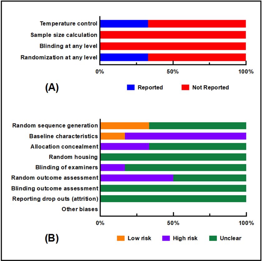
2.2. Risk of Bias Assessment
The results of the risk of bias [49] assessment in these studies are presented in Table 1 (individual
scores) and Figure 2. Randomization was only reported in two studies (33.3%) [33,46], but it was
unclear whether this occurred in the other studies. Similarly, temperature controls were only reported
in two studies (33.3%) [33,45]. No information on blinding or size calculations was reported in any
study (Figure 2A). Therefore, the risk of bias due to random housing and blinding of the assessor was
unclear. The risk of bias due to the concealment of group allocation during the experiment was assessed
as high for two studies, but was unclear for the others [33,45]. A high risk of bias was assessed as
insufficient information was reported for the baseline group characteristics in five out of the six studies.
No information was provided regarding attrition bias in these papers. Random group allocation was
reported for two studies, but was not clear in the others [33,46]. The blinding of examiners was high in
one study, but unclear in the others [45].Int.Int.
J. Mol. Sci.Sci.
J. Mol. 2020, 21,21,
2020, 6265
x FOR PEER REVIEW 4 of 15 15
4 of
Figure 1. Preferred Reporting Items for Systematic Reviews and Meta-Analyses (PRISMA) flow chart.
Figure 1. Preferred Reporting Items for Systematic Reviews and Meta-Analyses (PRISMA) flow
chart.
2.2. Risk of Bias Assessment
The results of the risk of bias [49] assessment in these studies are presented in Table 1 (individual
scores) and Figure 2. Randomization was only reported in two studies (33.3%) [33,46], but it was
unclear whether this occurred in the other studies. Similarly, temperature controls were only
reported in two studies (33.3%) [33,45]. No information on blinding or size calculations was reported
in any study (Figure 2A). Therefore, the risk of bias due to random housing and blinding of the
assessor was unclear. The risk of bias due to the concealment of group allocation during the
experiment was assessed as high for two studies, but was unclear for the others [33,45]. A high risk
of bias was assessed as insufficient information was reported for the baseline group characteristics in
five out of the six studies. No information was provided regarding attrition bias in these papers.
Random group allocation was reported for two studies, but was not clear in the others [33,46]. The
blinding of examiners was high in one study, but unclear in the others [45].Int. J. Mol. Sci. 2020, 21, 6265 5 of 15
Table 1. Assessment of the risk of bias in animal studies using SYstematic Review Centre for Laboratory animal Experimentation (SYRCLE) *.
Selection Bias Performance Bias Detection Bias Other Biases
First Author Attrition
(Year) Random Baseline Random Blinding Bias
Allocation Random Blinding of Any Any Size Temp
[Reference] Group Group Outcome of (Drop-Outs)
Concealed Housing Examiners Randomization Blinding Calculation Control
Allocation Characteristic Selection Assessor
Morell
L H H ? ? H ? ? Y N N Y
(2016) [33]
De Petrocellis
? H H ? H H ? ? N N N Y
(2013) [45]
Roberto
L H ? ? ? ? ? ? Y N N N
(2019) [46]
Olea-Herrero
? L ? ? ? ? ? ? N N N N
(2009) [50]
Mukhtar
? H ? ? ? ? ? ? N N N N
(2007) [51]
Morales
? H ? ? ? H ? ? N N N N
(2013) [52]
* H = high risk of bias; L = low risk of bias; ? = unclear; Y = yes reported; N = not reported.Int.
Int. J.J. Mol.
Mol. Sci. 2020, 21,
Sci. 2020, 21, 6265
x FOR PEER REVIEW 66 of
of 15
15
Figure2.2. Risk
Figure Risk of
of bias
biasand
andquality
qualityassessments.
assessments. (A)
(A) Quality
Quality indicators;
indicators; (B)
(B) Risk
Risk of
of bias
biasassessment
assessment
accordingto
according toeach
eachofofthe
theSYRCLE
SYRCLEcriteria.
criteria.
2.3.
2.3. Study
Study Characteristics
Characteristics
The
The characteristics
characteristics ofof the
the six
six studies
studies are
are summarized
summarized in in Table
Table 2.2. These
These inin vivo
vivo studies
studies were
were
performed on different male mice strains [45,46]. These included athymic
performed on different male mice strains [45,46]. These included athymic nude mice aged between 4 nude mice aged between
4and
and7 7weeksweeks andand athymic
athymic nude-Foxn1
nude-Foxn1 (nu/nu),
(nu/nu), MF-1 MF-1
nude nude
mice,mice, and BALB/cOlaHsd-Foxn1nu
and BALB/cOlaHsd-Foxn1nu nude
nude
mice. mice.
Human Human
prostateprostate
tumor tumor
cell linescellLNCaP,
lines LNCaP,
DU145,DU145,PC3, and PC3, and CWR22Rv1
CWR22Rv1 were used were used to
to generate
generate
tumors in tumors in these animals.
these animals. The tumors Thewere
tumors were induced
induced in the
in the flank flank
of the of the
mice via mice via subcutaneous
subcutaneous injection
injection [33,45,46]. Mice were injected with a minimum of 1 × 10 6 prostate cancer cells to a maximum
[33,45,46]. Mice were injected with a minimum of 1 × 10 prostate cancer cells to a maximum of 1–2 ×
6
of
101–2 × 10⁷ prostate
7 prostate cancer
cancer cells, in cells,
orderintoorder to establish
establish the tumor the in
tumor in reported
reported studies.studies. In Olea-Herrero’s
In Olea-Herrero’s study,
study, mice were injected with 2 × 10 6 PC3 cells [50]; in Mukhtar’s study, the mice were injected
mice were injected with 2 × 10 PC3 cells [50]; in Mukhtar’s study, the mice were injected with 1 × 10⁶
6
with
LNCaP 1 ×cell106[51];
LNCaP cell [51];
in Morell’s in Morell’s
study, mice were study, micewith
injected were5 injected
× 106 PC3with × 10in
cells5 [33];
6 PC3 cells [33];
De Petrocellis’
in De Petrocellis’ study, 1–2 × 10 7 LNCaP or DU145 cells were injected into the mice [45,52]; in Roberto’s
study, 1–2 × 10 LNCaP or DU145 cells were injected into the mice [45,52]; in Roberto’s study,
7 1 × 10⁶
study, 1 × 10 6 PC3 cells were injected into the mice [46]; and in the study by Morales, it is mentioned
PC3 cells were injected into the mice [46]; and in the study by Morales, it is mentioned that the mice
that
werethe mice were
injected withinjected
LNCaP with
and PC3LNCaP andcells
cancer PC3subcutaneously
cancer cells subcutaneously
[52]. [52].
Cannabinoid treatment was initiated once the required
Cannabinoid treatment was initiated once the required tumor volume was tumor volume was between
between 70 and
70 and 150
150 mm 3 in these subcutaneous xenografts [33,46,50–52]. These tumors were treated with a range of
mm in these subcutaneous xenografts [33,46,50–52]. These tumors were treated with a range of
3
cannabinoids,
cannabinoids,including
includingWIN55,212-2,
WIN55,212-2, CBD-BDS
CBD-BDS (biological
(biologicaldrug substance)
drug plusplus
substance) Docetaxel, CBD-BDS
Docetaxel, CBD-
plus bicalutamide, JWH015, JWH015 plus SA2 (specific
BDS plus bicalutamide, JWH015, JWH015 plus SA2 (specific antagonist), and antagonist), and Chromenopyrazoledione 4
Chromenopyrazoledione 4 (PM49, a synthetic cannabinoid). The duration of treatment varied fromInt. J. Mol. Sci. 2020, 21, 6265 7 of 15
(PM49, a synthetic cannabinoid). The duration of treatment varied from 14 to 38 days. Cannabinoids
were administered orally, intraperitoneally, subcutaneously, or intravenously at doses between 0.5 and
100 mg/kg body weight.
3. Discussion
This systematic review highlights six studies reporting the effectiveness of both synthetic and
phytocannabinoids in in vivo/xenograft models for prostate cancer. In this review, we have gathered
detailed information on the study population, group size, cell lines that were used for inoculation and
tumor generation, intervention that was used for treatment (natural/synthetic cannabinoid), dose size,
route of administration, duration of the study, and reduction of the tumor size (Table 2). Two earlier
systematic reviews [45,53,54] briefly explored the effect of cannabis in experimental models of prostate
cancer, such as in vitro studies [42,47] and urological tumors [53,54].
Recently, cannabinoids have been shown to inhibit cell proliferation, migration, and angiogenesis,
as well as arrest the cell cycle and induce apoptosis in prostate cancer cells [46]. Furthermore, a number
of in vitro studies have reported that synthetic cannabinoids such as WIN55,212, JWH-133, and JWH-015
can reduce the size of prostate cancer cell-derived tumors [55,56]. Of the six papers examined, five used
synthetic cannabinoids [33,46,50–52], while the other used natural cannabinoids [45]. Most studies
demonstrated a reduction in tumor size post-cannabinoid administration. In each of these studies,
there was a minimum of eight mice/treatment group. The minimum dose required to start the tumor
was 1 × 10⁶ prostate cancer cells (PC3, DU145, or LNCaP), which was injected into either the left or right
flank of the mice. All six studies reported that the prostate cancer cells were injected subcutaneously
into mice to induce tumors.
Once the tumors had established themselves, the cannabinoids were administered intraperitoneally,
as seen in four of the six studies [33,45,51,52]. Englund et al. [57] stated that when the phytocannabinoid
THC was administered intravenously to the human participants, they showed different psychosomatic
symptoms, such as anxiety, hallucinations, psychotomimetic effects, a blunted effect, paranoia,
conceptual disorganization, illusions, depersonalization, slowing of time, emotional withdrawal,
lack of spontaneity, and many more. However, when participants were given CBD intravenously, they
did not feel any of the above symptoms and no side effects were reported. No comment was made on
any behavioral changes in the animals receiving treatment.
Among the included studies, the synthetic cannabinoid agonist WIN55,212-2 was used in three
studies and was shown to inhibit tumor growth [33,46,51]. Roberto et al. [46] observed that the
synthetic cannabinoid WIN55,212-2 reduced the size of PC3-, DU145-, and LNCaP-induced tumors by
46–69%, and this reduction in size was dose dependent. Morell et al. [33] did not report the actual
percentage of reduction in tumor size; however, they reported treating athymic mice for 15 days with a
dose of 0.5 mg/kg WIN55,212-2, and noted that the WIN55,212-2-treated xenografts were smaller in
size compared to the untreated controls. Correspondingly, βIII Tub levels (a neuroendocrine marker
expressed in cancer cells, such as prostate cancer, non-small-cell lung carcinoma, and breast and
ovarian cancer [58]) were lower in the WIN55,212-2-treated tumors compared to the untreated tumors.
Additionally, Morales et al. [52] demonstrated that, when LNCaP and PC3 xenografts were treated
with 2 mg/kg of PM49 (the most potent derivative of the synthetic cannabinoid quinone), treatment
almost totally blocked the growth of LNCaP tumors, whereas it inhibited the growth of PC3 tumors
by 40%. They also stated that treatment with PM49 was more effective in LNCaP xenografts and is
androgen sensitive, unlike that of PC3 [52]. However, Olea-Herrero et al. [50] reported a reduction in
tumor size, but did not quantify the difference.Int. J. Mol. Sci. 2020, 21, 6265 8 of 15
Table 2. Characteristics of the in vivo identified studies. All of these studies were performed in male animal models.
First Author Study Population
Tumor Induction Study Intervention Cannabinoid Dose Duration
(Year) (Animals) Anticancer Outcomes
(Cell Line) (Route)
[Reference] Strain Age Number Intervention Control
In PC3 xenograft,
dose of 5 µM = 50%, 10 µM = 55%,
and 20 µM = 64% reduction in cell
For PC3 and DU145 cell
Xenograft proliferation.
10 mice lines, dose given was 5,
Roberto, Male athymic PC-3 cells, DU145 Vehicle In DU145 xenograft, dose of 5 µM =
6 weeks (n = 5 per WIN55,212-2 10, and 20 µM. 38 days
2019 [46] nu/nu mice cells, and LNCaP (DMSO) 46%, 10 µM = 51%, and 20 µM = 65%
group) For LNCaP cell line, dose
cells reduction in cell proliferation.
given was 20 and 30 µM.
In LNCaP xenograft,
dose of 20 µM = 69% and 30 µM =
66% reduction in cell proliferation.
WIN55,212-2-treated xenografts
grew slower and the size of the
tumor was smaller than that of
Athymic
Morell, Xenograft Vehicle (not Daily (i.p.) vehicle-treated xenografts (%
nude-Foxn1 4 weeks 8 mice WIN55,212-2 15 days
2016 [33] PC3 cells mentioned) 0.5 mg/kg (s.c.) reduction in tumor size not
(nu/nu)
mentioned).
βIII Tub levels decreased in
WIN55,212-2-treated tumors.
Vehicle (not
Daily (i.p.)
mentioned)
Control Grp 1—vehicle only
Grp 2—1 mg/kg daily CBD-BDS dose-dependently
CBD-BDS alone
Grp 2—10 mg/kg daily inhibited the growth of xenografts
Grp 2—100 mg/kg daily from LNCaP, but not DU145, cells.
60 mice At 100 mg/kg, extract exerted a
De Petrocellis, Xenograft Grp 3—5 mg/kg (i.v.) similar effect on both LNCaP and
MF-1 nude mice 4–7 weeks (n = 10 per 1× week
35 days
2013 [45] LNCaP cells Docetaxel DU145
group) Grp 4—25–50 mg/kg
Bicalutamide CBD-BDS plus bicalutamide
3× week (p.o.) significantly prolonged survival
CBD-BDS +
Grp 5—100 mg/kg (i.p.) + compared with bicalutamide or
Docetaxel
5 mg/kg (i.v.) 1× week CBD-BDS alone.
CBD-BDS +
Grp 6—100 mg/kg (i.p.) +
Bicalutamide
25–50 mg/kg (p.o.) 3×
weekInt. J. Mol. Sci. 2020, 21, 6265 9 of 15
Table 2. Cont.
First Author Study Population
Tumor Induction Study Intervention Cannabinoid Dose Duration
(Year) (Animals) Anticancer Outcomes
(Cell Line) (Route)
[Reference] Strain Age Number Intervention Control
Vehicle
(not Daily (i.p.)
mentioned)
Control Grp 1—vehicle only Tumor growth potentiation
CBD-BDS alone Grp 2—1 mg/kg daily CBD-BDS + Docetaxel (exact % of
Grp 2—10 mg/kg daily reduction not mentioned).
Grp 2—100 mg/kg daily CBD-BDS + bicalutamide at
60 mice 25 mg/kg significantly inhibited
(n = 10 per Xenograft Grp 3—5 mg/kg (i.v.) 35 days
MF-1 nude mice 4–7 weeks xenograft growth (exact % of
group) DU145 cells 1× week
Docetaxel reduction not mentioned).
Grp 4—25–50 mg/kg CBD-BDS + bicalutamide
Bicalutamide
3× week (p.o.) significantly prolonged survival
CBD-BDS +
Grp 5—100 mg/kg (i.p.) + compared with bicalutamide or
Docetaxel
5 mg/kg (i.v.) 1× week CBD-BDS alone.
CBD-BDS +
Grp 6—100 mg/kg (i.p.) +
Bicalutamide
25–50 mg/kg (p.o.) 3×
week
Treatment with PM49 almost totally
Xenograft
blocked the growth of LNCaP
Athymic nu/nu LNCaP cells
PM49 (synthetic Control tumors.
Morales, mice 16 mice (n = 8
5 weeks cannabinoid (vehicle not 2 mg/kg (i.p) 15 days
2013 [52] (BALB/cOlaHsd- per group) 40% tumor growth inhibition and
Xenograft quinone) mentioned)
Foxn1nu) final tumor volume was smaller in
PC3 cells
all four treated mice.
1.5 mg/mL (s.c.)
JWH-015 Final tumor volume and tumor
Olea-Herrero, Athymic nu/nu 24 mice (n = 8 Control 1.5 mg/mL (s.c.) weight were significantly lower in
6 weeks Xenograft PC3 cells 14 days
2009 [50] mice per group) (saline) the treatment group (exact % of
1.5 mg/mL + 1.5 mg/kg
JWH-015 + SR2 reduction not mentioned).
(s.c.)
Inhibition of tumor growth and
decrease in Serum PSA levels to
24 mice 1.86 ng/mL, whereas that of control
Mukhtar, Athymic nu/nu Xenograft 0.5 mg/kg (i.p) alternate
6–8 weeks (n = 8 per WIN55,212-2 Control 35 days group was 7.1 ng/mL. PSA secretion
2007 [51] mice 22Rν1 cells day
group) was correlated with tumor growth
inhibition (exact % of reduction not
mentioned).
i.p. = intraperitoneal; S.C. = subcutaneous; i.v. = intravenous; p.o. = per os (oral administration); PSA = prostate-specific antigen; CBD-BDS = cannabidiol-botanical drug substance.Int. J. Mol. Sci. 2020, 21, 6265 10 of 15
In the study by De Petrocellis et al. [45], a phytocannabinoid CBD-botanical drug substance was
used as the active compound. In this study, CBD was used in combination with anticancer agents, such
as Docetaxel and Bicalutamide, which effectively inhibited tumor growth. Similarly, Scott et al. [59]
studied combinations of the cannabinoids CBD, cannabigerol (CBG), and cannabigevarin (CBGV) in
their neutral forms in leukemia cells. They demonstrated that CBD acts non-antagonistically with other
cannabinoids to reduce the cell number and that the cannabinoid activity is influenced by the drug
combination and treatment schedule. Other studies have also highlighted the toxicity of cannabinoids
on tumor cells [60–62]. Müller et al. [60] showed that WIN55,212-2 caused a significant dose-dependent
effect on the viability of A549 lung cancer cells, HoTu-10 testicular cancer cells, and IMR-5 neuroblastoma
cells. Casanova [61] has also reported similar outcomes, showing that WIN55,212-2 and JWH-133
caused a 75% reduction of skin tumor growth in vivo. Finally, Sanchez et al. [62] observed that the
synthetic cannabinoid JWH-133 significantly inhibited the proliferation of brain tumors compared to
untreated controls.
In particular, emerging evidence suggests that cannabinoids have a dual role in counteracting
prostate cancer progression, as well as the proliferation of stromal cells in the prostate tumor
microenvironment. In a recent study, Pietrovito et al. [36] reported that the cannabinoid treatment
of prostate cancer cells (LNCaP, PC3, and DU145) selectively impaired cell-survival, while at the
same time regulating prostrate stromal fibroblast phenotypes under in vitro conditions. The authors
further showed that the activity of the synthetic cannabinoid WIN 55-212-2 was mediated by the
increased expression of CB2 receptors, which are normally downregulated in healthy prostate fibroblast
cells. The expression of both CB1 and CB2 receptors was elevated in LNCaP cells compared to PC3
and DU145 cells [36]. Similar results were also found by Roberto et al. [46], who demonstrated that
WIN55,212-2 substantially reduced cell proliferation, invasion, and migration, as well as inducing
G0/G1 cell cycle arrest apoptosis, in a dose-dependent manner in cultured PC3, DU145, and LNCaP
prostate cancer cells. These effects were mediated through a pathway involving the cell cycle regulators
p27, Cdk4, and pRb [46]. Collectively, the evidence from in vitro and in vivo studies highlights the
anti-cancer characteristics of phyto-, endo-, and synthetic cannabinoids in prostate cancer.
Interestingly in recent years, cannabinoids have been extensively studied for their potential
anticancer effects, as well as for symptomatic management in cancer patients. They are known to
interact with the components of the endocannabinoid system or other cellular pathways and thus affect
both tumor development and progression. Cannabidiol has been shown to exert chemo-preventive
effects in preclinical models of prostate cancer [63]. In a recent clinical trial, Kenyon et al. [64] reported
that an initial dose of 10 drops (10 mg) twice a day of cannabinoids (three days on and three days
off) reduced the numbers of circulating prostate tumor cells when compared to the effects elicited
with cannabidiol.
When evaluating in vivo studies, an important aspect is to identify bias that might be present and
ways to reduce it, if possible. For selection bias, it was not clear based on which baseline grouping
characteristics the authors grouped animals and if the identity of the allocated animals was concealed.
The performance bias item random housing of animals was not reported adequately and thus represents
a potential risk of bias. The blinding of investigators, as well as risk of bias due to dropout/attrition,
were also not reported in the included studies.
This systematic review has some limitations. The searches were only conducted in PubMed,
Scopus, Web of Science, and Embase. While unlikely, it is possible that additional articles/information
would have been discovered had other databases been included, and if the search strategy had
included gray literature resources, dissertations and theses, conference proceedings, and non-English
language articles. Due to insufficient in vivo evidence, further comprehensive in vivo studies are
required to fully understand the synergistic effect and molecular pathways that lead to anticancer
effects of combinatorial therapies, such as cannabidiol and DNA-damaging agents (temozolomide,
or cisplatin) [65]. Although cannabinoids may potentially assist with the management of prostateInt. J. Mol. Sci. 2020, 21, 6265 11 of 15
cancer, there is still a pressing need to identify the most effective combination(s) of drugs for the
treatment of prostate cancer, as well as other cancers.
In the past decade, extensive research has been undertaken to identify the therapeutic potential
of cannabinoids. This research has resulted in considerable data related to cancer, albeit most
findings being obtained from in vitro experiments. Despite the lack of clinical studies, the potential
use of cannabinoids in the treatment of various cancers, such as prostate, breast, and colon cancer,
cannot be discounted. There is substantial experimental evidence that supports the positive role that
cannabinoids play in cancer cell apoptosis, in preventing metastasis and in the reduction of tumor
growth. However, there is not much data available on the pharmacodynamics and pharmacokinetics
of cannabinoids. Such studies will provide more information about the dose, route of administration,
and in vivo effects when used to treat prostate cancer patients. This will enable us to further explore
the unrevealed properties of various cannabinoids, such as the phytocannabinoids, endogenous
cannabinoids, and synthetic cannabinoids that may be responsible for the anti-cancer effect. With such
knowledge, cannabinoids could become a therapy of choice in the contemporary oncological treatment
of prostate cancers.
4. Material and Methods
4.1. Search Strategy
We searched the PubMed, Embase, Scopus, and Web of Science electronic databases for all relevant
studies that have been published from inception to August 2020. The key terms used were related to
cannabis combined with prostate cancer, such as cannabinoids OR cannabis AND ‘prostate cancer’
(details of other search terms and search strategies are presented in supplementary file, Table S1).
A manual search of all references and citations from the relevant articles was also performed.
4.2. Inclusion Criteria
Articles were included in this review if they met the following criteria: (1) In vivo/xenograft
studies that reported a clear association between cannabinoids and cannabinoid receptor (CB1 and
CB2) activation and further induced cell cycle arrest and apoptosis; (2) cannabinoids inhibiting
tumor growth, proliferation, migration, and invasion in animal models; (3) phytocannabinoids or
synthetic cannabinoids as an intervention with any dose or duration and that reported at least one
tumor-related outcome; and (4) all study designs except reviews, commentaries, case-studies, and the
expert opinions. All clinical and in vitro studies and those that did not use cannabinoids were excluded.
All identified studies from electronic databases searched were screened according to the inclusion and
exclusion criteria.
4.3. Study Selection and Data Extraction
Reference citations were exported to Endnote for the removal of duplicates. Eligible studies were
identified after independent screening (N.J. and K.S.) of the titles and abstracts based on eligibility
criteria. Any disagreements were resolved after discussion or by a third author. Data were extracted
independently by two authors (N.J. and K.S.) after conducting full-text screening with a focus on
identifying anti-tumor activities of cannabinoid administration in vivo related to prostate cancer.
For each study, data was extracted on the first study author, publication year, study design, animal
species/strain used, age, numbers used, tumor induction (stating the cell line used), cannabinoid
intervention(s), dose size, duration of the study, and relevant outcomes.
4.4. Risk of Bias Assessment
Two authors (N.J. and K.S.) independently assessed the risk of bias for each study included in this
review. The assessment was performed according to SYRCLE’s ROB tool [49]. We assessed the risk
of bias for random group allocation, baseline group characteristics, allocation concealment, randomInt. J. Mol. Sci. 2020, 21, 6265 12 of 15
housing, the blinding of examiners and assessors, and random outcome selection. The risk of bias due
to dropout/attrition (column#8, Table 1) was not assessed as it was not reported in any study. To assess
whether studies were free of other risks of bias, aspects such as increasing the number of animals in
groups while conducting the experiment and any possible conflicts of interest were accounted for while
reviewing the included studies. We also assessed reporting of the following study quality indicators:
Blinding at any level; any randomization; sample size calculation; and temperature control (see Table 1
and Figure 2).
5. Conclusions
In summary, cannabinoids were shown to reduce the size of prostate tumors in mice and as such,
it can be concluded that they possess anticancer properties. As for their effectiveness, it is not possible
to evaluate this, as it depends on the cannabinoid itself or combinations thereof that are used for
treatment, as both synthetic and natural cannabinoids have shown anti-cancer outcomes. As reported
by Baram [44], not all THC-rich cannabis extracts have the same effect on a particular cell line at a
similar concentration. Other compounds that are present in total cannabis extracts besides THC and
CBD need to be identified and analyzed to understand their efficacy as anti-tumor agents. Further
in vitro and in vivo analyses are required to identify which cannabinoid compounds are better and in
what combination. Therefore, further controlled and longer-duration animal studies are warranted to
quantify these findings.
Supplementary Materials: The following are available online at http://www.mdpi.com/1422-0067/21/17/6265/s1.
Author Contributions: K.S., N.M. and N.J. designed and performed the study, analyzed data, prepared figures,
and drafted the manuscript. R.Z., T.J.P., and N.M. edited and finally approved the manuscript. All authors have
read and agreed to the published version of the manuscript.
Funding: This research study is partly supported by MGC Pharmaceuticals Limited, Australia.
Acknowledgments: K.S. is the recipient of the RMIT Research Stipend Scholarship from RMIT University, Australia.
Conflicts of Interest: The authors declare no conflict of interest.
References
1. Bray, F.; Ferlay, J.; Soerjomataram, I.; Siegel, R.L.; Torre, L.A.; Jemal, A. Global cancer statistics 2018:
GLOBOCAN estimates of incidence and mortality worldwide for 36 cancers in 185 countries. CA Cancer J.
Clin. 2018, 68, 394–424. [CrossRef] [PubMed]
2. Leitzmann, M.F.; Rohrmann, S. Risk factors for the onset of prostatic cancer: Age, location, and behavioral
correlates. Clin. Epidemiol. 2012, 4, 1–11. [CrossRef] [PubMed]
3. Barbieri, C.E.; Baca, S.C.; Lawrence, M.S.; Demichelis, F.; Blattner, M.; Theurillat, J.-P.; White, T.A.; Stojanov, P.;
Van Allen, E.; Stransky, N. Exome sequencing identifies recurrent SPOP, FOXA1 and MED12 mutations in
prostate cancer. Nat. Genet. 2012, 44, 685–689. [CrossRef]
4. Ku, S.Y.; Rosario, S.; Wang, Y.; Mu, P.; Seshadri, M.; Goodrich, Z.W.; Goodrich, M.M.; Labbé, D.P.; Gomez, E.C.;
Wang, J. Rb1 and Trp53 cooperate to suppress prostate cancer lineage plasticity, metastasis, and antiandrogen
resistance. Science 2017, 355, 78–83. [CrossRef] [PubMed]
5. Montgomery, B.T.; Young, C.Y.F.; Bilhartz, D.L.; Andrews, P.E.; Thompson, N.F.; Tindall, D.J.; Prescott, J.L.
Hormonal regulation of prostate-specific antigen (PSA) glycoprotein in the human prostatic adenocarcinoma
cell line, LNCaP. Prostate 1992, 21, 63–73. [CrossRef] [PubMed]
6. Sharma, M.; Hudson, J.B.; Adomat, H.; Guns, E.; Cox, M.E. In vitro anticancer activity of plant-derived
cannabidiol on prostate cancer cell lines. Pharmacol. Pharm. 2014, 5, 806–820. [CrossRef]
7. Lee, C.; Sutkowski, D.; Sensibar, J.; Zelner, D.; Kim, I.; Amsel, I.; Shaw, N.; Prins, G.S.; Kozlowski, J.M.
Regulation of proliferation and production of prostate-specific antigen in androgen-sensitive prostatic cancer
cells, LNCaP, by dihydrotestosterone. Endocrinology 1995, 136, 796–803. [CrossRef]
8. Nguyen, C.; Lairson, D.R.; Swartz, M.D.; Du, X.L. Risks of major long-term side effects associated with
androgen-deprivation therapy in men with prostate cancer. Pharmacotherapy 2018, 38, 999–1009. [CrossRef]Int. J. Mol. Sci. 2020, 21, 6265 13 of 15
9. Bhutani, K.K.; Gohil, V.M. Natural products drug discovery research in India: Status and appraisal. Indian J.
Exp. Biol. 2010, 48, 199–207.
10. Wang, X.; Fang, G.; Pang, Y. Chinese medicines in the treatment of prostate cancer: From formulas to extracts
and compounds. Nutrients 2018, 10, 283. [CrossRef]
11. Yang, H.; Chen, D.; Cui, Q.C.; Yuan, X.; Dou, Q.P. Celastrol, a triterpene extracted from the Chinese “Thunder
of God Vine,” is a potent proteasome inhibitor and suppresses human prostate cancer growth in nude mice.
Cancer Res. 2006, 66, 4758–4765. [CrossRef] [PubMed]
12. Liu, J.-M.; Lin, P.-H.; Hsu, R.-J.; Chang, Y.-H.; Cheng, K.-C.; Pang, S.-T.; Lin, S.-K. Complementary traditional
Chinese medicine therapy improves survival in patients with metastatic prostate cancer. Medicine 2016, 95,
e4475. [CrossRef] [PubMed]
13. Moyad, M.; Hathaway, S.; Ni, H. Traditional Chinese medicine, acupuncture, and other alternative medicines
for prostate cancer: An introduction and the need for more research. Semin. Urol. Oncol. 1999, 17, 103–110.
[PubMed]
14. Jiang, R.-W.; Zhou, J.-R.; Hon, P.-M.; Li, S.-L.; Zhou, Y.; Li, L.-L.; Ye, W.-C.; Xu, H.-X.; Shaw, P.-C.; But, P.P.-H.
Lignans from Dysosma versipellis with inhibitory effects on prostate cancer cell lines. J. Nat. Prod. 2007, 70,
283–286. [CrossRef] [PubMed]
15. Way, T.-D.; Lee, J.-C.; Kuo, D.-H.; Fan, L.-L.; Huang, C.-H.; Lin, H.-Y.; Shieh, P.-C.; Kuo, P.-T.; Liao, C.-F.;
Liu, H. Inhibition of epidermal growth factor receptor signaling by Saussurea involucrata, a rare traditional
Chinese medicinal herb, in human hormone-resistant prostate cancer PC-3 cells. J. Agric. Food Chem. 2010,
58, 3356–3365. [CrossRef]
16. Zuardi, A.W. History of cannabis as a medicine: A review. Braz. J. Psychiatry 2006, 28, 153–157. [CrossRef]
17. Ramos, J.A.; Bianco, F.J. The role of cannabinoids in prostate cancer: Basic science perspective and potential
clinical applications. Indian J. Urol. 2012, 28, 9–14. [CrossRef]
18. Dariš, B.; Verboten, M.T.; Knez, Ž.; Ferk, P. Cannabinoids in cancer treatment: Therapeutic potential and
legislation. Bosn. J. Basic Med. Sci. 2019, 19, 14–23. [CrossRef]
19. Lafaye, G.; Karila, L.; Blecha, L.; Benyamina, A. Cannabis, cannabinoids, and health. Dialogues Clin. Neurosci.
2017, 19, 309–316.
20. Massi, P.; Solinas, M.; Cinquina, V.; Parolaro, D. Cannabidiol as potential anticancer drug. Br. J. Clin.
Pharmacol. 2013, 75, 303–312. [CrossRef]
21. Pacher, P. Towards the use of non-psychoactive cannabinoids for prostate cancer. Br. J. Pharmacol. 2013, 168,
76–78. [CrossRef]
22. Bogdanović, V.; Mrdjanović, J.; Borišev, I. A review of the therapeutic antitumor potential of cannabinoids.
J. Altern. Complement. Med. 2017, 23, 831–836. [CrossRef] [PubMed]
23. Śledziński, P.; Zeyland, J.; Słomski, R.; Nowak, A. The current state and future perspectives of cannabinoids
in cancer biology. Cancer Med. 2018, 7, 765–775. [CrossRef] [PubMed]
24. Flygare, J.; Sander, B. The endocannabinoid system in cancer—Potential therapeutic target? Semin. Cancer
Biol. 2008, 18, 176–189. [CrossRef] [PubMed]
25. Guzmán, M. Cannabinoids: Potential anticancer agents. Nat. Rev. Cancer 2003, 3, 745–755. [CrossRef]
[PubMed]
26. Bifulco, M.; Laezza, C.; Pisanti, S.; Gazzerro, P. Cannabinoids and cancer: Pros and cons of an antitumour
strategy. Br. J. Pharmacol. 2006, 148, 123–135. [CrossRef] [PubMed]
27. Sarfaraz, S.; Adhami, V.M.; Syed, D.N.; Afaq, F.; Mukhtar, H. Cannabinoids for cancer treatment: Progress
and promise. Cancer Res. 2008, 68, 339–342. [CrossRef]
28. Velasco, G.; Hernández-Tiedra, S.; Dávila, D.; Lorente, M. The use of cannabinoids as anticancer agents.
Prog. Neuro-Psychopharmacol. Biol. Psychiatry 2016, 64, 259–266. [CrossRef]
29. Fraguas-Sanchez, A.I.; Fernandez-Carballido, A.; Torres-Suarez, A.I. Phyto-, endo- and synthetic cannabinoids:
Promising chemotherapeutic agents in the treatment of breast and prostate carcinomas. Expert Opin. Investig.
Drugs 2016, 25, 1311–1323. [CrossRef]
30. Svíženská, I.; Dubový, P.; Šulcová, A. Cannabinoid receptors 1 and 2 (CB1 and CB2), their distribution,
ligands and functional involvement in nervous system structures—A short review. Pharmacol. Biochem.
Behav. 2008, 90, 501–511. [CrossRef]
31. Console-Bram, L.; Marcu, J.; Abood, M.E. Cannabinoid receptors: Nomenclature and pharmacological
principles. Prog. Neuro-Psychopharmacol. Biol. Psychiatry 2012, 38, 4–15. [CrossRef] [PubMed]Int. J. Mol. Sci. 2020, 21, 6265 14 of 15
32. Matsuda, L.A.; Lolait, S.J.; Brownstein, M.J.; Young, A.C.; Bonner, T.I. Structure of a cannabinoid receptor
and functional expression of the cloned cDNA. Nature 1990, 346, 561–564. [CrossRef] [PubMed]
33. Morell, C.; Bort, A.; Vara, D.; Ramos-Torres, A.; Rodriguez-Henche, N.; Diaz-Laviada, I. The cannabinoid
WIN 55,212-2 prevents neuroendocrine differentiation of LNCaP prostate cancer cells. Prostate Cancer
Prostatic Dis. 2016, 19, 248–257. [CrossRef] [PubMed]
34. Barbado, M.V.; Medrano, M.; Caballero-Velázquez, T.; Álvarez-Laderas, I.; Sánchez-Abarca, L.I.;
García-Guerrero, E.; Martín-Sánchez, J.; Rosado, I.V.; Piruat, J.I.; Gonzalez-Naranjo, P. Cannabinoid derivatives
exert a potent anti-myeloma activity both in vitro and in vivo. Int. J. Cancer 2017, 140, 674–685. [CrossRef]
35. Khan, M.I.; Sobocinska, A.A.; Brodaczewska, K.K.; Zielniok, K.; Gajewska, M.; Kieda, C.; Czarnecka, A.M.;
Szczylik, C. Involvement of the CB2 cannabinoid receptor in cell growth inhibition and G0/G1 cell cycle arrest
via the cannabinoid agonist WIN 55,212-2 in renal cell carcinoma. BMC Cancer 2018, 18, 1–17. [CrossRef]
36. Pietrovito, L.; Iozzo, M.; Bacci, M.; Giannoni, E.; Chiarugi, P. Treatment with Cannabinoids as a Promising
Approach for Impairing Fibroblast Activation and Prostate Cancer Progression. Int. J. Mol. Sci. 2020, 21, 787.
[CrossRef]
37. Coke, C.J.; Scarlett, K.A.; Chetram, M.A.; Jones, K.J.; Sandifer, B.J.; Davis, A.S.; Marcus, A.I.; Hinton, C.V.
Simultaneous Activation of Induced Heterodimerization between CXCR4 Chemokine Receptor and
Cannabinoid Receptor 2 (CB2) Reveals a Mechanism for Regulation of Tumor Progression. J. Biol. Chem.
2016, 291, 9991–10005. [CrossRef]
38. Katchan, V.; David, P.; Shoenfeld, Y. Cannabinoids and autoimmune diseases: A systematic review.
Autoimmun. Rev. 2016, 15, 513–528. [CrossRef]
39. Velasco, G.; Galve-Roperh, I.; Sánchez, C.; Blázquez, C.; Guzmán, M. Hypothesis: Cannabinoid therapy for
the treatment of gliomas? Neuropharmacology 2004, 47, 315–323. [CrossRef]
40. Hardie, D.G. AMPK—Sensing energy while talking to other signaling pathways. Cell Metab. 2014, 20,
939–952. [CrossRef]
41. Hermanson, D.J.; Marnett, L.J. Cannabinoids, endocannabinoids, and cancer. Cancer Metastasis Rev. 2011, 30,
599–612. [CrossRef] [PubMed]
42. Orellana-Serradell, O.; Poblete, C.; Sanchez, C.; Castellon, E.; Gallegos, I.; Huidobro, C.; Llanos, M.;
Contreras, H. Proapoptotic effect of endocannabinoids in prostate cancer cells. Oncol. Rep. 2015, 33,
1599–1608. [CrossRef] [PubMed]
43. Guindon, J.; Hohmann, A.G. The endocannabinoid system and cancer: Therapeutic implication. Br. J.
Pharmacol. 2011, 163, 1447–1463. [CrossRef] [PubMed]
44. Baram, L.; Peled, E.; Berman, P.; Yellin, B.; Besser, E.; Benami, M.; Louria-Hayon, I.; Lewitus, G.M.; Meiri, D.
The heterogeneity and complexity of Cannabis extracts as antitumor agents. Oncotarget 2019, 10, 4091–4106.
[CrossRef]
45. De Petrocellis, L.; Ligresti, A.; Schiano Moriello, A.; Iappelli, M.; Verde, R.; Stott, C.G.; Cristino, L.; Orlando, P.;
Di Marzo, V. Non-THC cannabinoids inhibit prostate carcinoma growth in vitro and in vivo: Pro-apoptotic
effects and underlying mechanisms. Br. J. Pharmacol. 2013, 168, 79–102. [CrossRef]
46. Roberto, D.; Klotz, L.H.; Venkateswaran, V. Cannabinoid WIN 55,212-2 induces cell cycle arrest and apoptosis,
and inhibits proliferation, migration, invasion, and tumor growth in prostate cancer in a cannabinoid-receptor
2 dependent manner. Prostate 2019, 79, 151–159. [CrossRef]
47. Nithipatikom, K.; Gomez-Granados, A.D.; Tang, A.T.; Pfeiffer, A.W.; Williams, C.L.; Campbell, W.B.
Cannabinoid receptor type 1 (CB1) activation inhibits small GTPase RhoA activity and regulates motility of
prostate carcinoma cells. Endocrinology 2012, 153, 29–41. [CrossRef]
48. Milano, W.; Padricelli, U.; Capasso, A. Recent advances in research and therapeutic application of cannabinoids
in cancer disease. Pharmacol. Online 2017, 3, 66–78.
49. Hooijmans, C.R.; Rovers, M.M.; de Vries, R.B.; Leenaars, M.; Ritskes-Hoitinga, M.; Langendam, M.W.
SYRCLE’s risk of bias tool for animal studies. BMC Med. Res. Methodol. 2014, 14, 43. [CrossRef]
50. Olea-Herrero, N.; Vara, D.; Malagarie-Cazenave, S.; Diaz-Laviada, I. Inhibition of human tumour prostate
PC-3 cell growth by cannabinoids R (+)-Methanandamide and JWH-015: Involvement of CB 2. Br. J. Cancer
2009, 101, 940–950. [CrossRef]
51. Mukhtar, H.; Afaq, F.; Sarfaraz, S. Cannabinoid Receptors: A Novel Target for Therapy for Prostate Cancer
(Award number W81XWH-04-1-0217). Defense Technical Information Center, 2008. Available online:
https://apps.dtic.mil/dtic/tr/fulltext/u2/a482403.pdf (accessed on 21 March 2020).Int. J. Mol. Sci. 2020, 21, 6265 15 of 15
52. Morales, P.; Vara, D.; Goméz-Cañas, M.; Zúñiga, M.C.; Olea-Azar, C.; Goya, P.; Fernández-Ruiz, J.;
Díaz-Laviada, I.; Jagerovic, N. Synthetic cannabinoid quinones: Preparation, in vitro antiproliferative
effects and in vivo prostate antitumor activity. Eur. J. Med. Chem. 2013, 70, 111–119. [CrossRef] [PubMed]
53. Roberto, D.; Klotz, L.H.; Venkateswaran, V. Cannabinoids as an Anticancer Agent for Prostate Cancer.
J. Urol. Res. 2017, 4, 1090–1097.
54. Gandhi, S.; Vasisth, G.; Kapoor, A. Systematic review of the potential role of cannabinoids as antiproliferative
agents for urological cancers. Can. Urol. Assoc. J. 2017, 11, E138–E142. [CrossRef]
55. Qamri, Z.; Preet, A.; Nasser, M.W.; Bass, C.E.; Leone, G.; Barsky, S.H.; Ganju, R.K. Synthetic cannabinoid
receptor agonists inhibit tumor growth and metastasis of breast cancer. Mol. Cancer Ther. 2009, 8, 3117–3129.
[CrossRef] [PubMed]
56. Gholizadeh, F.; Ghahremani, M.H.; Aliebrahimi, S.; Shadboorestan, A.; Ostad, S.N. Assessment of Cannabinoids
Agonist and Antagonist in Invasion Potential of K562 Cancer Cells. Iran. Biomed. J. 2019, 23, 153–158. [CrossRef]
57. Englund, A.; M Stone, J.; D Morrison, P. Cannabis in the arm: What can we learn from intravenous
cannabinoid studies? Curr. Pharm. Des. 2012, 18, 4906–4914. [CrossRef]
58. Terry, S.; Ploussard, G.; Allory, Y.; Nicolaiew, N.; Boissière-Michot, F.; Maillé, P.; Kheuang, L.; Coppolani, E.;
Ali, A.; Bibeau, F.; et al. Increased expression of class III β-tubulin in castration-resistant human prostate
cancer. Br. J. Cancer 2009, 101, 951–956. [CrossRef]
59. Scott, K.A.; Shah, S.; Dalgleish, A.G.; Liu, W.M. Enhancing the activity of cannabidiol and other cannabinoids
in vitro through modifications to drug combinations and treatment schedules. Anticancer Res. 2013, 33,
4373–4380.
60. Mueller, L.; Radtke, A.; Decker, J.; Koch, M.; Belge, G. The synthetic cannabinoid WIN 55,212-2 elicits death
in human cancer cell lines. Anticancer Res. 2017, 37, 6341–6345.
61. Casanova, M.L.; Blázquez, C.; Martínez-Palacio, J.; Villanueva, C.; Fernández-Aceñero, M.J.; Huffman, J.W.;
Jorcano, J.L.; Guzmán, M. Inhibition of skin tumor growth and angiogenesis in vivo by activation of
cannabinoid receptors. J. Clin. Investig. 2003, 111, 43–50. [CrossRef]
62. Sánchez, C.; de Ceballos, M.L.; del Pulgar, T.G.; Rueda, D.; Corbacho, C.; Velasco, G.; Galve-Roperh, I.;
Huffman, J.W.; y Cajal, S.R.; Guzmán, M. Inhibition of glioma growth in vivo by selective activation of the
CB2 cannabinoid receptor. Cancer Res. 2001, 61, 5784–5789.
63. Kis, B.; Ifrim, F.C.; Buda, V.; Avram, S.; Pavel, I.Z.; Antal, D.; Paunescu, V.; Dehelean, C.A.; Ardelean, F.;
Diaconeasa, Z. Cannabidiol—From plant to human body: A promising bioactive molecule with multi-target
effects in cancer. Int. J. Mol. Sci. 2019, 20, 5905. [CrossRef] [PubMed]
64. Kenyon, J.; Liu, W.; Dalgleish, A. Report of objective clinical responses of cancer patients to
pharmaceutical-grade synthetic cannabidiol. Anticancer Res. 2018, 38, 5831–5835. [CrossRef] [PubMed]
65. Deng, L.; Ng, L.; Ozawa, T.; Stella, N. Quantitative analyses of synergistic responses between cannabidiol
and DNA-damaging agents on the proliferation and viability of glioblastoma and neural progenitor cells in
culture. J. Pharmacol. Exp. Ther. 2017, 360, 215–224. [CrossRef] [PubMed]
© 2020 by the authors. Licensee MDPI, Basel, Switzerland. This article is an open access
article distributed under the terms and conditions of the Creative Commons Attribution
(CC BY) license (http://creativecommons.org/licenses/by/4.0/).You can also read