Approaching In Vivo Models of Pneumococcus-Host Interaction: Insights into Surface Proteins, Capsule Production, and Extracellular Vesicles
←
→
Page content transcription
If your browser does not render page correctly, please read the page content below
pathogens
Article
Approaching In Vivo Models of Pneumococcus–Host
Interaction: Insights into Surface Proteins, Capsule
Production, and Extracellular Vesicles
Alfonso Olaya-Abril 1 , José A. González-Reyes 2                     and Manuel J. Rodríguez-Ortega 1, *

                                          1   Departamento de Bioquímica y Biología Molecular, Campus de Excelencia Internacional CeiA3,
                                              Universidad de Córdoba, 14071 Córdoba, Spain; b22olaba@uco.es
                                          2   Departamento de Biología Celular, Fisiología e Inmunología, Campus de Excelencia Internacional CeiA3,
                                              Universidad de Córdoba, 14071 Córdoba, Spain; bc1gorej@uco.es
                                          *   Correspondence: mjrodriguez@uco.es; Tel.: +34-957-218519

                                          Abstract: Infections caused by the Gram-positive bacterium Streptococcus pneumoniae have become a
                                          major health problem worldwide because of their high morbidity and mortality rates, especially in
                                          developing countries. This microorganism colonizes the human upper respiratory tract and becomes
                                          pathogenic under certain circumstances, which are not well known. In the interaction with the host,
                                          bacterial surface structures and proteins play major roles. To gain knowledge into gradual changes
                                          and adaptive mechanisms that this pathogen undergoes from when it enters the host, we mimicked
                                several in vivo situations representing interaction with epithelial and macrophage cells, as well as a

                                          condition of presence in blood. Then, we analyzed, in four pneumococcal strains, two major surface
Citation: Olaya-Abril, A.; González-      structures, the capsule and extracellular vesicles produced by the pneumococci, as well as surface
Reyes, J.A.; Rodríguez-Ortega, M.J.       proteins by proteomics, using the “shaving” approach, followed by LC-MS/MS. We found important
Approaching In Vivo Models of             differences in both surface ultrastructures and proteins among the culture conditions and strains
Pneumococcus–Host Interaction:
                                          used. Thus, this work provides insights into physiological adaptations of the pneumococcus when it
Insights into Surface Proteins,
                                          interacts with the host, which may be useful for the design of strategies to combat infections caused
Capsule Production, and
                                          by this pathogen.
Extracellular Vesicles. Pathogens 2021,
10, 1098. https://doi.org/10.3390/
                                          Keywords: host-pathogen interaction; capsule; membrane vesicles; surface proteins; proteomics
pathogens10091098

Academic Editors: Bindu Nanduri
and Ed Swiatlo
                                          1. Introduction
Received: 1 August 2021                        Infections by Streptococcus pneumoniae (the pneumococcus), a major agent of pneumo-
Accepted: 26 August 2021                  nia worldwide, cause high rates of morbidity and mortality mostly in developing countries,
Published: 28 August 2021                 with children, the elderly, and immunocompromised patients the most susceptible groups
                                          of the population [1]. In fact, around one million children
Pathogens 2021, 10, 1098 2 of 13
their physiology and responses to switch from a colonizing to a pathogenic status. In this
regard, comparative studies that simulate different host–interaction conditions constitute a
valuable choice to gain insight into adaptive mechanisms of microbes to the environment.
The microbial surface is the preferential site of interaction between cells and their environ-
ment [11] as it acts as both a barrier and the site to interplay with the host at multiple levels.
In this interaction, numerous structures and molecules play major roles, being especially
relevant to the capsule (which surrounds many bacterial cells), and surface proteins, which
have key functions and have the highest chances to be recognized by the host immune
system [12,13]. In addition, extracellular vesicles (EVs) released from the bacterial surface
have been revealed as a critical way to interact with the surroundings, and in the case of
pathogenic microorganisms, they play important roles during the infection process [14–16].
In this work, we examined the host–pathogen interaction using different culture
conditions for the pneumococcus, in order to understand how the major surface-attached
and surface-derived ultrastructures and proteins adapt to these environments. We analyzed,
in four pneumococcal strains, the amount of capsule and EVs, as well as the surface
proteins using the “shaving” approach, followed by LC-MS/MS in a condition resembling
bacteremia, and in other conditions that simulate interaction with host macrophages and
epithelial cells. We found strain-dependent differences in EV production and surface
protein profiles, and a general increase in capsule amount in the bacteremia-like model, as
well as a decrease in capsule production when the bacteria contact the host cells. Thus, this
study provides new insights into adaptation in the physiology of pneumococcus when it
interacts with the host.
2. Results
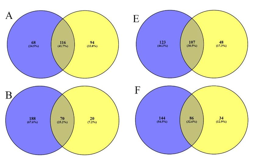
We analyzed the capsule and EV production in different culture media and models of
interaction with host cells in four pneumococcal strains (Figure 1): the avirulent and non-
encapsulated R6 reference strain and three virulent strains pertaining to three capsulated
serotypes: 1, 6B, and 8. First, we compared the growth of these strains in five liquid culture
media: THBB as representative of a model of bacteremia, and THB as its control; and
Pathogens 2021, 10, x FOR PEER REVIEW 3 ofthe
14
eukaryotic cell DMEM medium conditioned either by macrophages (MCM) or by epithelial
cells (ECM) as models of interaction with host cells during colonization for which DMEM
was used as control.
Figure 1. Sample preparation workflow: MCM, macrophages conditioned medium; ECM, epithelial conditioned medium;
Figure 1. Sample preparation workflow: MCM, macrophages conditioned medium; ECM, epithelial conditioned medium;
MC, co-culture with J774 macrophages (macrophages contact); EC, co-culture with A549 epithelial cells (epithelial contact);
MC, co‐culture with J774 macrophages (macrophages contact); EC, co‐culture with A549 epithelial cells (epithelial contact);
DMEM,
DMEM, Dulbecco’s
Dulbecco’s modified
modifiedEagle’s
Eagle’smedium;
medium;THB,
THB,Todd–Hewitt
Todd–Hewitt broth; THBB,
broth; Todd–Hewitt
THBB, Todd–Hewittbroth supplemented
broth supplemented with 5%
with
sheep blood.
5% sheep blood.
Significant differences were observed in the growth curves for the tested media (Fig‐
ure 2A). There was a faster growth in THBB than in THB, except for the ST1 strain. The
growth in THBB was also faster than in the other media analyzed, i.e., MCM and ECM, as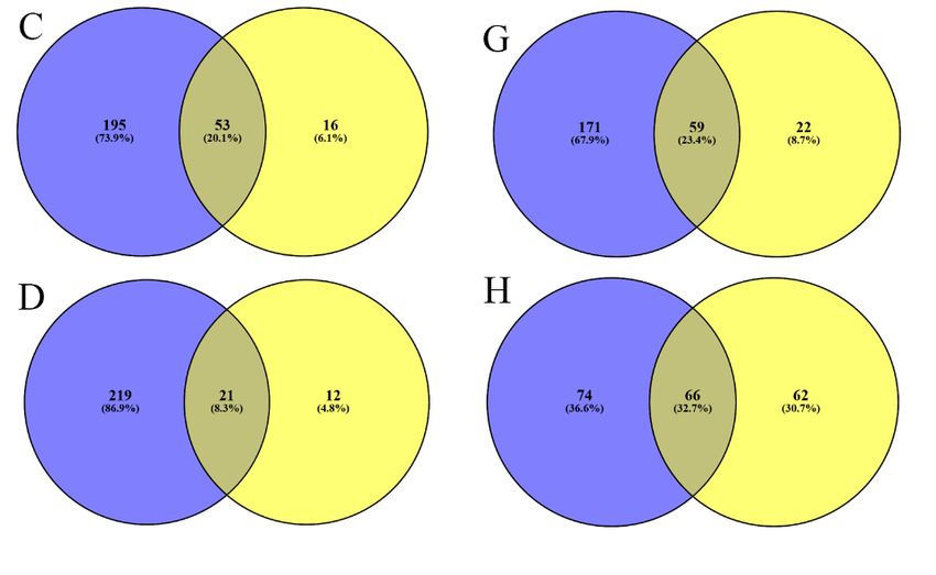
Pathogens 2021, 10, 1098                                                                                                          3 of 13

                                       Significant differences were observed in the growth curves for the tested media
                                  (Figure 2A). There was a faster growth in THBB than in THB, except for the ST1 strain.
                                  The growth in THBB was also faster than in the other media analyzed, i.e., MCM and
                                  ECM, as well as their control DMEM. In these last three media, representing direct host–
 Pathogens 2021, 10, x FOR PEER REVIEW                                                                                 4 of 14
                                  pathogen interaction, R6 showed the lowest growth, while the ST1 strain exhibited the best
                                  adaptation to these media.

      Figure 2. Effect of culture media on Streptococcus pneumoniae growth, capsule, and extracellular vesicles production:
       Figure
      (A)     2. Effect ofgrowth
          pneumococcal     culture curves
                                   media on   Streptococcus
                                           in the           pneumoniae
                                                  culture media  used; growth, capsule,
                                                                       (B) capsule      and extracellular
                                                                                   production  measured vesicles   production:
                                                                                                           by the stain-all     (A)
                                                                                                                            assay;
       pneumococcal growth curves in the culture media used; (B) capsule production measured by the stain‐all assay; (C) pro‐
      (C) production of extracellular vesicles (EVs) by pneumococcal strains per culture media. * p < 0.05.
       duction of extracellular vesicles (EVs) by pneumococcal strains per culture media. * p < 0.05.
                                        Next, we studied capsule production by the three encapsulated serotypes (Figure 2B).
                                  The growth in THBB caused a clear increase in the amount of capsule produced in the
                                  three strains when compared to the absence of blood, i.e., THB. However, the measured
                                  capsule clearly diminished after growing the pneumococci either in a medium previously
                                  conditioned by macrophages or epithelial cells, and a higher decrease was observed in
                                  bacterial cells recovered after direct contact with both macrophages (MC) and epithelial
                                  cells (EC) when comparing these conditions to their corresponding control DMEM. In all
                                  cases, the amount of capsule in cultured cell-conditioned media or after direct contact
                                  with cells was lower than in the bacteremia-like model of THBB. We visualized this effect
                                  by electron microscopy in the ST1 strain (Figure 3), in which a clear decrease in capsule
                                  amount was appreciated in bacterial cells in direct contact with cultured eukaryotic cells
                                  (EC and MC), compared to pneumococci cultured in an environment conditioned by the

                                   Figure 3. Transmission electron microscopy of serotype 1 Streptococcus pneumoniae showing the dif‐
                                   ferential amount of capsule depending on culture media. Panels show detailed pictures of pneumo‐
                                   coccal bacterial cells in each of the seven conditions used in this study, i.e., culture in THB medium,
                                   culture in THB with blood (THBB), cultured in DMEM medium, cultured in DMEM that was con‐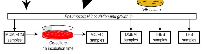
Pathogens 2021, 10, 1098                                                                                                                      4 of 13

                                            cultured
Figure 2. Effect of culture media on Streptococcus     cells (ECM
                                                   pneumoniae       and MCM),
                                                                growth,  capsule,as well
                                                                                  and    as in comparison
                                                                                      extracellular           with bacteria
                                                                                                    vesicles production: (A)grown in THBB. In
                                            this case,  the increase in capsule  for THBB-cultured     bacteria was  not
pneumococcal growth curves in the culture media used; (B) capsule production measured by the stain‐all assay; (C) pro‐   so evident, compared
                                            to those cultured    in THB,  as shown   previously
duction of extracellular vesicles (EVs) by pneumococcal strains per culture media. * p < 0.05.    in the colorimetric  assays (Figure 2B).

                             Figure 3. Transmission
               Figure 3. Transmission                   electron microscopy
                                           electron microscopy      of serotypeof1 serotype  1 Streptococcus
                                                                                    Streptococcus pneumoniae pneumoniae
                                                                                                                 showing theshowing    the dif‐amount of
                                                                                                                               differential
                             ferentialon
               capsule depending       amount
                                         cultureofmedia.
                                                    capsulePanels
                                                             depending
                                                                    show on  culturepictures
                                                                          detailed    media. of
                                                                                              Panels  show detailed
                                                                                                 pneumococcal          pictures
                                                                                                                   bacterial cellsofinpneumo‐
                                                                                                                                       each of the seven
                             coccal
               conditions used      bacterial
                                 in this study,cells
                                                i.e.,in each of
                                                      culture in the
                                                                  THBseven conditions
                                                                       medium,   cultureused  in this
                                                                                          in THB      study,
                                                                                                   with bloodi.e., culturecultured
                                                                                                                (THBB),    in THB in  medium,
                                                                                                                                        DMEM medium,
                             culture in THB with blood (THBB), cultured in DMEM medium, cultured in DMEM that was con‐
               cultured in DMEM that was conditioned either by epithelial cells (ECMs) or macrophages (MCMs), and bacteria recovered
                             ditioned either by epithelial cells (ECMs) or macrophages (MCMs), and bacteria recovered after
               after direct contact with either epithelial cells (ECs) or macrophages.
                             direct contact with either epithelial cells (ECs) or macrophages.
                                                    We also analyzed the amount of EVs produced by the four tested strains in the different
                                              media (Figure 2C). The effect of the culture media was not homogeneous among the strains,
                                              as the presence of blood in the medium (THBB) caused an overproduction of EVs except
                                              in the ST1 strain, when compared to its control THB. However, the exposure to cellular
                                              factors released by cultured cells was dissimilar in the four pneumococcal strains: the
                                              growth in ECM caused a decrease in pneumococcal EVs produced by three strains, but
                                              those produced by ST8 were not affected, compared to its control DMEM. On the other
                                              hand, EVs of pneumococci grown in MCM decreased in R6 and ST6B and increased in
                                              ST1 and ST8. Pneumococcal EVs in cultures after direct contact with host cells were not
                                              measured because of the technical difficulties to separate them from potential exosomes
                                              released by the eukaryotic cells.
                                                    Next, we analyzed the surface proteins of the four strains in the seven culture con-
                                              ditions tested in this work, using the approach of “shaving” intact living bacterial cells
                                              with trypsin, followed by LC-MS/MS analysis. In total, 279 different surface proteins were
                                              identified (Supplemental Dataset 1), of which 23 were cell-wall proteins with an LPXTG
                                              motif; another 23 proteins were classified as lipoproteins; 218 were membrane proteins, of
                                              which 69 had only 1 transmembrane domain and 149 were multi transmembrane; 15 were
                                              secreted proteins. Considering the 28 different strains and culture conditions examined,
                                              the most widely distributed proteins were those with the LPXTG cell-wall-anchoring mo-
                                              tif: the beta-galactosidase precursor Spr0565, the ZmpB Spr0581, and the IgA1 protease
                                              Spr1042 were identified in 17, 17, and 16 samples, respectively. Additionally, the cell-wall
                                              proteins Spr0561 and Spr0328 were both identified in 13 samples. Another 2 proteins, CbpA
                                              (Spr1995), which is predicted as secreted, and PspA, a membrane protein, were identified
                                              in 15 samples. Remarkably, the prophage-encoded protein PblB was also identified in all
                                              the strains except R6, being found in all the studied conditions.
                                                    In order to investigate changes in surface protein profiles, we established different
                                              comparisons based on host–pathogen interaction levels according to our proposed models
                                              (see all the comparison categories and identifications in Supplemental Dataset 1). Thus,
                                              the most general comparison was “host interaction,” using as controls THB and DMEM;
                                              in “contact,” the conditions described as “macrophage contact (MC)” and “epithelial cell
                                              contact (EC)” were compared with the rest. The comparisons for only MC and EC were
                                              also carried out. We also expanded this comparison to “interaction with cells,” which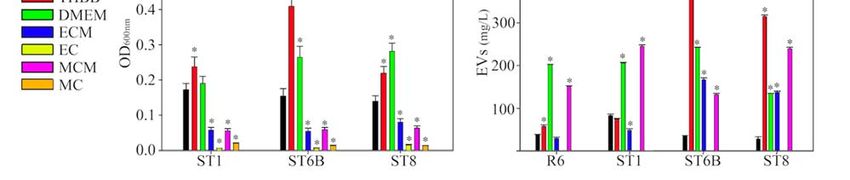
Pathogens 2021, 10, 1098                                                                                                                  5 of 13

                                   included the culture media previously conditioned by the cells. As before, this category
                                   was also subdivided to compare only interaction with macrophages or with epithelial
                                   cells. Finally, we analyzed the model of bacteremia, comparing THBB with its control
                                   THB. Figure 4 summarizes the proteins identified exclusively or in common for each
                                   category, in all the comparisons performed. Although the changes in protein profiles were
Pathogens 2021, 10, x FOR PEER REVIEW                                                                                       6 of 14
                                   profuse, we highlight that, in the model of “epithelial cells contact,” there was a generalized
                                   disappearance of most cell wall proteins, as well as the transmembrane hypothetical protein
                                   Spr1584.

                                 Figure 4. Comparison of identified surface proteins in different host-pathogen interaction models.
                                Figure
                                 Each Venn4. Comparison     of identified
                                               diagram represents          surfaceproteins
                                                                     the surface    proteinsidentified
                                                                                              in different  host‐pathogen
                                                                                                       in common            interaction
                                                                                                                    or exclusive  for eachmodels.
                                                                                                                                            group
                                Each
                                 in all Venn    diagram of
                                        the categories    represents   the(assurface
                                                             interaction      definedproteins   identified
                                                                                       in the text          in common orDataset
                                                                                                   and in Supplemental       exclusive
                                                                                                                                     1), for
                                                                                                                                         witheach
                                                                                                                                               the
                                group    in all
                                 left blue      theas
                                            circle  categories  of interaction
                                                      the control               (as defined
                                                                   (no interaction)  and theinright
                                                                                                the text andcircle
                                                                                                     yellow   in Supplemental   Dataset
                                                                                                                   as the cultures        1), with
                                                                                                                                    representing
                                the  left blue (A)
                                 interaction:    circle as the
                                                     “host     control (no
                                                           interaction”;  (B)interaction)  and“contact
                                                                               “contact”, (C)   the right  yellow
                                                                                                        with       circle as the
                                                                                                              macrophages”;      cultures
                                                                                                                               (D) “contactrepre‐
                                                                                                                                              with
                                senting interaction: (A) “host interaction”; (B) “contact”, (C) “contact with macrophages”; (D) “con‐
                                 epithelial cells”; (E) “interaction with cells”; (F) “interaction with macrophages”; (G) “interaction
                                tact with epithelial cells”; (E) “interaction with cells”; (F) “interaction with macrophages”; (G) “in‐
                                with epithelial cells”; (H) “interaction with blood.”
                                teraction with epithelial cells”; (H) “interaction with blood.”
                                      A gene ontology (GO) enrichment analysis of this model performed with all the
                                      A gene ontology (GO) enrichment analysis of this model performed with all the iden‐
                                 identified proteins (including also the cytoplasmic proteins, removed from the categories
                                tified proteins (including also the cytoplasmic proteins, removed from the categories of
                                 of surface proteins) revealed, among others, a clear enrichment of the GO terms “cell
                                surface   proteins)
                                 adhesion,”          revealed,
                                               as well         among
                                                        as other       others,
                                                                 related       a clear
                                                                         to zinc       enrichment
                                                                                  ion binding      of the GO(“response
                                                                                               or transport   terms “celltoadhe‐
                                                                                                                            zinc
                                sion,”  as  well  as other related to zinc ion  binding  or transport  (“response
                                 ion”, “regulation of sequestering of zinc ion”, and “zinc II ion transmembrane   totransport”)
                                                                                                                     zinc ion”,
                                “regulation
                                 (Figure 5). of sequestering of zinc ion”, and “zinc II ion transmembrane transport”) (Fig‐
                                ure 5).
                                      Likewise, two membrane proteins (Spr0343 and Spr1333) appeared after cell interac‐
                                tion. It is noteworthy that the predicted secreted protein Spr2021, also known as PcsB, was
                                identified only in two strains, R6 and ST6B, and only in THB and THBB but not in the
                                conditions simulating contact with cells.
ns 2021, 10,Pathogens
x FOR PEER
2021,REVIEW
10, 1098 7 of 14 6 of 13
Figure
Figure 5. Gene 5. Gene
ontology ontology
groups groups
affected by affected by Streptococcus
Streptococcus pneumoniae pneumoniae after with
after the contact the contact with
epithelial epithelial
cells. Only GO groups
cells. Only GO groups (biological function, third level) that change significantly, with a p‐value < analysis
(biological function, third level) that change significantly, with a p-value < 0.05 after a hypergeometric distribution
(E(GOi)) are0.05
shownafterasathe
hypergeometric
actual sample distribution analysis (E(GOi))
enrichment (ASE)/expected are shown
sample as the (ESE)
enrichment actualratio.
sample enrich‐
If the ratio was lower
ment (ASE)/expected sample enrichment (ESE) ratio. If the ratio was lower than 1 (over‐represented
than 1 (over-represented in the ESE), the inverse value with a negative symbol is shown. The size of the bubble corresponds
in the ESE), the inverse value with a negative symbol is shown. The size of the bubble corresponds
to the absolute value of the ASE/ESE or ESE/ASE ratio. The whole genome of S. pneumoniae R6 was used as reference. The
to the absolute value of the ASE/ESE or ESE/ASE ratio. The whole genome of S. pneumoniae R6 was
GO enrichmentusedparameter E(GOi)
as reference. wasenrichment
The GO calculated by the formula:
parameter (E(GOi))
E(GOi) = sampleby
was calculated size/genome
the formula: size × GOi.=
(E(GOi))
sample size/genome size GOi.
Likewise, two membrane proteins (Spr0343 and Spr1333) appeared after cell inter-
3. Discussion action. It is noteworthy that the predicted secreted protein Spr2021, also known as PcsB,
was identified only in two strains, R6 and ST6B, and only in THB and THBB but not in the
The purpose of this work
conditions consisted
simulating of providing
contact with cells. insights into the pneumococcal sur‐
face structure and protein variations when comparing different culture conditions resem‐
bling in vivo models of infection. The capsule is a widely studied and characterized struc‐
3. Discussion
ture of the pneumococcal
The purposesurface,ofwhich acts also
this work as a major
consisted virulence insights
of providing factor [17,18]. Re‐pneumococcal
into the
cently, the production of EVs by this microorganism has been described. These
surface structure and protein variations when comparing different culture conditions re- structures
are immunoreactive
sembling andinprotective
vivo models against infectionThe
of infection. [14], in addition
capsule to having
is a widely studiedan and
im‐ characterized
munomodulatory effect [19].
structure of theThe set of surfacesurface,
pneumococcal proteins, known
which acts asalso
the “surfome,” has also factor [17,18].
as a major virulence
been describedRecently,
in collections of clinical isolates to define potential candidates
the production of EVs by this microorganism has been described. These struc- for vaccines
and diagnosticstures
[20,21].
are However,
immunoreactive there isand a lack of studies
protective focusing
against on how
infection capsule,
[14], EVs, to having an
in addition
and surface proteins vary when the pathogen interacts with the
immunomodulatory effect [19]. The set of surface proteins, known as the host. For this purpose, we“surfome,” has
defined five models of host–pathogen interaction, which, although with limitations
also been described in collections of clinical isolates to define potential candidates for since
they are not real in vivoand
vaccines infections,
diagnosticscan help to understand
[20,21]. However, there the physiological
is a lack of adaptation
studies focusing on how
of the pneumococcus
capsule, when
EVs,itand contacts
surfacetheproteins
host. One model
vary when simulates bacteremia
the pathogen (THBB);
interacts with the host. For
two simulate thethispneumococcus
purpose, we defined in an environment
five models of close to target cells
host–pathogen (represented
interaction, which,by although with
DMEM conditioned by macrophages or epithelial cells, i.e., MCM and ECM),
limitations since they are not real in vivo infections, can help to understand the physio- and the
other two simulate
logicalthe bacteria inofdirect
adaptation contact with host
the pneumococcus when cells (MC and
it contacts theEC).
host.WeOneused
model simulates
THB as the control for the first
bacteremia model
(THBB); twoandsimulate
DMEM the as the control for theinother
pneumococcus four models. close to target
an environment
In the bacteremia model,
cells all the strains
(represented by DMEM except ST1 grew much
conditioned faster in THBB,
by macrophages compared
or epithelial cells, i.e., MCM
to the other media. It is evident
and ECM), and the that this two
other difference
simulate is due
the to the presence
bacteria in directof contact
blood, as thehost cells (MC
with
growth in THBand wasEC).
slower.
We usedHowever,THB we alsocontrol
as the recognize thatfirst
for the this model
model andmight be some‐
DMEM as the control for
the other four models. In the bacteremia model,
how far from a real bacteremia situation, which is defined as the presence of bacteria in all the strains except ST1 grew much
the bloodstream.faster
Usingin THBB, compared medium
a DMEM‐based to the other media. It
containing is evident
blood could that
havethis difference
been an is due to
the presence
alternative model, but even of blood,
in such as the growth
a medium, in THB was
the bacteremia is slower. However,
not completely we also recognize that
emulated.
We chosethis model
three mightstrains
virulent be somehow
belonging far from a realrepresentative
to three bacteremia situation,serotypes:which
ST1,is defined as the
ST6B, and ST8.presence of bacteria
ST1 is highly invasivein the
and,bloodstream.
although includedUsing a in DMEM-based mediumhas
conjugate vaccines, containing blood
could have
been highly prevalent among been theancirculating
alternativeisolates
model,in but even inelderly
Spanish such apeople
medium, thelast
in the bacteremia is not
completely emulated.
decade [22]. ST6B has been prevalent in childrenPathogens 2021, 10, 1098 7 of 13
We chose three virulent strains belonging to three representative serotypes: ST1,
ST6B, and ST8. ST1 is highly invasive and, although included in conjugate vaccines, has
been highly prevalent among the circulating isolates in Spanish elderly people in the last
decade [22]. ST6B has been prevalent in childrenPathogens 2021, 10, 1098 8 of 13
have lower metal cation levels available and have to synthesize more transporters and
binding proteins to uptake such ions.
It is noteworthy that we also identified the prophage-encoded PblB in nine out of the
28 combinations of this study (four strains, seven culture conditions each), in three out of the
four strains analyzed. This protein was first described as a platelet-binding and activator
in S. mitis [43,44]. In the pneumococcus, PblB possesses a galactose-binding domain that
mediates adhesion to host epithelial cells [45]. We previously identified this protein in the
“surfome” of approximately two-thirds of the clinical isolates of a previous study using
the “shaving” approach. Moreover, we have demonstrated that this protein is recognized
by human sera and showed the highest discrimination capacity between pneumococcus-
infected children and controls in a protein chip array platform [21]. Additionally, a positive
correlation has been reported between the presence of this protein and mortality in patients
with invasive pneumococcal disease (IPD) [46,47].
In conclusion, this work shows evidence of gradual changes in the surface ultra-
structures and surface proteins of the pneumococcus from a more planktonic to a more
host-interacting model, using conditions that resemble different stages of host–pathogen
interactions, even though the models used in this study might not totally resemble what
really happens in vivo. The capsule shows its maximum thickness in a model resembling
bacteremia and decreases when the pathogen contacts host cell surfaces. Surface proteins
also adapt to the medium. Production of EVs varies according to the strain and infection
model. Future strategies to study host–pathogen interactions should incorporate a global
analysis of surface structures and proteins in different models to gain insight into changes
evolving from non-interacting to a more pathogenic status of the microorganisms, in order
to design more effective strategies to fight against bacterial infections.
4. Materials and Methods
4.1. Cell Lines, Bacterial Strains, and Growth
J774 macrophages and A549 epithelial cells were cultured at 37 ◦ C in a 5% CO2
atmosphere in the air, in Dulbecco’s modified Eagle’s medium (DMEM) supplemented
with 10% fetal bovine serum, 10% NCTC, and 1% non-essential amino acids. Conditioned
media from macrophages and epithelial cells (macrophages-conditioned media (MCM);
epithelial cells-conditioned media (ECM)) were recovered as supernatant, centrifuged at
800× g for 3 min, filtered with 0.22 µm pore-size filters, centrifuged at 100,000× g and
stored at −20 ◦ C until use. Streptococcus pneumoniae strains (R6, serotype 2; ST1, serotype 1;
ST6B, serotype 6B; ST8, serotype 8) were grown at 37 ◦ C in different media: Todd–Hewitt
broth (THB); THB supplemented with 5% sheep blood (THBB) lysed with distilled water,
centrifuged to remove cell debris and filtered with 0.22 µm pore-size filters; DMEM; MCM;
ECM; in co-culture with macrophages (MC) or with epithelial cells (EC) during 1 h of
incubation. In this case, pneumococci were harvested by centrifugation, first at 800× g
for 3 min, to remove eukaryotic cell debris, and later at 5000× g for 10 min, checking the
absence of eukaryotic cells by optical microscopy visualization.
4.2. “Shaving” of Pneumococcal Living Cells
Pneumococcal strains were “shaved” for surface protein identification, as already
described [20,21,30], but with some modifications. Briefly, 10 mL of each strain were grown
in the corresponding media (THB, THBB, DMEM, MCM, or ECM) to an OD600 = 0.3, which
corresponds to approximately 108 bacterial cells/mL. When growth did not reach the
OD, the cultures were diluted to the lowest OD value for each individual strain. In the
case of bacteria in contact with eukaryotic cells, the bacterial cells were resuspended in a
volume of DMEM yielding the lowest OD for each individual strain. Under the growth
conditions tested, equal volumes of each strain were further processed in order to normalize
the number of bacterial cells. Afterward, the bacteria were pelleted by centrifugation at
3500 × g for 10 min. Bacterial pellets were washed twice with PBS, resuspended in 1 mL of
PBS containing 30% sucrose (pH 7.4), and digested with 5 µg trypsin (Promega, Madison,Pathogens 2021, 10, 1098 9 of 13
WI) for 30 min at 37 ◦ C. The resulting digestion mixtures were redigested with 2 µg trypsin
overnight at 37 ◦ C. Samples were cleaned using Oasis HLB extraction cartridges (Waters,
Milford, MA, USA) as described [48].
4.3. LC-MS/MS Analysis
All analyses were performed with a Surveyor HPLC System in tandem with an LTQ-
Orbitrap mass spectrometer (Thermo Fisher Scientific, San Jose, CA, USA) equipped with
a nanoelectrospray ionization interface (nESI). The separation column was 150 mm ×
0.150 mm ProteoPep2 C18 (New Objective, MA, USA) at a post-split flow rate of 1 µL/min.
For trapping of the digest, a 5 mm × 0.3 mm precolumn Zorbax 300 SB-C18 (Agilent
Technologies, Germany) was used. One-fourth of the total sample volume, i.e., 5 µL,
was trapped at a flow rate of 10 µL/min for 10 min and 5% acetonitrile/0.1% formic
acid. Afterward, the trapping column was switched online with the separation column,
and the gradient was started. Peptides were eluted with a 60 min gradient of 5–40% of
acetonitrile/0.1% formic acid solution at a 250 nL/min flow rate. All separations were
performed using a gradient of 5–40% solvent B for 60 min. MS data (Full Scan) were
acquired in the positive ion mode over the 400–1500 m/z range. MS/MS data were
acquired in a dependent scan mode, selecting automatically the five most intense ions for
fragmentation, with dynamic exclusion set to on. In all cases, a nESI spray voltage of 1.9 kV
was used.
4.4. Protein Identification by Database Searching
Tandem mass spectra were extracted using Proteome Discoverer 1.0 (Thermo Fisher
Scientific). All MS/MS samples were analyzed using Sequest (Thermo Fisher Scientific, ver-
sion v.27), applying the following search parameters: peptide tolerance, 10 ppm; tolerance
for fragment ions, 0.8 Da; b- and y-ion series; oxidation of methionine and deamidation of
asparagine and glutamine were considered as variable modifications; maximum trypsin
missed cleavage sites, 3. The raw data were searched against an in-house joint database con-
taining the protein sequences from all the sequenced and annotated S. pneumoniae strains
available at the NCBI ftp site. Peptide identifications were accepted if they exceeded the
filter parameter Xcorr score vs. charge state with SequestNode Probability Score (+1 = 1.5,
+2 = 2.0, +3 = 2.25, +4 = 2.5). With these search and filter parameters, no false-positive hits
were obtained. For proteins identified from only one peptide, fragmentations were checked
manually. Strain R6 was used as a reference for providing the accession numbers of the
identified proteins; whenever a protein belonging to another strain was found, homology
with a corresponding protein of strain R6 was given by using protein-BLAST. If homology
with R6 proteins was not observed, then the protein accession numbers of the other strains
were used. Primary predictions of subcellular localization were assigned by using the
LocateP web-based algorithm [49].
4.5. Pneumococcal EVs Production and Quantification
EVs were isolated as described [14], by using a series of Optiprep gradient layers
with concentrations ranging from 35–5% (w/v). Briefly, cells at different ODs were pelleted
from 1 L cultures and the supernatants were filtered through a 0.22 µm pore size filter
(Millipore, Burlington, MA, USA). The supernatants were then centrifuged at 100,000 × g
for 1.5 h at 4 ◦ C to sediment the vesicular fraction. The pellets were mixed with 2 mL
of Optiprep solution (Sigma-Aldrich, St. Louis, MO, USA), yielding 35% (w/v) Optiprep
final concentration. The crude vesicle sample was then overlaid with a series of Optiprep
gradient layers with concentrations ranging from 35% to 5% (w/v). The gradients were
centrifuged (100,000 × g, 16 h), and 1 mL fractions were removed from the top. The
fractions were then centrifuged at 100,000 × g for 1 h at 4 ◦ C and recovered. Finally,
vesicles were air-dried, weighed, and resuspended in 1 mL PBS.Pathogens 2021, 10, 1098 10 of 13
4.6. Capsule Quantification
The amount of capsule was determined using the stains-all assay (Sigma) for detecting
acidic polysaccharides, as described [50]. The bacteria were cultured to late-exponential
phase, then 5 mL was centrifuged for 10 min at 5000× g, washed with PBS, and resuspended
in 0.5 mL 0.85% NaCl. A total of 10 µL were removed to make dilutions in PBS for plating
out to quantify the number of bacteria. To the remaining bacterial suspension, 2 mL
of a solution containing 20 mg 1-ethyl-2(3-(1-ethylnaphthho- (1,2-d)thiazolin-2-ylidene)-
2methylpropenyl)naphthho-(1,2d)thiazoliumbromide (stains-all) and 60 mL glacial acetic
acid in 100 mL 50% formamide was added, and the OD640 determined; 0.5 mL NaCl with
2 mL stains-all solution was used as a blank.
4.7. Electron Microscopy
The pneumococcal capsule was observed by transmission electron microscopy as
described [50,51] but without lysin or acetate in the cacodylate buffer. Bacteria were
harvested by centrifugation for 10 min at 5000× g, then washed twice, resuspended in PBS,
and fixed with 2% paraformaldehyde and 2.5% glutaraldehyde in 0.1 M cacodylate buffer
(pH 7) with 0.075% ruthenium red for 20 min on ice. The samples were fixed again with
the fixing solution for 3 h, washed with cacodylate buffer containing 0.075% ruthenium
red, and then post-fixed with 1% osmium tetroxide in cacodylate buffer containing 0.075%
ruthenium red for 1 h at room temperature. After dehydration in an ascendant series
of ethanol, the pieces were transferred to propylene oxide and sequentially infiltrated
in EMbed 812 resin (EMS, USA). Blocks were formed in fresh resin that was allowed to
polymerize for 48 h at 65 ◦ C. Blocks were cut in an Ultracut Reicher ultramicrotome to
obtain 40–60 nm width sections using a diamond knife. The sections were observed and
photographed in a Jeol JEM 1400 Transmission Electron Microscope.
4.8. Statistics and Data Analysis
Statistical analyses were performed using SPSS v 21.0.0.0. Student’s t-test (2-tailed)
was applied for experiments involving pairwise comparisons. p < 0.05 was considered
significant. The BLAST2GO platform was used to assign gene ontology annotations to
gene products [52]. Gene ontology analysis was performed using the web-based algorithm
ComparativeGO [53].
Supplementary Materials: The following are available online at https://www.mdpi.com/article/
10.3390/pathogens10091098/s1, Supplementary Dataset 1: Predicted surface proteins identified
after “shaving” the living cells of the four Streptococcus pneumoniae strains in each of the seven
culture conditions.
Author Contributions: Conceptualization, A.O.-A. and M.J.R.-O.; methodology, J.A.G.-R. and
M.J.R.-O.; software, A.O.-A. and M.J.R.-O.; validation, A.O.-A., J.A.G.-R. and M.J.R.-O.; formal
analysis, A.O.-A. and M.J.R.-O.; investigation, A.O.-A., J.A.G.-R. and M.J.R.-O.; resources, J.A.G.-R.
and M.J.R.-O.; data curation, A.O.-A. and M.J.R.-O.; writing—original draft preparation, A.O.-A.
and M.J.R.-O.; writing—review and editing, A.O.-A., J.A.G.-R. and M.J.R.-O.; visualization, A.O.-A.,
J.A.G.-R. and M.J.R.-O.; supervision, M.J.R.-O.; project administration, M.J.R.-O.; funding acquisition,
M.J.R.-O. All authors have read and agreed to the published version of the manuscript.
Funding: This research was funded by Programa Propio de Investigación 2017 from University of
Córdoba (MOD4.2 AGR164-AGR256) to MJRO.
Institutional Review Board Statement: Not applicable.
Informed Consent Statement: Not applicable.
Data Availability Statement: The proteomics data have been deposited into the ProteomeXchange
Consortium [54] (http://proteomecentral.proteomexchange.org) via the PRIDE partner reposi-
tory [55] with the data set identifier PXD008885.
Acknowledgments: LC-MS/MS analysis and electron microscopy observations were performed
at the Proteomics and Microscopy Units (Central Facilities for Research Support, SCAI, UniversityPathogens 2021, 10, 1098 11 of 13
of Córdoba), respectively. We are indebted to members of the AGR-164 group, headed by Jesús
V. Jorrín-Novo, University of Córdoba, for lab support. We also especially thank Neil Murfin for
reading and correcting the manuscript.
Conflicts of Interest: The authors declare no conflict of interest.
References
1. Blasi, F.; Mantero, M.; Santus, P.; Tarsia, P. Understanding the burden of pneumococcal disease in adults. Clin. Microbiol. Infect.
2012, 18, 7–14. [CrossRef]
2. Johnson, H.L.; Deloria-Knoll, M.; Levine, O.S.; Stoszek, S.K.; Freimanis Hance, L.; Reithinger, R.; Muenz, L.R.; O’Brien, K.L.
Systematic Evaluation of Serotypes Causing Invasive Pneumococcal Disease among Children Under Five: The Pneumococcal
Global Serotype Project. PLoS Med. 2010, 7, e1000348. [CrossRef]
3. Pittet, L.; Posfay-Barbe, K.M. Pneumococcal vaccines for children: A global public health priority. Clin. Microbiol. Infect. 2012, 18,
25–36. [CrossRef]
4. Lagousi, T.; Basdeki, P.; Routsias, J.; Spoulou, V. Novel Protein-Based Pneumococcal Vaccines: Assessing the Use of Distinct
Protein Fragments Instead of Full-Length Proteins as Vaccine Antigens. Vaccines 2019, 7, 9. [CrossRef]
5. Kuster, S.; Rudnick, W.; Shigayeva, A.; Green, K.; Baqi, M.; Gold, W.L.; Lovinsky, R.; Muller, M.P.; Powis, J.; Rau, N.; et al. Previous
Antibiotic Exposure and Antimicrobial Resistance in Invasive Pneumococcal Disease: Results From Prospective Surveillance.
Clin. Infect. Dis. 2014, 59, 944–952. [CrossRef] [PubMed]
6. Bondi, T.; Canessa, C.; Lippi, F.; Iacopelli, J.; Nieddu, F.; Azzari, C. Streptococcus pneumoniae: Elusive mechanisms of the body’s
defense systems. J. Prev. Med. Hyg. 2012, 53, 89–93. [CrossRef] [PubMed]
7. Wantuch, P.L.; Avci, F.Y. Current status and future directions of invasive pneumococcal diseases and prophylactic approaches to
control them. Hum. Vaccines Immunother. 2018, 14, 2303–2309. [CrossRef]
8. Porat, N.; Trefler, R.; Godoy, D.; Bilek, N.; Arguedas, A.; Spratt, B.G.; Brilla, E.; Loaiza, C.; Dagan, R. Emergence of Penicillin—
Nonsusceptible Streptococcus pneumoniae Clones Expressing Serotypes Not Present in the Antipneumococcal Conjugate Vaccine.
J. Infect. Dis. 2004, 190, 2154–2161. [CrossRef] [PubMed]
9. Weiser, J.N.; Ferreira, D.M.; Paton, J.C. Streptococcus pneumoniae: Transmission, colonization and invasion. Nat. Rev. Genet.
2018, 16, 355–367. [CrossRef] [PubMed]
10. Morimura, A.; Hamaguchi, S.; Akeda, Y.; Tomono, K. Mechanisms Underlying Pneumococcal Transmission and Factors
Influencing Host-Pneumococcus Interaction: A Review. Front. Cell. Infect. Microbiol. 2021, 11, 639450. [CrossRef]
11. Navarre, W.W.; Schneewind, O. Surface Proteins of Gram-Positive Bacteria and Mechanisms of Their Targeting to the Cell Wall
Envelope. Microbiol. Mol. Biol. Rev. 1999, 63, 174–229. [CrossRef] [PubMed]
12. Grandi, G. Genomics and Proteomics in Reverse Vaccines. Methods Biochem. Anal. 2005, 49, 379–393. [CrossRef]
13. Abril, A.O.; Jiménez-Munguía, I.; Gascón, L.G.; Rodríguez-Ortega, M.J. Surfomics: Shaving live organisms for a fast proteomic
identification of surface proteins. J. Proteom. 2014, 97, 164–176. [CrossRef] [PubMed]
14. Abril, A.O.; Prados-Rosales, R.; McConnell, M.J.; Martín-Peña, R.; González-Reyes, J.A.; Jiménez-Munguía, I.; Gascón, L.G.;
Fernández, J.; Luque-Garcia, J.L.; García-Lidón, C.; et al. Characterization of protective extracellular membrane-derived vesicles
produced by Streptococcus pneumoniae. J. Proteom. 2014, 106, 46–60. [CrossRef] [PubMed]
15. Mitsuwan, W.; Jiménez-Munguía, I.; Visutthi, M.; Sianglum, W.; Rodríguez-Ortega, M.J.; Voravuthikunchai, S.P.; Jover, A.;
Barcenilla, F.; García, M.; Pujol, M.; et al. Rhodomyrtone decreases Staphylococcus aureus SigB activity during exponentially
growing phase and inhibits haemolytic activity within membrane vesicles. Microb. Pathog. 2019, 128, 112–118. [CrossRef]
16. Cao, Y.; Lin, H. Characterization and function of membrane vesicles in Gram-positive bacteria. Appl. Microbiol. Biotechnol. 2021,
105, 1795–1801. [CrossRef]
17. Hammerschmidt, S.; Wolff, S.; Hocke, A.; Rosseau, S.; Müller, E.; Rohde, M. Illustration of Pneumococcal Polysaccharide Capsule
during Adherence and Invasion of Epithelial Cells. Infect. Immun. 2005, 73, 4653–4667. [CrossRef]
18. Paton, J.C.; Trappetti, C. Streptococcus pneumoniae Capsular Polysaccharide. Microbiol. Spectr. 2019, 7. [CrossRef]
19. Codemo, M.; Muschiol, S.; Iovino, F.; Nannapaneni, P.; Plant, L.; Wai, S.N.; Henriques-Normark, B. Immunomodulatory Effects of
Pneumococcal Extracellular Vesicles on Cellular and Humoral Host Defenses. mBio 2018, 9, e00559-18. [CrossRef] [PubMed]
20. Olaya-Abril, A.; Jiménez-Munguía, I.; Gómez-Gascón, L.; Obando, I.; Rodríguez-Ortega, M.J. Identification of Potential New
Protein Vaccine Candidates through Pan-Surfomic Analysis of Pneumococcal Clinical Isolates from Adults. PLoS ONE 2013, 8,
e70365. [CrossRef] [PubMed]
21. Abril, A.O.; Jiménez-Munguía, I.; Gómez-Gascón, L.; Obando, I.; Rodríguez-Ortega, M.J. A Pneumococcal Protein Array as a
Platform to Discover Serodiagnostic Antigens Against Infection. Mol. Cell. Proteom. 2015, 14, 2591–2608. [CrossRef]
22. Ardanuy, C.; Marimón, J.M.; Calatayud, L.; Giménez, M.; Alonso, M.; Grau, I.; Pallarés, R.; Pérez-Trallero, E.; Liñares, J.
Epidemiology of Invasive Pneumococcal Disease in Older People in Spain (2007–2009): Implications for Future Vaccination
Strategies. PLoS ONE 2012, 7, e43619. [CrossRef]
23. Fu, J.; Yi, R.; Jiang, Y.; Xu, S.; Qin, P.; Liang, Z.; Chen, J. Serotype distribution and antimicrobial resistance of Streptococcus
pneumoniae causing invasive diseases in China: A meta-analysis. BMC Pediatr. 2019, 19, 424. [CrossRef]Pathogens 2021, 10, 1098 12 of 13
24. Massora, S.; Lessa, F.C.; Moiane, B.; Pimenta, F.C.; Mucavele, H.; Chaúque, A.; Cossa, A.; Verani, J.R.; Tembe, N.; Carvalho,
M.D.G.; et al. Invasive disease potential of Streptococcus pneumoniae serotypes before and after 10-valent pneumococcal
conjugate vaccine introduction in a rural area, southern Mozambique. Vaccine 2019, 37, 7470–7477. [CrossRef]
25. Vanderkooi, O.G.; Church, D.L.; MacDonald, J.; Zucol, F.; Kellner, J. Community-Based Outbreaks in Vulnerable Populations
of Invasive Infections Caused by Streptococcus pneumoniae Serotypes 5 and 8 in Calgary, Canada. PLoS ONE 2011, 6, e28547.
[CrossRef] [PubMed]
26. Hausdorff, W.P.; Feikin, D.R.; Klugman, K.P. Epidemiological differences among pneumococcal serotypes. Lancet Infect. Dis. 2005,
5, 83–93. [CrossRef]
27. Hyams, C.; Camberlein, E.; Cohen, J.M.; Bax, K.; Brown, J.S. The Streptococcuspneumoniae Capsule Inhibits Complement Activity
and Neutrophil Phagocytosis by Multiple Mechanisms. Infect. Immun. 2010, 78, 704–715. [CrossRef] [PubMed]
28. Gilley, R.P.; Orihuela, C.J. Pneumococci in biofilms are non-invasive: Implications on nasopharyngeal colonization. Front. Cell.
Infect. Microbiol. 2014, 4, 163. [CrossRef]
29. Rodríguez-Ortega, M.J.; Norais, N.; Bensi, G.; Liberatori, S.; Capo, S.; Mora, M.; Scarselli, M.; Doro, F.; Ferrari, G.; Garaguso, I.;
et al. Characterization and identification of vaccine candidate proteins through analysis of the group A Streptococcus surface
proteome. Nat. Biotechnol. 2006, 24, 191–197. [CrossRef] [PubMed]
30. Olaya-Abril, A.; Gascón, L.G.; Jiménez-Munguía, I.; Obando, I.; Rodríguez-Ortega, M.J. Another turn of the screw in shaving
Gram-positive bacteria: Optimization of proteomics surface protein identification in Streptococcus pneumoniae. J. Proteom. 2012,
75, 3733–3746. [CrossRef]
31. Rodríguez-Ortega, M.J.; Luque, I.; Tarradas, C.; Bárcena, J.A. Overcoming function annotation errors in the Gram-positive
pathogen Streptococcus suis by a proteomics-driven approach. BMC Genom. 2008, 9, 588. [CrossRef]
32. Garibaldi, M.; Rodríguez-Ortega, M.J.; Mandanici, F.; Cardaci, A.; Midiri, A.; Papasergi, S.; Gambadoro, O.; Cavallari, V.; Teti, G.;
Beninati, C. Immunoprotective activities of a Streptococcus suis pilus subunit in murine models of infection. Vaccine 2010, 28,
3609–3616. [CrossRef]
33. Mandanici, F.; Gascón, L.G.; Garibaldi, M.; Olaya-Abril, A.; Luque, I.; Tarradas, C.; Mancuso, G.; Papasergi, S.; Bárcena, J.A.; Teti,
G.; et al. A surface protein of Streptococcus suis serotype 2 identified by proteomics protects mice against infection. J. Proteom.
2010, 73, 2365–2369. [CrossRef]
34. Gascón, L.G.; Luque, I.; Abril, A.O.; Jiménez-Munguía, I.; Orbegozo-Medina, R.A.; Peralbo, E.; Tarradas, C.; Rodríguez-Ortega,
M.J. Exploring the pan-surfome of Streptococcus suis: Looking for common protein antigens. J. Proteom. 2012, 75, 5654–5666.
[CrossRef] [PubMed]
35. De La Torre, E.P.; Rodríguez-Franco, A.; Rodríguez-Ortega, M.J. Proteomic and Bioinformatic Analysis of Streptococcus suis
Human Isolates: Combined Prediction of Potential Vaccine Candidates. Vaccines 2020, 8, 188. [CrossRef]
36. Solis, N.; Larsen, M.R.; Cordwell, S.J. Improved accuracy of cell surface shaving proteomics in Staphylococcus aureus using a
false-positive control. Proteomics 2010, 10, 2037–2049. [CrossRef] [PubMed]
37. Solis, N.; Cain, J.A.; Cordwell, S.J. Comparative analysis of Staphylococcus epidermidis strains utilizing quantitative and cell
surface shaving proteomics. J. Proteom. 2016, 130, 190–199. [CrossRef] [PubMed]
38. Monteiro, R.; Hebraud, M.; Chafsey, I.; Chambon, C.; Viala, D.; Torres, C.; Poeta, P.; Igrejas, G. Surfaceome and exoproteome of a
clinical sequence type 398 methicillin resistant Staphylococcus aureus strain. Biochem. Biophys. Rep. 2015, 3, 7–13. [CrossRef]
[PubMed]
39. Galán-Relaño, Á.; Gómez-Gascón, L.; Rodríguez-Franco, A.; Luque, I.; Huerta, B.; Tarradas, C.; Rodríguez-Ortega, M.J. Search of
Potential Vaccine Candidates against Trueperella pyogenes Infections through Proteomic and Bioinformatic Analysis. Vaccines
2020, 8, 314. [CrossRef] [PubMed]
40. Giefing, C.; Meinke, A.L.; Hanner, M.; Henics, T.; Minh, D.B.; Gelbmann, D.; Lundberg, U.; Senn, B.M.; Schunn, M.; Habel, A.; et al.
Discovery of a novel class of highly conserved vaccine antigens using genomic scale antigenic fingerprinting of pneumococcus
with human antibodies. J. Exp. Med. 2008, 205, 117–131. [CrossRef] [PubMed]
41. Mills, M.F.; Marquart, M.E.; McDaniel, L.S. Localization of PcsB of Streptococcus pneumoniae and Its Differential Expression in
Response to Stress. J. Bacteriol. 2007, 189, 4544–4546. [CrossRef] [PubMed]
42. Jiménez-Munguía, I.; Calderón-Santiago, M.; Franco, A.R.; Priego-Capote, F.; Rodríguez-Ortega, M.J. Multi-omic profiling to
assess the effect of iron starvation inStreptococcus pneumoniaeTIGR4. PeerJ 2018, 6, e4966. [CrossRef]
43. Bensing, B.A.; Rubens, C.E.; Sullam, P.M. Genetic Loci of Streptococcus mitis That Mediate Binding to Human Platelets. Infect.
Immun. 2001, 69, 1373–1380. [CrossRef] [PubMed]
44. Siboo, I.R.; Bensing, B.A.; Sullam, P.M. Genomic Organization and Molecular Characterization of SM1, a Temperate Bacteriophage
of Streptococcus mitis. J. Bacteriol. 2003, 185, 6968–6975. [CrossRef] [PubMed]
45. Hsieh, Y.-C.; Lin, T.-L.; Lin, C.-M.; Wang, J.-T. Identification of PblB mediating galactose-specific adhesion in a successful
Streptococcus pneumoniae clone. Sci. Rep. 2015, 5, 12265. [CrossRef]
46. Tunjungputri, R.N.; Mobegi, F.M.; Cremers, A.; Jongh, C.E.v.d.G.-d.; Ferwerda, G.; Meis, J.F.; Roeleveld, N.; Bentley, S.D.; Pastura,
A.S.; van Hijum, S.; et al. Phage-Derived Protein Induces Increased Platelet Activation and Is Associated with Mortality in
Patients with Invasive Pneumococcal Disease. mBio 2017, 8, e01984-16. [CrossRef] [PubMed]Pathogens 2021, 10, 1098 13 of 13
47. Càmara, J.; Cubero, M.; Martín-Galiano, A.J.; García, E.; Grau, I.; Nielsen, J.B.; Worning, P.; Tubau, F.; Pallares, R.; Domínguez,
M.A.; et al. Evolution of the β-lactam-resistant Streptococcus pneumoniae PMEN3 clone over a 30 year period in Barcelona,
Spain. J. Antimicrob. Chemother. 2018, 73, 2941–2951. [CrossRef] [PubMed]
48. Rodríguez-Ortega, M.J. “Shaving” Live Bacterial Cells with Proteases for Proteomic Analysis of Surface Proteins. Methods Mol.
Biol. 2018, 1722, 21–29. [CrossRef] [PubMed]
49. Zhou, M.; Boekhorst, J.; Francke, C.; Siezen, R.J. LocateP: Genome-scale subcellular-location predictor for bacterial proteins. BMC
Bioinform. 2008, 9, 173. [CrossRef]
50. Mitsuwan, W.; Olaya-Abril, A.; Calderón-Santiago, M.; Munguía, I.J.; González-Reyes, J.A.; Priego-Capote, F.; Voravuthikunchai,
S.P.; Rodríguez-Ortega, M.J. Integrated proteomic and metabolomic analysis reveals that rhodomyrtone reduces the capsule in
Streptococcus pneumoniae. Sci. Rep. 2017, 7, 2715. [CrossRef]
51. Hammerschmidt, S.; Rohde, M. Electron Microscopy to Study the Fine Structure of the Pneumococcal Cell. Methods Mol. Biol.
2019, 1968, 13–33. [CrossRef] [PubMed]
52. Conesa, A.; Götz, S.; García-Gómez, J.M.; Terol, J.; Talón, M.; Robles, M. Blast2GO: A universal tool for annotation, visualization
and analysis in functional genomics research. Bioinformatics 2005, 21, 3674–3676. [CrossRef]
53. Fruzangohar, M.; Ebrahimie, E.; Ogunniyi, A.D.; Mahdi, L.; Paton, J.C.; Adelson, D.L. Comparative GO: A Web Application for
Comparative Gene Ontology and Gene Ontology-Based Gene Selection in Bacteria. PLoS ONE 2013, 8, e58759. [CrossRef]
54. Deutsch, E.W.; Csordas, A.; Sun, Z.; Jarnuczak, A.; Perez-Riverol, Y.; Ternent, T.; Campbell, D.S.; Llinares, M.B.; Okuda, S.;
Kawano, S.; et al. The ProteomeXchange consortium in 2017: Supporting the cultural change in proteomics public data deposition.
Nucleic Acids Res. 2017, 45, D1100–D1106. [CrossRef] [PubMed]
55. Perez-Riverol, Y.; Csordas, A.; Bai, J.; Bernal-Llinares, M.; Hewapathirana, S.; Kundu, D.J.; Inuganti, A.; Griss, J.; Mayer, G.;
Eisenacher, M.; et al. The PRIDE database and related tools and resources in 2019: Improving support for quantification data.
Nucleic Acids Res. 2018, 47, D442–D450. [CrossRef] [PubMed]You can also read