ADHESION PROTEINS PROFILE AND LOCALIZATION IN OVARIAN CARCINOMA CELL LINES
←
→
Page content transcription
If your browser does not render page correctly, please read the page content below
ADHESION PROTEINS PROFILE AND LOCALIZATION
IN OVARIAN CARCINOMA CELL LINES

O.M. Kutova, A.D. Pospelov, L.V. Krylova, E.N. Gorshkova, I.V. Balalaeva*

Institute of Biology and Biomedicine, Lobachevsky State University of Nizhny Novgorod, Nizhny Novgorod,
603950,Russia.

* Corresponding author: irin-b@mail.ru

Abstract. The three-dimensional structure of tumor tissue and particularly cell-cell and cell-extracellular matrix adhe-
sion is an important factor that can determine the phenotype of tumor cells. In this work, we have investigated the
abundance profile of actin-binding adhesion proteins in human ovarian adenocarcinoma cell lines SKOV-3 and SKOV-
3.ip. We have investigated levels of total and superficially localized adherens junctions proteins E- and N-cadherin, gap
junction protein сonnexin-43 and cell-extracellular matrix contacting integrin beta-1. Our results indicate a complete
absence of epithelial marker E-cadherin, a low level of mesenchymal N-cadherin and high levels of connexin-43 and
integrin beta-1. Modest superficial localization of the represented proteins was observed, indicating their mislocaliza-
tion. SKOV-3 cell line was characterized by higher levels of the total content of studied cell-cell contacts proteins and
a lower level of superficially localized integrin beta-1, which is both considered to be associated with lower tumor
aggressiveness. The revealed differences in the profile of adhesion proteins are in line with the accepted view on SKOV-
3.ip cell line having a more aggressive phenotype than that of SKOV-3. The revealed features of the total abundance of
the adhesion proteins and their superficially localized pool made it possible to supplement the information on the nature
of phenotypic differences between the studied cell lines.

Keywords: cell–cell contacts, cell-extracellular matrix contacts, cancer cell phenotype, adherens junctions, gap junc-
tions, integrin-based junctions.

   List of Abbreviations                                       tumor process lies in the disorder of the inter-
   SMT – somatic mutation theory                               action between all of the participants of the
   TOFT – tissue organization field theory                     tissue microenvironment (Soto, Sonnen-
   ECM – extracellular matrix                                  schein., 2020; Neophytou et al., 2021). Dis-
   RF – relative fluorescence                                  turbance in cellular communication has been
   EMT – epithelial-to-mesenchymal transition                  widely described for tumors of various loca-
   WNT – Wingless-integrated                                   tions and stages of development and is being
   GJIC – gap junction intercellular communi-                  considered as a potential target for various
cation                                                         treatment approaches (Brücher & Jamall.,
   EGF – epidermal growth factor                               2014; Song et al., 2019; Dominiak et al.,
   Bax – Bcl2 associated X protein                             2020).
   FAK – focal adhesion kinase                                    Communication within the tissue is medi-
                                                               ated by cell-cell contacts (adherens, tight, gap
   Introduction                                                junctions and desmosomes) and cell-extracellu-
   Contribution of the three-dimensional                       lar matrix (ECM) contacts (mainly integrin-
structure of the tumor to its development and                  based) comprised by complexes of integral pro-
drug resistance is now a cutting edge in can-                  teins. Adherens junctions are comprised by cal-
cer research. Thus the prevailing ‘somatic                     cium-dependent proteins cadherins which or-
mutation theory’ (SMT) of carcinogenesis                       ganize in belt-like adhesion plaques thus
which declares that cancer stems from one                      providing the organization of the epithelial
compromised cell is now being revised and                      sheet and participate in maintaining its polari-
complemented by elements of ‘tissue organi-                    zation along with tight junctions and cell-ECM
zation field theory’ (TOFT) of cancer, which                   contacts (Harris & Tepass, 2010). Tight junc-
in turn suggests that the development of the                   tions are comprised by caludins and occludin

                                                                             Opera Med Physiol. 2021. Vol. 8 (2) | 43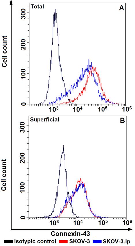
O.M. Kutova, A.D. Pospelov, L.V. Krylova, E.N. Gorshkova, I.V. Balalaeva
which form the most prohibitive junctions ization and thus may be considered as the basic
strictly regulating paracellular transport (Tsu- member of the majority of integrin-based con-
kita et al., 2001). Gap junctions constituted by tacts (Hynes, 1992). A summary of the studied
connexins on contrary act as direct cell connect- proteins and their common binding partners
ors as they form hexameric transmembrane among adapter proteins are represented in
channels (Nielsen et al., 2012). Desmosomes Fig. 1.
are formed by desmogleins and desmocollins It should be noted that to date a considerable
which are calcium-independent non-classical amount of data was obtained indicating mislo-
cadherins (Garrod & Chidgey, 2008). Integrin- calization of adhesion proteins (Wang & Li,
based contacts to ECM are dimers of various al- 2014; Alaga et al., 2017; Seraya-Bareket et al.,
pha- and beta-integrin subunits, where the spec- 2020) which shifts cancer cells towards aggres-
ificity of adhesion is defined by the composi- siveness. Our work aims to investigate the ex-
tion of a dimer (Takada et al., 2007). Concern- pression profile and localization of actin bind-
ing intracellular connections of integral adhe- ing integral adhesion proteins representing cell-
sion proteins, they bind to various components cell and cell-matrix contacts in two lines of hu-
of the cytoskeleton: desmosomes bind to inter- man ovarian adenocarcinoma cells with differ-
mediate filaments (Hatzfeld et al., 2017), while ent aggressiveness.
proteins of adherens and tight junctions bind to
the actin cytoskeleton (Campbell et al., 2017; Materials and Methods
Vicente-Manzanares et al., 2009). Emerging Cell lines. Cells of human ovarian adenocar-
data testifies that gap junction proteins con- cinoma cell lines SKOV-3 and SKOV-3.ip
nexins are also connected to actin cytoskeleton were used. Cells were cultured in DMEM me-
via adapter proteins which bind to specific sites dium containing 2 mM glutamine (PanEco,
on their cytoplasmatic C-tail (Herve et al., Russia), 10% (v/v) fetal bovine serum (Hy-
2007; Ambrosi et al., 2016). Clone, USA), 50μg/ml of Penicillin and
Actin cytoskeleton is recognized as one of 50 μg/ml of Streptomycin (PanEco, Russia) at
the central components orchestrating cancer 37 C in 5% CO2. For passaging, cells were de-
progression and metastasis (Galluzzi & tached with Versene solution (PanEco, Russia).
Thomas, 2020 a,b), thus profile assessment of
actin-connected adhesion proteins may be a rel- Cell preparation protocol to assess localiza-
evant tool in cancer research. Integral proteins tion of adhesion proteins. In order to perform
in general are the first-line contributors to adhe- flow cytometry analysis, monolayer culture
sion and communication and in these terms was detached from the substrate by incubation
their representation profile serves as a reflection with TrypLE solution (Thermo Fischer Scien-
of interactions in tumor microenvironment in a tific, USA) for 20 min at 37 C in 5% CO2.
certain time point and becomes a potent charac- Cells were fixed in 4% formaldehyde (Appli-
teristic which is appropriate to rely on both in chem, Germany) for 20 min at room tempera-
fundamental cancer research and drug effi- ture to exclude a possibility of protein profile
ciency evaluation (Farahani et al., 2014; changes during the analysis. In order to assess
Kutova et al., 2020). In this study we focus on the simultaneous superficial and intracellular
representative integral actin-binding proteins of abundance of studied proteins fixed cells were
adherens junctions, namely epithelial marker E- permeabilized with 0,02% Triton X-100 (VWR
cadherin and mesenchymal marker N-cadherin Life Science, USA) for 20 min at room temper-
(Loh et al., 2019); gap junctions, namely con- ature. Cells intended for assessing the abun-
nexin-43 which is the most widely represented dance of superficially located proteins of inter-
connexin in mammalian tissues (Bonacquisti & est proceeded to staining directly after fixation.
Nguyen, 2019); and integrin-based cell-ECM Cells were thoroughly washed with PBS to re-
contacts, namely integrin beta-1, which is the move formaldehyde and Triton-X100 before
most preferential partner in α-β integrin dimer- staining.
44 | doi: 10.24412/2500-2295-2021-2-43-54
ADHESION PROTEINS PROFILE AND LOCALIZATION IN OVARIAN CARCINOMA CELL LINES

 Fig. 1. Summary of studied proteins: E-cadherin, N-cadherin (cell-cell adherens junctions); connexin-43
 (cell-cell gap junctions); integrin beta-1 (cell-matrix junctions) with common adapter proteins

    Cell staining and flow cytometry analysis. In        Results
order to minimize non-specific binding, cells            Two ovarian carcinoma cell lines SKOV-3
were blocked in a solution of 3% milk (Appli-         and SKOV-3.ip were used in this study.
chem, Germany) in PBS for 1 h at room temper-         SKOV-3 cell line was obtained from the malig-
ature and incubated with antibodies specific to       nant ascites of a 64-year-old Caucasian female
the proteins of interest and antibodies of corre-     (Hung et al., 1992) and SKOV-3.ip cell line
sponding isotypic control according to manufac-       was obtained from the malignant ascites of tu-
turer’s instructions. The following specific anti-    mor bearing nu/nu mice which were intraperi-
bodies were used: E-cadherin Monoclonal Anti-         toneally inoculated with SKOV-3 cells.
body (67A4), FITC (Thermo Fischer Scientific,         SKOV-3.ip cells were indicated to possess
USA, Cat#A15757), N-cadherin Monoclonal               higher DNA synthesis rates, accelerated prolif-
Antibody (8C11), PE (Thermo Fischer Scien-            eration, increased colony-formation in soft
tific, USA, Cat#12-3259-42), connexin-43 Mon-         agar, formation of larger subcutaneous tumors
oclonal Antibody (CX-1B1), Alexa Fluor 488            and reduced survival of nu/nu mice bearing in-
(Thermo Fischer Scientific, USA, Cat#138388),         traperitoneally inoculated tumors (Yu et al.,
integrin beta-1 Monoclonal Antibody (TS2/16),         1993; Dar et al., 2017).
PE (Thermo Fischer Scientific, USA, Cat#12-              To evaluate the localization of adhesion pro-
0299-42). Excess antibodies were washed out           teins in monolayer cultures in ovarian carci-
from the specimens with 1% BSA (Sigma-Al-             noma cell lines we have conducted flow cytom-
drich, USA) solution in PBS and analyzed by           etry analysis of adhesion proteins participating
flow cytometry using a CytoFlex S (Beckman            in formation of cell-cell contacts, namely E-
Coulter, USA). Fluorescence of FITC, Alexa-           cadherin and N-cadherin (adherens junctions),
Fuor 488 and PE was excited with a 488-nm la-         connexin-43 (gap junctions) and cell-ECM con-
ser and the signal was detected with 525/40           tacts namely integrin beta-1. The abundance
(FITC, AlexaFluor488) and 585/42 (PE) band            level of each protein of interest is represented
pass filters.                                         by relative fluorescence (RF) value calculated

                                                                   Opera Med Physiol. 2021. Vol. 8 (2) | 45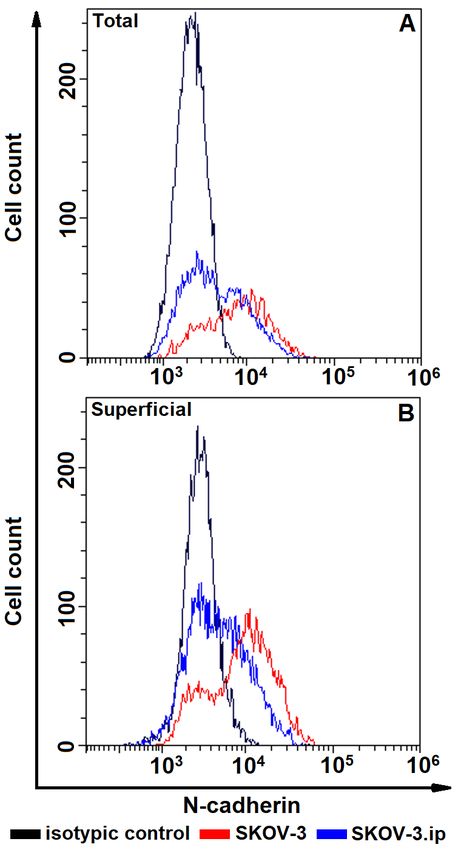
O.M. Kutova, A.D. Pospelov, L.V. Krylova, E.N. Gorshkova, I.V. Balalaeva

Fig. 2. The distributions of SKOV-3 and SKOV-3.             Fig. 3. The distributions of SKOV-3 and SKOV-3.
ip cells according to presence of total (A, upper plot)     ip cells according to presence of total (A, upper plot)
and superficially located (B, lower plot) E-cadherin.       and superficially located (B, lower plot)
Cells were stained with E-cadherin-specific anti-           N-cadherin. Cells were stained with N-cadherin-
bodies and analyzed by flow cytometry                       specific antibodies and analyzed by flow cytometry

 as a ratio of mean fluorescence intensity of cells              N-cadherin was represented at very low
 stained with antibodies specific to proteins of              level in both cell lines. Along with that, pop-
 interest to mean fluorescence intensity of cells             ulation heterogeneity was detected: a consid-
 stained with antibodies of corresponding iso-                erable part of the cells demonstrated the com-
 typic control. RF value equal to 1 indicates that            plete absence of N-cadherin (Fig. 3). Only a
 no analyzed protein is present in the cells.                 modest part of total N-cadherin was super-
                                                              ficially located (Fig. 6). Of note, the total
     E cadherin. The obtained data indicate that              abundance of N-cadherin in N-cadherin-
 the studied cells do not possess E-cadherin at               positive cells was significantly higher in
 all; relative fluorescence values were equal or              SKOV-3 cells in comparison to SKOV-3.
 close to 1 (Figs 2, 6, 7).                                   ip cells (Fig. 7,B).

 46 | doi: 10.24412/2500-2295-2021-2-43-54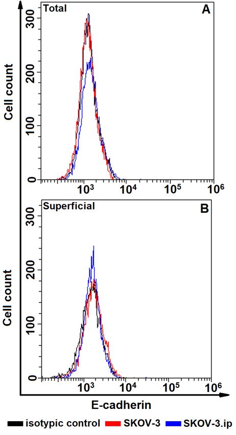
ADHESION PROTEINS PROFILE AND LOCALIZATION IN OVARIAN CARCINOMA CELL LINES
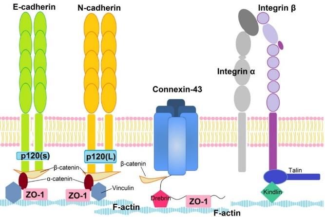
Fig. 4. The distributions of SKOV-3 and SKOV-3. Fig. 5. The distributions of SKOV-3 and SKOV-3.
ip cells according to presence of total (A, upper plot) ip cells according to presence of total (A, upper plot)
and superficially located (B, lower plot) connexin- and superficially located (B, lower plot) integrin
43. Cells were stained with connexin-43 -specific beta-1. Cells were stained with integrin beta-1-spe-
antibodies and analyzed by flow cytometry cific antibodies and analyzed by flow cytometry
Connexin-43 total representation was at a the total integrin beta-1 content was superfi-
relatively high level both in SKOV-3 and cially localized. Also the studied cell lines
SKOV-3.ip cell lines, at the same time similarly showed heterogeneity as a minor part of the
to N-cadherin, only a modest level of connexin- population represented loss of integrin beta-1
43 was represented at the cell surface (not more (Figs 5, 6). Statistically significant differ-
than 20% of total amount) (Figs 4, 6) and also ences between the studied cell lines were de-
similarly to N-cadherin, connexin-43 was sig- tected, moreover, the peculiar phenomenon
nificantly more abundant in SKOV-3 cell line was revealed for integrin beta-1: SKOV-3 cell
compared to SKOV-3.ip (Fig. 7, B). line possessed higher total integrin beta-1
Integrin beta-1 was the most abundant of level (Fig. 2,A) while superficially located in-
the studied proteins both in SKOV-3 and tegrin beta-1 prevailed in SKOV-3.ip cell line
SKOV-3.ip cell lines and again only a part of (Fig. 2,B).
Opera Med Physiol. 2021. Vol. 8 (2) | 47O.M. Kutova, A.D. Pospelov, L.V. Krylova, E.N. Gorshkova, I.V. Balalaeva
Fig. 6. The abundance of total and superficially located Fig. 7. The abundance of adhesion proteins in ovarian ad-
adhesion proteins in SKOV-3 (A) and SKOV-3.ip (B) enocarcinoma cell lines totally represented (A) and lo-
cell lines. Expression levels of E-cadherin, N-cadherin, cated superficially (B). Expression levels of E-cadherin,
connexin-43 and integrin beta-1 denoted as relative fluo- N-cadherin, connexin-43 and integrin beta-1 denoted as
rescence values calculated as a ratio of mean fluores- relative fluorescence values calculated as a ratio of mean
cence intensity of cells stained with specific antibodies to fluorescence intensity of cells stained with specific anti-
mean fluorescence intensity of cells stained with antibod- bodies to mean fluorescence intensity of cells stained
ies of corresponding isotypic control. Data are presented with antibodies of corresponding isotypic control. Data
as mean ± SD (n = 3). “*” indicates significant difference are presented as mean ± SD (n = 3). “*” indicates signif-
in RF between total and superficially located protein (Un- icant difference in RF between protein abundance in
paired t-test with Welch correction, p < 0.05) studied cell lines (Unpaired t-test with Welch correction,
p < 0.05)
Discussion drive cell invasiveness or motility (Moh &
Intercellular contact proteins are usually Shen, 2009; Tang et al., 2018; Mulkearns-Hu-
considered to be tumor suppressors, but there is bert et al., 2020).
more and more conflicting evidence of their in- In present work we have analyzed the total
volvement in the formation of an aggressive abundance and the abundance of superficially
phenotype and triggering tumor progression localized representative proteins of actin-con-
and invasion. An intriguing potential reason of nected cell-cell and cell-ECM contacts in two
such controversy is the context in which these ovarian carcinoma cell lines with different ag-
proteins function, e.g. a protein which is con- gressiveness. In case of adherens junctions we
sidered to be proliferation suppressor might have showed that E-cadherin was absent in
48 | doi: 10.24412/2500-2295-2021-2-43-54ADHESION PROTEINS PROFILE AND LOCALIZATION IN OVARIAN CARCINOMA CELL LINES
both studied cell lines. Concerning N-cadherin tient survival (Liu et al., 2015) and in excep-
the studied cell cultures showed heterogeneity tional cases of invasive lobular breast cancer
as a part of the cells did not express N-cadherin characterized by tubular elements formation
and the rest of the population expressed N-cad- (Christgen et al., 2020). In addition, the ob-
herin at a relatively low level. Loss of E-cad- served difference in the amount of total
herin, which is widely recognized as an epithe- N-cadherin and superficially localized can be
lial marker, triggers the Epithelial-to-Mesen- explained by the transition of N-cadherin into
chymal Transition (EMT) of cells and contrib- a soluble form, which has pro-angiogenic
utes to the development of an aggressive phe- properties and is at high level detected in bi-
notype. During EMT, there is a switch from ological fluids of cancer patients (Derycke et
the synthesis of E-cadherin to the synthesis of al., 2006 a, b).
N-cadherin, which is a mesenchymal marker According to the obtained data gap junction
and is involved in intracellular signaling pro- protein connexin-43 was represented at rela-
moting invasion and metastasis (Gravdal et al., tively high level in both studied cell lines yet
2007; Loh et al., 2019). It is worth noting that significantly prevailed in SKOV-3 cell line.
the cells of the studied lines are characterized Functional connexin-based gap junctions being
by the formation of loose three-dimensional properly organized provide Gap Junction Inter-
aggregates in vitro; which is in line with an as- cellular Communication (GJIC), which in turn
sumption of their mesenchymal state (Winner maintains coordinated work of the cells within
et al., 2016; Sokolova et al., 2019; Kutova et the tissue. Thus, connexins are considered to
al., 2020). hinder tumor progression via GJIC (Krutov-
Currently, more and more evidence is accu- skikh et al., 2002, Zefferino et al., 2019). An-
mulating that EMT is not a binary process, but other plausible mechanism of connexin-medi-
a continual one, implying a huge number of var- ated tumor suppression is the participation of
iants of the intermediate states of the cell, when connexins in intracellular signaling interfering
it can express epithelial and mesenchymal with proliferative and invasive signals (Aasen
markers simultaneously, which contributes to et al., 2019; Mulkearns-Hubert et al., 2020).
the development of cancer plasticity (Sha et al., Concerning tumor suppressing potencies of
2019). It has been hypothesized and supported connexin-43 it is reported that it can inhibit pro-
by mathematical modeling that such intermedi- liferation by affecting the Wingless-Integrated
ate states can accelerate EMT and exacerbate its (Wnt)/β-catenin pathway (Shima et al., 2006),
consequences (Goetz et al., 2020). The data on decreasing the activity of proto-oncogene tyro-
the intermediate position occupied by the sine-protein kinase Src (acronym of ‘sarcoma’)
SKOV-3 and SKOV-3.ip lines vary, because or epidermal growth factor (EGF) (Herrero-
despite the fact that these lines are called inter- Gonzalez et al., 2010; Qui et al., 2016) or by
mediate mesenchymal, this can mean both the triggering apoptosis via binding to pro-apop-
simultaneous presence of E-cadherin and N- totic Bcl2-associated X protein (Bax) (Sun et
cadherin (Rosso et al., 2017; Teng et al., 2015) al., 2012). Of note, our data indicate that the
or the absence of E-cadherin with presence of level of superficially localized connexin-43 did
N-cadherin (Klymenko et al., 2017), which was not exceed 20% of total abundance. It is possi-
observed in our work. Such inconsistency of the bly due to the disrupted connexin-43 trafficking
data might be due to the high plasticity of tumor to plasma membrane. Translocation to the cyto-
cells, which results in subtle differences in the plasm was reported for connexin-43 and may be
phenotype evoked by different cultivation con- mediated by Wnt signaling pathway (Hou et al.,
ditions. It should also be noted that the repre- 2019); it was also shown for other connexins
sentation of N-cadherin in our experiments was (Krutovskikh et al., 1994; Thiagarajan et al.,
rather low. Simultaneous loss of adherens junc- 2018). Aberrant localization of connexins
tions’ proteins has been described for very which was observed in our study is reported to
poorly differentiated hepatocellular carcinoma, be associated with the triggering of EMT
which was accompanied by extremely low pa- (Crespin et al., 2016; Kotini et al., 2018).
Opera Med Physiol. 2021. Vol. 8 (2) | 49O.M. Kutova, A.D. Pospelov, L.V. Krylova, E.N. Gorshkova, I.V. Balalaeva
The most abundant of all observed proteins uously change over time during different
was integrin beta-1. The majority of research phases of an invasion process (Mol et al., 2007;
testifies that high levels of integrin beta-1 are Xu et al., 2008).
represented in tumor cells compared to normal
epithelium (Min et al., 2020). Up-regulated in- Conclusions
tegrin beta-1 is associated with increased cancer In this work we have analyzed total content
cell survival, proliferation, migration and col- and superficially localized pool of adhesion
ony formation in vitro (Pardo et al., 2002; proteins in two ovarian carcinoma cell lines.
Chang et al., 2019) and with advanced stage of The absolute loss of epithelial marker E-cad-
the tumor and lower life expectancy in patients herin and overall mislocalization of the rest an-
(Lin et al., 2014; Lawson et al., 2010). Our data alyzed proteins and hence their impaired adhe-
indicate that superficial localization of integrin sive function, allow us to speculate about mes-
beta-1 is low compared to total protein. It is in- enchymal characteristics of the studied cell
teresting to note that higher total abundance of lines. At the same time, we have detected het-
integrin beta-1 was detected in SKOV-3 cell erogeneity of the studied cell cultures in terms
line, yet superficially localized integrin beta-1 of N-cadherin and integrin beta-1 abundance.
was more represented in SKOV-3.ip cell line. This is in line with the inconsistency of pub-
Superficially located integrins being activated lished data on SKOV-3 and SKOV-3.ip pheno-
are shown to promote tumors towards increased type and thus may be a strong evidence of can-
malignancy mainly via focal adhesion kinase cer cells plasticity even in controllable condi-
(FAK) signaling axis (Yang et al., 2014; Zhang tions of laboratory maintenance. According to
& Zou, 2015; Xu et al., 2017). the obtained data SKOV-3.ip cell line pos-
The profile of adhesion proteins expression sessed lower overall adhesion proteins abun-
appears to be more adequate means of tumor dance which is in line with a mainstream notion
phenotype identification compared to assess- that loss of cellular adhesion underlies tumor
ment of individual proteins due to their com- aggressiveness. At the same time higher repre-
plex crosstalk and reciprocal regulation. For ex- sentation of superficially localized integrin
ample, it was shown that diapedesis-mediated beta-1 might be an additional sign of SKOV-
tumor invasiveness was registered in tumors 3.ip superlative aggressiveness. Our data may
with simultaneous loss of E-cadherin and high be useful to expand an understanding of the na-
expression of connexin-43 (Mol et al., 2007) or ture of the exceeding aggressiveness of SKOV-
integrin beta-1 (Shu et al., 2013, Symowicz et 3.ip cell line in comparison to SKOV-3.
al., 2007). It should be noted that protein ex-
pression and their representation on the cell sur- Acknowledgments
face is a dynamic process which is highly de- This research was financially supported by
pendent on the interplay of microenvironmental the Russian Science Foundation (project No.
context and ongoing physiological processes. 19-74-20168). O.M.K. thanks the Russian
For example, along with high expression of Foundation for Basic Research for personal
connexin-43, E-cadherin abundance can contin- financial support (project No. 19-34-90159).
References
AASEN T., LEITHE E., GRAHAM S.V., KAMERITSCH P., MAYAN M.D., MESNIL M., POGODA K.
& TABERNERO A. (2019): Connexins in cancer: bridging the gap to the clinic. Oncogene, 38, 4429–
4451.
ALAGA K.C., CRAWFORD M., DAGNINO L., & LAIRD D.W. (2017): Aberrant Cx43 Expression and
Mislocalization in Metastatic Human Melanomas. Journal of Cancer, 8(7), 1123–1128.
AMBROSI C., REN C., SPAGNOL G., CAVIN G., CONE A., GRINTSEVICH E.E., SOSINSKY G.E. &
SORGEN P.L. (2016): Connexin43 Forms Supramolecular Complexes through Non-Overlapping Bind-
ing Sites for Drebrin, Tubulin, and ZO-1. PloS one, 11(6), e0157073.
50 | doi: 10.24412/2500-2295-2021-2-43-54ADHESION PROTEINS PROFILE AND LOCALIZATION IN OVARIAN CARCINOMA CELL LINES
BONACQUISTI E.E. & NGUYEN J. (2019): Connexin 43 (Cx43) in cancer: Implications for therapeutic
approaches via gap junctions. Cancer Letters, 442, 439–444.
BRÜCHER B.L. & JAMALL I.S. (2014): Cell-cell communication in the tumor microenvironment, carcin-
ogenesis, and anticancer treatment. Cellular physiology and biochemistry: international journal of ex-
perimental cellular physiology, biochemistry, and pharmacology, 34(2), 213–243.
CAMPBELL H.K., MAIERS J.L. & DEMALI K.A. (2017): Interplay between tight junctions & adherens
junctions. Experimental cell research, 358(1), 39–44.
CHANG Y.N., LIANG Y., GU W., WANG J., QIN Y., CHEN K., LI J., BAI X., ZHANG J. & XING G.
(2019): Microfluidic Analysis for Separating and Measuring the Deformability of Cancer Cell Subpop-
ulations. ACS Omega, 4(5), 8318–8323.
CHRISTGEN M., BARTELS S., VAN LUTTIKHUIZEN J.L., BUBLITZ J., RIEGER L.U., CHRISTGEN
H., STARK H., SANDER B., LEHMANN U., STEINEMANN D., DERKSEN P. & KREIPE H. (2020).
E-cadherin to P-cadherin switching in lobular breast cancer with tubular elements. Modern pathology:
an official journal of the United States and Canadian Academy of Pathology, Inc, 33(12), 2483–2498.
CRESPIN S., FROMONT G., WAGER M., LEVILLAIN P., CRONIER L., MONVOISIN A., DEFAMIE
N., & MESNIL M. (2016): Expression of a gap junction protein, connexin43, in a large panel of human
gliomas: new insights. Cancer medicine, 5(8), 1742–1752.
DAR S., CHHINA J., MERT I., CHITALE D., BUEKERS T., KAUR H., GIRI S., MUNKARAH A. &
RATTAN R. (2017): Bioenergetic Adaptations in Chemoresistant Ovarian Cancer Cells. Scientific re-
ports, 7(1), 8760.
DERYCKE L., DE WEVER O., STOVE V., VANHOECKE B., DELANGHE J., DEPYPERE H. &
BRACKE M. (2006): Soluble N-cadherin in human biological fluids. International journal of cancer,
119(12), 2895–2900.
DERYCKE L., MORBIDELLI L., ZICHE M., DE WEVER O., BRACKE M. & VAN AKEN E. (2006): Soluble
N-cadherin fragment promotes angiogenesis. Clinical & experimental metastasis, 23(3-4), 187–201.
DOMINIAK A., CHEŁSTOWSKA B., OLEJARZ W. & NOWICKA G. (2020): Communication in the Can-
cer Microenvironment as a Target for Therapeutic Interventions. Cancers, 12(5), 1232.
FARAHANI E., PATRA H.K., JANGAMREDDY J.R., RASHEDI I., KAWALEC M., RAO PARITI R.K.,
BATAKIS P. & WIECHEC E. (2014): Cell adhesion molecules and their relation to (cancer) cell stem-
ness. Carcinogenesis, 35(4), 747–59.
GALLUZZI L. & THOMAS C. (2020a): Actin Cytoskeleton in Cancer Progression and Metastasis. Interna-
tional Review of Cell and Molecular Biology, 355(Pt A), 254.
GALLUZZI L. & THOMAS C. (2020b): Actin Cytoskeleton in Cancer Progression and Metastasis. Interna-
tional Review of Cell and Molecular Biology, 356(Pt B), 330.
GARROD D., & CHIDGEY M. (2008): Desmosome structure, composition and function. Biochimica et Bi-
ophysica Acta, 1778, 572–587.
GOETZ H., MELENDEZ-ALVAREZ J.R., CHEN L. & TIAN X. J. (2020): A plausible accelerating function
of intermediate states in cancer metastasis. PLoS computational biology, 16(3), e1007682.
GRAVDAL K., HALVORSEN O.J., HAUKAAS S.A. & AKSLEN L.A. (2007): A switch from E-cadherin
to N-cadherin expression indicates epithelial to mesenchymal transition and is of strong and independent
importance for the progress of prostate cancer. Clinical cancer research: an official journal of the Amer-
ican Association for Cancer Research, 13(23), 7003–7011.
HARRIS T. J. & TEPASS U. (2010): Adherens junctions: from molecules to morphogenesis. Nature reviews.
Molecular cell biology, 11(7), 502–514.
HATZFELD M., KEIL R. & MAGIN T.M. (2017): Desmosomes and Intermediate Filaments: Their Conse-
quences for Tissue Mechanics. Cold Spring Harbor perspectives in biology, 9(6), a029157.
HERRERO-GONZALEZ S., GANGOSO E., GIAUME C., NAUS, C.C., MEDINA J.M. & TABERNERO
A. (2010): Connexin43 inhibits the oncogenic activity of c-Src in C6 glioma cells. Oncogene, 29, 5712–
5723.
HERVÉ J.C., BOURMEYSTER N., SARROUILHE D. & DUFFY H.S. (2007): Gap junctional complexes:
from partners to functions. Progress in biophysics and molecular biology, 94(1-2), 29–65.
HOU X., KHAN M.R.A., TURMAINE M., THRASIVOULOU C., BECKER D.L. & AHMED A. (2019):
Wnt signaling regulates cytosolic translocation of connexin 43. American Journal of Physiology-Regu-
latory, 317, R248–R261.
Opera Med Physiol. 2021. Vol. 8 (2) | 51O.M. Kutova, A.D. Pospelov, L.V. Krylova, E.N. Gorshkova, I.V. Balalaeva
HUNG M.C., ZHANG X., YAN D.H., ZHANG H.Z., HE G.P., ZHANG T.Q. & SHI D.R. (1992): Aberrant
expression of the c-erbB-2/neu protooncogene in ovarian cancer. Cancer letters, 61(2), 95–103.
HYNES R.O. (1992): Integrins: versatility, modulation, and signaling in cell adhesion. Cell, 69(1), 11–25.
KHAN Z.S. & HUSSAIN F. (2020): Shear Stress Increases V-H + -ATPase and Acidic Vesicle Number
Density, and p-mTORC2 Activation in Prostate Cancer Cells. Cellular and molecular bioengineering,
13(6), 591–604.
KLYMENKO Y., JOHNSON J., BOS B., LOMBARD R., CAMPBELL L., LOUGHRAN E. & STACK
M.S. (2017): Heterogeneous Cadherin Expression and Multicellular Aggregate Dynamics in Ovarian
Cancer Dissemination. Neoplasia (New York, N.Y.), 19(7), 549–563.
KOTINI M., BARRIGA E.H., LESLIE J., GENTZEL M., RAUSCHENBERGER V., SCHAMBONY A. &
MAYOR R. (2018): Gap junction protein connexin-43 is a direct transcriptional regulator of N-cadherin
in vivo. Nature communications, 9(1), 3846.
KRUTOVSKIKH V., PICCOLI C. & YAMASAKI H. (2002): Gap junction intercellular communication
propagates cell death in cancerous cells. Oncogene, 21, 1989–1999.
KUTOVA O.M., SENCHA L.M., POSPELOV A.D., DOBRYNINA O.E., BRILKINA A.A., CHERKASOVA
E.I. & BALALAEVA I.V. (2020). Comparative Analysis of Cell-Cell Contact Abundance in Ovarian Car-
cinoma Cells Cultured in Two- and Three-Dimensional In Vitro Models. Biology, 9(12), 446.
LAWSON M.H., CUMMINGS N.M., RASSL D.M., VOWLER S.L., WICKENS M., HOWAT W.J., BREN-
TON J.D., MURPHY G. & RINTOUL R.C. (2010): Bcl-2 and β1-integrin predict survival in a tissue
microarray of small cell lung cancer. British journal of cancer, 103(11), 1710–1715.
LIN H.C., WU C.L., CHEN Y.L., HUANG J.S., WONG T.Y. & YUAN K. (2014): High-level β1-integrin
expression in a subpopulation of highly tumorigenic oral cancer cells. Clinical Oral Investigations,
18(4), 1277–1284.
LIU Y.A., LIANG B.Y., GUAN Y., YOU J., ZHU L., CHEN X.P. & HUANG Z.Y. (2015): Loss of N-
cadherin is associated with loss of E-cadherin expression and poor outcomes of liver resection in hepa-
tocellular carcinoma. The Journal of surgical research, 194(1), 167–176.
LOH C.Y., CHAI J.Y., TANG T.F., WONG W.F., SETHI G., SHANMUGAM M.K., CHONG P.P. & LOOI
C.Y. (2019): The E-Cadherin and N-Cadherin Switch in Epithelial-to-Mesenchymal Transition: Signal-
ing, Therapeutic Implications, and Challenges. Cells, 8(10), 1118.
MIN W., ZOU C., DAI D., ZUO Q., CHEN C., XU J., LI Y. & YUE Z. (2020): Integrin Beta 1 Promotes
Glioma Cell Proliferation by Negatively Regulating the Notch Pathway. Journal of Oncology, 15,
8297017.
MOH M.C. & SHEN S. (2009): The roles of cell adhesion molecules in tumor suppression and cell migration:
a new paradox. Cell adhesion & migration, 3(4), 334–336.
MOL A.J., GELDOF A.A., MEIJER G.A., VAN DER POEL H.G. & VAN MOORSELAAR R.J. (2007):
New experimental markers for early detection of high-risk prostate cancer: role of cell-cell adhesion and
cell migration. Journal of cancer research and clinical oncology, 133(10), 687–695.
MOU Y.Y., ZHAO G.Q., LIN J.Y., ZHAO J., LIN H., HU L.T., XU Q., WANG Q. & SUN W.R. (2011):
Expression of connexin 43 and E-cadherin in choroidal melanoma. International journal of ophthalmol-
ogy, 4(2), 156–161.
MULKEARNS-HUBERT E.E., REIZES O. & LATHIA J.D. (2020): Connexins in Cancer: Jekyll or Hyde?
Biomolecules, 10(12), 1654.
NEOPHYTOU C.M., PANAGI M., STYLIANOPOULOS T. & PAPAGEORGIS P. (2021): The Role of
Tumor Microenvironment in Cancer Metastasis: Molecular Mechanisms and Therapeutic Opportunities.
Cancers, 13(9), 2053.
NIELSEN M.S., AXELSEN L.N., SORGEN P.L., VERMA V., DELMAR M. & HOLSTEIN-RATHLOU
N.H. (2012): Gap junctions. Comprehensive Physiology, 2, 1981–2035.
NISHIMURA M. (2003): Suppression of gap junctional intercellular communication via 5' CpG island meth-
ylation in promoter region of E-cadherin gene in endometrial cancer cells. Carcinogenesis, 24(10),
1615–1623.
PARDO O.E., ARCARO A., SALERNO G., RAGUZ S., DOWNWARD J. & SECKL M.J. (2002): Fibro-
blast growth factor-2 induces translational regulation of Bcl-XL and Bcl-2 via a MEK-dependent path-
way: correlation with resistance to etoposide-induced apoptosis. Journal of Biological Chemistry, 277,
12040–12046.
52 | doi: 10.24412/2500-2295-2021-2-43-54ADHESION PROTEINS PROFILE AND LOCALIZATION IN OVARIAN CARCINOMA CELL LINES
QIU X., CHENG J.C., KLAUSEN C., CHANG H.M., FAN Q. & LEUNG P.C. (2016): EGF-Induced Con-
nexin43 Negatively Regulates Cell Proliferation in Human Ovarian Cancer. Journal of Cellular Physi-
ology, 231, 111–119.
ROSSO M., MAJEM B., DEVIS L., LAPYCKYJ L., BESSO M.J., LLAURADÓ M., ABASCAL M.F.,
MATOS M.L., LANAU L., CASTELLVÍ J., SÁNCHEZ J.L., PÉREZ BENAVENTE A., GIL-
MORENO A., REVENTÓS J., SANTAMARIA MARGALEF A., RIGAU M. & VAZQUEZ-LEVIN
M.H. (2017): E-cadherin: A determinant molecule associated with ovarian cancer progression, dissemi-
nation and aggressiveness. PloS one, 12(9), e0184439.
SANDQUIST E.J., SOMJI S., DUNLEVY J.R., GARRETT S.H., ZHOU X.D., SLUSSER-NORE A. &
SENS D.A. (2016): Loss of N-Cadherin Expression in Tumor Transplants Produced From As+3- and
Cd+2-Transformed Human Urothelial (UROtsa) Cell Lines. PloS one, 11(5), e0156310.
SERAYA-BAREKET C., WEISZ A., SHINDERMAN-MAMAN E., TEPER-ROTH S., STAMLER D., AR-
BIB, N., KADAN Y., FISHMAN A., KIDRON D., EDELSTEIN E., ELLIS M. & ASHUR-FABIAN
O. (2020): The identification of nuclear αvβ3 integrin in ovarian cancer: non-paradigmal localization
with cancer promoting actions. Oncogenesis, 9(7), 69.
SHA Y., HAENSEL D., GUTIERREZ G., DU H., DAI X. & NIE Q. (2019): Intermediate cell states in
epithelial-to-mesenchymal transition. Physical biology, 16(2), 021001.
SHIMA K., MURAMATSU T., ABIKO Y., YAMAOKA Y., SASAKI H. & SHIMONO M. (2006): Con-
nexin 43 transfection in basaloid squamous cell carcinoma cells. Oncology Reports, 16, 285–288.
SHU H., CHEN H., YANG B., CHANG Z., XIONG M. & CHEN W. (2013): Aberrant expression of E-
cadherin and integrin β-1 in trophoblasts is associated with malignant gestational trophoblastic diseases.
International journal of gynecological cancer: official journal of the International Gynecological Can-
cer Society, 23(4), 749–754.
SOKOLOVA E., KUTOVA O., GRISHINA A., POSPELOV A., GURYEV E., SCHULGA A., DEYEV S.,
& BALALAEVA I. (2019). Penetration Efficiency of Antitumor Agents in Ovarian Cancer Spheroids:
The Case of Recombinant Targeted Toxin DARPin-LoPE and the Chemotherapy Drug, Doxorubicin.
Pharmaceutics, 11(5), 219.
SONG D., YANG D., POWELL C.A. & WANG X. (2019): Cell-cell communication: old mystery and new
opportunity. Cell biology and toxicology, 35(2), 89–93.
SONNENSCHEIN C. & SOTO A.M. (2020): Over a century of cancer research: Inconvenient truths and
promising leads. PLoS biology, 18(4), e3000670.
SUN Q., KANEHIRA K. & TANIGUCHI A. (2018): PEGylated TiO2 nanoparticles mediated inhibition of
cell migration via integrin beta 1. Science and technology of advanced materials, 19(1), 271–281.
SUN Y., ZHAO X., YAO Y., QI X., YUAN Y. & HU Y. (2012): Connexin 43 interacts with Bax to regulate
apoptosis of pancreatic cancer through a gap junction-independent pathway. International journal of
oncology, 41(3), 941–948.
SYMOWICZ J., ADLEY B.P., GLEASON K.J., JOHNSON J.J., GHOSH S., FISHMAN D.A., HUDSON
L.G. & STACK M.S. (2007): Engagement of collagen-binding integrins promotes matrix metallopro-
teinase-9-dependent E-cadherin ectodomain shedding in ovarian carcinoma cells. Cancer research,
67(5), 2030–2039.
TAKADA Y., YE X. & SIMON S. (2007): The integrins. Genome biology, 8(5), 215.
TANG M., YUE P., IP P.P., HUANG R.L., LAI H.C., CHEUNG A., TSE K.Y., NGAN H. & WONG A.
(2018): Soluble E-cadherin promotes tumor angiogenesis and localizes to exosome surface. Nature com-
munications, 9(1), 2270.
TENG L., PENG S., GUO H., LIANG H., XU Z., SU Y. & GAO L. (2015). Conditioned media from human
ovarian cancer endothelial progenitor cells induces ovarian cancer cell migration by activating epithelial-
to-mesenchymal transition. Cancer gene therapy, 22(11), 518–523.
TSUKITA S., FURUSE M. & ITOH M. (2001): Multifunctional strands in tight junctions. Nature Reviews
Molecular Cell Biology, 2, 285–293.
VICENTE-MANZANARES M., CHOI C.K. & HORWITZ A.R. (2009): Integrins in cell migration--the actin
connection. Journal of cell science, 122(Pt 2), 199–206.
WANG X. & LI S. (2014): Protein mislocalization: mechanisms, functions and clinical applications in cancer.
Biochimica et biophysica acta, 1846(1), 13–25.
Opera Med Physiol. 2021. Vol. 8 (2) | 53O.M. Kutova, A.D. Pospelov, L.V. Krylova, E.N. Gorshkova, I.V. Balalaeva
WELLS A., YATES C. & SHEPARD C.R. (2008): E-cadherin as an indicator of mesenchymal to epithelial
reverting transitions during the metastatic seeding of disseminated carcinomas. Clinical & experimental
metastasis, 25(6), 621–628.
WINNER K.K., STEINKAMP M.P., LEE R.J., SWAT M., MULLER C.Y., MOSES M.E., JIANG Y. &
WILSON B.S. (2016): Spatial Modeling of Drug Delivery Routes for Treatment of Disseminated Ovar-
ian Cancer. Cancer research, 76(6), 1320–1334.
XU H.T., LI Q.C., ZHANG Y.X., ZHAO Y., LIU Y., YANG Z.Q. & WANG E. H. (2008): Connexin 43
recruits E-cadherin expression and inhibits the malignant behaviour of lung cancer cells. Folia histo-
chemica et cytobiologica, 46(3), 315–321.
XU J., ZHENG T., HONG W., YE H., HU C. & ZHENG Y. (2018): Mechanism for the Decision of Ovarian
Surface Epithelial Stem Cells to Undergo Neo-Oogenesis or Ovarian Tumorigenesis. Cell Physiol Bio-
chem, 50(1), 214–232.
YANG Z., ZHOU X., LIU Y., GONG C., WEI X., ZHANG T., MA D. & GAO Q. (2014). Activation of
integrin β1 mediates the increased malignant potential of ovarian cancer cells exerted by inflammatory
cytokines. Anti-cancer agents in medicinal chemistry, 14(7), 955–962.
YU D., WOLF J.K., SCANLON M., PRICE J.E. & HUNG M.C. (1993): Enhanced c-erbB-2/neu expression
in human ovarian cancer cells correlates with more severe malignancy that can be suppressed by E1A.
Cancer Research, 53(4), 891–898.
ZEFFERINO R., PICCOLI C., GIOIA S. D., CAPITANIO N. & CONESE M. (2019): Gap Junction Inter-
cellular Communication in the Carcinogenesis Hallmarks: Is This a Phenomenon or Epiphenomenon?
Cells, 8(8), 896.
ZEMLJIC-HARPF A.E., GODOY J.C., PLATOSHYN O., ASFAW E.K., BUSIJA A.R., DOMENIGHETTI
A.A. & ROSS R.S. (2014): Vinculin directly binds zonula occludens-1 and is essential for stabilizing
connexin-43-containing gap junctions in cardiac myocytes. Journal of cell science, 127(Pt 5), 1104–
1116.
ZHANG L. & ZOU W. (2015). Inhibition of integrin β1 decreases the malignancy of ovarian cancer cells
and potentiates anticancer therapy via the FAK/STAT1 signaling pathway. Molecular medicine reports,
12(6), 7869–7876.
54 | doi: 10.24412/2500-2295-2021-2-43-54You can also read