Cancer Susceptibility Candidate 9 (CASC9) Promotes Colorectal Cancer Carcinogenesis via mTOR-Dependent Autophagy and Epithelial-Mesenchymal ...
←
→
Page content transcription
If your browser does not render page correctly, please read the page content below
ORIGINAL RESEARCH
                                                                                                                                              published: 04 May 2021
                                                                                                                                     doi: 10.3389/fmolb.2021.627022

                                               Cancer Susceptibility Candidate 9
                                               (CASC9) Promotes Colorectal
                                               Cancer Carcinogenesis via
                                               mTOR-Dependent Autophagy and
                                               Epithelial–Mesenchymal Transition
                                               Pathways
                                               Md Zahirul Islam Khan and Helen Ka Wai Law*
                                               Department of Health Technology and Informatics, Faculty of Health and Social Sciences, The Hong Kong Polytechnic
                                               University, Kowloon, Hong Kong

                                               Background: Colorectal cancer (CRC) is the third most common cancer worldwide.
                         Edited by:            Many recent studies have demonstrated that different long non-coding RNAs (lncRNAs)
                        Xuefeng Liu,           are involved in the initiation, advancement, and metastasis of many cancers including
Georgetown University, United States
                                               CRC. Cancer susceptibility candidate 9 (CASC9) is an lncRNA that has been reported
                           Reviewed by:
                           Paola Valentini,    in many cancers, but its role in CRC is poorly understood. In this study, we aimed to
Italian Institute of Technology (IIT), Italy   examine the expression of CASC9 in CRC cell lines and to determine the mechanism of
                               Yujie Yuan,
      The First Affiliated Hospital of Sun
                                               action of CASC9 in CRC carcinogenesis.
                Yat-sen University, China
                                               Methods: The expression of CASC9 in CRC tissues was compared with normal
                    *Correspondence:
                                               samples from publicly available datasets in The Cancer Genome Atlas (TCGA) and The
                     Helen Ka Wai Law
                  hthelen@polyu.edu.hk         Encyclopedia of RNA Interactomes (ENCORI). CASC9 expression was further verified
                                               in four CRC cell lines (DLD1, HT-29, SW480, and HCT-116) and normal colorectal cell
                    Specialty section:
                                               line (CCD-112CoN) by real-time quantitative polymerase chain reaction (RT-qPCR). After
          This article was submitted to
           Protein and RNA Networks,           gene silencing in HCT-116 and SW480, Cell Counting Kit-8 assay, clonogenic assay, and
                 a section of the journal      wound healing assay were performed to evaluate cell proliferation, viability, and migration
    Frontiers in Molecular Biosciences
                                               index of cells. Western blotting was used to explore the key pathways involved.
        Received: 07 November 2020
           Accepted: 09 March 2021             Results: CASC9 was significantly upregulated as analyzed from both public datasets
            Published: 04 May 2021
                                               TCGA and ENCORI where its overexpression was associated with poor survival
                              Citation:
       Islam Khan MZ and Law HKW
                                               of CRC patients. Similarly, CASC9 was significantly overexpressed in the CRC cell
          (2021) Cancer Susceptibility         lines compared with normal cells studied. The silencing of CASC9 in HCT-116 and
     Candidate 9 (CASC9) Promotes              SW480 attenuated cell proliferation and migration significantly. Furthermore, pathways
Colorectal Cancer Carcinogenesis via
        mTOR-Dependent Autophagy               investigations showed that silencing of CASC9 significantly induced autophagy,
          and Epithelial–Mesenchymal           promoted AMP-activated protein kinase (AMPK) phosphorylation, inhibited mTOR and
                 Transition Pathways.
         Front. Mol. Biosci. 8:627022.
                                               AKT signaling pathways, and altered epithelial–mesenchymal transition (EMT) marker
    doi: 10.3389/fmolb.2021.627022             protein expression.

Frontiers in Molecular Biosciences | www.frontiersin.org                            1                                            May 2021 | Volume 8 | Article 627022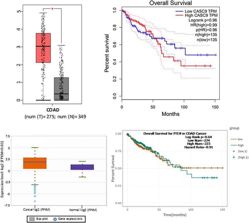
Islam Khan and Law                                                                                         CASC9 Promotes Colorectal Cancer Carcinogenesis

                                            Conclusion: We demonstrated that silencing of CASC9 contributes to the reduced
                                            CRC cell proliferation and migration by regulating autophagy and AKT/mTOR/EMT
                                            signaling. Therefore, CASC9 plays an important role in carcinogenesis, and its
                                            expression may act as a prognostic biomarker and a potential therapeutic target
                                            of CRC management.
                                            Keywords: cancer susceptibility candidate 9, long non-coding RNA, colorectal cancer, autophagy, epithelial-
                                            mesenchymal transition

INTRODUCTION                                                                       carcinogenesis by enhancing aerobic glycolysis and stabilization
                                                                                   of hexokinase 2 gene (Chen C. et al., 2020). Likewise, FOXC2-
Colorectal cancer (CRC) is the third most commonly diagnosed                       AS1 promoted CRC progression via stabilizing FOXC2 and
malignancy worldwide (Bray et al., 2018; Rawla et al., 2019).                      Ca2+ channel–controlled FAK signaling pathway (Pan and Xie,
Statistics revealed that in 2018, nearly 1.8 million new CRC cases                 2020). Bin et al., 2021 demonstrated that overexpression of
were reported with ∼0.9 million CRC deaths worldwide (Araghi                       EPB41L4A-AS1 is associated with CRC development. It activates
et al., 2019). In the past decade, CRC treatment has progressed                    Rho/Rho-associated protein kinase to promote CRC cell growth,
remarkably, but late diagnosis and development of metastasis are                   proliferation, and migration (Bin et al., 2021). Shan et al. (2016)
the main obstacles leading to failure in CRC treatments (Islam                     revealed that Linc-POU3F3 acts as an oncogenic gene in CRC
Khan et al., 2019; Liu et al., 2020). Therefore, it is important to                to promote initiation, progression, and metastasis in vitro. In
identify novel targets for early diagnosis and design new therapy                  contrast, silencing of linc-POU3F3 reduced CRC carcinogenesis
to minimize the CRC mortality globally.                                            by inducing autophagy-mediated apoptosis process (Shan et al.,
    Long non-coding RNAs (lncRNAs) are fragments of RNA                            2016). Another study showed that TTN-AS1 silencing exerts its
that lack protein coding transcript. They are members of non-                      tumor suppressor activity through the reduction of epithelial–
coding RNAs (ncRNAs). More specifically, lncRNAs contain                           mesenchymal transition (EMT) process and PI3K/AKT/mTOR
more than 200 nucleotides and are routinely transcribed by RNA                     signaling (Cui et al., 2019). Overall, this indicates that lncRNA
polymerase-II in the human genome (Zampetaki et al., 2018;                         expression may promote or suppress CRC independently by
Islam Khan et al., 2019). In recent years, accumulating evidence                   regulating diverse molecular pathways.
suggested that lncRNAs sometimes behaved like regulatory                               Cancer susceptibility candidate 9 (CASC9), a recently
molecules to control gene expressions. They are involved in the                    discovered lncRNA, consists of four transcript variants CASC9-
signaling pathways responsible for cell growth, development, and                   201, CASC9-202, CASC9-203, and CASC9-204. CASC9 earned
metabolic processes (Lin and He, 2017; Sparber et al., 2019).                      significant attention of researchers because of the potential roles
In cancer, lncRNAs are associated with each stage of tumor                         of its transcript variants in association with the pathogenesis
initiation, progression, and poor prognosis by enabling drug                       of various cancer (Sharma et al., 2020). Recently, Luo et al.
resistance (Galamb et al., 2019; Qi et al., 2020). The aberrant                    (2019) revealed that upregulation of CASC9 is associated with
expression of lncRNAs alters the major oncogenic signaling                         advanced TNM stage and poor prognosis of CRC. In addition,
cascades, for example, WNT/B-catenin, P53, mTOR, PI3K/Akt,                         CASC9 exerts its oncogenic activity through the phosphorylation
AMP-activated protein kinase (AMPK), EGFR, NOTCH, and                              of SMAD3 and TGF-β signaling in vitro (Luo et al., 2019).
MAPK pathways (Kessler et al., 2013; Khan et al., 2015; Sever and                  Another study performed by Ding et al. (2020) reported
Brugge, 2015).                                                                     that CASC9 upregulation promotes CRC carcinogenesis by
    In the past decade, many investigators concluded that                          regulating miR-193a-5p and ERBB2 expression. Although these
abnormal expression of lncRNAs may be responsible for                              two studies demonstrated some roles of CASC9 in CRC, the
CRC inception, progression, and poor treatment outcomes of                         molecular mechanisms of CASC9 in promoting carcinogenesis
patients (Esmaeili et al., 2020; Qi et al., 2020). For instance,                   still remain largely unknown. Our present study aimed to
UNC5B antisense lncRNA 1 (UNC5B-AS1) has been shown                                explore the expression of CASC9 in CRC cell lines and to
to reduce apoptosis, accelerate CRC progression, and result                        determine the roles of CASC9 in mTOR-dependent autophagy
in metastasis (Zhang Y. et al., 2020). Liu et al. (2020)                           and EMT, which are associated with CRC progression. Our
illustrated that higher expression of KCNQ1OT1 promotes CRC                        findings suggest that CASC9 might be used to evaluate CRC
                                                                                   prognosis, and it may be used as a novel therapeutic target
Abbreviations: AMPK, AMP-activated protein kinase; ATCC, American Type             for CRC patients.
Culture Collection; CASC9, Cancer susceptibility candidate 9; COAD, Colon
adenocarcinoma; CRC, Colorectal cancer; DMEM, Dulbecco modified eagle
medium; Dsi-CASC9, Dicer-substrate mediated CASC9; Dsi-NC, Dicer-substrate
negative control; EMT, Epithelial–mesenchymal transition; ENCORI, The              MATERIALS AND METHODS
Encyclopedia of RNA Interactomes; FBS, Fetal bovine serum; HR, Hazard ratio;
IDT, Integrated DNA technologies; lncRNAs, Long non-coding RNAs; ncRNAs,           Data Mining and Analysis
Non-coding RNAs; qRT-PCR, Quantitative reverse transcriptase–polymerase
chain reaction; TCGA, The Cancer Genome Atlas; TPM, Transcript per million;        The differential expressions of CASC9 in CRC and the adjacent
UNC5B-AS1, The UNC5B antisense lncRNA 1.                                           normal tissues were obtained from two publicly available dataset:

Frontiers in Molecular Biosciences | www.frontiersin.org                       2                                        May 2021 | Volume 8 | Article 627022
Islam Khan and Law CASC9 Promotes Colorectal Cancer Carcinogenesis
The Cancer Genome Atlas (TCGA)1 program under the National The RNA concentration was measured by NanoDrop 200
Cancer Institute and The Encyclopedia of RNA Interactomes (Thermo Fisher Scientific, United States). Following standard
(ENCORI) (Li et al., 2013). From TCGA–colon adenocarcinoma protocol, first-strand cDNA was synthesized using Superscript
(COAD) analysis, there were 275 tumor samples compared II and Random Hexamer (Invitrogen, United States). Master
with 349 adjacent normal tissues for the CASC9 expression Mix LightCycler 480 SYBR Green I (Roche, Switzerland)
profile analysis. P < 0.01 and fold change Log2 FC > 2.00 were was used to complete the quantitative reaction using
considered as cutoff values to plot CASC9 boxplot. The overall LightCycler 480 Instrument II (Roche, Switzerland). Here,
survival of CASC9 was determined by using TCGA-COAD GAPDH was considered as the housekeeping gene, and
dataset with median cutoff and 95% confidence interval, counting relative expression was calculated by 2−11Ct method. The
the number of transcript per million (TPM), and considering the following self-designed primer sequences were used in this
hazard ratio (HR). Likewise, the CASC9 expression was extracted study: GAPDH, forward: 50 -TGCCATCAATGACCCCTTC-30
from the ENCORI-COAD dataset containing 471 tumor samples, and reverse, 50 -CATCGCCCCACTTGATTTTG-30 ; CASC9,
and 41 normal tissues were used to generate the boxplot. forward: 50 -TTGGTCAGCCACATTCATGGT-30 and reverse, 50 -
The log-rank P < 0.05, HR, and high/low expression number AGTGCCAATGACTCTCCAGC-30 ; HPRT1, forward: 50 -TGC
were used to plot the survival curve from ENCORI-Pan-Cancer TCGAGATGTGATGAAGG-30 , and reverse, 50 -TCCCCTGTT
(Li et al., 2013). GACTGGTCATT-30 . All primers were purchased from IDT
(United States).
Cell Culture
Human normal colon cells, CCD-112CoN, were acquired from Cell Viability Assay
American Type Culture Collection (ATCC) (Manassas, VA, After a day of transfection, cells were trypsinized and counted
United States), and human CRC HT-29 cell was acquired from by hemocytometer for seeding and performing cell proliferation
PerkinElmer, Inc. (Waltham, MA, United States). In addition, assay using Cell Counting Kit-8 (CCK-8, Dojindo). Cells, 3 × 10³ ,
three more human CRC cell lines, namely, DLD-1, HCT-116, and in 100 µL of complete medium were seeded and cultured in a 96-
SW480, were kindly provided by our collaborator, Prof. Jun Yu, well plate. According to CCK-8 cell proliferation assay protocol,
Department of Medicine and Therapeutics, Institute of Digestive 10 µL of CCK-8 solution was added to the well. After 3-h
Disease, The Chinese University of Hong Kong. CCD-112CoN incubation at 37◦ C + 5% CO2 , the amount of formazan that
cells were maintained with 10% fetal bovine serum (FBS) (Gibco, represents the number of live cells was measured at an absorbance
United States) in Eagle minimum essential medium (ATCC), of 450 nM using a SPECTROstar Nano Microplate Reader (BMG
whereas HT-29, DLD-1, HCT-116, and SW480 were cultured in Labtech, Germany).
Dulbecco modified eagle medium (DMEM; Gibco, United States)
with 10% FBS. Cell culture was maintained at 37◦ C in 5% CO2 Colony Formation Assay
in 100% humidity. The colony formation assay was performed to measure the cell
proliferation potential in vitro. After being transfected for 24 h,
Dicer-Substrate Mediated Transfection 1 × 10³ cells were seeded and cultured for around 2 weeks in
To achieve Dicer-substrate–mediated CASC9 (Dsi-CASC9) a six-well plate in triplicates. At the endpoint, the colonies were
silencing, SW480 and HCT116 cells were seeded and cultured fixed with a 3:1 mixture of methanol and acetic acid. A solution
in a six-well plate. Transfection experiment was performed of 0.5% crystal violet in methanol was used to stain and visualized
when the cell density reached 60–70% confluence. A lipid- the colonies. The images were taken, and the numbers of colonies
based in vitro transfection was carried out using Lipofectamine were counted using the ImageJ software [National Institutes
2000 (Invitrogen, United States) according to the manufacturer’s of Health (NIH)].
protocol. TriFECTa kits were purchased from Integrated DNA
Technologies (IDT, United States), which contained a Dicer- Migration Assay
substrate negative control (DSi-NC), positive control (Dsi- In the migration assay, 5 × 10⁴ cells in 70 µL DMEM with 10%
HPRT-S1), transfection control (Dsi-TYE 563), and predesigned FBS were carefully placed in both compartments of the Culture-
Dsi-CASC9 duplex. The duplex sequences for Dsi-CASC9 Insert 2 Well (Ibidi LLC, Germany). After 24 h of cell settling, the
were as follows: 50 -GAGAGUCAUUGGCACUAUCAAGAAA- culture inserts were gently removed to create a gap of ∼500 µm
30 and 30 -ACCUCUCAGUAACCGUGAUAGUUCUUU-50 . The for measuring the cell migration ability. Then, each well was filled
Dsi-NC and Dsi-HPRT-S1 sequences were not provided by with 1.5 mL of complete medium. The photographs of the wound
the manufacturer. areas were taken using an inverted microscope (Nikon, Japan) at
various time points of 0, 24, and 48 h, respectively. The migration
Complementary DNA Synthesis and index indicating the size of the gap was measured using the MRI
RT-qPCR Wound Healing Tool in ImageJ (NIH).
Total RNA from the colon cells was extracted using RNeasy mini
kit (Qiagen, Germany) according to manufacturer guidelines. Western Blotting
Western blotting was performed using standard, established
1
http://gepia.cancer-pku.cn/detail.php protocol as previously published (Tam et al., 2020). Briefly,
Frontiers in Molecular Biosciences | www.frontiersin.org 3 May 2021 | Volume 8 | Article 627022Islam Khan and Law CASC9 Promotes Colorectal Cancer Carcinogenesis
protein isolation was performed using RIPA lysis and extraction the disease and reduced overall survival (Figure 1B). To further
buffer (Thermo Fisher Scientific, United States) with a confirm our findings, we explored CASC9 expression in another
supplement of cOmplete ULTRA Tablets, Mini EDTA-free, publicly available dataset, ENCORI-COAD (Figure 1C). CASC9
Easy pack Protease Inhibitor Cocktail (Roche, Switzerland). expression was found to be overexpressed in 417 CRC tumor
Protein concentration was quantified using BCA Protein Assay samples compared with 41 normal tissues (Li et al., 2013).
Kit (Thermo Fisher Scientific, United States), and similar Similarly, we plotted the survival curve for CRC patients based
amounts of proteins were loaded and run on 8–12% sodium on log-rank P < 0.05, HR, and high/low expression profiles
dodecyl sulfate–polyacrylamide gel electrophoresis at ambient of CASC9 in the dataset. Higher expression of CASC9 from
temperature. Proteins were then transferred onto Immun-Blot ENCORI-COAD dataset showed a reduced overall survival of
PVDF membrane (Bio-Rad Laboratories, Inc., United States), patients compared to normal (Figure 1D).
followed by 2-h blocking in 5% bovine serum albumin (BSA)
(Hyclone BSA; GE Healthcare Life Science, United States) in CASC9 Can Be Effectively and
Tris-buffered saline with a supplement of 0.1% Tween 20. Then, Consistently Silenced by Dicer-Substrate
the blocked membrane was incubated overnight with primary siRNA Techniques in CRC Cells
antibodies: β-actin [#8457, Cell Signaling Technology, Inc., Cancer susceptibility candidate 9 expression was measured in
(CST, United States)], GAPDH (#2118, CST), AKT (#9272, human CRC cell lines (DLD-1, HT-29, SW480, and HCT-
AKT, Phosphor-AKT (#9271, CST), AMPKα (#5832, CST), 116) and normal colon cell line CCD-112CoN (Figure 2A)
phosphor-AMPKα (#2535, CST), E-cadherin (#3195, CST), by RT-qPCR. Similar to the observation in public datasets,
N-cadherin (#13116S, CST), LC3B (#2775, CST), mTOR (#2972, the expression of CASC9 was significantly upregulated in CRC
CST), Phosphor-mTOR (#2535, CST), and vimentin (#5741S, cell lines compared to normal CCD-112CoN cells (P < 0.001,
CST) at 4◦ C. The secondary anti-rabbit immunoglobulin G n = 8). This result indicated that higher expression of CASC9
(IgG), horseradish peroxide (HRP)–linked, or anti-mouse IgG- may play a role in CRC carcinogenesis. Significantly higher
HRP–linked (#7076, CST) antibody were added and incubated level of CASC9 was detected in HCT-116 > SW480 > HT-
with the membrane for 2 h. Afterward, Western Lightning Plus- 29 cells (Figure 2A). Therefore, HCT-116 and SW480 were
Electrochemiluminescence (PerkinElmer, Inc., United States) chosen for gene silencing assay. The qRT-PCR results showed
was added to the membrane to visualize protein bands in a excellent knockdown efficiency of Dsi-CASC9 in HCT-116 and
ChemiDoc MP Imaging System (Bio-Rad Laboratories, Inc., SW480 cells to be 63.25 ± 8.42% and 58.0 ± 6.20%, respectively
United States). The relative protein expressions were quantified (Figures 2B,C). To validate our knockdown method, gene of
using ImageJ software (NIH) with β-actin or GAPDH as positive control (HPRT-1) was performed in both cell lines,
internal control. and we confirmed a knockdown efficiency of more than 60%
(Figures 2B,C).
Statistical Analysis
The mean ± standard error of mean (SEM) of at least three Silencing of CASC9 Reduced Cell
or more independent experiments were included for analysis.
Viability, Colony Formation, and
The statistical level of the experimental data was calculated by
Student t test or one-way analysis of variance using GraphPad Migratory Index of CRC Cells
Prism version 8.0 (GraphPad Software, Inc., San Diego, CA, To evaluate the biological and physiological function of CASC9
United States). P < 0.05 is considered statistically significant. in CRC cells, HCT-116 and SW480 cells were silenced by Dsi-
CASC9 with Dsi-NC as negative control. By performing CCK-8
assay, we confirmed that CASC9 silencing significantly decreased
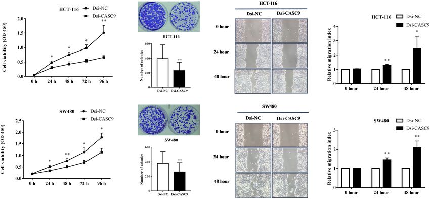
RESULTS the cell proliferation capacity in HCT-116 and SW480 cells
(Figure 3A). Corresponding to the decrease in cell proliferation,
CASC9 Overexpression Correlates With significantly reduced cell growth of HCT-116 and SW480 was
Poor Survival in CRC shown by Dsi-CASC9 from colony formation assay (Figure 3B).
The migration assay was conducted in both cells to evaluate the
To explore the role of CASC9 in CRC, we first searched the
migration ability of cells. Significant increase of migration index
publicly available TCGA-COAD dataset (Figure 1A). Boxplot
was shown in HCT-116 and SW480 after Dsi-CASC9 treatment
analysis of CASC9 showed that it was significantly upregulated
(Figure 3C) at 24 and 48 h after transfection.
in CRC samples (n = 275) compared with adjacent normal
tissues (n = 349) (Figure 1A). Furthermore, we evaluated the
relationship between CASC9 expression and clinical outcomes
Silencing of CASC9-Induced Autophagy
of patients. To do so, we plotted the survival curve of CRC in CRC Cells
patients according to their CASC9 expression level, number of Autophagy is a very crucial pathway for cell to survive during
TPM, and HR% using TCGA-COAD dataset in Gene Expression energy-deficient and hypoxic conditions. The Western blot in
Profiling Interactive Analysis bioinformatics tool2 . We found Figures 4A,C showed that the expression of LC3B-II (autophagy
that the patients with higher CASC9 have poor prognosis of marker protein) was significantly upregulated in HCT-116 and
SW480 cells after CASC9 silencing. Another autophagy marker
2
http://gepia.cancer-pku.cn protein p62 is a negative regulator of autophagy process. The
Frontiers in Molecular Biosciences | www.frontiersin.org 4 May 2021 | Volume 8 | Article 627022Islam Khan and Law CASC9 Promotes Colorectal Cancer Carcinogenesis
FIGURE 1 | CASC9 overexpression is correlated with poor survival in CRC. (A) Boxplot CASC9 expressions of CRC tissues (n = 275) compared with normal samples
(n = 349) from TCGA-COAD dataset (http://gepia.cancer-pku.cn/detail.php) showing that CASC9 was significantly upregulated in CRC tissues with a Log2FC cutoff
value 2.0 and P < 0.01 (*). (B) The expression profiles, number of TPM, and HR (%) were used to plot overall survival. Higher expression of CASC9 in CRC tissue is
associated with poor overall survival. (C) CASC9 expression was extracted from ENCORI-COAD dataset (Li et al., 2013). Four hundred seventeen tumor samples
and 41 normal tissues were used from the dataset. Boxplot analysis showed that CASC9 was upregulated in tumor tissues. (D) The log-rank P < 0.05, HR, and
high/low expression numbers were used to plot survival curve from ENCORI-Pan-Cancer. Higher expression of CASC9 reduced the overall survival of patients.
TCGA, The Cancer Genome Atlas; COAD, colon adenocarcinoma; ENCORI, The Encyclopedia of RNA Interactomes; TPM, transcript per million, HR, hazard ratio.
silencing of CASC9 significantly reduced the expression of p62 to GAPDH were being evaluated in HCT-116 (Figures 5A–
protein level in both cell lines (Figures 4B,D), suggesting that D) and SW480 cells (Figures 5E–H). Dsi-CASC9 significantly
Dsi-CASC9 promotes autophagy in HCT-116 and SW480 cells. promotes AMPK signaling in HCT-116 and SW480 cells
compared to Dsi-NC. In contrast, CASC9 silencing significantly
Silencing of CASC9 Promoted the AMPK downregulated AKT and mTOR signaling pathways in both cell
lines (Figures 5C,D,G,H).
Signaling Pathway but Downregulated
the AKT/mTOR Signaling Pathway in
CRC Cells CASC9 Silencing Altered the Expression
We subsequently analyze more signaling pathway proteins to of EMT Marker Proteins in CRC Cells
explore the role of CASC9 in the AKT and mTOR pathway. The Epithelial–mesenchymal transition is one of the key steps of
ratio of p-AMPKα/AMPKα, p-AKT/AKT, and p-mTOR/mTOR metastasis in cancer. We explored whether CASC9 silencing
Frontiers in Molecular Biosciences | www.frontiersin.org 5 May 2021 | Volume 8 | Article 627022Islam Khan and Law CASC9 Promotes Colorectal Cancer Carcinogenesis FIGURE 2 | CASC9 expressions in colon cell lines and effective silencing by Dicer-substrate siRNA. (A) The expression of CASC9 in CRC cell lines DLD-1, HT-29, SW480, and HCT-116 was compared with normal colon cell line CCD-112CoN using RT-qPCR. The data are shown as mean ± SEM of eight independent experiments. (B,C) Effective and consistent silencing of CASC9 by Dicer-substrate siRNA techniques was observed. The data are shown as mean ± SEM compared to the negative control Dsi-NC. n = 6, ***P < 0.001. FIGURE 3 | Silencing of CASC9 reduced cell viability and colony formation and increased migration index of CRC cells. (A) The silencing of CASC9 led to significant decrease in HCT-116 and SW480 cell proliferation (n = 4). (B) As determined by colony formation assay, the numbers of colonies in HCT-116 and SW480 were significantly reduced after CASC9 silencing (n = 4). (C) After knockdown of CASC9, the migration index of HCT-116 and SW480 was significantly increased at both the 24- and 48-h time points (n = 6), suggesting decrease in migration of cells to the gap. The data are shown as mean ± SEM compared to the negative control Dsi-NC (*P < 0.05, **P < 0.01). would alter the expression of key EMT regulatory proteins, N-cadherin in HCT-116 (Figure 6C) did not reach statistical such as E-cadherin, N-cadherin, and vimentin, in HCT-116 and significance, but the trend was observed in all experiments. SW480 cells. As shown in Figure 6, the expression of epithelial marker E-cadherin was significantly upregulated in Dsi-CASC9– treated HCT-116 and SW480 cells. On the other hand, DISCUSSION the mesenchymal marker protein vimentin was significantly downregulated in HCT-116 and SW480 cells (Figures 6D,H). With the advancement of next-generation sequencing The N-cadherin expression was also significantly downregulated technology, an increasing number of lncRNAs have been in SW480 cell (Figure 6G). However, the downregulation of revealed. LncRNAs are not non-functional by-products or Frontiers in Molecular Biosciences | www.frontiersin.org 6 May 2021 | Volume 8 | Article 627022
Islam Khan and Law                                                                                              CASC9 Promotes Colorectal Cancer Carcinogenesis

  FIGURE 4 | Silencing of CASC9 enhanced autophagy in CRC cells. The expressions of autophagy marker proteins LC3B and p62 were measured by Western
  blotting in HCT-116 (A,B) and SW480 (C,D) cells. After Dsi-CASC9–mediated silencing, the ratio of autophagy marker LC3B-II to LC3B-I significantly increased in
  HCT-116 and SW480 cells with corresponding decrease in p62 expression. The data are shown as mean ± SEM using β-actin as housekeeping control (*P < 0.05,
  **P < 0.01, and n = 4).

junk molecules of the body (Yao et al., 2019). They play very                        demonstrated that HOTAIR and MALAT1 are associated with
important roles in epigenetics and have multiple functions in                        poor prognosis of CRC by accelerating metastasis process. So far,
cell growth and development. They are involved in various                            a large number of lncRNAs have been reported as an oncogenic
physiological process related to metabolism including gene                           or tumor-suppressor in CRC such as CDKNIA, PANDAR,
mutation, regulation of transcription and translational processes,                   MALAT1, CCAT1, CCAT2, UCA1, MEG3, HOTAIR, and GAS5.
and regulation of cell cycles (Zhu et al., 2013). In carcinogenesis,                 In addition, ncRuPAR and lincRNA-p21 are considered to be a
lncRNAs play crucial roles in gene expression process steering                       negative regulator of CRC in the process of radioresistance and
structural stability and transcriptional process of nucleus,                         metastasis (Zhai et al., 2013; Yan et al., 2014).
regulating stability of mRNA, and maintaining transcriptional                            In this present study, we first explored the expression of
and post-transcriptional modification in the cytoplasm (Wilusz                       CASC9 in CRC patient samples in two open databases, TCGA-
et al., 2009; Marchese et al., 2017; Zampetaki et al., 2018; Yao                     COAD and ENCORI-COAD, which collected cases in the
et al., 2019). So far, approximately 3,000 lncRNAs have been                         United States and Mainland China, respectively. Even though
identified from the human genome with their regulatory impact                        the plot functions are different in these databases yielding very
on cancer development, progression, metastasis, and poor                             different graphs (Figure 1), the conclusions are consistent. We
prognosis (Gao et al., 2019).                                                        observed that CASC9 expression was significantly upregulated
   The role of lncRNAs in CRC was first reported by Tsang                            in CRC tissues compared to its adjacent normal samples. In
et al. (2010). They revealed that H19-derived miR-675 plays                          addition, the expression of CASC9 is associated with reduced
an oncogenic role in CRC development and progression by                              survival of CRC patients. We also demonstrated that CASC9 was
targeting retinoblastoma proteins (Tsang et al., 2010). Subsequent                   overexpressed in CRC cell lines compared to normal colon cell
studies performed by Kogo et al. (2011) and Xu et al. (2011)                         line. All these and previous findings on CASC9 suggested that

Frontiers in Molecular Biosciences | www.frontiersin.org                         7                                             May 2021 | Volume 8 | Article 627022
Islam Khan and Law                                                                                          CASC9 Promotes Colorectal Cancer Carcinogenesis

  FIGURE 5 | Silencing of CASC9 promoted the AMPK signaling pathway but downregulated the AKT and mTOR pathways. The ratio of p-AMPKα/AMPKα,
  p-AKT/AKT, and p-mTOR/mTOR to GAPDH were evaluated by Western blotting in HCT-116 (A–D) and SW480 cells (E–H). Dsi-CASC9 significantly promotes
  AMPK signaling in HCT-116 and SW480 cells compared to Dsi-NC. In contrast, CASC9 silencing significantly downregulated AKT and mTOR signaling pathways in
  HCT-116 and SW480 cells. The data are shown as mean ± SEM using GAPDH as housekeeping gene (*P < 0.05, and n = 3).

CASC9 might be a novel marker for CRC prognosis. In fact,                         autophagy and EMT (Vellai et al., 2008; Wang and Levine,
there are many reports that CASC9 is involved in non-small cell                   2010; Sever and Brugge, 2015; Mathiassen et al., 2017; Brabletz
lung carcinoma, bladder cancer, thyroid cancer, hepatocellular                    et al., 2018; Pavel et al., 2018; Pastushenko and Blanpain,
carcinoma, nasopharyngeal cancer, and lung cancer (Jin et al.,                    2019).
2019; Zeng et al., 2019; Chen Y. et al., 2020; Huo et al., 2020;                     The self-degradation mechanism called autophagy is a
Zhao et al., 2020). It has been suggested that CASC9 is a                         major intracellular process that maintains the balance between
novel diagnostic, prognostic, and therapeutic target in cancer                    cell death and survival in response to nutritional stress,
treatment (Qian et al., 2020; Sharma et al., 2020). Our findings                  hypoxia, and growth factor deprivation (Tam et al., 2019;
are in line with previous articles in CRC and other cancers                       Noguchi et al., 2020). Autophagy is a dual-edged sword that
(Luo et al., 2019; Ding et al., 2020) and hence leading us to                     can inhibit or promote carcinogenesis by regulating mTOR
hypothesize that CASC9 may be involved in specific pathogenesis                   and apoptosis process (Levine, 2007). Along with, it is well
of CRC carcinogenesis.                                                            established that lncRNAs promote or inhibit carcinogenesis
    Many studies have reported that CASC9 knockdown or                            by regulating autophagy either through mTOR-dependent or -
silencing reduced cell proliferation, invasion, and migration                     independent pathways (Peng et al., 2020; Zhang X. Z. et al.,
(Jin et al., 2019; Zhang et al., 2019; Chen Y. et al., 2020;                      2020). To explore the autophagy process, we determined
Ding et al., 2020; Fang et al., 2020; Huo et al., 2020). We are                   the expression of autophagy marker proteins LC3B and p62
the first to perform similar experiments in CRC cell lines. By                    before and after silencing. Dsi-CASC9 significantly increased
performing a series of in vitro experiments, including CCK-8                      LC3B-II and reduced p62 expression. The increased LC3-
assay, colony formation assay, and migration assay after Dsi-                     II is regarded as the standard marker for autophagy. It is
CASC9 silencing, we confirmed that CASC9 played malignant                         directly associated with the number of autophagosomes and
roles in CRC cell survival, proliferation, and migration. To                      considered as the most commonly used autophagic marker
decipher the role of CASC9 in CRC growth, proliferation, and                      protein (Zheng et al., 2012). The ubiquitin-associated protein
migration, we examined the potential pathways related to cell                     p62 protein itself is degraded through autophagy and can also
growth, apoptosis, and metastasis and decided to focus on                         serve as a marker of autophagic flux (Cohen-Kaplan et al., 2016;

Frontiers in Molecular Biosciences | www.frontiersin.org                      8                                           May 2021 | Volume 8 | Article 627022
Islam Khan and Law CASC9 Promotes Colorectal Cancer Carcinogenesis FIGURE 6 | CASC9 silencing altered the expressions of EMT marker proteins in CRC cells. The EMT markers E-cadherin, N-cadherin, and vimentin were evaluated by Western blotting in HCT-116 (A–D) and SW480 cells (E–H). In both cell lines, CASC9 significantly upregulated E-cadherin and downregulated vimentin expressions. The N-cadherin expression was also downregulated but did not reach statistical significance in HCT-116 cells. The data are shown as mean ± SEM using GAPDH as housekeeping gene (*P < 0.05, and n = 3). Liu et al., 2016). Here, we clearly demonstrated the promotion revealed for the first time that abnormal expression of CASC9 of autophagy in CRC after silencing CASC9 (Mizushima, 2004; promoted carcinogenesis of CRC cells through activating Jiang and Mizushima, 2015). We believe the induction of AKT/mTOR signaling and reduced phosphorylation of AMPK autophagy may be related to the reduced cell growth observed and inhibiting autophagy. after gene silencing. The poor prognosis for most cancer is due to the development To further our investigation of molecular pathways in of metastasis where EMT eventuates to enhance the cellular relation with reduced CRC cell proliferation and migration, migration properties (Brabletz et al., 2018; Wang et al., 2020). we explored key signaling molecules, AMPK, mTOR, and In case of CRC, more than 20% of the patients were diagnosed AKT, which are linked to the autophagy pathway. In mTOR- when the tumor has already metastasized to distant organs dependent autophagy process, AMPK phosphorylates to activate (van der Geest et al., 2015). Targeted therapy with or without upon energy starvation, leading to phosphorylation of Ser317, chemotherapy is mostly recommended to patients with advanced Ser777, and Ser555 to activate ULK1 and inhibition of stages of CRC for eradication of the tumor (Xie et al., 2020). mTORC1 signaling pathway (Paquette et al., 2018; Wang The traditional targeted or immune therapy mostly targets the and Zhang, 2019). The protein kinase B or AKT is one abnormal oncogenic proteins or strands of DNAs. Recently, of the most critical intracellular pathways associated with researchers are focusing on using lncRNAs as a novel set of mTOR signaling, and it has been considered as the master therapeutic targets (Mitra and Chakrabarti, 2018). Our study regulator for most of the cancers (Porta et al., 2014; Yang revealed that CASC9 potentially induced EMT. Here in our et al., 2019). Inhibition of AKT/mTOR signaling promotes evaluation, silencing CASC9 upregulated the epithelial marker autophagy and sensitizes tumor cells to anticancer drugs by protein E-cadherin and downregulated mesenchymal marker reducing cell growth, cell cycle, cell survival, differentiation, protein N-cadherin and vimentin expressions. These results and metabolism (Paquette et al., 2018; Terracciano et al., suggest that CASC9 may be involved in CRC progression 2019). In this study, we revealed that the silencing of CASC9 and metastasis by enhancing the EMT-dependent migratory potentially promotes mTOR-dependent autophagy where it characteristics of CRC. These findings guide us to propose significantly enhances phosphorylation of AMPK and reduces a novel therapeutic approach that silencing of CASC9 for phosphorylation of AKT and mTOR. The inhibition of metastatic CRC patients improves the therapeutic outcomes. AKT and mTOR pathways may lead to the attenuated However, we still need to wait for the next leap of technology to cell growth and migration. Taken together, these findings reach this goal. Frontiers in Molecular Biosciences | www.frontiersin.org 9 May 2021 | Volume 8 | Article 627022
Islam Khan and Law                                                                                                             CASC9 Promotes Colorectal Cancer Carcinogenesis

CONCLUSION                                                                                     AUTHOR CONTRIBUTIONS
In conclusion, our findings demonstrated that CASC9 was                                        MI and HL conceived and designed the project. MI conducted
aberrantly upregulated in CRC cells and tissues. Our study                                     the experiments, analyzed the data, and wrote the manuscript. HL
revealed that silencing of CASC9 suppressed CRC proliferation,                                 interpret the results and reviewed the manuscript. Both authors
growth, and migration via activation of mTOR-dependent                                         read, approved, and finalized the manuscript.
autophagy and inhibition of EMT in vitro. This is in line
with some recent reports that suggested autophagy/mTOR/EMT
pathways may be the crucial targets for the understanding of CRC
carcinogenesis and novel therapeutic targets (Song et al., 2019;
                                                                                               FUNDING
Wang et al., 2019; Zheng et al., 2019). CASC9 expression in tumor                              This project was partially supported by the research grant to HL
might be a novel prognostic biomarker and CASC9 might be a                                     including Departmental Seeding Fund and Internal Institutional
potential therapeutic target for the management of CRC.                                        Research Fund (P0031318-UAHS) and Postgraduate studentship
                                                                                               from The Hong Kong Polytechnic University for MI.
DATA AVAILABILITY STATEMENT
The datasets presented in this study can be found in                                           ACKNOWLEDGMENTS
online repositories. The names of the repository/repositories
and accession number(s) can be found below: https://                                           We thank Prof. Jun Yu, Department of Medicine and
www.ncbi.nlm.nih.gov/,     NR_103850.2,   https://www.ncbi.                                    Therapeutics, Institute of Digestive Disease, The Chinese
nlm.nih.gov/, NR_103849.2, and https://www.ncbi.nlm.nih.                                       University of Hong Kong for providing of DLD-1, HCT-116, and
gov/, NR_103848.1.                                                                             SW480 cells for this project.

REFERENCES                                                                                     Fang, J., Chen, W., and Meng, X. L. (2020). LncRNA CASC9 suppressed the
                                                                                                   apoptosis of gastric cancer cells through regulating BMI1. Pathol. Oncol. Res.
Araghi, M., Soerjomataram, I., Jenkins, M., Brierley, J., Morris, E., Bray, F., et al.             26, 475–482. doi: 10.1007/s12253-019-00703-3
   (2019). Global trends in colorectal cancer mortality: projections to the year               Galamb, O., Barták, B. K., Kalmár, A., Nagy, Z. B., Szigeti, K. A., Tulassay, Z.,
   2035. Int. J. Cancer 144, 2992–3000. doi: 10.1002/ijc.32055                                     et al. (2019). Diagnostic and prognostic potential of tissue and circulating long
Bin, J., Nie, S., Tang, Z., Kang, A., Fu, Z., Hu, Y., et al. (2021). Long noncoding                non-coding RNAs in colorectal tumors. World J. Gastroenterol. 25, 5026–5048.
   RNA EPB41L4A-AS1 functions as an oncogene by regulating the Rho/ROCK                            doi: 10.3748/wjg.v25.i34.5026
   pathway in colorectal cancer. J. Cell. Physiol. 236, 523–535. doi: 10.1002/jcp.             Gao, L., Guo, Y.-N., Zeng, J.-H., Ma, F.-C., Luo, J., Zhu, H.-W., et al. (2019). The
   29880                                                                                           expression, significance and function of cancer susceptibility candidate 9 in lung
Brabletz, T., Kalluri, R., Nieto, M. A., and Weinberg, R. A. (2018). EMT in cancer.                squamous cell carcinoma: a bioinformatics and in vitro investigation. Int. J.
   Nat. Rev. Cancer 18, 128–134.                                                                   Oncol. 54, 1651–1664.
Bray, F., Ferlay, J., Soerjomataram, I., Siegel, R. L., Torre, L. A., and Jemal, A.            Huo, W., Tan, D., and Chen, Q. (2020). CASC9 facilitates cell proliferation in
   (2018). Global cancer statistics 2018: GLOBOCAN estimates of incidence and                      bladder cancer by regulating CBX2 expression. Nephron 144, 388–399. doi:
   mortality worldwide for 36 cancers in 185 countries. CA Cancer J. Clin. 68,                     10.1159/000507828
   394–424. doi: 10.3322/caac.21492                                                            Islam Khan, M. Z., Tam, S. Y., and Law, H. K. W. (2019). Autophagy-modulating
Chen, C., Wei, M., Wang, C., Sun, D., Liu, P., Zhong, X., et al. (2020). Long                      long non-coding RNAs (LncRNAs) and their molecular events in cancer. Front.
   noncoding RNA KCNQ1OT1 promotes colorectal carcinogenesis by enhancing                          Genet. 9:750. doi: 10.3389/fgene.2018.00750
   aerobic glycolysis via hexokinase-2. Aging 12, 11685–11697. doi: 10.18632/                  Jiang, P., and Mizushima, N. (2015). LC3- and p62-based biochemical methods for
   aging.103334                                                                                    the analysis of autophagy progression in mammalian cells. Methods 75, 13–18.
Chen, Y., Li, Y., and Gao, H. (2020). Long noncoding RNA CASC9 promotes the                        doi: 10.1016/j.ymeth.2014.11.021
   proliferation and metastasis of papillary thyroid cancer via sponging miR-488-              Jin, Y., Xie, H., Duan, L., Zhao, D., Ding, J., and Jiang, G. (2019). Long non-
   3p. Cancer Med. 9, 1830–1841. doi: 10.1002/cam4.2839                                            coding RNA CASC9 And HIF-1α form a positive feedback loop to facilitate cell
Cohen-Kaplan, V., Livneh, I., Avni, N., Fabre, B., Ziv, T., Kwon, Y. T., et al. (2016).            proliferation and metastasis in lung cancer. Onco Targets Ther. 12, 9017–9027.
   p62- and ubiquitin-dependent stress-induced autophagy of the mammalian 26S                      doi: 10.2147/ott.s226078
   proteasome. Proc. Natl. Acad. Sci. U.S.A. 113, E7490–E7499.                                 Kessler, T., Hache, H., and Wierling, C. (2013). Integrative analysis of cancer-
Cui, Z., Han, B., Wang, X., Li, Z., Wang, J., and Lv, Y. (2019). Long non-coding                   related signaling pathways. Front. Physiol. 4:124. doi: 10.3389/fphys.2013.00124
   RNA TTN-AS1 promotes the proliferation and invasion of colorectal cancer                    Khan, M., Maryam, A., Qazi, J. I., and Ma, T. (2015). Targeting apoptosis and
   cells by activating miR-497-mediated PI3K/Akt/mTOR signaling. OncoTargets                       multiple signaling pathways with icariside II in cancer cells. Int. J. Biol. Sci. 11,
   Ther. 12, 11531–11539. doi: 10.2147/ott.s229104                                                 1100–1112. doi: 10.7150/ijbs.11595
Ding, Y., Li, X., Zhang, Y., and Zhang, J. (2020). Long non-coding RNA cancer                  Kogo, R., Shimamura, T., Mimori, K., Kawahara, K., Imoto, S., Sudo, T.,
   susceptibility 9 (CASC9) up-regulates the expression of ERBB2 by inhibiting                     et al. (2011). Long noncoding RNA HOTAIR regulates polycomb-dependent
   miR-193a-5p in colorectal cancer. Cancer Manag. Res. 12, 1281–1292. doi:                        chromatin modification and is associated with poor prognosis in colorectal
   10.2147/cmar.s234620                                                                            cancers. Cancer Res. 71, 6320–6326. doi: 10.1158/0008-5472.can-11-1021
Esmaeili, M., Keshani, M., Vakilian, M., Esmaeili, M., Peymani, M., Seyed                      Levine, B. (2007). Cell biology: autophagy and cancer. Nature 446, 745–747.
   Forootan, F., et al. (2020). Role of non-coding RNAs as novel biomarkers for                Li, J.-H., Liu, S., Zhou, H., Qu, L.-H., and Yang, J.-H. (2013). starBase v2.0: decoding
   detection of colorectal cancer progression through interaction with the cell                    miRNA-ceRNA, miRNA-ncRNA and protein–RNA interaction networks from
   signaling pathways. Gene 753:144796. doi: 10.1016/j.gene.2020.144796                            large-scale CLIP-Seq data. Nucleic Acids Res. 42, D92–D97.

Frontiers in Molecular Biosciences | www.frontiersin.org                                  10                                                   May 2021 | Volume 8 | Article 627022
Islam Khan and Law CASC9 Promotes Colorectal Cancer Carcinogenesis Lin, C.-P., and He, L. (2017). Noncoding RNAs in cancer development. Annu. Rev. in colorectal cancer cells. Cancers 12:224. doi: 10.3390/cancers1201 Cancer Biol. 1, 163–184. 0224 Liu, W. J., Ye, L., Huang, W. F., Guo, L. J., Xu, Z. G., Wu, H. L., et al. (2016). Tam, S. Y., Wu, V. W. C., and Law, H. K. W. (2019). Dynamics of oxygen level- p62 links the autophagy pathway and the ubiqutin–proteasome system upon driven regulators in modulating autophagy in colorectal cancer cells. Biochem. ubiquitinated protein degradation. Cell. Mol. Biol. Lett. 21:29. Biophys. Res. Commun. 517, 193–200. doi: 10.1016/j.bbrc.2019.07.043 Liu, X., Dong, C., Ma, S., Wang, Y., Lin, T., Li, Y., et al. (2020). Terracciano, L. M., Piscuoglio, S., and Ng, C. K. (2019). “Hepatocellular carcinoma: Nanocomplexes loaded with miR-128-3p for enhancing chemotherapy effect pathology and genetics,” in Reference Module in Biomedical Sciences, eds J. E. of colorectal cancer through dual-targeting silence the activity of PI3K/AKT Riviere and N. A. Monteiro-Riviere (Amsterdam: Elsevier). and MEK/ERK pathway. Drug Deliv. 27, 323–333. doi: 10.1080/10717544.2020. Tsang, W. P., Ng, E. K., Ng, S. S., Jin, H., Yu, J., Sung, J. J., et al. (2010). Oncofetal 1716882 H19-derived miR-675 regulates tumor suppressor RB in human colorectal Luo, K., Geng, J., Zhang, Q., Xu, Y., Zhou, X., Huang, Z., et al. (2019). LncRNA cancer. Carcinogenesis 31, 350–358. doi: 10.1093/carcin/bgp181 CASC9 interacts with CPSF3 to regulate TGF-β signaling in colorectal cancer. van der Geest, L. G., Lam-Boer, J., Koopman, M., Verhoef, C., Elferink, M. A., and J. Exp. Clin. Cancer Res. 38:249. De Wilt, J. H. (2015). Nationwide trends in incidence, treatment and survival Marchese, F. P., Raimondi, I., and Huarte, M. (2017). The multidimensional of colorectal cancer patients with synchronous metastases. Clin. Exp. Metastasis mechanisms of long noncoding RNA function. Genome Biol. 18:206. 32, 457–465. doi: 10.1007/s10585-015-9719-0 Mathiassen, S. G., De Zio, D., and Cecconi, F. (2017). Autophagy and the cell cycle: Vellai, T., Bicsák, B., Tóth, M. L., Takács-Vellai, K., and Kovács, A. L. (2008). a complex landscape. Front. Oncol. 7:51. doi: 10.3389/fonc.2017.00051 Regulation of cell growth by autophagy. Autophagy 4, 507–509. doi: 10.4161/ Mitra, S., and Chakrabarti, J. (2018). “Chapter 9 - transfer RNA in cancer,” in auto.5670 Cancer and Noncoding RNAs, eds D. J. Chakrabarti and D. S. Mitra (Boston: Wang, L. L., Zhang, L., and Cui, X. F. (2019). Downregulation of long noncoding Academic Press), 151–161. doi: 10.1016/b978-0-12-811022-5.00009-7 RNA LINC01419 inhibits cell migration, invasion, and tumor growth and Mizushima, N. (2004). Methods for monitoring autophagy. Int. J. Biochem. Cell promotes autophagy via inactivation of the PI3K/Akt1/mTOR pathway in Biol. 36, 2491–2502. gastric cancer. Ther. Adv. Med. Oncol. 11:1758835919874651. Noguchi, M., Hirata, N., Tanaka, T., Suizu, F., Nakajima, H., and Chiorini, J. A. Wang, R. C., and Levine, B. (2010). Autophagy in cellular growth control. FEBS (2020). Autophagy as a modulator of cell death machinery. Cell Death Dis. Lett. 584, 1417–1426. doi: 10.1016/j.febslet.2010.01.009 11:517. Wang, X., Gao, X., Tian, J., Zhang, R., Qiao, Y., Hua, X., et al. (2020). LINC00261 Pan, K., and Xie, Y. (2020). LncRNA FOXC2-AS1 enhances FOXC2 mRNA stability inhibits progression of pancreatic cancer by down-regulating miR-23a-3p. Arch. to promote colorectal cancer progression via activation of Ca(2+)-FAK signal Biochem. Biophys. 689:108469. doi: 10.1016/j.abb.2020.108469 pathway. Cell Death Dis. 11, 434–434. Wang, Y., and Zhang, H. (2019). “Regulation of autophagy by mTOR signaling Paquette, M., El-Houjeiri, L., and Pause, A. (2018). mTOR pathways in cancer and pathway,” in Autophagy: Biology and Diseases: Basic Science, ed. Z.-H. Qin autophagy. Cancers 10:18. (Singapore: Springer), 67–83. doi: 10.1007/978-981-15-0602-4_3 Pastushenko, I., and Blanpain, C. (2019). EMT transition states during tumor Wilusz, J. E., Sunwoo, H., and Spector, D. L. (2009). Long noncoding RNAs: progression and metastasis. Trends Cell Biol. 29, 212–226. doi: 10.1016/j.tcb. functional surprises from the RNA world. Genes Dev. 23, 1494–1504. doi: 2018.12.001 10.1101/gad.1800909 Pavel, M., Renna, M., Park, S. J., Menzies, F. M., Ricketts, T., Füllgrabe, J., Xie, Y.-H., Chen, Y.-X., and Fang, J.-Y. (2020). Comprehensive review of targeted et al. (2018). Contact inhibition controls cell survival and proliferation via therapy for colorectal cancer. Signal. Transduct. Target. Therapy 5:22. YAP/TAZ-autophagy axis. Nat. Commun. 9:2961. Xu, C., Yang, M., Tian, J., Wang, X., and Li, Z. (2011). MALAT-1: a long non-coding Peng, Y., Tang, D., Zhao, M., Kajiyama, H., Kikkawa, F., and Kondo, Y. (2020). RNA and its important 3’ end functional motif in colorectal cancer metastasis. Long non-coding RNA: a recently accentuated molecule in chemoresistance Int. J. Oncol. 39, 169–175. in cancer. Cancer Metastasis Rev. 39, 825–835. doi: 10.1007/s10555-020- Yan, B., Gu, W., Yang, Z., Gu, Z., Yue, X., Gu, Q., et al. (2014). Downregulation of 09910-w a long noncoding RNA-ncRuPAR contributes to tumor inhibition in colorectal Porta, C., Paglino, C., and Mosca, A. (2014). Targeting PI3K/Akt/mTOR signaling cancer. Tumor Biol. 35, 11329–11335. doi: 10.1007/s13277-014-2465-0 in cancer. Front. Oncol. 4:64. doi: 10.3389/fonc.2014.00064 Yang, J., Nie, J., Ma, X., Wei, Y., Peng, Y., and Wei, X. (2019). Targeting PI3K in Qi, F.-F., Yang, Y., Zhang, H., and Chen, H. (2020). Long non-coding RNAs: key cancer: mechanisms and advances in clinical trials. Mol. Cancer 18:26. regulators in oxaliplatin resistance of colorectal cancer. Biomed. Pharmacother. Yao, R.-W., Wang, Y., and Chen, L.-L. (2019). Cellular functions of long noncoding 128:110329. doi: 10.1016/j.biopha.2020.110329 RNAs. Nat. Cell Biol. 21, 542–551. Qian, P., Xu, Z., Chen, H., Yue, S., and Lv, Y. (2020). Abnormally expressed Zampetaki, A., Albrecht, A., and Steinhofel, K. (2018). Long non-coding RNA lncRNAs in the prognosis and clinicopathology of oesophageal cancer: a structure and function: is there a link?. Front. Physiol. 9:1201. doi: 10.3389/ systematic review and meta-analysis. J. Genet. 99:43. fphys.2018.01201 Rawla, P., Sunkara, T., and Barsouk, A. (2019). Epidemiology of colorectal cancer: Zeng, Y. L., Guo, Z. Y., Su, H. Z., Zhong, F. D., Jiang, K. Q., and Yuan, G. D. (2019). incidence, mortality, survival, and risk factors. Przeglad Gastroenterologiczny Diagnostic and prognostic value of lncRNA cancer susceptibility candidate 9 in 14, 89–103. doi: 10.5114/pg.2018.81072 hepatocellular carcinoma. World J. Gastroenterol. 25, 6902–6915. doi: 10.3748/ Sever, R., and Brugge, J. S. (2015). Signal transduction in cancer. Cold Spring Harb. wjg.v25.i48.6902 Perspect. Med. 5:a006098. Zhai, H., Fesler, A., Schee, K., Fodstad, Ø, Flatmark, K., and Ju, J. (2013). Clinical Shan, T.-D., Xu, J.-H., Yu, T., Li, J.-Y., Zhao, L.-N., Ouyang, H., et al. (2016). significance of long intergenic noncoding RNA-p21 in colorectal cancer. Clin. Knockdown of linc-POU3F3 suppresses the proliferation, apoptosis, and Colorectal Cancer 12, 261–266. doi: 10.1016/j.clcc.2013.06.003 migration resistance of colorectal cancer. Oncotarget 7, 961–975. doi: 10.18632/ Zhang, J., Wang, Q., and Quan, Z. (2019). Long non-coding RNA CASC9 enhances oncotarget.5830 breast cancer progression by promoting metastasis through the meditation Sharma, U., Barwal, T. S., Acharya, V., Tamang, S., Vasquez, K. M., and Jain, of miR-215/TWIST2 signaling associated with TGF-β expression. Biochem. A. (2020). Cancer susceptibility candidate 9 (CASC9): a novel targetable long Biophys. Res. Commun. 515, 644–650. doi: 10.1016/j.bbrc.2019.05.080 noncoding RNA in cancer treatment. Transl. Oncol. 13:100774. doi: 10.1016/j. Zhang, X. Z., Liu, H., and Chen, S. R. (2020). Mechanisms of long non-coding tranon.2020.100774 RNAs in cancers and their dynamic regulations. Cancers 12:1245. doi: 10.3390/ Song, F., Li, L., Liang, D., Zhuo, Y., Wang, X., and Dai, H. (2019). Knockdown cancers12051245 of long noncoding RNA urothelial carcinoma associated 1 inhibits colorectal Zhang, Y., Li, Z., and Lan, Z. (2020). Silencing UNC5B antisense lncRNA 1 cancer cell proliferation and promotes apoptosis via modulating autophagy. represses growth and metastasis of human Colon cancer cells via raising miR- J. Cell. Physiol. 234, 7420–7434. doi: 10.1002/jcp.27500 622. Artif. Cells Nanomed. Biotechnol. 48, 60–67. doi: 10.1080/21691401.2019. Sparber, P., Filatova, A., Khantemirova, M., and Skoblov, M. (2019). The role of 1699809 long non-coding RNAs in the pathogenesis of hereditary diseases. BMC Med. Zhao, W., Chen, T., and Zhao, Y. (2020). Upregulated lncRNA CASC9 contributes Genomics 12:42. doi: 10.1186/s12920-019-0487-6 to progression of non-small cell lung cancer through inhibition of miR-335- Tam, S. Y., Wu, V. W., and Law, H. K. (2020). JNK pathway mediates low oxygen 3p and activation S100A14 expression. Onco Targets Ther. 13, 6027–6036. doi: level induced epithelial–mesenchymal transition and stemness maintenance 10.2147/ott.s249973 Frontiers in Molecular Biosciences | www.frontiersin.org 11 May 2021 | Volume 8 | Article 627022
Islam Khan and Law                                                                                                    CASC9 Promotes Colorectal Cancer Carcinogenesis

Zheng, H.-Y., Zhang, X.-Y., Wang, X.-F., and Sun, B.-C. (2012). Autophagy                Conflict of Interest: The authors declare that the research was conducted in the
  enhances the aggressiveness of human colorectal cancer cells and their ability         absence of any commercial or financial relationships that could be construed as a
  to adapt to apoptotic stimulus. Cancer Biol. Med. 9, 105–110.                          potential conflict of interest.
Zheng, Y., Tan, K., and Huang, H. (2019). Long noncoding RNA HAGLROS
  regulates apoptosis and autophagy in colorectal cancer cells via sponging miR-         Copyright © 2021 Islam Khan and Law. This is an open-access article distributed
  100 to target ATG5 expression. J. Cell. Biochem. 120, 3922–3933. doi: 10.1002/         under the terms of the Creative Commons Attribution License (CC BY). The use,
  jcb.27676                                                                              distribution or reproduction in other forums is permitted, provided the original
Zhu, J., Fu, H., Wu, Y., and Zheng, X. (2013). Function of lncRNAs and approaches        author(s) and the copyright owner(s) are credited and that the original publication
  to lncRNA-protein interactions. Sci. China Life Sci. 56, 876–885. doi: 10.1007/        in this journal is cited, in accordance with accepted academic practice. No use,
  s11427-013-4553-6                                                                      distribution or reproduction is permitted which does not comply with these terms.

Frontiers in Molecular Biosciences | www.frontiersin.org                            12                                                May 2021 | Volume 8 | Article 627022
You can also read