Recursive ensemble feature selection provides a robust mRNA expression signature for myalgic encephalomyelitis/chronic fatigue syndrome
←
→
Page content transcription
If your browser does not render page correctly, please read the page content below
www.nature.com/scientificreports
OPEN Recursive ensemble feature
selection provides a robust mRNA
expression signature for myalgic
encephalomyelitis/chronic fatigue
syndrome
Paula I. Metselaar1, Lucero Mendoza‑Maldonado2, Andrew Yung Fong Li Yim3,
Ilias Abarkan4, Peter Henneman3, Anje A. te Velde1, Alexander Schönhuth5, Jos A. Bosch6,7,
Aletta D. Kraneveld4 & Alejandro Lopez‑Rincon4,8*
Myalgic encephalomyelitis/chronic fatigue syndrome (ME/CFS) is a chronic disorder characterized by
disabling fatigue. Several studies have sought to identify diagnostic biomarkers, with varying results.
Here, we innovate this process by combining both mRNA expression and DNA methylation data. We
performed recursive ensemble feature selection (REFS) on publicly available mRNA expression data
in peripheral blood mononuclear cells (PBMCs) of 93 ME/CFS patients and 25 healthy controls, and
found a signature of 23 genes capable of distinguishing cases and controls. REFS highly outperformed
other methods, with an AUC of 0.92. We validated the results on a different platform (AUC of 0.95) and
in DNA methylation data obtained from four public studies on ME/CFS (99 patients and 50 controls),
identifying 48 gene-associated CpGs that predicted disease status as well (AUC of 0.97). Finally, ten of
the 23 genes could be interpreted in the context of the derailed immune system of ME/CFS.
Myalgic encephalomyelitis/chronic fatigue syndrome (ME/CFS) is a complex disorder characterized by a per-
sistent and debilitating fatigue that lasts for at least six months. According to the 1994 Fukuda case definition,
ME/CFS includes four or more of the following symptoms: memory or concentration impairment, sore throat,
tender glands, muscle pain, multi-joint pain, headaches, unrefreshing sleep and post-exertion malaise. The latter
implies that fatigue symptoms worsen upon minimal mental or physical efforts1. Three types of ME/CFS can be
distinguished, based on the medical history: 1) post-infection related, 2) (auto)immune disease related, and 3)
of unknown origin. The worldwide prevalence of ME/CFS is approximately 0.76–3.28%2. Spontaneous recovery
is less than 5% and current treatment options are considered suboptimal. ME/CFS is thus a chronic disorder
that severely impacts patients’ quality of life. The etiology remains elusive, although ME/CFS appears to have a
heritable component3 with genome-wide association studies identifying multiple risk loci4,5. Recent evidence
suggests that non-genetic factors like infections6 can induce epigenetic c hanges7,8 that might be involved in
etiology as well. Currently, there is no prognostic or diagnostic test a vailable9,10.
Several studies focused on mRNA expression to find biomarkers that support diagnosis and better under-
standing of etiology. Unfortunately, the results show little consistency. Fang et al.11 found 164 mRNAs that were
significantly differentially expressed in peripheral blood mononuclear cells (PBMCs) from fatigued versus non-
fatigued participants (n = 167). Presson et al.12 restructured this data and recoded fatigue severity for the 167
participants.They selected 118 participants for an Integrated Weighted Gene Co-expression Network Analysis
1
Tytgat Institute for Liver and Intestinal Research, AGEM, Amsterdam UMC, University of Amsterdam, Amsterdam,
The Netherlands. 2Unidad de Citogenética, Hospital Civil de Guadalajara “Juan I. Menchaca”, Guadalajara,
Jalisco, Mexico. 3Department of Clinical Genetics, Amsterdam UMC, University of Amsterdam, Amsterdam, The
Netherlands. 4Division of Pharmacology, Utrecht Institute for Pharmaceutical Sciences, Faculty of Science, Utrecht
University, Utrecht, The Netherlands. 5Genome Data Science, Faculty of Technology, Bielefeld University, Bielefeld,
Germany. 6Department of Psychology, University of Amsterdam, Amsterdam, The Netherlands. 7Department of
Medical Psychology, Amsterdam UMC, University of Amsterdam, Amsterdam, The Netherlands. 8Department of
Data Science, Julius Center for Health Sciences and Primary Care, University Medical Center Utrecht, Utrecht, The
Netherlands. *email: a.lopezrincon@uu.nl
Scientific Reports | (2021) 11:4541 | https://doi.org/10.1038/s41598-021-83660-9 1
Vol.:(0123456789)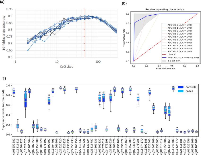
www.nature.com/scientificreports/
(IWGCNA) that identified 20 candidate genes related to ME/CFS severity. In other cohorts, Gow et al.13 found
366 differentially expressed mRNAs in PBMCs of eight post-infectious ME/CFS patients versus seven healthy
controls and Nguyen et al.14 identified 176 mRNAs in whole blood of adolescents (18 healthy vs. 29 patients).
Between those 366 and 176 mRNAs, measured in slightly different cell populations, only ten overlapped. Finally,
Byrnes et al.15 found no differential expression in peripheral blood leukocytes (PBLs) of 44 monozygotic twin
pairs discordant for ME/CFS. In the results from four additional differential expression studies in PBLs, only
one gene, MSN overlapped between two of them15.
Consideration of epigenetic factors may help resolve some of the inconsistencies. Epigenetics performs
transcriptional regulation in a mitotically heritable fashion, yet can be influenced by non-genetic factors16. It
encompasses a wide variety of modifications that do not change the DNA sequence, but are thought to affect the
accessibility and hence readability thereof. One modification is DNA methylation, the addition of a methyl group
to the fifth carbon of cytosine. This process has been described predominantly for cytosines followed by guanine
residues (CpG)17. An increased concentration of CpGs is often observed in the promoter region of genes, where
such clusters are called CpG islands18. Promoter methylation is typically inversely correlated with the expression
of the associated gene, where a high density of methylated CpGs correlates with lower mRNA expression and vice
versa19,20. De Vega et al. have performed differential methylation analyses on PMBCs of ME/CFS p atients21–23 and
reported between 1,192 and 12,608 differentially methylated CpG sites, related to 826 to 5,544 annotated genes.
Using another microarray method with more probes, Trivedi et al.24 found 17,296 differentially methylated CpG
sites related to 6,368 genes. To the best of our knowledge, no study has yet integrated mRNA expression data
and DNA methylation data of ME/CFS.
In light of the preceding discussion, we sought to integrate publicly available mRNA expression and DNA
methylation data to innovate biomarker research in ME/CFS. To this end, we made use of recursive ensemble
feature selection (REFS), a classification pipeline which, when applied in near-analogous settings, has proven
to reliably shed light on the corresponding relationships25. REFS was used to identify a robust mRNA expres-
sion signature that could differentiate between cases and controls. Subsequently, we investigated whether genes
found to be differentially expressed in ME/CFS were indicative of differential DNA methylation as well. To
equate methods and results of previous studies, we also compared the performance of REFS to IWGCNA and
univariate analyses. Finally, we sought to interpret the biological function and relevance of the genes identified
by REFS as candidate biomarkers for ME/CFS.
Results
mRNA expression feature selection. We ran the REFS algorithm ten times on the CAMDA mRNA
expression dataset containing 118 samples and the algorithm found the optimal number of predictor genes
to be 23 (Fig. 1a; Supplemental Table S1). Multivariate analysis of variance (MANOVA)26 indicated a statisti-
cally significant difference between healthy controls and ME/CFS patients ( F(23, 95) = 5.15, p < .0001; Wilk’s
= 0.445, R-squared (uncentered) = 0.555). mRNA expression of all 23 predictor genes was down-regulated in
ME/CFS patients (Fig. 1b). Next, we compared our results with IWGCNA and univariate feature reduction using
χ 2, which were used by Presson et al.12 and Byrnes et al.15, respectively, to identify genes that were associated
with ME/CFS.
IWGCNA constructs a co-expression network and prioritizes modules of genes based on their association
with a phenotype in combination with the presence of a disease-associated genetic variant. Essentially, IWGCNA
selects the most important genes by p − value score. Presson et al.12 reported 20 candidate genes based on mRNA
expression data from the CAMDA dataset, the same dataset in which we found 23 predictor genes using REFS.
The results of Presson et al. and our REFS algorithm could thus be directly compared. Univariate feature selection
reduces the number of features using univariate score metrics, in this case, χ 2. For the comparison, we applied
the sci-kit learning implementation27 to the CAMDA dataset, and selected features according to the highest χ 2
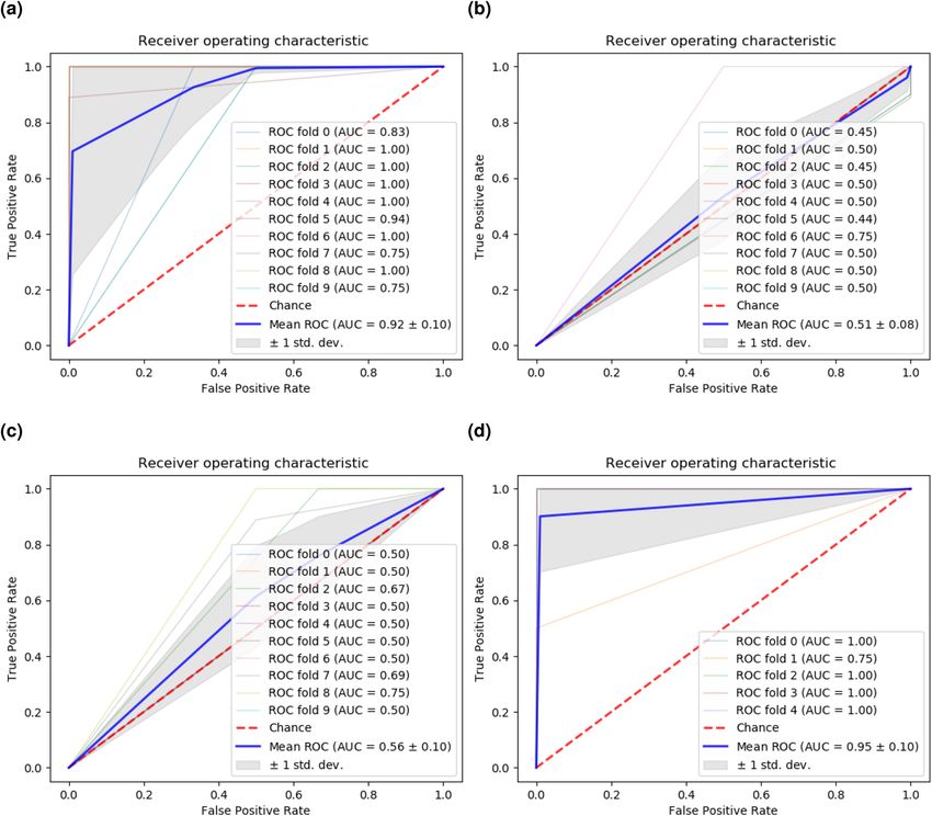
scores with a fixed k = 23. Next, we calculated the accuracy and the associated receiver-operating characteristic
(ROC) curve for all three algorithms. The REFS algorithm had an average accuracy of 91.57% differentiating
controls and patients with ME/CFS globally, with the Passive Aggressive classifier showing a 95.87% accuracy
using only the 23 candidate genes (Table 1). Moreover, the area under the curve (AUC) of the ROC curve for
REFS was 0.92 (Fig. 2a), which is considered o utstanding28,29. The compared methods were only slightly better
than chance (IWGCNA: AUC = 0.51 (Fig. 2b), univariate analysis: AUC = 0.56 (Fig. 2c). Comparing the genes
of interest each of the three methods returned, we found that REFS outperformed both IWGCNA and univariate
analysis based on the ROC curve and every performance classifier (Table 1).
Cross platform validation. We applied the resulting mRNA expression signature to a separate mRNA
dataset (GSE14577) as a cross platform validation. Due to the small sample size (n = 15), we did not perform
feature reduction, but applied the 23 candidate genes directly to the dataset. As the data was measured with a
different platform, not all genes overlapped with the CAMDA dataset. Using the DAVID gene functional tool30,
we retrieved eighteen genes. After training the eighteen-gene based model through 5-fold cross validation, we
achieved a global classification accuracy of 91.11% differentiating controls and patients with ME/CFS (Table 1)
with an ROC AUC of 0.95 (Fig. 2d).
DNA methylation feature selection. We reduced the number of CpG probes to 278 by selecting for
probes associated with the 23 predictor genes. Of these 278 CpGs, REFS identified 48 CpGs as predictive of
ME/CFS after ten runs on the combined methylation data (Fig. 3a). MANOVA analysis showed a statistically
significant difference between healthy controls and ME/CFS patients (F(48, 101) = 50.6783, p < .0001; Wilk’s
= 0.034, R-squared (uncentered) = 0.960). The 48 CpGs reached a global accuracy of 90.17% distinguishing
Scientific Reports | (2021) 11:4541 | https://doi.org/10.1038/s41598-021-83660-9 2
Vol:.(1234567890)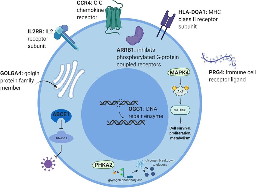
www.nature.com/scientificreports/

                                  Figure 1.  Results of the REFS algorithm run ten times on the mRNA expression data of 118 samples from the
                                  CAMDA dataset. (a) The optimal number of predictor genes to distinguish 93 cases from 25 controls was 23
                                  (red vertical line). (b) mRNA expression levels of the 23 predictor genes for 93 cases (light blue) and 25 controls
                                  (dark blue) in box-and-whisker plots. Outliers were omitted for visualization purposes.

                                                                                                                                mRNA              DNA
                                                                          REFS              IWGCNA            χ2                validation        methylation
                                   Classifier                             µ        σ        µ        σ        µ        σ        µ        σ        µ        σ
                                   Gradient boosting (n_estimators=300)   0.898    0.0647   0.7417   0.0975   0.7801   0.0775   0.8111   0.1641   0.7852   0.1023
                                   Random forest (n_estimators=300)       0.8575   0.0714   0.7298   0.0576   0.799    0.0459   0.9333   0.0943   0.8181   0.0867
                                   Logistic regression                    0.9595   0.0533   0.7138   0.0913   0.8052   0.0530   0.9333   0.0943   0.9457   0.042
                                   Passive aggressive                     0.9587   0.0675   0.6057   0.1744   0.8151   0.0693   0.9333   0.0943   0.98     0.0306
                                   SGD                                    0.9595   0.0533   0.6264   0.0659   0.7564   0.0914   0.9333   0.0943   0.9324   0.0437
                                   SVC (linear)                           0.9421   0.0754   0.7472   0.0848   0.7962   0.0408   0.9333   0.0943   0.9667   0.0683
                                   Ridge                                  0.9023   0.0691   0.7214   0.0858   0.8143   0.0616   0.9333   0.0943   0.9733   0.0327
                                   Bagging (n_estimators=300)             0.8478   0.0495   0.7144   0.0841   0.7801   0.0940   0.8778   0.0875   0.8119   0.0586
                                   Average                                0.9157   0.0630   0.7001   0.0927   0.7933   0.0667   0.9111   0.1022   0.9017   0.0581

                                  Table 1.  Classification accuracy of the 23 predictor genes obtained with REFS, the 20 genes obtained with
                                  IWGCNA, and the 23 genes obtained with univariate analysis based on the same mRNA expression data of 118
                                  samples from the CAMDA dataset Classification accuracy of eighteen of the 23 predictor genes applied to a
                                  validation mRNA dataset (GSE14577), and of 48 predictor CpGs obtained with REFS based on merged DNA
                                  methylation data. µ, mean; σ , standard deviation.

                                  patients and healthy controls (Table 1). When comparing CpG methylation between cases and controls, most
                                  CpG sites showed enhanced methylation in ME/CFS patients (Fig. 3c). Finally, the calculated ROC curve had
                                  an AUC of 0.97 (Fig. 3b), pointing to a clear distinction between patients and controls when applying the 48
                                  predictor CpG sites.

                                  Biological interpretation of the gene signature. To put our findings in a biological context, we inter-
                                  rogated the existing literature on the proteins encoded by the 23 predictor genes produced by the REFS algo-
                                  rithm. These results were obtained in PBMCs, therefore we focused on ten proteins, MAPK4, ARRB1, GOLGA4,

Scientific Reports |   (2021) 11:4541 |                   https://doi.org/10.1038/s41598-021-83660-9                                                                3

                                                                                                                                                            Vol.:(0123456789)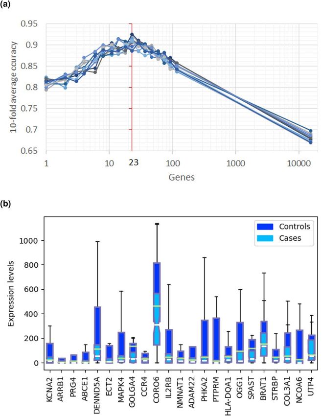
www.nature.com/scientificreports/
Figure 2. ROC curves for (a) REFS, (b) IWGCNA, and (c) univariate analysis applied to the same mRNA
expression data of 118 samples from the CAMDA dataset. (d) ROC curve for the validation of the obtained
23-gene signature on a separate dataset (GSE14577). The 5-fold cross validation was performed with the
eighteen genes available in GSE14577.
ABCE1, PHKA2, IL2RB, CCR4, HLA-DQA1, PRG4 and OGG1, that acted in immune pathways. The mRNA
transcripts encoding these proteins were all downregulated in ME/CFS patients compared to healthy controls
(Fig. 1a). Protein functions are visualized in Fig. 4 and briefly described below.
Intracellular signaling. In response to extracellular stimuli binding to immune cell receptors, mitogen-activated
protein kinases (MAPKs) transduce intracellular signals through protein phosphorylation cascades to medi-
ate the cellular response. MAPK4 (also known as ERK4) directly phosphorylates AKT, subsequently activating
the mTOR signaling pathway and ultimately regulating cell survival, proliferation and metabolism. Decreased
MAPK4 activity is associated with decreased AKT activity and proliferation31. Another intracellular mediator
of the immune response is ARRB1 (β-arrestin 1, also known as arrestin 2). ARRB1 is found in high levels in
PBLs where it inhibits G-protein coupled receptors that are phosphorylated by β-adrenergic receptor kinase.
GOLGA4 (golgin A4) is a member of the golgin protein family. Upon macrophage LPS activation, formation of
GOLGA4-demarked carriers is upregulated, which was found to increase TNF trafficking and subsequent secre-
tion. Depletion of GOLGA4 consequently impairs TNF s ecretion32. ATP-binding cassette sub-family E member
1 (ABCE1), a cell membrane transporter, inhibits RNase L activity. RNase L is important in immune defense,
as it degrades all RNA within the cell upon viral infection and releases antiviral IFNγ 33. ABCE1 was downregu-
lated in PBLs of ME/CFS patients, which correlated with upregulated RNase L34. Finally, PHKA2 is the hepatic
isoform of the alpha subunit of phosphorylase b kinase. This enzyme phosphorylates glycogen phosphorylase b,
converting it to the active glycogen phosphorylase a, which then breaks down stored glycogen to glucose. This
glucose is converted through glycolysis to ATP, to meet metabolic demands. PHKA2 is thus important in provid-
Scientific Reports | (2021) 11:4541 | https://doi.org/10.1038/s41598-021-83660-9 4
Vol:.(1234567890)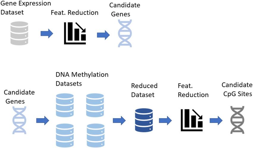
www.nature.com/scientificreports/
Figure 3. Results of the REFS algorithm run ten times on the merged DNA methylation datasets restricted to
278 probes associated with the 23 candidate genes. (a) The optimal number of predictor CpGs to distinguish 99
cases from 50 controls was 48 (red vertical line). (b) ROC curve of the 48 predictor CpGs. c) DNA methylation
levels (normalized using Standard scaler) of the 48 predictor CpG sites for 99 cases (light blue) and 50 controls
(dark blue).
ing energy to cells by maintaining glucose levels. Mutations in PHKA2 gene cause glycogen storage disease type
IXa, characterized by hypoglycemia, hepatomegaly, elevated liver enzymes, growth retardation and motor delay,
hypercholesterolemia and hypertriglyceridemia. Some patients report fatigue and muscle w eakness35.
Immune cell receptors and ligands. IL2RB is the β-subunit of the IL2 receptor, a receptor for IL2 that is involved
in the differentiation of regulatory-, effector-, memory-, type 1 and 2 helper T cells. IL2RB-deficient patients show
dysregulated IL2 and IL15 signaling, enhanced natural killer cell levels, and subsequent immunodeficiency and
impaired antiviral immunity36. A SNP in the 3-prime untranslated region of the gene was associated with ME/
CFS, interfering with miRNA binding which could reduce IL2 receptor f unction37. CCR4 (CC chemokine type
4 receptor) is a G protein-coupled receptor expressed on regulatory and type 2 helper T cells that binds CCL17
and CCL22. These chemokines cause chemotaxis of the cell, traffic leukocytes and are involved in development,
homeostasis and function of the immune system38. Blockage of CCR4 by mogamulizumab induced fatigue in
human subjects39. HLA-DQA1 is the α-chain of the MHC class II receptor expressed on antigen-presenting cells
such as macrophages, B lymphocytes and dendritic cells. The MHC class II receptor presents peptides to T cell
receptors to activate the immune system upon viral infection. Proteoglycan 4 (PRG4) is an immune cell ligand. It
is a lubricant, reducing friction between joints or boundaries, and altered expression is implicated in rheumatoid
arthritis. In addition, PRG4 was found to ‘coat’ neutrophils and macrophages, perhaps by binding TLR2, -4, -5
or CD44, thereby preventing activation by low levels of pro-inflammatory cytokines. Upon inflammation, when
an immune response is required, PRG4 was shed from the immune cells to allow a ctivation40.
DNA damage repair. DNA repair enzyme 8-oxoguanine DNA glycosylase (OGG1) specifically excises the most
pre-mutagenic oxidative base lesion 8-oxoguanine. Such DNA damage caused by exposure to reactive oxygen
Scientific Reports | (2021) 11:4541 | https://doi.org/10.1038/s41598-021-83660-9 5
Vol.:(0123456789)
www.nature.com/scientificreports/
Figure 4. Visualization of the functions and locations of ten proteins in a hypothetical immune cell setting.
All ten mRNA transcripts were downregulated in PBMCs of ME/CFS patients compared to healthy controls.
Created with BioRender.com.
species (ROS) leads to mutagenesis or cell death. OGG1 activity prevents mutations and Alzheimer’s disease
patients with an OGG1 polymorphism show increased DNA damage41.
Discussion
ME/CFS is a chronic disorder characterized by persistent, disabling fatigue for which no diagnostic or prog-
nostic test nor complete treatment is available. Several studies have sought to define biomarkers for ME/CFS
by performing differential mRNA expression or DNA methylation analysis. However, as Byrnes et al.15 pointed
out, these results were study-dependent and no definitive biomarkers were found. We used a state-of-the-art
machine learning technique to distinguish ME/CFS patients from healthy controls across different platforms,
several cohorts and on different levels of gene expression regulation. To our knowledge, this was the first time
such a technique was used in mRNA expression data and validated in DNA methylation data.
In this study, we implemented the REFS algorithm on public mRNA expression data and found 23 genes
whose changes in expression levels were able to distinguish ME/CFS patients from healthy controls. The 23 pre-
dictor genes differentiated between cases and controls with 91.57% global accuracy and returned a ROC AUC of
0.92. In addition, 48 CpG methylation sites associated with these genes were predictive of ME/CFS in four merged
DNA methylation studies. Moreover, all 23 candidate genes were downregulated in ME/CFS patients while
DNA methylation of almost all 48 CpG sites was enhanced. This inverse correlation between mRNA expression
and DNA methylation, across different samples and studies, legitimizes the results of our study. As previously
demonstrated25, REFS identifies a more accurate, robust gene signature than previous methods. Comparing the
gene signature returned by three different methods, based on the same data, REFS outperformed both IWGCNA
and univariate analysis in separating ME/CFS patients and healthy controls with a ROC AUC of 0.92. The AUC
of the gene signature applied to a different platform was 0.95, and the AUC even reached 0.97 when plotting the
sensitivity and specificity of the 48 predictor CpG sites.
To show the relevance of the returned predictor genes, we investigated the biological functions of ten encoded
proteins active in immune pathways. This decision was based on the mRNA expression being measured in
PBMCs and the literature pointing towards an important role for the immune system in ME/CFS. Sotzny et al.42
reviewed autoimmunity in ME/CFS, concluding that immunologic and metabolic alterations were often reported.
The authors stress the potential importance of autoantibodies in the disorder and the proposed role of preceding
infections. Downregulation of ABCE1, one of the encoded proteins identified in our study, concurs with the pres-
ence of previous viral infections, as the protein inhibits RNase L’s viral RNA degrading activity. Similarly, ARRB1
protein was decreased after Epstein-Barr virus-infection in m ice43. Its downregulation in our study concurs with
this finding. Recently, Mandarano et al.44 described evidence of immune involvement in their study of 53 ME/CFS
patients. They specifically focused on T cells, showing that CD8+ T cells derived from patients had lower mito-
chondrial membrane potential, which points towards T cell exhaustion. PHKA2 is necessary for the first step in
breaking down glycogen to glucose. Its downregulation could contribute to impaired glycolysis in immune cells.
In ME/CFS, CD4+ and CD8+ T cells had impaired resting glycolysis, and plasma glucose was reduced. CD8+
Scientific Reports | (2021) 11:4541 | https://doi.org/10.1038/s41598-021-83660-9 6
Vol:.(1234567890)www.nature.com/scientificreports/
T cells showed an impaired metabolic response to a ctivation44. Another study found that glycogen metabolism
regulates the immune functions of dendritic c ells45. Inhibiting glycogen phosphorylase impaired their ability to
produce inflammatory cytokines and stimulate T cells. These findings combined suggest that reduced PHKA2
in ME/CFS might inhibit glycogen phosphorylase activation and thus dendritic cell functioning.
Several (subunits of) immune cell receptors were also part of our gene signature and downregulated in ME/
CFS. IL2RB, CCR4 and HLA-DQA1 are vital elements in proper immune response, dysregulation, whether up or
down, could be evidence of a disturbed immune system or be the cause of it. The same holds true for decreased
MAPK4 expression, an ubiquitous transducer of intracellular signals in response to immune cell receptor bind-
ing. Further downstream, GOLGA4 was upregulated in response to macrophage LPS activation to increase TNF
secretion. TNF is the main pro-inflammatory cytokine secreted by inflammatory macrophages, and its release is
important for enhancing the activation and recruitment of T cells, ensuring robust innate and adaptive immune
responses. In our study however, GOLGA4 was downregulated in PBMCs of ME/CFS patients, potentially caus-
ing impaired TNF secretion and subsequently an impaired immune response to inflammation. Furthermore,
decreased expression of PRG4 leads to reduced anti-inflammatory action of this protein. By binding immune
cells receptors, PRG4 prevents activation by low levels of circulating pro-inflammatory cytokines. PRG4 could
thus be important in low-grade inflammation causing ME/CFS46.
Finally, evidence has emerged that oxidative stress levels are raised in ME/CFS, for example in response to
exercise, perhaps causing some of the symptoms seen in ME/CFS47,48. DNA damage caused by exposure to ROS
leads to mutagenesis or cell death, OGG1 specifically repairs this DNA damage. OGG1 depletion in human
monocyte-derived dendritic cells inhibited enhanced cell surface molecule-expression and secretion of pro-
inflammatory cytokines upon exposure to 8-oxoguanine base lesions. This suggests that OGG1 is important for
dendritic cell activation in response to R OS49. Concurrently, 8-oxoguanine base lesions did not cause acute or
systemic inflammation in Ogg1-deficient mice50. As we found that OGG1 is downregulated in ME/CFS patients,
while oxidative stress levels are increased, DNA damage might be increased, which in turn causes the release of
danger associated molecular patterns (DAMPs) and activates the innate immune system. We can conclude, from
the various roles of these ten genes, that their downregulation may not only contribute to immune activation, but
also towards a general dysregulation of the immune response. Whether all genes are causative of the ME/CFS
phenotype, or some are mere consequences of immune mayhem in ME/CFS patients remains to be investigated.
Our results return a promising gene signature for ME/CFS that needs to be validated in a well characterized
clinical cohort to study its use as a diagnostic tool. In this cohort, the number of cases and controls should be
balanced, as the current study suffers from cases outnumbering controls in both the mRNA expression and DNA
methylation datasets. We compensated for the imbalance with stratified folds in the cross-validation. Finally,
the investigation of the predictor genes has thus far been limited to a literature review. In vitro experiments with
PBMCs should provide additional information regarding gene function.
To conclude, we found a mRNA expression signature of 23 genes for ME/CFS capable of separating cases and
controls. These candidate genes could potentially be used as biomarkers for diagnostic purposes. In addition, ten
of these genes could be interpreted in the context of a derailed immune system in ME/CFS. Those genes could
be investigated further for target finding and development of future treatments for ME/CFS.
Methods
mRNA expression data. The mRNA expression data used in this study was retrieved as provided by
Presson et al.12, which was made available by the CDC for the 2006 Critical Assessment of Microarray Data
Analysis (CAMDA) conference (https://horvath.genetics.ucla.edu/html/CoexpressionNetwork/CFS/). We refer
to this dataset as the CAMDA dataset. The CAMDA- and subsequent datasets were chosen because they are
the only large, publicly available studies performed on PBMCs. PBMCs are thought to be involved in ME/CFS
pathophysiology42,44. Included in the CAMDA dataset are mRNA expression levels in PBMCs and fatigue sever-
ity status from 118 participants. ME/CFS was determined based on the Fukuda case definition criteria1, and
fatigue severity was estimated based on clustered scores from the SF-36 fatigue score, Multidimensional Fatigue
Inventory, and Symptom Inventory Case Definition Score12. mRNA expression was measured on the MWG
Biotech microarray platform, containing probes for approximately 20,000 transcripts.
To analyze the data, we first re-annotated the probes with HGNC gene symbols using D AVID51, yielding
15,419 gene-associated probes. The data was then normalized using the Standard scaler from sci-kit learning
toolbox27. Samples with low fatigue severity were encoded as 0 (controls) and samples with moderate or high
fatigue severity were encoded as 1 (cases), based on the clustered scores described by Presson et al.12. Altogether,
93 cases and 25 controls were included for REFS. A 10-fold cross-validation with stratified folds was performed
to accommodate the unbalanced classes.
As a test dataset for the algorithm, mRNA expression levels in PBMCs from post-infectious, male ME/
CFS patients (n = 8) and healthy male controls (n = 7) were included as provided by Gow et al.13 in the Gene
Expression Omnibus (GEO)52 GSE14577. ME/CFS was determined with the Fukuda case definition c riteria1
and mRNA expression was measured with Affymetrix Human Genome U133A Array. To analyze the GSE14577
data, the series matrix file was employed and scaled using the Standard scaler. Given the low number of samples,
a 5-fold cross-validation was performed only for the analysis. A summary of the datasets is available in Table 2.
DNA methylation data. DNA methylation data was retrieved from GEO (Table 2). The DNA methylation
datasets pertained methylation profiles in PBMCs of ME/CFS patients and healthy controls measured with the
Illumina HumanMethylation Infinium 450k BeadChip array (450k) and its successor, the Illumina Human-
Methylation Infinium EPIC BeadChip array (EPIC). The 450k and the EPIC measured the methylation status
of 485,577 and 865,859 CpGs, respectively, with approximately 90% of the 450k probes being present on the
Scientific Reports | (2021) 11:4541 | https://doi.org/10.1038/s41598-021-83660-9 7
Vol.:(0123456789)www.nature.com/scientificreports/
GEO accession number/name Reference Samples (n) Measuring platform Probes (n) Type
CAMDA Presson et al.12 118 MWG Biotech microarray 15,419 mRNA expression
Affymetrix Human Genome
GSE14577 Gow et al.13 15 22,284 mRNA expression
U133A Array
21
GSE59489 de Vega et al. 24 450k (GPL13534) 485,577 DNA methylation
GSE93266 de Vega et al.22 75 450k (GPL13534) 485,512 DNA methylation
GSE102504 de Vega et al.23 25 450k (GPL13534) 467,971 DNA methylation
GSE111183 Trivedi et al.24 25 EPIC (GPL21145) 866,895 DNA methylation
Table 2. Characteristics of the mRNA expression and DNA methylation datasets.
EPIC53. In all four studies, ME/CFS was determined with the Fukuda and Canadian case definitions1,54. From the
GEO repositories, series matrix files containing the normalized percentage methylation per CpG were obtained.
The data was pre-processed by scaling the percentage methylation per sample using the Standard scaler, after
which the individual datasets were merged. Subsequently, the data was split for 10-fold cross-validation with
stratified folds. In total, the methylation data included 149 samples (99 cases and 50 controls).
REFS on mRNA expression data. Several studies have employed univariate feature selection, with sta-
tistic metrics such as χ 2 or F − value , that reduce the features using the highest scores, yielding study-specific
results. To overcome this issue, REFS was used to identify a mRNA expression signature that was able to clas-
sify disease status in a PMBC study of ME/CFS patients and healthy controls. The utility and accuracy of REFS
compared to other methods was previously demonstrated in miRNA and mRNA datasets25,55. In short, the REFS
algorithm reduces the number of features to the most significant ones by combining the results of separate clas-
sifiers of distinct topologies. The algorithm gives each feature a score, based on how each different algorithm
used it. In case of a tree-based algorithm, it depends on how many times the feature appeared in the tree. If it is
a coefficient based algorithm, it depends on the value of the coefficient, the highest being the most important.
This scoring is consistent with the Borda method56, the difference being that 10-fold cross validation is included
for scoring. After each iteration, the most important features are selected (Fig. 5).
The REFS algorithm (Algorithm 1) was implemented on the 15,419 gene-associated probes through 10-fold
cross-validation with the following classifiers: Bagging, Gradient Boosting, Random Forest, Logistic Regression,
Passive Aggressive, SGD, SVC (linear), and Ridge. During the first iteration, the algorithm reduced the total
features to the most significant ones, after which it reduced the number of variables by 20% per step with a stop
parameter of 70% accuracy as indicated in Fig. 5.
Scientific Reports | (2021) 11:4541 | https://doi.org/10.1038/s41598-021-83660-9 8
Vol:.(1234567890)www.nature.com/scientificreports/
Figure 5. Overview of the REFS algorithm using the 10-fold global accuracy > 70% as a stop parameter.
Figure 6. Overview of the pipeline to reduce the number of methylation data probes for REFS by selecting
CpGs associated with the candidate genes.
REFS on DNA methylation data of candidate genes. The DNA methylation platforms were repre-
sented by a large amount of features, 485,577 and 865,859 features by the 450k and EPIC, respectively. Therefore,
the search space was reduced by adopting a candidate gene approach, in which CpGs were only selected if they
associated with the genes found using REFS on the CAMDA dataset. Practically, CpG probes were selected
from the datasheet of the platform GPL13534 (https://www.ncbi.nlm.nih.gov/geo/query/acc.cgi?acc/=GPL13
534) when the UCSC_RefGene_Name field matched any of the genes previously selected by the ensemble feature
selection approach. This process is explained in Fig. 6.
Biological interpretation of the mRNA expression signature. To put the genes identified with the
REFS algorithm in the mRNA expression dataset in a biological perspective, the protein encoded by each gene
was investigated in the existing literature available on Pubmed and Google Scholar (March 2020). Because the
analyses were performed with genetic material derived from PBMCs, protein function was investigated in the
context of the immune system. After initial analysis, ten proteins that functioned in immune pathways were
selected for further literature review.
Received: 22 August 2020; Accepted: 27 January 2021
References
1. Fukuda, K. et al. The chronic fatigue syndrome: A comprehensive approach to its definition and study. Ann. Intern. Med. 121,
953–959. https://doi.org/10.7326/0003-4819-121-12-199412150-00009 (1994).
2. Johnston, S., Brenu, E. W., Staines, D. & Marshall-Gradisnik, S. The prevalence of chronic fatigue syndrome/myalgic encephalo-
myelitis: A meta-analysis. Clin. Epidemiol. 5, 105. https://doi.org/10.2147/CLEP.S39876 (2013).
3. Walsh, C. M., Zainal, N. Z., Middleton, S. J. & Paykel, E. S. A family history study of chronic fatigue syndrome. Psychiatr. Genet.
11, 123–128. https://doi.org/10.1097/00041444-200109000-00003 (2001).
4. Schlauch, K. A. et al. Genome-wide association analysis identifies genetic variations in subjects with myalgic encephalomyelitis/
chronic fatigue syndrome. Transl. Psychiatry 6, e730–e730. https://doi.org/10.1038/tp.2015.208 (2016).
Scientific Reports | (2021) 11:4541 | https://doi.org/10.1038/s41598-021-83660-9 9
Vol.:(0123456789)www.nature.com/scientificreports/
5. Perez, M. et al. Genetic predisposition for immune system, hormone, and metabolic dysfunction in myalgic encephalomyelitis/
chronic fatigue syndrome: A pilot study. Front. Pediatr. 7, 206. https://doi.org/10.3389/fped.2019.00206 (2019).
6. Rasa, S. et al. Chronic viral infections in myalgic encephalomyelitis/chronic fatigue syndrome (me/cfs). J. Transl. Med. 16, 268.
https://doi.org/10.1186/s12967-018-1644-y (2018).
7. Almenar-Pérez, E. et al. Epigenetic components of myalgic encephalomyelitis/chronic fatigue syndrome uncover potential trans-
posable element activation. Clin. Therap. 41, 675–698. https://doi.org/10.1016/J.CLINTHERA.2019.02.012 (2019).
8. Herrera, S., de Vega, W. C., Ashbrook, D., Vernon, S. D. & McGowan, P. O. Genome-epigenome interactions associated with myalgic
encephalomyelitis/chronic fatigue syndrome. Epigenetics 13, 1174–1190. https://doi.org/10.1080/15592294.2018.1549769 (2018).
9. Smith, M. E. B. et al. Treatment of myalgic encephalomyelitis/chronic fatigue syndrome: A systematic review for a national institutes
of health pathways to prevention workshop. Ann. Intern. Med. 162, 841–50. https://doi.org/10.7326/M15-0114 (2015).
10. Cortes Rivera, M., Mastronardi, C., Silva-Aldana, T. C., Arcos-Burgos, M. & Lidbury, A. B. Myalgic encephalomyelitis/chronic
fatigue syndrome: A comprehensive review. Diagnosticshttps://doi.org/10.3390/diagnostics9030091 (2019).
11. Fang, H. et al. Gene expression profile exploration of a large dataset on chronic fatigue syndrome. Pharmacogenomicshttps://doi.
org/10.2217/14622416.7.3.429 (2006).
12. Presson, A. P. et al. Integrated weighted gene co-expression network analysis with an application to chronic fatigue syndrome.
BMC Syst. Biol. 2, 95. https://doi.org/10.1186/1752-0509-2-95 (2008).
13. Gow, J. W. et al. A gene signature for post-infectious chronic fatigue syndrome. BMC Med. Genom. 2, 38. https://doi.
org/10.1186/1755-8794-2-38 (2009).
14. Nguyen, C. B. et al. Whole blood gene expression in adolescent chronic fatigue syndrome: An exploratory cross-sectional study
suggesting altered b cell differentiation and survival. J. Transl. Med. 15, 102. https://doi.org/10.1186/s12967-017-1201-0 (2017).
15. Byrnes, A. et al. Gene expression in peripheral blood leukocytes in monozygotic twins discordant for chronic fatigue: No evidence
of a biomarker. PLoS ONE 4, e5805. https://doi.org/10.1371/journal.pone.0005805 (2009).
16. Nanney, D. L. Epigenetic control systems. Proc. Natl. Acad. Sci. 44, 712–717. https://doi.org/10.1073/pnas.44.7.712 (1958).
17. Jang, H., Shin, W., Lee, J. & Do, J. Cpg and non-cpg methylation in epigenetic gene regulation and brain function. Genes 8, 148.
https://doi.org/10.3390/genes8060148 (2017).
18. Bird, A. P. Cpg-rich islands and the function of DNA methylation. Nature 321, 209–213. https://doi.org/10.1038/321209 a0 (1986).
19. Schübeler, D. Function and information content of DNA methylation. Nature 517, 321. https: //doi.org/10.1038/nature 14192 (2015).
20. Stepper, P. et al. Efficient targeted DNA methylation with chimeric dcas9-dnmt3a-dnmt3l methyltransferase. Nucleic Acids Res.
45, 1703–1713. https://doi.org/10.1093/nar/gkw1112 (2017).
21. de Vega, W. C., Vernon, S. D. & McGowan, P. O. DNA methylation modifications associated with chronic fatigue syndrome. PLoS
ONE 9, e104757. https://doi.org/10.1371/journal.pone.0104757 (2014).
22. de Vega, W. C., Herrera, S., Vernon, S. D. & McGowan, P. O. Epigenetic modifications and glucocorticoid sensitivity in myalgic
encephalomyelitis/chronic fatigue syndrome (me/cfs). BMC Med. Genom. 10, 11. https://doi.org/10.1186/s12920-017-0248-3
(2017).
23. de Vega, W. C., Erdman, L., Vernon, S. D., Goldenberg, A. & McGowan, P. O. Integration of DNA methylation & health scores
identifies subtypes in myalgic encephalomyelitis/chronic fatigue syndrome. Epigenomics 10, 539–557. https://doi.org/10.2217/
epi-2017-0150 (2018).
24. Trivedi, M. S. et al. Identification of myalgic encephalomyelitis/chronic fatigue syndrome-associated DNA methylation patterns.
PLoS ONE 13, e0201066. https://doi.org/10.1371/journal.pone.0201066 (2018).
25. Lopez-Rincon, A., Martinez-Archundia, M., Martinez-Ruiz, G. U., Schoenhuth, A. & Tonda, A. Automatic discovery of 100-mirna
signature for cancer classification using ensemble feature selection. BMC Bioinform. 20, 480. https://doi.org/10.1186/s12859-019-
3050-8 (2019).
26. O’Brien, R. G. & Kaiser, M. K. Manova method for analyzing repeated measures designs: An extensive primer. Psychol. Bull. 97,
316. https://doi.org/10.1037/0033-2909.97.2.316 (1985).
27. Pedregosa, F. et al. Scikit-learn: Machine learning in python. J. Mach. Learn. Res. 12, 2825–2830 (2011).
28. Šimundić, A.-M. Measures of diagnostic accuracy: Basic definitions. EJIFCC 19, 203 (2009).
29. Mandrekar, J. N. Receiver operating characteristic curve in diagnostic test assessment. J. Thorac. Oncol. 5, 1315–1316. https://doi.
org/10.1097/JTO.0b013e3181ec173d (2010).
30. Huang, D. W. et al. The David gene functional classification tool: a novel biological module-centric algorithm to functionally
analyze large gene lists. Genome Biol. 8, R183. https://doi.org/10.1186/gb-2007-8-9-r183 (2007).
31. Wang, W. et al. Mapk4 overexpression promotes tumor progression via noncanonical activation of akt/mtor signaling. J. Clin.
Investig. 129, 66. https://doi.org/10.1172/JCI97712 (2019).
32. Lieu, Z. Z. et al. A trans-golgi network golgin is required for the regulated secretion of TNF in activated macrophages in vivo. Proc.
Natl. Acad. Sci. USA 105, 3351–3356. https://doi.org/10.1073/pnas.0800137105 (2008).
33. Tian, Y., Han, X. & Tian, D. L. The biological regulation of abce1. IUBMB Life 64, 795–800. https: //doi.org/10.1002/iub.1071 (2012).
34. Vojdani, A., Choppa, P. C. & Lapp, C. W. Downregulation of rnase l inhibitor correlates with upregulation of interferon-induced
proteins (2–5a synthetase and rnase l) in patients with chronic fatigue immune dysfunction syndrome. J. Clin. Lab. Immunol. 50,
1–16 (1998).
35. Beauchamp, N. J. et al. Glycogen storage disease type ix: High variability in clinical phenotype. Mol. Genet. Metab. 92, 88–99. https
://doi.org/10.1016/j.ymgme.2007.06.007 (2007).
36. Fernandez, I. Z. et al. A novel human il2rb mutation results in t and nk cell-driven immune dysregulation. J. Exp. Med. 216,
1255–1267. https://doi.org/10.1084/jem.20182015 (2019).
37. Rajeevan, M. S., Dimulescu, I., Murray, J., Falkenberg, V. R. & Unger, E. R. Pathway-focused genetic evaluation of immune
and inflammation related genes with chronic fatigue syndrome. Hum. Immunol. 76, 553–560. https://doi.org/10.1016/j.humim
m.2015.06.014 (2015).
38. Yoshie, O. & Matsushima, K. Ccr4 and its ligands: From bench to bedside. Int. Immunol. 27, 11–20. https://doi.org/10.1093/intim
m/dxu079 (2014).
39. Kasamon, Y. L. et al. Fda approval summary: Mogamulizumab-kpkc for mycosis fungoides and sezary syndrome. Clin. Cancer
Res. 25, 7275–7280. https://doi.org/10.1158/1078-0432.Ccr-19-2030 (2019).
40. Das, N., Schmidt, T. A., Krawetz, R. J. & Dufour, A. Proteoglycan 4: From mere lubricant to regulator of tissue homeostasis and
inflammation: Does proteoglycan 4 have the ability to buffer the inflammatory response?. BioEssays 41, 1800166. https://doi.
org/10.1002/bies.201800166 (2019).
41. Dinçer, Y. et al. DNA repair gene ogg1 polymorphism and its relation with oxidative DNA damage in patients with Alzheimer’s
disease. Neurosci. Lett. 709, 134362. https://doi.org/10.1016/j.neulet.2019.134362 (2019).
42. Sotzny, F. et al. Myalgic encephalomyelitis/chronic fatigue syndrome—Evidence for an autoimmune disease. Autoimmun. Rev. 17,
601–609. https://doi.org/10.1016/j.autrev.2018.01.009 (2018).
43. Williams, M. V., Cox, B., Lafuse, W. P. & Ariza, M. E. Epstein-barr virus dutpase induces neuroinflammatory mediators: Implications
for myalgic encephalomyelitis/chronic fatigue syndrome. Clin. Ther. 41, 848–863. https://doi.org/10.1016/j.clinthera.2019.04.009
(2019).
44. Mandarano, A. H. et al. Myalgic encephalomyelitis/chronic fatigue syndrome patients exhibit altered t cell metabolism and cytokine
associations. J. Clin. Investig. 130, 1491–1505. https://doi.org/10.1172/JCI132185 (2020).
Scientific Reports | (2021) 11:4541 | https://doi.org/10.1038/s41598-021-83660-9 10
Vol:.(1234567890)www.nature.com/scientificreports/
45. Thwe, P. M. et al. Cell-intrinsic glycogen metabolism supports early glycolytic reprogramming required for dendritic cell immune
responses. Cell Metab. 26, 558-567.e5. https://doi.org/10.1016/j.cmet.2017.08.012 (2017).
46. Montoya, J. G. et al. Cytokine signature associated with disease severity in chronic fatigue syndrome patients. Proc. Natl. Acad.
Sci. 114, E7150–E7158. https://doi.org/10.1073/pnas.1710519114 (2017).
47. Jammes, Y. & Retornaz, F. Understanding neuromuscular disorders in chronic fatigue syndrome. F1000Reshttps: //doi.org/10.12688
/f1000research.18660.1 (2019).
48. Kennedy, G. et al. Oxidative stress levels are raised in chronic fatigue syndrome and are associated with clinical symptoms. Free
Radic. Biol. Med. 39, 584–9. https://doi.org/10.1016/j.freeradbiomed.2005.04.020 (2005).
49. Pazmandi, K. et al. Oxidized base 8-oxoguanine, a product of DNA repair processes, contributes to dendritic cell activation. Free
Radic. Biol. Med. 143, 209–220. https://doi.org/10.1016/j.freeradbiomed.2019.08.010 (2019).
50. Visnes, T. et al. Small-molecule inhibitor of ogg1 suppresses proinflammatory gene expression and inflammation. Science 362,
834–839. https://doi.org/10.1126/science.aar8048 (2018).
51. Dennis, G. et al. David: Database for annotation, visualization, and integrated discovery. Genome Biol. 4, R60. https://doi.
org/10.1186/gb-2003-4-9-r60 (2003).
52. Edgar, R. Gene expression omnibus: Ncbi gene expression and hybridization array data repository. Nucleic Acids Res. 30, 207–210.
https://doi.org/10.1093/nar/30.1.207 (2002).
53. Pidsley, R. et al. Critical evaluation of the illumina methylationepic beadchip microarray for whole-genome DNA methylation
profiling. Genome Biol. 17, 208. https://doi.org/10.1186/s13059-016-1066-1 (2016).
54. Carruthers, B. M. et al. Myalgic encephalomyelitis: International consensus criteria. J. Intern. Med. 270, 327–338. https://doi.org
/10.1111/j.1365-2796.2011.02428.x (2011).
55. Lopez-Rincon, A. et al. Machine learning-based ensemble recursive feature selection of circulating mirnas for cancer tumor clas-
sification. Cancers 12, 1785 (2020).
56. Sarkar, C., Cooley, S. & Srivastava, J. Robust feature selection technique using rank aggregation. Appl. Artif. Intell. 28, 243–257.
https://doi.org/10.1080/08839514.2014.883903 (2014).
Acknowledgements
The funding for the study was provided by the division of Pharmacology, Department of Pharmaceutical Sciences,
Faculty of Science, Utrecht University and Tytgat Institute for Liver and Intestinal Research, AGEM, Amsterdam
UMC. This work was carried out on the Dutch national e-infrastructure with the support of SURF Cooperative.
Author contributions
A.D.K., A.S., A.A.V., A.L.R. suggested the problem, A.L.R. prepared the datasets, performed the analyses and
coded. P.I.M. wrote the manuscript and contributed the biological interpretation. L.M.M. contributed the
biological interpretation, A.L.Y. and P.H. on epigenetics, I.A. on genetics, and A.A.V., A.S. and A.D.K. on all
fields. A.D.K., A.S., A.A.V., and J.B. provided insights on ME/CFS. All authors reviewed and approved the final
manuscript.
Competing interests
The authors declare no competing interests.
Additional information
Supplementary Information The online version contains supplementary material available at https://doi.
org/10.1038/s41598-021-83660-9.
Correspondence and requests for materials should be addressed to A.L.-R.
Reprints and permissions information is available at www.nature.com/reprints.
Publisher’s note Springer Nature remains neutral with regard to jurisdictional claims in published maps and
institutional affiliations.
Open Access This article is licensed under a Creative Commons Attribution 4.0 International
License, which permits use, sharing, adaptation, distribution and reproduction in any medium or
format, as long as you give appropriate credit to the original author(s) and the source, provide a link to the
Creative Commons licence, and indicate if changes were made. The images or other third party material in this
article are included in the article’s Creative Commons licence, unless indicated otherwise in a credit line to the
material. If material is not included in the article’s Creative Commons licence and your intended use is not
permitted by statutory regulation or exceeds the permitted use, you will need to obtain permission directly from
the copyright holder. To view a copy of this licence, visit http://creativecommons.org/licenses/by/4.0/.
© The Author(s) 2021
Scientific Reports | (2021) 11:4541 | https://doi.org/10.1038/s41598-021-83660-9 11
Vol.:(0123456789)You can also read


