Stem Cell-Induced Inflammation in Cholesteatoma is Inhibited by the TLR4 Antagonist LPS-RS - PUB
←
→
Page content transcription
If your browser does not render page correctly, please read the page content below
Article
Stem Cell-Induced Inflammation in Cholesteatoma is
Inhibited by the TLR4 Antagonist LPS-RS
Matthias Schürmann 1,†, Johannes F. W. Greiner 2,†, Verena Volland-Thurn 1,3,†, Felix Oppel 1,
Christian Kaltschmidt 2,‡, Holger Sudhoff 1,‡ and Barbara Kaltschmidt 2,3,*,‡
 1 Department of Otolaryngology, Head and Neck Surgery, Klinikum Bielefeld, 33604 Bielefeld, Germany;
   MATTHIAS.SCHUERMANN@klinikumbielefeld.de (M.S.); Verena-Volland-Thurn@gmx.de (V.V.-T.);
   Felix.Oppel@klinikumbielefeld.de (F.O.); holger.sudhoff@rub.de (H.S.)
 2 Department of Cell Biology, University of Bielefeld, 33619 Bielefeld, Germany;

   johannes.greiner@uni-bielefeld.de (J.F.W.G.); c.kaltschmidt@uni-bielefeld.de (C.K.)
 3 AG Molecular Neurobiology, University of Bielefeld, 33619 Bielefeld, Germany

 * Correspondence: barbara.kaltschmidt@uni-bielefeld.de; Tel.: +49-521-106-5624
 † Authors contributed equally to this work.

 ‡ Authors contributed equally to this work.

 Received: 11 November 2019; Accepted: 9 January 2020; Published: 14 January 2020

 Abstract: Cholesteatoma is a severe non-cancerous lesion of the middle ear characterized by
 massive inflammation, tissue destruction, and an abnormal growth of keratinized squamous
 epithelium. We recently demonstrated the presence of pathogenic stem cells within cholesteatoma
 tissue, unfortunately their potential roles in regulating disease-specific chronic inflammation
 remain poorly understood. In the presented study, we utilized our established human in vitro
 cholesteatoma stem cell model for treatments with lipopolysaccharides (LPS), tumor necrosis factor
 α (TNFα), and the TLR4-antagonist LPS from R. sphaeroides (LPS-RS) followed by qPCR, western
 blot, and immunocytochemistry. Middle ear cholesteatoma stem cells (ME-CSCs) showed a
 significantly increased expression of TLR4 accompanied by a significantly enhanced
 LPS-dependent pro-inflammatory gene expression pattern of TNFα, IL-1α, IL-1ß, IL-6, and IL-8
 compared to non-pathogenic control cells. LPS-dependent pro-inflammatory gene expression in
 ME-CSCs was driven by an enhanced activity of NF-κB p65 leading to a TNFα-mediated
 feed-forward-loop of pro-inflammatory NF-κB target gene expression. Functional inactivation of
 TLR4 via the TLR4-antagonist LPS-RS blocked chronic inflammation in ME-CSCs, resulting in a
 nearly complete loss of IL-1ß, IL-6, and TNFα expression. In summary, we determined that
 ME-CSCs mediate the inflammatory environment of cholesteatoma via TLR4-mediated
 NF-κB-signaling, suggesting a distinct role of ME-CSCs as drivers of cholesteatoma progression
 and TLR4 on ME-CSCs as a therapeutic target.

 Keywords: cholesteatoma; stem cells; inflammation; TRL4; NF-κB; LPS-RS; IL-6

1. Introduction
     Cholesteatoma is a potentially life-threatening inflammatory lesion of abnormally growing
keratinizing squamous epithelium in the middle ear (Figure 1A) resulting in clinical symptoms like
hearing loss, ear discharge, and ear pain [1]. The non-cancerous but intensely proliferating
squamous epithelium can also locally invade and destroy nearby structures like the temporal bone
and the auditory ossicles [2]. Severe complications of cholesteatoma may further include sigmoid
sinus thrombosis, epidural abscess, encephalitis, or meningitis. Approximately 9.2 new
cholesteatoma cases in 100,000 people are reported per year in northern Europe [1]. Medical
management of cholesteatoma is challenging (reviewed in [3]) and most often limited to surgical
removal of cholesteatoma. Cholesteatoma can be classified into acquired and congenital

Cells 2020, 9, 199; doi:10.3390/cells9010199                                        www.mdpi.com/journal/cells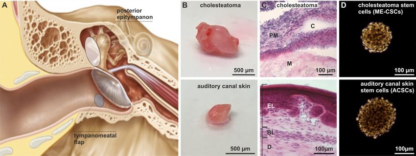
Cells 2020, 9, 199                                                                             2 of 17

cholesteatoma [4], with congenital cholesteatoma representing 2–4% of all cases and occurring only
in children between the ages of 4–6 years [5]. In contrast, acquired cholesteatoma affects both
children and adults and is closely associated with long lasting inflammation and infection of the
middle ear, e.g., chronic otitis media [2,4]. Even though there are different existing theories
regarding the formation of acquired cholesteatoma (reviewed in [3,6]), massive inflammation is one
of its major characteristics and is also discussed as a pre-requisite for cholesteatoma formation and
recurrence [7]. Hence the presence of inflammatory mediators in cholesteatoma tissue was studied
and exhibits a higher presence of the cytokines TNFα, IL-6, IL-8, IL-1α, and IL-1ß in comparison to
non-inflamed external auditory canal skin tissue [8,9]. Bone resorption and destruction was also
shown to be positively correlated to inflammation, the presence of bacterial lipopolysaccharides
(LPS), and expression of inflammatory mediators like TNFα, IL-1α, and IL-6 in cholesteatoma [10–
12]. In addition, an enhanced expression of the Toll-like receptor 4 (TLR4) was observed in
cholesteatoma tissue compared to external auditory canal skin tissue [13]. Interestingly, in a mouse
model, TLR4 was demonstrated to promote not only local inflammation but also bone destruction
[14]. Notably, the activation of the transcription factor NF-κB was shown to be elevated in
cholesteatoma tissue [15] [16]. NF-κB is not only recognized to mediate cell survival, but is also
known as a key regulator in the pathogenesis of inflammatory diseases [17]. For example, the
hypertrophy of nasal mucosa was linked to enhanced activity of NF-κB [18] in chronic rhinosinusitis
exhibiting overexpression of TLR4 [19]. However, the cellular mediators potentially linking
NF-κB-signaling and the pro-inflammatory environment of cholesteatoma still remain unknown.
      In the present study, we aimed to determine these cellular mediators of the highly
pro-inflammatory environment in cholesteatoma. We recently demonstrated the presence of stem
cells contributing to cholesteatoma pathogenesis within cholesteatoma tissue [20]. Middle ear
cholesteatoma derived stem cells (ME-CSCs) and stem cells isolated from auditory canal skin
(ACSCs), which were utilized as control cells, were successfully isolated and cultivated in vitro,
where they showed the ability for spheres formation, self-renewal, and multipotent differentiation.
Exposure of factors mimicking the microenvironment of cholesteatoma further resulted in
differentiation of ME-CSCs into keratinocyte-like cells, suggesting a potential novel disease
mechanism [20]. Extending these promising findings, our present observations demonstrate a
significantly increased expression of TRL4 accompanied by a significantly enhanced LPS-dependent
pro-inflammatory gene expression pattern in ME-CSCs compared to ACSCs. ME-CSCs treated with
LPS further showed an elevated activity of NF-κB p65 in comparison to ACSCs, promoting a
TNFα-mediated feed-forward-loop of chronic NF-κB target gene expression. Interestingly, the TNFα
response to this feed-forward-loop is specifically more sensitive in ME-CSCs. As a clinical
perspective, we functionally blocked the TLR4 using the TLR4-antagonist LPS from R. sphaeroides
(LPS-RS), resulting in a nearly complete loss of IL-1α, IL-1ß, IL-6, and TNFα expression in in
ME-CSCs. In summary, we determined that ME-CSCs regulate the inflammatory environment of
cholesteatoma via TLR4-mediated NF-κB-signaling, suggesting a distinct role of ME-CSCs as drivers
of cholesteatoma progression in an inflammation-dependent manner.

2. Materials and Methods

2.1. Ethics Statement and Human Samples
     Acquired cholesteatomas and external auditory canal skin specimens were obtained from
patients undergoing middle ear surgery at Klinikum Bielefeld Mitte (Bielefeld, Germany). Fully
informed written consent was obtained prior to surgery and all clinical investigations were ethically
approved (Reg. no. 2235) and conducted according to the principles of the Declaration of Helsinki
(1964) and local guidelines (Bezirksregierung Detmold/Münster). The removed tissue samples were
used for isolation of stem cells and paraffin sectioning.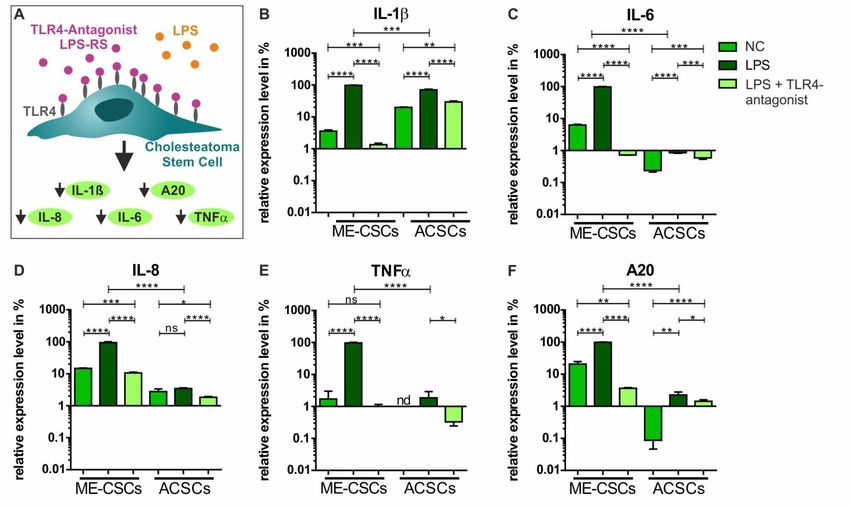
Cells 2020, 9, 199                                                                                      3 of 17

2.2. Isolation and Culture of Cholesteatoma and Auditory Canal Skin Stem Cells
     Middle ear cholesteatoma stem cells (ME-CSCs) and auditory canal skin stem cells (ACSCs)
were isolated and cultivated with addition of 10% human blood plasma or as free-floating spheres
according to our previously described protocol [20]. Briefly, tissue samples were digested with
Collagenase I (0.375 U/mL in PBS, SERVA Electrophoresis GmbH, Heidelberg, Germany) for at least
1 h and mechanically disintegrated followed by stem cell isolation at 37 °C and 5% CO2 in standard
medium comprising DMEM/F-12 (Sigma-Aldrich, Merck KGaA, Darmstadt, Germany), L-Glutamin
(200 mM, Sigma Aldrich), penicillin (10 U/mL, Sigma Aldrich), streptomycin (10 U/mL, Sigma
Aldrich), amphotericin B (25 µg/mL, Sigma Aldrich), EGF (20 ng/mL, Peprotech, Hamburg,
Germany), bFGF (40 ng/mL, Peprotech), and B27 supplement (Gibco, Thermo Fisher Scientific,
Waltham, MA, United States). This medium was either supplemented with 10% human blood
plasma for efficient expansion according to [21] or heparin (2 µg/mL, Sigma-Aldrich), to allow
sphere formation.

2.3. Haematoxylin and Eosin Staining of Cryostat Sections
    Frozen 10 µm thick paraffin sections of cholesteatoma tissue and external auditory canal skin
were subjected to H&E staining followed by microscopically examination.

2.4. Treatment of ME-CSCs and ACSCs with LPS, heat-killed bacteria, TNFα, or LPS-RS
      ME-CSCs and ACSCs were seeded in 6-well plates (CytoOne® Multiple Well Plates, STARLAB
GmbH, Hamburg, Germany, 5.3 x 10³ cells/cm2) and cultivated overnight at 37 °C and 5% CO2 in
Dulbeccos’s Modified Eagle Medium (Sigma Aldrich) containing L-Glutamin (200 mM, Sigma
Aldrich), Amphotericin B (25 µg/mL, Sigma Aldrich), fetal calf serum (FCS, 10%, Sigma Aldrich)
penicillin, and streptomycin (10 U/mL, Sigma Aldrich). After overnight culture, ME-CSCs and
ACSCs were treated with LPS (LPS, 100 ng/mL, rough strain from Salmonella enterica Re 595, Sigma
Aldrich), TNFα (10 ng/mL, PeproTech) or heat killed bacteria (HBK, 108 cells/mL of heat killed
Staphylococcus aureus, InvivoGen, Toulouse, France). Commercially purchased LPS from
Rhodobacter sphaeroides (LPS-RS, 10,000 ng/mL, InvivoGen) served as TLR4 antagonist and was
applied simultaneously to LPS from Salmonella enterica Re 595 (100 ng/mL, Sigma Aldrich). Controls
were treated within medium described above without additional stimuli. For gene expression
analysis, treatments were performed for 4 h, while a 2 h treatment was done for
immunocytochemistry in accordance to our previous studies [22], and a 5 h treatment for western
blot.

2.5. qPCR
     RNA isolation was done with the innuPREP RNA mini Kit (Analytik Jena, Jena, Germany) and
cDNA was synthesized using RevertAid First Strand cDNA Synthesis Kit (Thermo Fisher Scientific)
according to the manufacturer’s guidelines. qPCR was performed as technical triplicates using the
Luna® Universal qPCR Master Mix (BioLabs, Frankfurt am Main, Germany) according to
manufacturer’s guidelines in the MIC qPCR cycler (Bio Molecular Systems, San Francisco, USA).
GAPDH served for normalization of cycle threshold values. GraphPad Prism Software (GraphPad
Software, La Jolla, CA, USA) was used for statistical analysis. Expression levels were normalized
to 100% for each target gene and donor. Primer sequences are depicted in Table 1.

                                        Table 1. Primer sequences.

          Primer                     Sequence (5′ to 3′)                         Size of Product (bp)
            A20                TACCCTTGGTGACCCTGAAG                                      175
                               CCTTGGACGGGGATTTCTAT
         GAPDH                 CTGCACCACCAACTGCTTAG                                      108
                               GTCTTCTGGGTGGCAGTGAT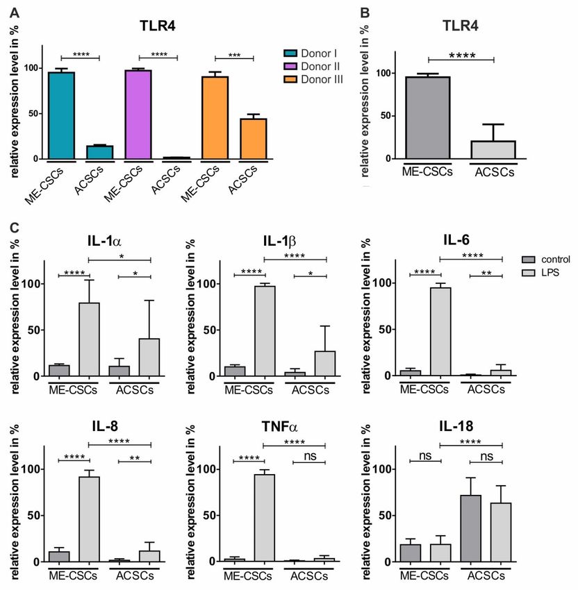
Cells 2020, 9, 199                                                                           4 of 17

           IL-18              GCAAGGATTGTCTCCCAGT                               125
                             CGATCTGGAAGGTCTGAGGT
           IL-1α             TGCCTGAGATACCCAAACC                                145
                             GCCAAGCACACCCAGTAGTC
           IL-1β           TGTACCTGTCCTGCGTGTTGAAAG                             149
                           CTGGGCAGACTCAAATTCCAGCTT
            IL-6           GCAAAGAGGCACTGGCAGAAAACA                             226
                           TTCTGCAGGAACTGGATCAGGACT
            IL-8            TCTCTTGGCAGCCTTCCTGATTTC                            227
                           AGTTTTCCTTGGGGTCCAGACAGA
           IκBα              AGACCTGGCCTTCCTCAACT                               127
                             GTCTCGGAGCTCAGGATCAC
           TLR2            AGATGCCTCCCTCTTACCCATGTT                             186
                            AAGACTTTGGCCAGTGCTTGCT
           TLR4            CACAGACTTGCGGGTTCTACATCA                             192
                           TGGACTTCTAAACCAGCCAGACCT
          TNFR1            AGGGGACAGGGAGAAGAGAGGTT                              181
                               TTCTGAAGCGGTGAAGG
          TNFR2              TCACCTCCAGCTCCACCTAT                               175
                              AGGCTCTGTGGCTTGTGG
          TNFα             AAGCCCTGGTATGAGCCCATCTAT                             137
                           AGGGCAATGATCCCAAAGTAGACC

2.6. Immunocytochemistry
     ME-CSCs and ACSCs, treated as described above, were fixed with 4% PFA (Sigma Aldrich) for
20 min followed by permeabilization and blocking in TritonX-100 (AppliChem, Darmstadt,
Germany) with 5% goat serum for 30 min. Primary antibody anti-NF-κB p65 (F-6) (sc-8008, Santa
Cruz Biotechnologies, Heidelberg, Germany) was applied for 3 h at RT or overnight at 4 °C, while
anti-phospho-c-Jun (S63) antibody (P05412, R&D Systems, Minneapolis, MN, United States) was
applied for 3 h at RT. Secondary fluorochrome-conjugated antibodies anti-rabbit Alexa 488 and
anti-mouse Alexa 555 (A32731, 21422, Molecular Probes, Thermo Fisher Scientific) were
subsequently applied for 1 h at RT. Finally, nuclear counter staining with
4′,6-Diamidin-2-phenylindol (DAPI, 1:2000, Sigma Aldrich) for 15 min at RT was followed by
mounting with Mowiol. Imaging was done using confocal laser scanning microscopy (LSM 780,
Carl Zeiss, Jena, Germany) with ZEN software. For analysis of images, Fiji [23] was applied and the
data was processed by GraphPad Prism Software (GraphPad Software).

2.7. Western Blot
     ME-CSCs and ACSCs were stimulated as described above followed by harvesting via
trypsination and subsequent lysis with l ysis buffer (0.01 M Tris, 3 mM EDTA, 1% SDS) for 10 min at
RT and denaturation for 10 min at 95 °C. The Protein quantification was performed using of
Roti-Quant (Carl Roth, Karlsruhe, Germany) following manufacturer’s guidelines. Samples were
mixed with 4× loading dye, incubated for 5 min at 95 °C and subjected to electrophoresis on 10%
denaturing SDS polyacrylamide gels and subsequently transferred to a nitrocellulose membrane
using the Biometra Fastblot B34 blotter (Analytik Jena AG, Jena, Germany). Blocking was
subsequently performed with 10% milk powder and 0.1% Tween-20 in PBS for 30 min followed by
application of the first antibody against A20 (A-12, sc-166692, Santa Cruz Biotechnologies) and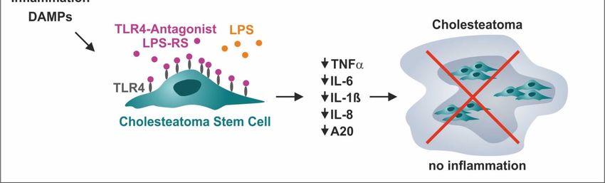
Cells 2020, 9, 199                                                                                        5 of 17

overnight incubation at 4 °C. Afterwards, HRP-linked secondary antibody (515-035-003, Dianova,
Hamburg, Germany) was applied for 1.5 h at RT. Visualization was performed via enhanced
chemiluminescence using Fusion Solo (PEQLAB, Erlangen, Germany), while GAPDH (sc-32233,
Santa Cruz Biotechnologies) served as loading control.

3. Results

3.1. Successful Isolation of Sphere-Forming Stem Cells from Middle Ear Cholesteatoma Tissue and Auditory
Canal Skin
      For isolation of stem cells, cholesteatoma tissue was obtained from the human posterior
epitympanon, while auditory canal skin originated from the tympanomeatal flap (Figure 1A).
Histological examination of the isolated cholesteatoma tissue revealed the presence of characteristic
cholesteatoma structures like the perimatrix, the matrix, and enclosed cystic content (Figure 1B–C).
On the contrary, a characteristic epithelial and a basal layer, as well as the dermis, were observable in
sectioned auditory canal skin (Figure 1B–C). In accordance to our previous findings [20], we
successfully isolated stem cells from the matrix and perimatrix of middle ear cholesteatoma (middle
ear cholesteatoma stem cells, ME-CSCs) and from the dermis of auditory canal skin (auditory canal
skin stem cells, ACSCs). Cultivated ME-CSCs and ACSCs revealed the ability of sphere formation
under serum-free culture conditions (Figure 1D).

      Figure 1. Isolation of middle ear cholesteatoma and auditory canal skin stem cells. (A) Schematic
      view on the localization of cholesteatoma obtained from the posterior epitympanon and the auditory
      canal skin removed from the tympanomeatal flap. Modified from [20] (Creative Commons
      Attribution 4.0 International License). (B) Obtained cholesteatoma and auditory canal skin tissue. (C)
      H&E staining of cryostat sections of cholesteatoma tissue revealed characteristic structures as the
      matrix (M), perimatrix (PM) and cystic content, while auditory canal skin revealed an epithelial layer
      (EL), a basal layer (BL), and a dermis (D). (D) Successfully isolated stem cells from cholesteatoma
      tissue and auditory canal skin were able to form spheres.

3.2. Stem Cells Derived from Middle Ear Cholesteatoma Show Significantly Elevated Expression of TRL4
Compared to Auditory Canal Skin Stem Cells
     To determine a potential contribution of ME-CSCs to the pro-inflammatory environment of
cholesteatoma, we investigated expression levels of TLR4 in ME-CSCs and ACSCs. Notably, a
significantly elevated expression of TLR4 was observable in ME-CSCs from three independent
donors in comparison to ACSCs derived from the corresponding donors (Figure 2A,B). On the
contrary, transcript levels of TLR2 were not significantly elevated in ME-CSCs compared to ACSCs
(Figure S1).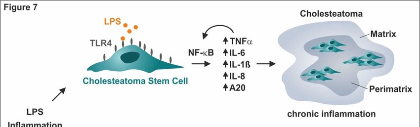
Cells 2020, 9, 199                                                                                      6 of 17

      Figure 2. Compared to ACSCs, ME-CSCs show significantly increased expression of TRL4
      accompanied by significantly enhanced LPS-dependent pro-inflammatory gene expression patterns.
      (A) qPCR analysis revealed highly increased expression levels of TLR4 in ME-CSCs compared to
      ACSCs in three different donors (**** ≤0.0001, *** ≤0.001, unpaired t-test, one-tailed, confidence
      interval: 95%) (B) Mean of the relative expression levels of TLR4 from ME-CSCs and ACSCs
      validated the increased expression levels of TLR4 in ME-CSCs compared to ACSCs (n = 3, ****
      ≤0.0001, Mann Whitney test, one-tailed, confidence interval: 95%). (C) Expression levels of the
      pro-inflammatory mediators IL-1α, IL-1ß, IL-6, IL-8, and TNFα were strongly increased in ME-CSCs
      treated with LPS from S. enterica compared to untreated control and LPS-treated ACSC. Expression
      level of IL-18 was not affected by LPS-treatment (mean of the relative expression levels from
      ME-CSCs and ACSCs (n = 3, **** ≤0.0001, ** ≤0.01, * ≤0.05, ns > 0.05, Mann Whitney test, one-tailed,
      confidence interval: 95%).

3.3. Treatment of ME-CSCs with LPS Results in Significantly Enhanced Expression Levels of
Pro-Inflammatory Mediators Compared to ACSCs
    Since bacterial infections are strongly associated to cholesteatoma growth and progression [24],
we found TLR4 to be highly upregulated in ME-CSCs. ME-CSCs and ACSCs were exposed to the
TLR4 agonist LPS from S. enterica in order to model the cholesteatoma microenvironment. Treatment
of ME-CSCs from three independent donors with LPS resulted in a strong and highly significant
Cells 2020, 9, 199 7 of 17
upregulation of pro-inflammatory mediators IL- 1α, IL-1ß, IL-6, and IL-8 in comparison to untreated
control (Figure 2C and Figure S2). LPS-treated ACSCs likewise revealed a slight increase in the
expression of IL- 1α, IL-1ß, IL-6, and IL-8 compared to untreated control. However, the expression
levels of these pro-inflammatory genes were significantly elevated in LPS-treated ME-CSCs in
comparison to LPS-treated ACSCs for all three independent donors (Figure S2). Notably, we also
observed a strong upregulation of TNFα expression in ME-CSCs induced by LPS-treatment in
comparison to untreated controls and particularly to LPS-treated ACSCs, which showed no
significant increase in the expression of TNFα after exposure to LPS (Figure 2C and Figure S2).
Interestingly, expression levels of IL-18 were not affected by LPS-treatment in ME-CSCs or ACSCs,
although ACSCs showed elevated basal expression levels in comparison to ME-CSCs (Figure 2C). To
exclude a potential impact of TLR2-dependent signaling in ME-CSCs, we exposed ME-CSCs and
ACSCs to heat killed bacteria (S. aureus, HKB). In addition to the unchanged expression levels of
TRL2 between ME-CSCs and ACSCs, neither of the stem cell populations showed an upregulation of
IL1-ß or IL-8 through stimulation with HBK (Figure S1). Hence, ME-CSCs are highly subjected to
TLR4-dependent pro-inflammatory signaling, which is much more pronounced in ME-CSCs in
relation to ACSCs.
3.4. LPS-Dependent Pro-Inflammatory Gene Expression in ME-CSCs is Mediated by an Enhanced Activity of
NF-κB
To determine potential downstream mediators of TLR4-dependent pro-inflammatory signaling
in ME-CSCs, we investigated the effects of LPS on the activity of NF-κB p65 in ME-CSCs and ACSCs
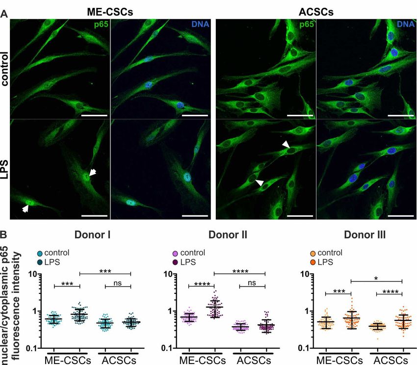
by immunocytochemistry. Upon treatment with LPS, ME-CSCs exhibited a distinct nuclear
translocation of NF-κB p65 protein compared to control (Figure 3A, left panels, arrows). As a
profound difference to ME-CSCs, only a minor nuclear translocation of NF-κB p65 was observable in
ACSCs even after exposure to LPS (Figure 3A, right panels, arrowheads). An excessive
quantification of the relative NF-κB p65 nuclear to cytoplasmic fluorescence intensities validated
these observations in three independent donors. It revealed a significantly increased nuclear
localization of NF-κB p65 in ME-CSCs when compared to control and LPS-treated ACSCs (Figure
3B). Notably, a slight basal translocation of NF-κB p65 could be detected in ME-CSCs without
LPS-dependent stimulation (Figure 3A,B).Cells 2020, 9, 199 8 of 17
Figure 3. LPS-dependent pro-inflammatory gene expression in ME-CSCs is mediated by
NF-κB. (A) Exemplary immunocytochemical staining reveals strong nuclear translocation of NF-κB
p65 protein in LPS-treated ME-CSCs (arrows) compared to control ME-CSCs, which was only
slightly detectable in both control and LPS-treated ACSCs (arrowheads). ME-CSCs and ACSCs were
derived from Donor II, LPS from S. enterica, scale bars: 50 µm. (B) Quantification of NF-κB p65
nuclear to cytoplasmic fluorescence intensities validated a significantly increased nuclear
localization of NF-κB p65 in ME-CSCs compared to control and to LPS-treated ACSCs (n = 3, ****
≤0.0001, *** ≤0.001, * ≤0.05, ns > 0.05, Mann Whitney test, one-tailed, confidence interval: 95%).
In addition, two out of three ACSC-populations derived from independent donors showed no
significant changes in the amount of nuclear NF-κB p65 protein even after LPS-treatment (Figure
3B). Interestingly, we also observed a significant translocation of AP1 protein upon LPS-treatment in
ME-CSCs compared to control and LPS-stimulated ACSCs, which displayed no increase in nuclear
AP1 protein by LPS (Figure S3).
In accordance to the elevated nuclear translocation of NF-κB p65, expression levels of the
specific NF-κB target genes A20 and IκBα were significantly increased in ME-CSCs upon
LPS-stimulation compared to control and LPS-treated ACSCs (Figure 4A,B,S4). Western Blot
analysis further confirmed the elevated expression of A20 in LPS-treated ME-CSCs on protein level.
In accordance to the transcriptional data, ME-CSCs exposed to LPS revealed a highly increased A20
protein amount compared to control and LPS-treated ACSCs. However, LPS-treated ACSCs showed
no increase in A20 protein in comparison to control ACSCs (Figure 4C). These findings demonstrate
NF-κB as the mediator of LPS-dependent pro-inflammatory gene expression exclusively in
ME-CSCs and not in ACSCs.Cells 2020, 9, 199 9 of 17
Figure 4. LPS-treatment of ME-CSCs results in significantly elevated levels of NF-κB target genes
compared to ACSCs. (A–B) qPCR analysis showed significantly increased transcript levels of the
NF-κB related targets A20 and IκBα in ME-CSCs after LPS-treatment (S. enterica) compared to
control and LPS-stimulated ACSCs (mean of the relative expression levels from ME-CSCs and
ACSCs, n = 3, **** ≤0.0001, * ≤0.05, ns > 0.05, Mann Whitney test, one-tailed, confidence interval:
95%). (C) Western blot analysis demonstrated highly increased amounts of A20 protein in
LPS-treated ME-CSCs in comparison to ACSCs exposed to LPS. ME-CSCs and ACSCs were derived
from donor IV.
3.5. ME-CSCs show a TNFα-Mediated Feed-Forward-Loop of Pro-Inflammatory NF-κB Target Gene
Expression
With TNFα being a major target gene of NF-κB and also an important cytokine involved in
cholesteatoma progression [8], we investigated a potential pro-inflammatory feed-forward-loop of
NF-κB target gene expression in ME-CSCs mediated by TNFα. We found that the expression of the
TNF receptors TNFR1 and TNFR2 did not differ significantly between ME-CSCs and ACSCs (Figure
5A). But besides that, stimulation of ME-CSCs with TNFα resulted in significantly increased
expression levels of IL-6, IL-8, and A20, and TNFα itself in comparison to control and TNFα-treated
ACSCs (Figure 5B and Figure S5). Interestingly, we observed no significant difference in the
expression levels of IL-1ß and IκBα between ME-CSCs and ACSCs after TNFα-dependent
stimulation, although the expression levels were significantly elevated compared to controls (Figure
5B). In comparison to untreated control, exposure to TNFα further resulted in a highly pronounced
and significant increase of TNFα expression almost exclusively in ME-CSCs and not in ACSCs
(Figure 5B and Figure S5). The bottom line is that TNFα-induced expression of TNFα was
significantly increased in ME-CSCs compared to ACSCs (Figure 5B and Figure S5). These
observations indicate the presence of an exaggerated pro-inflammatory TNFα-mediated
feed-forward-loop of NF-κB target gene expression in ME-CSCs.Cells 2020, 9, 199 10 of 17
Figure 5. ME-CSCs reveal a TNFα-mediated feed-forward-loop of pro-inflammatory NF-κB target
gene expression. (A) qPCR analysis revealed no significant difference in expression levels of TNFR1
and TNFR2 (mean of the relative expression levels from ME-CSCs and ACSCs, n = 3, ns > 0.05;
Mann Whitney test, one-tailed, confidence interval: 95%). (B) qPCR analysis showed a significant
increase in the expression levels of IL-6, IL-8, TNFα, and A20 in ME-CSCs stimulated with TNFα
compared to control and TNFα-stimulated ACSC (mean of the relative expression levels from
ME-CSCs and ACSCs, n = 3, **** ≤0.0001, *** ≤0.001, ** ≤0.01, * ≤0.05, ns > 0.05; Mann Whitney test,
one-tailed, confidence interval: 95%, nd: not detectable).Cells 2020, 9, 199 11 of 17
3.6. Functional Inactivation of TLR4 via a TLR4-Antagonist Blocks Pro-Inflammatory Signaling in ME-CSCs
To functionally block TLR4-dependent pro-inflammatory signaling in ME-CSCs, we applied the
TLR4 antagonist LPS from Rhodobacter sphaeroides (LPS-RS), which is structurally similar to the
drug Eritoran [25] (Figure 6A). In accordance to our observation in ME-CSCs from donors I-III, we
observed a strongly and significantly increased expression of IL-ß, IL-6, IL-8, TNFα, and
A20 in ME-CSCs derived from donor IV after treatment with LPS from S. enterica compared to
control and LPS-treated ACSCs (Figure 6B–F). Application of the TLR4 antagonist LPS-RS in
ME-CSCs simultaneously treated with LPS from S. enterica resulted in a strong reduction of TNFα
expression compared to LPS-treated ME-CSCs (Figure 6E). Although detected expression levels
were overall lower compared to ME-CSCs, ACSCs treated with LPS and LPS-RS likewise
showed a significantly impaired expression of IL-1ß, IL-6, IL-8, TNFα, and A20 compared to
exposure to LPS. Notably, we observed an significant downregulation of expression of IL-1ß, IL-6,
IL-8, and A20 in ME-CSCs exposed to LPS and the TRL4 antagonist LPS-RS compared to LPS-treated
and especially untreated ME-CSCs (Figure 6B,C,E).
Figure 6. Functional inactivation of TLR4 via the TLR4-antagonist LPS-RS blocks pro-inflammatory
signaling in ME-CSCs. (A) Schematic view on the mode of action of the TLR4 antagonist LPS-RS in
ME-CSCs. (B–F) ME-CSCs simultaneously treated with the TLR4 antagonist LPS from R. sphaeroides
(LPS-RS) and LPS from S. enterica (LPS) showed a significant reduction of IL-ß, IL-6 IL-8, TNFα, and
A20 expression compared to LPS-treated ME-CSCs. Although detected expression levels were overall
lower compared to ME-CSCs, ACSCs treated with LPS and LPS-RS likewise showed a significantly
impaired expression of IL-ß, IL-6, IL-8, TNFα, and A20 compared to stimulation with LPS (ME-CSCs
and ACSCs were derived from donor IV, **** ≤0.0001, *** ≤0.001, ** ≤0.01, * ≤0.05, ns > 0.05, unpaired
t-test, one-tailed, confidence interval: 95%, nd: not detectable).
4. Discussion
In the present study, we demonstrate that stem cells residing in cholesteatoma, a severe
expanding lesion in the middle ear, mediating its inflammatory environment in a
TLR4-NF-κB-dependent manner.
We took advantage of previously described cholesteatoma stem cells as a cellular in vitro model
of cholesteatoma inflammation [20]. In particular, we previously showed that Integrin-β1 ME-CSCs
are present in the matrix and perimatrix of middle ear cholesteatoma. These ME-CSCs were able toCells 2020, 9, 199 12 of 17
differentiate into keratinocyte-like cells after exposure to growth factors like hepatocyte growth
factor and keratinocyte growth factor present in cholesteatoma tissue [20]. In line with their
expression of markers characteristic for neural-crest-derived stem cells (NCSCs) [26] and the
commonly known contribution of NCSCs to middle ear development [20], ME-CSCs are proposed to
be epidermal stem cells of neural crest origin. Accordingly, ME-CSCs were able to grow as spheres
under serum-free conditions in previous studies [20] and the present study, thus showing a
characteristic of epidermal stem cells (reviewed in [27]). With regard to the identification of
epidermal stem cells in the tympanic membrane [28], ME-CSCs showed markers of epidermal stem
cells. Hence, they are particularly suggested to migrate from this stem cell niche to the lesion and
contribute to cholesteatoma through differentiation into proliferating keratinocyte-like cells [20].
Here, we extend these promising findings by demonstrating a role of ME-CSCs in cholesteatoma
formation as mediators of its inflammatory environment. In particular, ME-CSCs exhibited a
pronounced inflammatory footprint characterized by a significantly increased expression of the
TLR4, a receptor recognizing bacterial LPS in the first line defense against bacterial infections. An
enhanced expression of TLR4 was already shown in 2013 by Hirai and colleagues in middle ear
tissues obtained from five patients with acquired middle ear cholesteatoma [13]. In 2015, Si and
colleagues extended these findings by demonstrating TLR4 as a major driver of cholesteatoma
pathogenesis in terms of promoting both inflammation and bone destruction. Expression of TLR4
was particularly shown to be correlated with disease severity in terms of increased invasion, bone
destruction, and hearing loss [14]. Although an up-regulation of TLR2 was observable in
cholesteatoma tissue [13], deficiency in TLR2 in mice did not affect disease severity or inflammatory
responses [14]. Interestingly, we observed an upregulation of TLR4 gene expression in ME-CSCs
compared to ACSCs in the present study, whereas expression of TLR2 and TLR2-dependent
inflammatory signaling remained unaffected in ME-CSCs.
Aggressiveness of cholesteatoma is strongly associated with the presence of bacteria [24] and
bacterial LPS [10] as the common agonist of TLR4. LPS was also demonstrated to directly trigger
pathogenic characteristic of cholesteatoma [29,30]. Hence, we utilized LPS to investigate
pro-inflammatory signaling in ME-CSCs. LPS is well-described to initiate pro-inflammatory
signaling by binding to a heterodimer consisting of TLR-4 and myeloid differentiation factor 2
(MD-2), in turn leading to recruitment of MyoD88 and phosphorylation of the IκB kinases (IKKs).
Phosphorylated IKKs in turn phosphorylate IκBα, resulting in its polyubiquitylation and
26S-proteasome-mediated degradation. Loss of IκBα leads to unmasking of the nuclear translocation
signal region of the NF-κB p65/p50 heterodimer, resulting in its nuclear translocation and activation
of distinct target genes via binding to κB-sites [31,32]. NF-κB target genes particularly include
pro-inflammatory cytokines like TNFα, IL-1α, IL-1ß, IL-6, and IL-8 [33–36] and were shown to be
upregulated in cholesteatoma tissue [8,9,12]. Consequently, we demonstrate that exposure of
ME-CSCs to LPS resulted in significantly increased nuclear translocation of NF-κB p65 resulting in a
strongly enhanced pro-inflammatory gene expression pattern of TNFα, IL- 1α, IL-1ß, IL-6, and IL-8
compared to ACSCs. When compared to ACSCs, ME-CSCs further demonstrated a strongly elevated
expression of the NF-κB target genes IκBα and A20 if exposed to LPS. In this line, A20 was already
described to be higher expressed in cholesteatoma tissue using whole human genome microarrays
[37], which we could verify in case of the A20 expression in ME-CSCs on protein level. In addition,
we observed an increase in nuclear translocation of the transcription factor AP1, suggesting a
pro-inflammatory signaling mediated by both NF-κB and AP1 in ME-CSCs. Being a dimer of c-Jun
and c-Fos, AP-1, as well as NF-κB, are both well-described to drive transcription of cytokines like
TNFα and IL-1β in inflammatory diseases [38].
As a major target gene of NF-κB, TNFα itself is known to drive a pro-inflammatory
feed-forward-loop described in [39] by strongly activating the NF-κB transcriptional pathway [40].
Our present findings showed that stimulation of ME-CSCs with TNFα induces a distinctively
elevated expression levels of IL-6, IL-8, A20, and TNFα in comparison to TNFα-treated ACSCs. This
observation was very similar to the effect observed after stimulation with LPS. But in contrast to the
TLR4, we could not observe a higher expression of the corresponding TNF receptors in ME-CSC.Cells 2020, 9, 199 13 of 17
Hence, we suggest that the overlapping parts between the TNFα and LPS pathway [40] might be more
sensitive in ME-CSC. Anyhow, the resulting TNFα-driven pro-inflammatory feed-forward-loop of
NF-κB activation in ME-CSCs is in line with the already described involvement of TNFα in
cholesteatoma progression [8] and other pathogenesis via inflammatory cues [41]. Additionally, the
exaggerated inflammation might give rise to an enhanced epidermal differentiation observed in
other inflammatory diseases like eczema [42], psoriasis [43], or nasal polyps [18]. Consequently, this
might trigger the epidermal stem cells to differentiate into keratinized squamous epithelium
promoting cholesteatoma formation.
As a clinical perspective, we applied the TLR4 antagonist LPS of Rhodobacter sphaeroides
(LPS-RS), which is structurally similar to the drug Eritoran, a synthetic molecule derived from the
lipid A structure of LPS-RS [25]. In comparison to LPS-RS, the lipid A of Eritoran shows one
hydrocarbon chain combining structural components of two out of five LPS-RS chains, while two
further chains are nearly completely similar and one chain reveals complete structural similarity
[44]. Eritoran was shown to bind competitively to the hydrophobic pocket of MD-2, thus preventing
dimerization of TLR4-MD2 complexes (reviewed in [44]) and in turn intracellular downstream
activation of NF-κB and activation of pro-inflammatory gene expression [25,45,46]. Even though
there are other inhibitors of TLR4 signaling like Sulforaphane [47] we decided to focus on Eritoran
in this study. Firstly, because it lays most upstream of the initial problem, the contamination of
cholesteatoma tissue with LPS [11]. Secondly, because Eritoran was already successfully applied for
treating chronic airway response to inhaled lipopolysaccharide in mice [48] and acute severe liver
injury in rats [49]. Most importantly, Eritoran was applied in different clinical trials against sepsis
e.g., phase II [50] and even phase III [51], reviewed in [41], therefore making it a promising molecule
for new clinical application sometime soon. In the present study, we observed a pronounced
reduction of the expression of IL-8 and A20 and a near-complete loss of expression of IL-1ß, IL-6, and
TNFα in ME-CSCs exposed to both LPS and LPS-RS compared to LPS-treated and untreated
ME-CSCs. These findings suggest a local treatment strategy for cholesteatoma by specifically
targeting TLR4-mediated pro-inflammatory down-stream signaling in cholesteatoma stem cells as
mediators of inflammation and cholesteatoma progression (Figure 7). The local drug administration
after Cholesteatom surgery to the middle ear cavity might reduce recurrence of cholesteatoma. This
could be realized by applying carrier substances e.g., chitosan, a biodegradable polymer, which is
even applicable on the delicate inner ear [52].
Importantly, TLR4-deficiency in mice was already shown to be protective against
cholesteatoma-driven hearing loss and bone destruction by reduction of local expression of
pro-inflammatory cytokines and osteoclast formation [14]. In summary, we demonstrated that stem
cells residing in cholesteatoma are the cellular mediators of its highly pro-inflammatory
environment, suggesting a distinct role of ME-CSCs as drivers in cholesteatoma progression. On a
molecular level, LPS-dependent pro-inflammatory gene expression patterns observed in ME-CSCs
were driven by a TLR4-NF-κB signaling cascade, resulting in a TNFα-mediated feed-forward-loop
of pro-inflammatory NF-κB target gene expression (Figure 7). Functional inactivation of TLR4 via
the TLR4-antagonist LPS-RS blocked pro-inflammatory signaling in ME-CSCs (Figure 7), thus
providing a direct clinical perspective for pharmaceutical cholesteatoma treatment that is not present
today.Cells 2020, 9, 199 14 of 17
Figure 7. Schematic summary of cholesteatoma stem cells as cellular mediators of the highly
pro-inflammatory environment in cholesteatoma and drivers of cholesteatoma progression.
LPS-dependent pro-inflammatory gene expression in cholesteatoma stem cells was driven by a
TLR4-NF-κB signaling cascade resulting in a TNFα-mediated autocrine feed-forward-loop of
pro-inflammatory gene expression. As a clinical perspective, functional inactivation of TLR4 via the
TLR4-antagonist LPS-RS blocked pro-inflammatory signaling, suggesting its therapeutic
applicability for treating cholesteatoma.
Supplementary Materials: The following are available online at www.mdpi.com/2073-4409/9/1/199/s1,
Figure S1: Inflammatory signaling in ME-CSCs is not mediated via TLR2; Figure S2: Expression levels of
pro-inflammatory genes in ME-CSCs and ACSCs upon LPS-stimulation on single-donor level; Figure S3:
Significant translocation of AP1 protein in ME-CSCs upon LPS-treatment. Figure S4: Expression levels of NF-κB
target genes A20 and IκBα in ME-CSCs and ACSCs upon LPS-stimulation on single-donor level; Figure S5:
ME-CSCs show a TNFα-mediated feed-forward-loop of pro-inflammatory NF-κB target gene expression on
single-donor level.
Author Contributions: Conceptualization: B.K., H.S.; Validation: B.K., H.S., C.K., M.S., J.F.W.G.; Investigation:
M.S., V.V.-T.; Resources: B.K., C.K., H.S.; Data Curation: B.K., C.K., H.S.; Writing—Original Draft Preparation:
J.F.W.G., V.V.-T., M.S.; Writing—Review and Editing: M.S., V.V.-T., J.F.W.G., F.O., C.K., H.S., B.K.;
Visualization: J.G., M.S., V.V.-T.; Supervision: B.K., C.K., H.S.; Project Administration: B.K., C.K., H.S.; Funding
Acquisition: B.K., C.K., H.S. All authors have read and agreed to the published version of the manuscript.
Funding: This research received no external funding.
Acknowledgments: This study was funded by the University of Bielefeld and the Department of
Otolaryngology, Head and Neck Surgery at Klinikum Bielefeld.
Conflicts of Interest: The authors declare no conflict of interest.
References
1. Bhutta, M.F.; Williamson, I.G.; Sudhoff, H.H. Cholesteatoma. BMJ 2011, 342, d1088, doi:10.1136/bmj.d1088.
2. Aberg, B.; Westin, T.; Tjellstrom, A.; Edstrom, S. Clinical characteristics of cholesteatoma. Am. J.
Otolaryngol. 1991, 12, 254–258, doi:10.1016/0196-0709(91)90002-w.
3. Kuo, C.L. Etiopathogenesis of acquired cholesteatoma: prominent theories and recent advances in
biomolecular research. Laryngoscope 2015, 125, 234–240, doi:10.1002/lary.24890.Cells 2020, 9, 199 15 of 17
4. Olszewska, E.; Wagner, M.; Bernal-Sprekelsen, M.; Ebmeyer, J.; Dazert, S.; Hildmann, H.; Sudhoff, H.
Etiopathogenesis of cholesteatoma. Eur. Arch. Otorhinolaryngol. 2004, 261, 6–24,
doi:10.1007/s00405-003-0623-x.
5. Richter, G.T.; Lee, K.H. Contemporary assessment and management of congenital cholesteatoma. Curr.
Opin. Otolaryngol. Head Neck Surg. 2009, 17, 339–345, doi:10.1097/MOO.0b013e3283303688.
6. Sudhoff, H.; Tos, M. Pathogenesis of attic cholesteatoma: clinical and immunohistochemical support for
combination of retraction theory and proliferation theory. Am. J. Otol. 2000, 21, 786–792.
7. Louw, L. Acquired cholesteatoma pathogenesis: stepwise explanations. J. Laryngol. Otol. 2010, 124, 587–
593, doi:10.1017/S0022215109992763.
8. Marenda, S.A.; Aufdemorte, T.B. Localization of cytokines in cholesteatoma tissue. Otolaryngol. Head Neck
Surg. 1995, 112, 359–368, doi:10.1016/s0194-5998(95)70268-7.
9. Kim, C.S.; Lee, C.H.; Chung, J.W.; Kim, C.D. Interleukin-1α, Interleukin-1β and Interleukin-8 Gene
Expression in Human Aural Cholesteatomas. Acta Oto-Laryngologica 1996, 116, 302–306,
doi:10.3109/00016489609137846.
10. Peek, F.A.; Huisman, M.A.; Berckmans, R.J.; Sturk, A.; Van Loon, J.; Grote, J.J. Lipopolysaccharide
concentration and bone resorption in cholesteatoma. Otol. Neurotol. 2003, 24, 709–713,
doi:10.1097/00129492-200309000-00002.
11. Sudhoff, H.; Liebehenz, Y.; Aschenbrenner, J.; Jung, J.; Hildmann, H.; Dazert, S. A Murine Model of
Cholesteatoma-Induced Bone Resorption Using Autologous Dermal Implantation. The Laryngoscope 2003,
113, 1022–1026, doi:10.1097/00005537-200306000-00019.
12. Kuczkowski, J.; Sakowicz-Burkiewicz, M.; Izycka-Swieszewska, E.; Mikaszewski, B.; Pawelczyk, T.
Expression of tumor necrosis factor-alpha, interleukin-1alpha, interleukin-6 and interleukin-10 in chronic
otitis media with bone osteolysis. ORL J. Otorhinolaryngol. Relat. Spec. 2011, 73, 93–99,
doi:10.1159/000323831.
13. Hirai, H.; Kariya, S.; Okano, M.; Fukushima, K.; Kataoka, Y.; Maeda, Y.; Nishizaki, K. Expression of
toll-like receptors in chronic otitis media and cholesteatoma. Int. J. Pediatric Otorhinolaryngol. 2013, 77, 674–
676, doi:10.1016/j.ijporl.2013.01.010.
14. Si, Y.; Chen, Y.B.; Chen, S.J.; Zheng, Y.Q.; Liu, X.; Liu, Y.; Jiang, H.L.; Xu, G.; Li, Z.H.; Huang, Q.H., et al.
TLR4 drives the pathogenesis of acquired cholesteatoma by promoting local inflammation and bone
destruction. Sci. Rep. 2015, 5, 16683, doi:10.1038/srep16683.
15. Liu, W.; Yin, T.; Ren, J.; Li, L.; Xiao, Z.; Chen, X.; Xie, D. Activation of the EGFR/Akt/NF-κB/cyclinD1
survival signaling pathway in human cholesteatoma epithelium. Eur. Arch. Oto-Rhino-Laryngology 2014,
271, 265–273, doi:10.1007/s00405-013-2403-6.
16. Byun, J.Y.; Yune, T.Y.; Lee, J.Y.; Yeo, S.G.; Park, M.S. Expression of CYLD and NF-kappaB in human
cholesteatoma epithelium. Mediat. Inflamm. 2010, 2010, 796315, doi:10.1155/2010/796315.
17. Taniguchi, K.; Karin, M. NF-kappaB, inflammation, immunity and cancer: coming of age. Nat. Rev.
Immunol. 2018, 10.1038/nri.2017.142, doi:10.1038/nri.2017.142.
18. Fraczek, M.; Rostkowska-Nadolska, B.; Kapral, M.; Szota, J.; Krecicki, T.; Mazurek, U. Microarray analysis
of NF-kappaB-dependent genes in chronic rhinosinusitis with nasal polyps. Adv. Clin. Exp. Med. 2013, 22,
209–217.
19. Zhang, Q.; Wang, C.S.; Han, D.M.; Sy, C.; Huang, Q.; Sun, Y.; Fan, E.Z.; Li, Y.; Zhou, B. Differential
expression of Toll-like receptor pathway genes in chronic rhinosinusitis with or without nasal polyps. Acta
Otolaryngol. 2013, 133, 165–173, doi:10.3109/00016489.2012.717713.
20. Nagel, J.; Wollner, S.; Schurmann, M.; Brotzmann, V.; Muller, J.; Greiner, J.F.; Goon, P.; Kaltschmidt, B.;
Kaltschmidt, C.; Sudhoff, H. Stem cells in middle ear cholesteatoma contribute to its pathogenesis. Sci. Rep.
2018, 8, 6204, doi:10.1038/s41598-018-24616-4.
21. Greiner, J.F.; Hauser, S.; Widera, D.; Muller, J.; Qunneis, F.; Zander, C.; Martin, I.; Mallah, J.; Schuetzmann,
D.; Prante, C., et al. Efficient animal-serum free 3D cultivation method for adult human neural
crest-derived stem cell therapeutics. Eur. Cell Mater. 2011, 22, 403–419.
22. Müller, J.; Greiner, J.F.; Zeuner, M.; Brotzmann, V.; Schafermann, J.; Wieters, F.; Widera, D.; Sudhoff, H.;
Kaltschmidt, B.; Kaltschmidt, C. 1,8-Cineole potentiates IRF3-mediated antiviral response in human stem
cells and in an ex vivo model of rhinosinusitis. Clin. Sci. (Lond) 2016, 130, 1339–1352,
doi:10.1042/CS20160218.
23. Schindelin, J.; Arganda-Carreras, I.; Frise, E.; Kaynig, V.; Longair, M.; Pietzsch, T.; Preibisch, S.; Rueden,
C.; Saalfeld, S.; Schmid, B., et al. Fiji: an open-source platform for biological-image analysis. Nat. Methods
2012, 9, 676–682, doi:10.1038/nmeth.2019.
24. Ricciardiello, F.; Cavaliere, M.; Mesolella, M.; Iengo, M. Notes on the microbiology of cholesteatoma:
clinical findings and treatment. Acta Otorhinolaryngol. Ital. 2009, 29, 197–202.Cells 2020, 9, 199 16 of 17
25. Mullarkey, M.; Rose, J.R.; Bristol, J.; Kawata, T.; Kimura, A.; Kobayashi, S.; Przetak, M.; Chow, J.;
Gusovsky, F.; Christ, W.J., et al. Inhibition of endotoxin response by e5564, a novel Toll-like receptor
4-directed endotoxin antagonist. J. Pharmacol. Exp. Ther. 2003, 304, 1093–1102, doi:10.1124/jpet.102.044487.
26. Hauser, S.; Widera, D.; Qunneis, F.; Muller, J.; Zander, C.; Greiner, J.; Strauss, C.; Luningschror, P.;
Heimann, P.; Schwarze, H., et al. Isolation of novel multipotent neural crest-derived stem cells from adult
human inferior turbinate. Stem Cells Dev. 2012, 21, 742–756.
27. Pastrana, E.; Silva-Vargas, V.; Doetsch, F. Eyes wide open: a critical review of sphere-formation as an assay
for stem cells. Cell Stem Cell. 2011, 8, 486–498, doi:10.1016/j.stem.2011.04.007.
28. Kim, S.W.; Kim, J.; Seonwoo, H.; Jang, K.J.; Kim, Y.J.; Lim, H.J.; Lim, K.T.; Tian, C.; Chung, J.H.; Choung,
Y.H. Latent progenitor cells as potential regulators for tympanic membrane regeneration. Sci. Rep. 2015, 5,
11542, doi:10.1038/srep11542.
29. Iino, Y.; Toriyama, M.; Ogawa, H.; Kawakami, M. Cholesteatoma debris as an activator of human
monocytes. Potentiation of the production of tumor necrosis factor. Acta Otolaryngol. 1990, 110, 410–415,
doi:10.3109/00016489009107462.
30. Kobayashi, H.; Asano, K.; Kanai, K.; Suzaki, H. Suppressive activity of vitamin D3 on matrix
metalloproteinase production from cholesteatoma keratinocytes in vitro. Mediat. Inflamm. 2005, 2005, 210–
215, doi:10.1155/MI.2005.210.
31. Gilmore, T.D. Introduction to NF-kappaB: players, pathways, perspectives. Oncogene 2006, 25, 6680–6684,
doi:10.1038/sj.onc.1209954.
32. Wan, F.; Lenardo, M.J. The nuclear signaling of NF-kappaB: current knowledge, new insights, and future
perspectives. Cell Res. 2010, 20, 24–33, doi:10.1038/cr.2009.137.
33. Mori, N.; Prager, D. Transactivation of the interleukin-1alpha promoter by human T-cell leukemia virus
type I and type II Tax proteins. Blood 1996, 87, 3410–3417.
34. Hiscott, J.; Marois, J.; Garoufalis, J.; D’Addario, M.; Roulston, A.; Kwan, I.; Pepin, N.; Lacoste, J.; Nguyen,
H.; Bensi, G., et al. Characterization of a functional NF-kappa B site in the human interleukin 1 beta
promoter: evidence for a positive autoregulatory loop. Mol. Cell. Biol. 1993, 13, 6231–6240,
doi:10.1128/mcb.13.10.6231.
35. Greiner, J.F.; Muller, J.; Zeuner, M.T.; Hauser, S.; Seidel, T.; Klenke, C.; Grunwald, L.M.; Schomann, T.;
Widera, D.; Sudhoff, H., et al. 1,8-Cineol inhibits nuclear translocation of NF-kappaB p65 and
NF-kappaB-dependent transcriptional activity. Biochimica et biophysica acta 2013, 1833, 2866–2878,
doi:10.1016/j.bbamcr.2013.07.001.
36. Libermann, T.A.; Baltimore, D. Activation of interleukin-6 gene expression through the NF-kappa B
transcription factor. Mol. Cell. Biol. 1990, 10, 2327–2334, doi:10.1128/mcb.10.5.2327.
37. Klenke, C.; Janowski, S.; Borck, D.; Widera, D.; Ebmeyer, J.; Kalinowski, J.; Leichtle, A.; Hofestadt, R.;
Upile, T.; Kaltschmidt, C., et al. Identification of novel cholesteatoma-related gene expression signatures
using full-genome microarrays. PloS one 2012, 7, e52718, doi:10.1371/journal.pone.0052718.
38. Herlaar, E.; Brown, Z. p38 MAPK signalling cascades in inflammatory disease. Mol. Med. Today 1999, 5,
439–447, doi:https://doi.org/10.1016/S1357-4310(99)01544-0.
39. Pekalski, J.; Zuk, P.J.; Kochanczyk, M.; Junkin, M.; Kellogg, R.; Tay, S.; Lipniacki, T. Spontaneous
NF-kappaB activation by autocrine TNFalpha signaling: a computational analysis. PloS one 2013, 8, e78887,
doi:10.1371/journal.pone.0078887.
40. Schwamborn, J.; Lindecke, A.; Elvers, M.; Horejschi, V.; Kerick, M.; Rafigh, M.; Pfeiffer, J.; Prüllage, M.;
Kaltschmidt, B.; Kaltschmidt, C. Microarray analysis of tumor necrosis factor α induced gene expression in
U373 human glioblastoma cells. BMC Genomics. 2003, 4, 46, doi:10.1186/1471-2164-4-46.
41. Zhu, G.; Du, Q.; Wang, X.; Tang, N.; She, F.; Chen, Y. TNF-alpha promotes gallbladder cancer cell growth
and invasion through autocrine mechanisms. Int J. Mol. Med. 2014, 33, 1431–1440,
doi:10.3892/ijmm.2014.1711.
42. Proksch, E.; Folster-Holst, R.; Jensen, J.M. Skin barrier function, epidermal proliferation and differentiation
in eczema. J. Dermatol. Sci. 2006, 43, 159–169, doi:10.1016/j.jdermsci.2006.06.003.
43. Gaspari, A.A. Innate and adaptive immunity and the pathophysiology of psoriasis. J. Am. Acad. Dermatol.
2006, 54, S67-80, doi:10.1016/j.jaad.2005.10.057.
44. Barochia, A.; Solomon, S.; Cui, X.; Natanson, C.; Eichacker, P.Q. Eritoran tetrasodium (E5564) treatment
for sepsis: review of preclinical and clinical studies. Expert Opin. Drug Metab. Toxicol. 2011, 7, 479–494,
doi:10.1517/17425255.2011.558190.
45. Kim, H.M.; Park, B.S.; Kim, J.I.; Kim, S.E.; Lee, J.; Oh, S.C.; Enkhbayar, P.; Matsushima, N.; Lee, H.; Yoo,
O.J., et al. Crystal structure of the TLR4-MD-2 complex with bound endotoxin antagonist Eritoran. Cell.
2007, 130, 906–917, doi:10.1016/j.cell.2007.08.002.Cells 2020, 9, 199 17 of 17
46. Visintin, A.; Halmen, K.A.; Latz, E.; Monks, B.G.; Golenbock, D.T. Pharmacological inhibition of endotoxin
responses is achieved by targeting the TLR4 coreceptor, MD-2. J. Immunol. 2005, 175, 6465–6472,
doi:10.4049/jimmunol.175.10.6465.
47. Koo, J.E.; Park, Z.Y.; Kim, N.D.; Lee, J.Y. Sulforaphane inhibits the engagement of LPS with TLR4/MD2
complex by preferential binding to Cys133 in MD2. Biochem. Biophys. Res. Commun. 2013, 434, 600–605,
doi:10.1016/j.bbrc.2013.03.123.
48. Savov, J.D.; Brass, D.M.; Lawson, B.L.; McElvania-Tekippe, E.; Walker, J.K.; Schwartz, D.A. Toll-like
receptor 4 antagonist (E5564) prevents the chronic airway response to inhaled lipopolysaccharide. Am. J.
Physiol. Lung Cell Mol. Physiol. 2005, 289, L329-337, doi:10.1152/ajplung.00014.2005.
49. Kitazawa, T.; Tsujimoto, T.; Kawaratani, H.; Fukui, H. Therapeutic approach to regulate innate immune
response by Toll-like receptor 4 antagonist E5564 in rats with D-galactosamine-induced acute severe liver
injury. J. Gastroenterol. Hepatol. 2009, 24, 1089–1094, doi:10.1111/j.1440-1746.2008.05770.x.
50. Opal, S.M.; Scannon, P.J.; Vincent, J.L.; White, M.; Carroll, S.F.; Palardy, J.E.; Parejo, N.A.; Pribble, J.P.;
Lemke, J.H. Relationship between plasma levels of lipopolysaccharide (LPS) and LPS-binding protein in
patients with severe sepsis and septic shock. J. Infect. Dis. 1999, 180, 1584–1589, doi:10.1086/315093.
51. Opal, S.M.; Laterre, P.F.; Francois, B.; LaRosa, S.P.; Angus, D.C.; Mira, J.P.; Wittebole, X.; Dugernier, T.;
Perrotin, D.; Tidswell, M., et al. Effect of eritoran, an antagonist of MD2-TLR4, on mortality in patients
with severe sepsis: the ACCESS randomized trial. JAMA, 2013, 309, 1154–1162, doi:10.1001/jama.2013.2194.
52. Saber, A.; Strand, S.P.; Ulfendahl, M. Use of the biodegradable polymer chitosan as a vehicle for applying
drugs to the inner ear. Eur. J. Pharm. Sci. 2010, 39, 110–115, doi:10.1016/j.ejps.2009.11.003.
© 2020 by the authors. Licensee MDPI, Basel, Switzerland. This article is an open access
article distributed under the terms and conditions of the Creative Commons Attribution
(CC BY) license (http://creativecommons.org/licenses/by/4.0/).You can also read