Green Silver Nanoparticles Formed by Phyllanthus urinaria, Pouzolzia zeylanica, and Scoparia dulcis Leaf Extracts and the Antifungal Activity - MDPI
←
→
Page content transcription
If your browser does not render page correctly, please read the page content below
nanomaterials

Article
Green Silver Nanoparticles Formed by Phyllanthus
urinaria, Pouzolzia zeylanica, and Scoparia dulcis
Leaf Extracts and the Antifungal Activity
Dai Hai Nguyen 1,2 , Jung Seok Lee 3 , Ki Dong Park 4 , Yern Chee Ching 5 , Xuan Thi Nguyen 6 ,
V.H. Giang Phan 6, * and Thai Thanh Hoang Thi 6, *
 1   Institute of Applied Materials Science, Vietnam Academy of Science and Technology, 01 TL29 District 12,
     Ho Chi Minh City 700000, Vietnam; nguyendaihai0511@gmail.com
 2   Graduate University of Science and Technology, Vietnam Academy of Science and Technology, Hanoi 100000,
     Vietnam
 3   Biomedical Engineering, Malone Engineering Center 402A, Yale University, 55 Prospect St. New Haven,
     CT 06511, USA; jungseok.lee@yale.edu
 4   Department of Molecular Science and Technology, Ajou University, Suwon 16499, Korea; kdp@ajou.ac.kr
 5   Department of Chemical Engineering, Faculty of Engineering, University of Malaya, 50603 Kuala Lumpur,
     Malaysia; chingyc@um.edu.my
 6   Biomaterials and Nanotechnology Research Group, Faculty of Applied Sciences, Ton Duc Thang University,
     Ho Chi Minh City, 700000 Vietnam; 186002007@student.tdtu.edu.vn
 *   Correspondence: phanvuhoanggiang@tdtu.edu.vn (V.H.G.P.); hoangthithaithanh@tdtu.edu.vn (T.T.H.T.)

 Received: 1 March 2020; Accepted: 15 March 2020; Published: 17 March 2020

 Abstract: Phytoconstituents presenting in herbal plant broths are the biocompatible, regenerative, and
 cost-effective sources that can be utilized for green synthesis of silver nanoparticles. Different plant
 extracts can form nanoparticles with specific sizes, shapes, and properties. In the study, we prepared
 silver nanoparticles (P.uri.AgNPs, P.zey.AgNPs, and S.dul.AgNPs) based on three kinds of leaf
 extracts (Phyllanthus urinaria, Pouzolzia zeylanica, and Scoparia dulcis, respectively) and demonstrated
 the antifungal capacity. The silver nanoparticles were simply formed by adding silver nitrate to
 leaf extracts without using any reducing agents or stabilizers. Formation and physicochemical
 properties of these silver nanoparticles were characterized by UV-vis, Fourier transforms infrared
 spectroscopy, scanning electron microscope, transmission electron microscope, and energy dispersive
 X-ray spectroscopy. P.uri.AgNPs were 28.3 nm and spherical. P.zey.AgNPs were 26.7 nm with
 hexagon or triangle morphologies. Spherical S.dul.AgNPs were formed and they were relatively
 smaller than others. P.uri.AgNPs, P.zey.AgNPs and S.dul.AgNPs exhibited the antifungal ability
 effective against Aspergillus niger, Aspergillus flavus, and Fusarium oxysporum, demonstrating their
 potentials as fungicides in the biomedical and agricultural applications.

 Keywords: silver nanoparticles; green synthesis; Phyllanthus urinaria; Pouzolzia zeylanica; Scoparia
 dulcis; the antifungal activity

1. Introduction
     Metallic nanoparticles have attracted much attention because of their intrinsic properties that
are advantageous in many applications. The nanoscale dimension guides them to penetrate easily
into the hosts and cover entirely the substance surfaces. The high surface area to volume ratio [1]
and the inherent characteristic of surface plasmon resonance [2] are unique. Particularly, in the life
sciences, metallic nanoparticles have been rapidly developed for electronic devices, sensors, batteries,
abrasive materials, semiconductor materials, catalysis, paints, detergents, adhesives, pharmaceutical

Nanomaterials 2020, 10, 542; doi:10.3390/nano10030542                     www.mdpi.com/journal/nanomaterials
Nanomaterials 2020, 10, 542                                                                                2 of 13

products, drug delivery, and cosmetics [3,4]. Among them, silver nanoparticles (AgNPs) have the
longest research history and exhibit outstanding advantages in antimicrobial applications [5]. AgNPs
also inhibit formation of biofilm and antibiotic resistant strains [5]. In this antimicrobial respect,
cationic polymers [6–8], liquid metals [9], and nitric oxide [10] also exhibited the comparable or
more remarkable efficiency than AgNPs, but the complicated synthesis and high cost limited their
applications within biomedical or high-tech area. Thus, AgNPs were still chosen for widely developing
in all general fields. However, typical synthesis methods for AgNPs involve complicated steps and high
costs, which limit their applications in biomedical and agricultural areas where large scale production
of AgNPs should be considered. Typical methods to fabricate AgNPs have many drawbacks [11–15].
First, physical routes such as mechanical milling, laser, and thermal ablation to convert bulk materials
into nanoparticles involve high energy consumption, extensively long milling/laser ablation time, and
limits in modification of the formed nanoparticles (i.e., surficial functional groups and physicochemical
properties) [1,11]. Second, chemical reduction routes use reducing agents (sodium borohydrate,
ethylene glycol, and hydrazine hydrate) and capping agents (trisodium citrate and sodium lauryl
sulphate) that are cost-inefficient, impure, toxic, and have limited reducing ability [1,4,12]. Moreover,
capping agents using synthetic polymers have been become unfavorable, due to cumulative risks in
humans, leading to create anti-polymer antibodies, further causing accelerated blood clearance of
some pharmaceutical products [16]. These methods face further limitations when large scale synthesis
is necessitated [17].
      For these reasons, green synthesis of nanoparticles has been emerging as an alternative route
to traditional ones. Various green sources, including microorganisms, virus, naturally reducing
polysaccharides, polyols, and plant extracts were reported [4,12,18]. Microorganisms (yeasts, bacteria,
fungi, and actinomycetes) may intracellularly or extracellularly synthesize metallic nanoparticles [12,18],
but the disadvantages missing knowledge related to mechanisms and enzyme/protein functions, as well
as the complicated cultivation of microorganisms [4]. The polyol method also had the limitations about
the additives (e.g. hydroxyl ions, etc.) and the non-aqueous technique. In case of using polysaccharides
(heparin, hyaluronic acid, chitosan, cellulose derivatives, starch, pectin, or alginate) as a reducing
agent, metallic nanoparticles were successfully formed [18]. However, obstacles were reported as the
complex purification and the poor solubility of polysaccharides in water. In contrast, plant extracts
have been considered as a more favorable way, due to the biodiversity of plants, simple procedures,
and the medicinal activities [11]. Especially, biosynthesized nanoparticles from plant extracts were
more stable than that from microbes, and the reaction rate using plant extracts was more rapid [12].
Interestingly, the size and morphology through distinct parts/species or locations of plant species could
be highly tunable. More importantly, plant extract-mediated methods may be used for a large-scale
production [12].
      Previously, diverse plants have been investigated regarding forming silver nanoparticles [19–21].
More than sixty plants used for green AgNP synthesis were summarized [11,14], but Phyllanthus
urinaria (P. urinaria), Pouzolzia zeylanica (P. zeylanica), and Scoparia dulcis (S. dulcis) are plants of interest
that have not yet been explored. P. urinaria, P. zeylanica, and S. dulcis are traditional herbs distributed
mainly in tropical countries [22–27]. In folk medicine, P. urinaria is well known as a remedy for treating
many diseases, including diabetes, jaundice, malaria, cancer, infection, and liver diseases [24–26]. P.
zeylanica shows its effects in anti-bacteria, anti-inflammation, anti-allergic, antioxidant, anti-snake
venom activities, antiasthma, and pain relief [22,23,28]. S. dulcis is also known for its medicinal functions
in treatment of inflammation, hemorrhoids, bronchitis, urinary disorders, diabetes, hypertension,
kidney stones, stomach ailments, hepatosis, insect bites, fever, diarrhea, cancer, and ulcers [29,30].
      In the study, P. urinaria, P. zeylanica, and S. dulcis were used as natural compounds to formulate
silver nanoparticles. P.uri.AgNPs, P.zey.AgNPs, and S.dul.AgNPs were characterized by ultraviolet
visible spectroscopy (UV-vis), the Fourier transform infrared spectroscopy (FTIR), and energy dispersive
X-ray analysis (EDX) to confirm formation of nanoparticles, their functional groups, and elemental
compositions. Scanning electron microscopy (SEM) and transmission electron microscopy (TEM)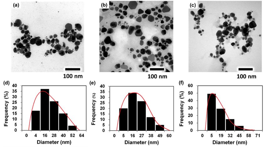
Nanomaterials 2020, 10, 542                                                                             3 of 13

were performed to elucidate the nanoparticle morphology, the agglomeration and to calculate their
dimension, especially to comprehend the difference between three types of these silver nanoparticles.
Finally, Aspergillus niger (A. niger), Fusarium oxysporum (F. oxysporum), and Aspergillus flavus (A. flavus)
were cultured on agar dishes containing P.uri.AgNPs, P.zey.AgNPs, and S.dul.AgNPs to investigate
antifungal activities.

2. Materials and Methods

2.1. Materials
      Silver nitrate (AgNO3 ) was obtained from Guanhao High-Tech Co., Ltd. (Zhanjiang, China).
Potassium bromide (KBr) were FT-IR grade and supplied by Sigma-Aldrich (Merck, Darmstadt,
Germany). Potato dextrose agar (PDA) was purchased from Millipore (Merck, Kenilworth, NJ, USA).
Deionized water (DIW) was obtained from Milli-Q HX 7150 systems (Merck Millipore, Alsace, France).
pH-indicator paper was purchased from Merck (Darmstadt, Germany).
      P. urinaria, S. dulcis, and P. zeylanica were planted in the medicinal garden of Tra Vinh University
(Tra Vinh province, Vietnam). A. niger, F. oxysporum, and A. flavus were isolated and cultured by
Institute of Applied Materials Science (Ho Chi Minh city, Vietnam). Deionized water (DIW) was used
for all experiments.
      Plant materials: P. urinaria, P. zeylanica, and S. dulcis leaves could not have been diseased, damaged,
or contaminated. The leaves were collected and cut into small pieces. Each species (1 g) was put into
the different Erlenmeyer flask and 50 mL of DIW was added. These Erlenmeyer flasks were heated
at 60 ◦ C for 60 min. Aqueous extracts of P. urinaria, P. zeylanica, and S. dulcis leaves (abbreviated as
P.uri.ext, P.zey.ext and S.dul.ext respectively) were obtained after filtration with Whatman No. 1 filter
paper (Figure 1). Their pH value was tested with pH-indicator papers. These extracts were kept at
4 ◦ C for further usage within 7 days.

2.2. Preparation of Silver Nanoparticles Using P. urinaria, P. zeylanica, and S. dulcis Leaf Extracts
      Silver nitrate of 1 mM was prepared in DIW. Three different Erlenmeyer flasks contained 8 mL
of the P.uri.ext, P.zey.ext and S.dul.ext solutions. The silver nitrate solution (0.8 mL) was dropped
slowly into each flask to form the silver nanoparticles at the rate of 30 drops per minute. Three silver
nanoparticles fabricated by P.uri.ext, P.zey.ext, and S.dul.ext were named as P.uri.AgNPs, P.zey.AgNPs,
and S.dul.AgNPs, respectively (Figure 1). The reactions were performed at room temperature. After
8 h, the reacted mixtures had yellowish-brown color, and were washed with DIW three times to collect
silver nanoparticles.

2.3. Characterization of Silver Nanoparticles
     UV-Vis spectrophotometer: To realize the formation of biosynthesized AgNPs, three silver
nanoparticle solutions (P.uri.AgNPs, P.zey.AgNPs, and S.dul.AgNPs) and three leaf broths (P.uri.ext,
P.zey.ext, and S.dul.ext) were respectively loaded into the quartz curvets to collect their UV-vis spectrum
by the Shimadzu UV-1800 machine (Shimadzu, Columbia, MD, USA). The resolution was set at 1 nm,
the wavelength range was 350–750 nm. DIW was used to adjust baseline.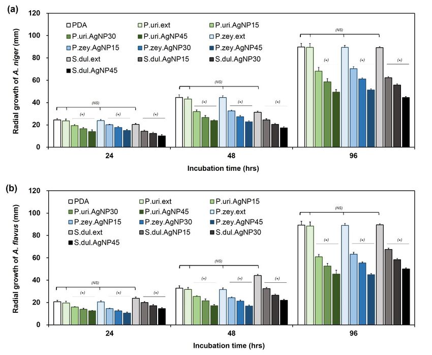
Nanomaterials 2020, 10, 542                                                                                            4 of 13

Nanomaterials 2020, 10, x FOR PEER REVIEW                                                                             4 of 13

      Figure 1.1.Biosynthesis
      Figure      Biosynthesisprocedure
                                procedure      of silver
                                           of silver      nanoparticles
                                                     nanoparticles  usingusing
                                                                          various various plant extracts:
                                                                                   plant extracts:         Phyllanthus
                                                                                                   Phyllanthus urinaria,
      urinaria, Pouzolzia  zeylanica,  and  Scoparia   dulcis leaf extracts symbolized    as P.uri.ext, P.zey.ext,
      Pouzolzia zeylanica, and Scoparia dulcis leaf extracts symbolized as P.uri.ext, P.zey.ext, and S.dul.ext wereand
      S.dul.ext  were reacted  with   silver nitrate  to form  three types  of silver nanoparticles  that were
      reacted with silver nitrate to form three types of silver nanoparticles that were named as P.uri.AgNP,    named
      as P.uri.AgNP,
      P.zey.AgNP,   andP.zey.AgNP,
                        S.dul.AgNP,and      S.dul.AgNP, respectively.
                                        respectively.

2.2. Preparation of Silver KBr
      FTIR spectroscopy:   Nanoparticles Using
                               was blended     P. urinaria,
                                             with           P. samples
                                                  each of all  zeylanica,including
                                                                          and S. dulcis
                                                                                    theLeaf Extracts P.uri.ext,
                                                                                        lyophilized
P.zey.ext,
      SilverS.dul.ext,
              nitrate ofP.uri.AgNPs,    P.zey.AgNPs
                         1 mM was prepared             andThree
                                                 in DIW.    S.dul.AgNPs
                                                                   differentin  turn at theflasks
                                                                             Erlenmeyer     weight   ratio of 100:1.
                                                                                                   contained  8 mL
These   mixtures   were   pelleted   and  recorded  by   FTIR  spectroscopy     (Frontier MIR/FIR,
of the P.uri.ext, P.zey.ext and S.dul.ext solutions. The silver nitrate solution (0.8 mL) was droppedPerkinElmer,
Hopkinton,     MA, flask
                    USA).toThe    wavenumber     was set in the                           −1 .
slowly into each              form  the silver nanoparticles   atrange   of of
                                                                  the rate  500–4000
                                                                               30 dropscmper   minute. Three silver
     EDX analysis:
nanoparticles         The biosynthesized
                  fabricated    by P.uri.ext, AgNPs  including
                                                 P.zey.ext,  andP.uri.AgNPs,
                                                                    S.dul.ext wereP.zey.AgNPs,
                                                                                        named and     S.dul.AgNPs
                                                                                                  as P.uri.AgNPs,
in succession were
P.zey.AgNPs,      and dissolved    in pure
                        S.dul.AgNPs,         ethanol and
                                         respectively      followed
                                                        (Figure        by ultrasonication.
                                                                 1). The   reactions were A      few microliters
                                                                                               performed         of
                                                                                                           at room
these  suspensions    were   loaded  on  copper  grid, then  dried  in the air, and  analyzed
temperature. After 8 hours, the reacted mixtures had yellowish-brown color, and were washed withwith EDX   (H-7593,
Horiba,
DIW three Kyoto,
             timesJapan).
                    to collect silver nanoparticles.
     SEM and TEM observation: The sample preparation was similar to EDX measurement.
The   P.uri.AgNPs, P.zey.AgNPs,
2.3. Characterization                  and S.dul.AgNPs were observed with TEM (JEOL-JEM-1400, JEOL
                        of Silver Nanoparticles
Ltd., Tokyo, Japan) and SEM (S-4800, Hitachi, Tokyo, Japan).
      UV-Vis spectrophotometer: To realize the formation of biosynthesized AgNPs, three silver
nanoparticle solutions (P.uri.AgNPs, P.zey.AgNPs, and S.dul.AgNPs) and three leaf broths (P.uri.ext,
P.zey.ext, and S.dul.ext) were respectively loaded into the quartz curvets to collect their UV-vis
spectrum by the Shimadzu UV-1800 machine (Shimadzu, Columbia, MD, USA). The resolution was
set at 1 nm, the wavelength range was 350–750 nm. DIW was used to adjust baseline.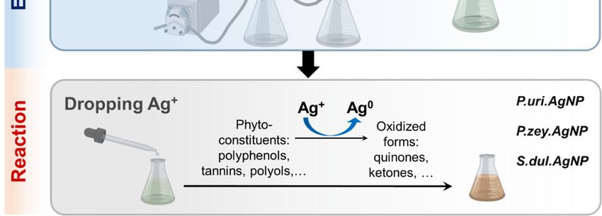
Nanomaterials 2020, 10, 542                                                                              5 of 13

2.4. Antifungal Assays
     A. niger, F. oxysporum, and A. flavus were grown on the media containing various compositions
to follow the fungal proliferation. The petri dishes containing pure PDA solution were used as the
control (named as PDA). The petri dishes containing PDA and each of extracts including P.uri.ext,
P.zey.ext, S.dul.ext were used to check the antifungal ability of each broth that was written in short
as P.uri.ext, P.zey.ext, S.dul.ext. The petri dishes containing PDA and each biosynthesized AgNP
at different concentrations were used to test the antifungal effect of these biosynthesized AgNPs.
The symbolization was explained in Table 1.

         Table 1. The compositions of media prepared for antifungal assay. PDS = Potato dextrose agar,

                              Symbolization                Description
                              P.uri.AgNP15       15 ppm of P.uri.AgNPs in PDA
                              P.uri.AgNP30       30 ppm of P.uri.AgNPs in PDA
                              P.uri.AgNP45       45 ppm of P.uri.AgNPs in PDA
                              P.zey.AgNP15       15 ppm of P.zey.AgNPs in PDA
                              P.zey.AgNP30       30 ppm of P.zey.AgNPs in PDA
                              P.zey.AgNP45       45 ppm of P.zey.AgNPs in PDA
                              S.dul.AgNP15       15 ppm of S.dul.AgNPs in PDA
                              S.dul.AgNP30       30 ppm of S.dul.AgNPs in PDA
                              S.dul.AgNP45       45 ppm of S.dul.AgNPs in PDA

     After preparing all dishes, fungal colonies were placed directly in the center of agar plate.
The fungal culture was carried out at room temperature. The diameter of fungal zone was measured
every 24 h for 4 days.

2.5. Statistical Analysis
     The results were replicated 3 times and represented as mean ± standard deviation. All experimental
data were analyzed by Student’s t test. P < 0.05 implied that two compared results were statistically
significant. P > 0.05 indicated non-statistical (NS) difference.

3. Results and Discussion

3.1. Biosynthesis of Silver Nnanoparticles from Plant Extracts
      Leaf extracts were obtained from three plant species including P. urinaria, P. zeylanica, and S. dulcis
(Figure 1); and their pH value was around 7. Then, silver nitrate was simply added drop-by-drop to
the extracts to form AgNPs under mild conditions (room temperature for 8 h). The mixture solution
turned to yellowish brown to dark brown, demonstrating reactions between extracts and silver (I)
ions. The UV-vis spectra of pure extracts (Figure 2a–c, dashed line) did not have any peaks in the
wavelength range of 350–700 nm, and the peaks of spectra were at the wavelength around 200–280 nm,
representing π − π* or n − π* transition of a myriad of organic compounds in plant extracts due to
an absorbing wavelength of the UV radiation. The UV-vis of the reacted mixtures (Figure 2a–c, solid
line) showed the peaks from 400–600 nm, demonstrating silver nanoparticles with a surface plasmon
resonance (SPR) [31]. The nanoparticle size strongly impacted on the peak position: the larger scale
had red-shifting (towards longer wavelengths), while the smaller one had blue-shifting (towards
shorter wavelengths), because the conductive electrons of the aggregated nanoparticle surface become
less flexible [31]. Concurrently, the pH value was reported as one of the influencing factors in the
green synthesis of nanoparticles: a different pH displayed a different SPR behavior. At a neutral pH,
the maximum wavelength was found in Figure 2a–c (dashed line) of three AgNPs that agreed with the
previous study of Singh et al. At pH 7.5, green silver nanoparticles showed a peak of 449 nm [32].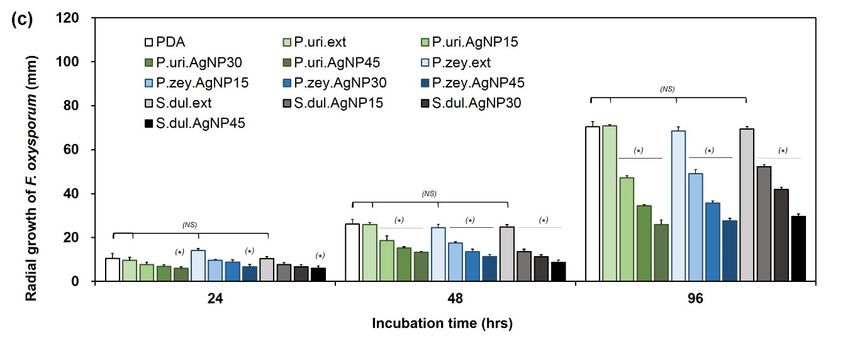
Nanomaterials 2020, 10, 542                                                                                      6 of 13
Nanomaterials 2020, 10, x FOR PEER REVIEW                                                                       7 of 13

     Figure 2. The UV-vis spectra of P.uri.ext and P.uri.AgNP (a), P.zey.ext and P.zey.AgNP (b), S.dul.ext and
     Figure 2. The UV-vis spectra of P.uri.ext and P.uri.AgNP (a), P.zey.ext and P.zey.AgNP (b), S.dul.ext
     S.dul.AgNP (c), in which the dashed line is the spectra of extract, and the solid line is that of AgNP; the
     and S.dul.AgNP (c), in which the dashed line is the spectra of extract, and the solid line is that of
     FTIR spectra of P.uri.ext and P.uri.AgNP (d), P.zey.ext and P.zey.AgNP (e), S.dul.ext and S.dul.AgNP (f),
     AgNP; the FTIR spectra of P.uri.ext and P.uri.AgNP (d), P.zey.ext and P.zey.AgNP (e), S.dul.ext and
     in which (i) line is the spectra of AgNP, (ii) line is the one of extract; the EDX spectra of P.uri.AgNP (g),
     S.dul.AgNP (f), in which (i) line is the spectra of AgNP, (ii) line is the one of extract; the EDX spectra
     P.zey.AgNP (h) and S.dul.AgNP (i).
     of P.uri.AgNP (g), P.zey.AgNP (h) and S.dul.AgNP (i).
     The UV-Vis spectrum of P.uri.AgNPs (Figure 2a, solid line) had one broad peak at 400–550 nm. The
      Figure 2d–f(i) shows the FTIR spectra of P.uri.AgNPs, P.zey.AgNPs, and S.dul.AgNPs. These
P.zey.AgNP UV-vis spectrum (Figure 2b, solid line) exhibited two close peaks centered        at 440 nm and
spectra indicated the presence of troughs at wavenumber region of 3500–3300 cm−1, 1630–1635 cm−1,
520 nm. The S.dul.AgNP       one (Figure 2c, solid line) indicated the pointed peak of 430 nm. These UV-vis
1380–1385 cm−1, and 1000–1100 cm−1, which were attributed to O–H stretching, O–H bending, C–H
results predicted P.uri.AgNPs might agglomerate, the P.zey.AgNPs had widely size distribution, and
bending, C–O stretching vibration, respectively. These troughs were also found in the FTIR spectra
the S.dul.AgNP dimension was smallest. This difference may be attributed by the phytoconstituent
of extracts (Figure 2d–f(ii)), but the OH troughs were broadening due to stronger hydrogen bonding.
variance of each plant extracts. Table S1 summarizes the phytochemicals that indicate P. urinaria, P.
In contrast, P.uri.AgNPs, P.zey.AgNPs, and S.dul.AgNPs showed relatively sharp O–H stretching
zeylanica, and S. dulcis have different compounds; even various classes. P. urinaria possessed lignans,
peaks, as compared to pure extracts, demonstrating that silver nanoparticles were capped by
tannins, flavonoids, phenolics, terpenoids, and others. P. zeylanica had nor-lignans, carotenoids, vitamin
phytoconstituents [12,15]. So it could be said that using plant extracts could stabilize the formed silver
C, malic acid, pectic acid, tartaric acid and gum beside tannins and flavonoids. S. dulcis mainly contained
nanoparticles within the synthesizing process. In fact, these biosynthesized silver nanoparticles
flavones, terpenes, and steroids. The underlying mechanism for the formation of silver nanoparticles
including P.uri.AgNPs, P.zey.AgNPs, and S.dul.AgNPs had zeta potentials of −15.6 mV, −24.3 mV,
in the plant extracts was an oxidation-reduction process. Generally, lignans, nor-lignans, tannins,
and −20.8 mV, respectively (Figure S1). The negative potential implied that these green silver
flavonoids, phenolics, terpenoids, carotenoids, and vitamin C are great antioxidants and reducing
nanoparticles achieved a stable stage. The negative charge of P.uri.AgNPs, P.zey.AgNPs, and
agents [22,24,28,33]. These compounds, mostly possessing phenol or hydroxyl groups, may inhibit
S.dul.AgNPs may explained by the carboxylic groups of phytochemicals from leaf extracts (Table S1).
oxidants by transferring their hydrogen atom and forming a transition state pending one remained
This value was also agreed with other AgNPs fabricated by phytochemicals [34]. In other words, the
electron. In addition, the unsaturated chains of these phytoconstituents also may act as a reducer. The
charge of AgNPs formed by gallic acid + quercetin, resveratrol, and mPEG-luteolin were −19.0 mV,
aldehyde functional groups of 5-hydroxymethyl-2-furaldehyde or p-hydroxybenzaldehyde may also
−20.6 mV, and −25.5 mV respectively. The presence of carbon, oxygen, and silver was confirmed by
react with oxidants to become carboxylic acids. P. urinaria contains the largest amount of antioxidants
EDX analysis (Figure 2g–i). The detected other elements such as Si and Cu were from the grid.
(Table S1) and quickly reduces Ag ions, forming relatively large nanoparticles.
     Figure 2d–f(i) shows the FTIR spectra of P.uri.AgNPs, P.zey.AgNPs, and S.dul.AgNPs. These
spectra indicated the presence of troughs at wavenumber region of 3500–3300 cm−1 , 1630–1635 cm−1 ,
Nanomaterials 2020, 10, 542 7 of 13
1380–1385 cm−1 , and 1000–1100 cm−1 , which were attributed to O–H stretching, O–H bending,
C–H bending, C–O stretching vibration, respectively. These troughs were also found in the FTIR
spectra of extracts (Figure 2d–f(ii)), but the OH troughs were broadening due to stronger hydrogen
bonding. In contrast, P.uri.AgNPs, P.zey.AgNPs, and S.dul.AgNPs showed relatively sharp O–H
stretching peaks, as compared to pure extracts, demonstrating that silver nanoparticles were capped
by phytoconstituents [12,15]. So it could be said that using plant extracts could stabilize the
formed silver nanoparticles within the synthesizing process. In fact, these biosynthesized silver
nanoparticles including P.uri.AgNPs, P.zey.AgNPs, and S.dul.AgNPs had zeta potentials of −15.6 mV,
−24.3 mV, and −20.8 mV, respectively (Figure S1). The negative potential implied that these green
silver nanoparticles achieved a stable stage. The negative charge of P.uri.AgNPs, P.zey.AgNPs,
and S.dul.AgNPs may explained by the carboxylic groups of phytochemicals from leaf extracts (Table
S1). This value was also agreed with other AgNPs fabricated by phytochemicals [34]. In other words,
the charge of AgNPs formed by gallic acid + quercetin, resveratrol, and mPEG-luteolin were −19.0 mV,
−20.6 mV, and −25.5 mV respectively. The presence of carbon, oxygen, and silver was confirmed by
EDX analysis (Figure 2g–i). The detected other elements such as Si and Cu were from the grid.
3.2. Size and Morphology of Biosynthesized Silver Nanoparticles (P.uri.AgNPs, P.zey.AgNPs and
S.dul.AgNPs)
Morphology and size are two of the most important properties of nanoparticles. SEM and TEM
were used to look at P.uri.AgNPs, P.zey.AgNPs, and S.dul.AgNPs (Figure 3). The TEM images in
Figure 3a reveals that P.uri.AgNPs were mostly spherical and oval. For the P.zey.AgNPs (Figure 3b),
various morphologies such as spherical, triangle, plate-like polyshaped, pentagonal, and hexagonal
shapes were found. S.dul.AgNPs were spherical and relatively smaller than P.uri.AgNPs and
P.zey.AgNPs (Figure 3c), consistent with the results from UV-Vis (Figure 2c) and size measurements
(Figure 3d–f). Also, their surfaces were smooth when observed by SEM (Figure S2). The size of
P.uri.AgNPs, P.zey.AgNPs, and S.dul.AgNPs were in the range of 4–52 nm, 5–49 nm, and 5–45 nm.
However, ~65% of P.uri.AgNPs had an average size of 28.3 nm and ~59% of P.zey.AgNPs was
26.7 nm, while ~50% of S.dul.AgNPs were of 5 nm. Taken together, the extract type impacted
the size, morphology, and distribution of biosynthesized AgNPs. Considering influence factors,
the reducing power [35] and capping agents [36] in synthesis process mainly decided nanoparticle
structure. The strongly reducing compounds could form the small nanoparticles, while the weakly
reducing agents led to making large and/or polydisperse nanoparticles [35]. However, the very rapid
reduction may not give enough time for capping silver nanoparticles with phytoconstituents, thus may
cause agglomeration. Hence, the S.dul.AgNPs were smallest due to the better harmony between the
reducing power and capping reaction of S.dul.ext, compared to P.uri.ext and P.zey.ext broths. P. urinaria
contains many well-known reductants such as rutin, kaempferol, quercetin, gallic acid, ellagic acid,
5-hydroxymethyl-2-furaldehyde, lignans, tannins, glycosides, rich of other polyols, and phenolics
(Table S1) that show strong reducing activity. Similarly, the highly reducing content of P. zeylanica was
exhibited by the presence of quercetin, epicatechin, ascorbic acid, gum, and alkaloids. Meanwhile,
S. dulcis was found to contain fewer reducing compounds of phenolics, lignans, and diols than two
aforementioned plants. Reasonably, S.dul.AgNPs had enough time to be grafted with its scoparic acid
and others through –C=O groups [34] to be stabilized, thus became less aggregated than P.uri.AgNPs
and P.zey.AgNPs. In addition, it may be understood that the size of P.uri.AgNPs and P.zey.AgNPs were
approximately the same and similar to the other AgNPs formed by kaempferol, quercetin, or gallic acid
reductant [34]. Three compounds were also found in P. urinaria and P. zeylanica (Table S1). Concerning
the relation between nanoparticle morphology and capping agents, when increasing the content of
capping agent, the diversified shapes such as rods, triangles, hexagon, cylinders and cubic would
be formed besides the sphere. [36]. Thus it may be inferred that the P.zey.ext contained the highest
capping compounds.P.zey.AgNPs were approximately the same and similar to the other AgNPs formed by kaempferol,
quercetin, or gallic acid reductant [34]. Three compounds were also found in P. urinaria and P.
zeylanica (Table S1). Concerning the relation between nanoparticle morphology and capping agents,
when increasing the content of capping agent, the diversified shapes such as rods, triangles, hexagon,
Nanomaterials
cylinders and 10, 542 would be formed besides the sphere. [36]. Thus it may be inferred that
2020,cubic 8 ofthe
13
P.zey.ext contained the highest capping compounds.
Figure TEM images
3. TEM
Figure 3. images of
ofP.uri.AgNP
P.uri.AgNP (a),
(a), P.zey.AgNP
P.zey.AgNP (b),
(b), and
and S.dul.AgNP
S.dul.AgNP (c);
(c); the
the graph of dimension
graph of dimension
distribution of P.uri.AgNP (d), P.zey.AgNP (e), and S.dul.AgNP
distribution of P.uri.AgNP (d), P.zey.AgNP (e), and S.dul.AgNP (f). (f).
3.3. Antifungal Activities of P.uri.AgNPs, P.zey.AgNPs and S.dul.AgNPs
P.uri.AgNPs, P.zey.AgNPs, and S.dul.AgNPs were tested for antifungal ability against A. niger,
A. flavus, and F. oxysporum at the concentration of 15, 30 and 45 ppm. The pure PDA and three
extracts (P.uri.ext, P.zey.ext, and S.dul.ext) were parallelly carried out as a control. Figure 4 shows
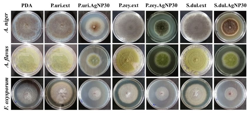
the growth of three fungal strains on various agar dishes for 96 h. The proliferation of A. niger, A.
flavus, and F. oxysporum was suppressed by the presence of AgNPs in the nanoparticle concentration
dependent manner, while the pure extracts were not influenced the proliferation of the fungus under
the same concentration, which demonstrated the anti-fungal activities of P.uri.AgNPs, P.zey.AgNPs,
and S.dul.AgNPs.(P.uri.ext, P.zey.ext, and S.dul.ext) were parallelly carried out as a control. Figure 4 shows the growth
of three fungal strains on various agar dishes for 96 hours. The proliferation of A. niger, A. flavus, and
F. oxysporum was suppressed by the presence of AgNPs in the nanoparticle concentration dependent
manner, while the pure extracts were not influenced the proliferation of the fungus under the same
concentration, which demonstrated the anti-fungal activities of P.uri.AgNPs, P.zey.AgNPs, and
Nanomaterials
S.dul.AgNPs.2020, 10, 542 9 of 13
Figure 4. Three fungal strains including A. niger, A. flavus, and F. oxysporum were culture in different
Figure 4. Three fungal strains including A. niger, A. flavus, and F. oxysporum were culture in different
agar matrix after 96 h.
agar matrix after 96 hours.
Quantitatively, the mycelium diameters of A. niger, A. flavus, and F. oxysporum on various agar
Quantitatively, the mycelium diameters of A. niger, A. flavus, and F. oxysporum on various agar
dishes were followed as a function of time interval until 96 h (Figure 5). The size of fungal zones was
dishes were followed as a function of time interval until 96 hours (Figure 5). The size of fungal zones
similar in P.uri.ext, P.zey.ext, and S.dul.ext, and gradually increased as incubated for a longer time period.
was similar in P.uri.ext, P.zey.ext, and S.dul.ext, and gradually increased as incubated for a longer
There were no significant differences in the A. niger proliferation between PDA, P.uri.ext, P.zey.ext, and
time period. There were no significant differences in the A. niger proliferation between PDA, P.uri.ext,
S.dul.ext dishes (Figure 5a). Similarly, A. flavus (Figure 5b), and F. oxysporum (Figure 5c) showed the
P.zey.ext, and S.dul.ext dishes (Figure 5a). Similarly, A. flavus (Figure 5b), and F. oxysporum (Figure
same
5c) showed A.same
trend ofthe niger.trend
In case
of A.ofniger.
silverInnanoparticles, the mycelium
case of silver nanoparticles, thediameters
myceliumbecamediameters significantly
became
smaller
significantly smaller compared to PDA control. When increasing the biosynthesized to
compared to PDA control. When increasing the biosynthesized AgNP concentration 30 and
AgNP
ppm, A. nigerto
45concentration was
30 and 45 ppm, A. niger was increasingly inhibited by P.uri.AgNPs, P.zey.AgNPs, an
increasingly inhibited by P.uri.AgNPs, P.zey.AgNPs, and S.dul.AgNPs. Taking
observation at Figure
and S.dul.AgNPs. 5b,c shows
Taking the antifungal
an observation at Figureresults againstthe
5b,c shows A.antifungal
flavus andresults
F. oxysporum, theflavus
against A. similar
comments with A. niger were withdrawn. So P.uri.AgNPs, P.zey.AgNPs, and
and F. oxysporum, the similar comments with A. niger were withdrawn. So P.uri.AgNPs, P.zey.AgNPs, S.dul.AgNPs may inhibit
effectively all three fungal
and S.dul.AgNPs strains
may inhibit that wasall
effectively explained by their
three fungal strainsnanosize
that was less than 60 nm.
explained Thisnanosize
by their dimension
supports
less thanthese
60 nm.nanoparticles
This dimensionto penetrate,
supportstotheseaccumulate, and to
nanoparticles tointeract
penetrate, with to cell membranes
accumulate, andeasily,
to
andinteract with cell membranes
thus inactivate easily, and
the protein activities thus
that inactivate
leading the protein
cell death [37]. Asactivities
a result,that
theleading
AgNPscell death
eco-friendly
[37]. As aby
fabricated result, the broths
the leaf AgNPsofeco-friendly
P. urinaria, P.fabricated
zeylanica,byandtheS.leaf broths
dulcis were of proven
P. urinaria,
theirP.antifungal
zeylanica, and
ability.
S. dulcis were
P.uri.AgNPs, proven theirand
P.zey.AgNPs, antifungal ability.were
S.dul.AgNPs P.uri.AgNPs,
less thanP.zey.AgNPs,
50 nm which and size S.dul.AgNPs
could exhibitwere less
an effective
than 50 nm which
antimicrobial abilitysize
[14].could exhibit an
In previous effective
studies, antimicrobial
green AgNPs with ability
size[14].
rangeIn previous
less than studies, greenalso
50 nm were
AgNPs with size range less than 50 nm were also formed by other plants
formed by other plants including Elephantopus scaber, Phyllanthus amarus, Alpinia katsumadai, Psidium including Elephantopus
scaber, Salvia
guajava, Phyllanthus amarus,
leriifolia, andAlpinia katsumadai,
Artocarpus Psidium
altilis. All guajava,
showed Salvia antimicrobial
the good leriifolia, and Artocarpus altilis. A.
activity against
All showed the good antimicrobial activity against A. flavus, and A. niger besides E. coli, Staphylococcus
flavus, and A. niger besides E. coli, Staphylococcus spp., Bacillus spp., Pseudomonas spp. [14]. Higher
spp., Bacillus spp., Pseudomonas spp.[14]. Higher antimicrobial activity was achieved when increasing
antimicrobial activity was achieved when increasing green AgNP concentration [14]. So, together with
green AgNP concentration [14]. So, together with previous ones, this study introduces three more
previous ones, this study introduces three more renewable materials to produce silver nanoparticles as
renewable materials to produce silver nanoparticles as effective fungicide for biomedical and
effective fungicide for biomedical and agricultural applications.
agricultural applications.Nanomaterials 2020, 10, 542 10 of 13
Nanomaterials 2020, 10, x FOR PEER REVIEW 10 of 13
Figure 5. The mycelium diameter of A. niger (a), A. flavus (b), and F. oxysporum (c) cultured on
Figure 5.agar
different Thematrices
mycelium asdiameter
a function ofof
A.time
nigerinterval.
(a), A. flavus (b),potato
(PDA: and F. dextrose
oxysporum (c) cultured
agar dishes ason different
a control;
agar matrices as a function of time interval. (PDA: potato dextrose agar dishes as a control;
P.uri.ext, P.zey.ext, and S.dul.ext: the dishes made from PDA containing leaf extracts of Phyllanthus P.uri.ext,
P.zey.ext,
urinaria, and S.dul.ext:
Pouzolzia the and
zeylanica, dishes made dulcis;
Scoparia from PDA containing P.uri.AgNP30,
P.uri.AgNP15, leaf extracts of and
Phyllanthus urinaria,
P.uri.AgNP45:
Pouzolzia
the dishes zeylanica,
made from andPDA
Scoparia dulcis; P.uri.AgNP15,
containing 15, 30 and 45P.uri.AgNP30, and P.uri.AgNP45:
ppm of P.uri.AgNPs the dishes
silver nanoparticles;
made from PDA
P.zey.AgNP15, containing 15,
P.zey.AgNP30, 30 and 45 ppm
P.zey.AgNP45: theofdishes
P.uri.AgNPs silver
made from nanoparticles;
PDA containing P.zey.AgNP15,
15, 30 and 45
P.zey.AgNP30, P.zey.AgNP45: the dishes made from PDA containing
ppm of P.zey.AgNPs silver nanoparticles; S.dul.AgNP15, S.dul.AgNP30, S.dul.AgNP45: the 15, 30 and 45 ppm
dishesof
P.zey.AgNPs
made from PDA silver nanoparticles;
containing 15, 30 andS.dul.AgNP15, S.dul.AgNP30,
45 ppm of S.dul.AgNPs silverS.dul.AgNP45: < 0.05;made
thePdishes
nanoparticles (*) NS:
from PDA containing
non-statistical different. 15, 30 and 45 ppm of S.dul.AgNPs silver nanoparticles (*) P < 0.05; NS: non-
statistical different.Nanomaterials 2020, 10, 542 11 of 13
4. Conclusions
In the study, three kinds of silver nanoparticles (P.uri.AgNPs, P.zey.AgNPs, and S.dul.AgNPs)
were successfully biosynthesized by simply adding silver (I) ions to herbal plant extracts (P. urinaria,
P. zeylanica, and S. dulcis) under mild conditions. Their formation and physicochemical properties
were well characterized by UV-Vis, FTIR, EDX, TEM, and SEM. The results revealed that P.uri.AgNPs,
P.zey.AgNPs, and S.dul.AgNPs were modified with variously organic compounds in biosynthesis
process. So the formation and the coating procedure of these silver nanoparticles were performed at
the same time. The plant species possessing the different phytoconstituents that lead to influence on
the size and morphology of silver nanoparticles. P. urinaria and P. zeylanica leaf extract could form
the silver nanoparticles about 28.3 and 26.7 nm, especially S. dulcis leaf extract could create almost
5 nm particles. Among the three AgNP types, only P.zey.AgNP showed a diversified morphology
including spherical, triangle, plate-like polyshaped, pentagonal, and hexagonal shapes, while the
others were in spherical morphology. In addition, the antifungal ability of P.uri.AgNPs, P.zey.AgNPs,
and S.dul.AgNPs against A. niger, A. flavus, and F. oxysporum were validated. This green method is
the simplest and most largely scalable method for the production of antifungal silver nanoparticles in
biomedical and agricultural applications.
Supplementary Materials: The following are available online at http://www.mdpi.com/2079-4991/10/3/542/s1,
Figure S1: The zeta potential of P.uri.AgNP (a), P.zey.AgNP (b) and S.dul.AgNP (c); Figure S2: The SEM images of
P.uri.AgNP (a), P.zey.AgNP (b) and S.dul.AgNP (c); Table S1: The summary of phytoconstituents in P. urinaria, P.
zeylanica, and S. dulcis.
Author Contributions: Conceptualization, D.H.N., J.S.L. and T.T.H.T.; data curation, J.S.L., X.T.N. and T.T.H.T.;
methodology, J.S.L., V.H.G.P. and T.T.H.T.; project administration, D.H.N. and K.D.P.; supervision, K.D.P., Y.C.C.,
V.H.G.P. and T.T.H.T.; validation, J.S.L.; writing—review & editing, J.S.L. and T.T.H.T. All authors have read and
agreed to the published version of the manuscript.
Acknowledgments: This research was supported by the Domestic Master/PhD Scholarship Programme of
Vingroup Innovation Foundation (Grant number: VINIF.2019.TS.72).
Conflicts of Interest: The authors declare no conflict of interest.
References
1. Jamkhande, P.G.; Ghule, N.W.; Bamer, A.H.; Kalaskar, M.G. Metal nanoparticles synthesis: An overview on
methods of preparation, advantages and disadvantages, and applications. J. Drug Deliv. Sci. Technol. 2019,
53, 101174. [CrossRef]
2. Gellé, A.; Moores, A. Plasmonic nanoparticles: Photocatalysts with a bright future. Curr. Opin. Green
Sustain. Chem. 2019, 15, 60–66. [CrossRef]
3. Srivastava, V.; Gusain, D.; Sharma, Y.C. Critical Review on the Toxicity of Some Widely Used Engineered
Nanoparticles. Ind. Eng. Chem. Res. 2015, 54, 6209–6233. [CrossRef]
4. Narayanan, K.B.; Sakthivel, N. Biological synthesis of metal nanoparticles by microbes. Adv. Colloid
Interface Sci. 2010, 156, 1–13. [CrossRef]
5. Medici, S.; Peana, M.; Nurchi, V.M.; Zoroddu, M.A. Medical Uses of Silver: History, Myths, and Scientific
Evidence. J. Med. Chem. 2019, 62, 5923–5943. [CrossRef]
6. Grace, J.L.; Huang, J.X.; Cheah, S.-E.; Truong, N.P.; Cooper, M.A.; Li, J.; Davis, T.P.; Quinn, J.F.; Velkov, T.;
Whittaker, M.R. Antibacterial low molecular weight cationic polymers: Dissecting the contribution of
hydrophobicity, chain length and charge to activity. RSC Adv. 2016, 6, 15469–15477. [CrossRef]
7. Barbon, S.M.; Truong, N.P.; Elliott, A.G.; Cooper, M.A.; Davis, T.P.; Whittaker, M.R.; Hawker, C.J.;
Anastasaki, A. Elucidating the effect of sequence and degree of polymerization on antimicrobial properties
for block copolymers. Polym. Chem. 2020, 11, 84–90. [CrossRef]
8. Grace, J.L.; Elliott, A.G.; Huang, J.X.; Schneider, E.K.; Truong, N.P.; Cooper, M.A.; Li, J.; Davis, T.P.; Quinn, J.F.;
Velkov, T.; et al. Cationic acrylate oligomers comprising amino acid mimic moieties demonstrate improved
antibacterial killing efficiency. J. Mater. Chem. B 2017, 5, 531–536. [CrossRef]Nanomaterials 2020, 10, 542 12 of 13
9. Elbourne, A.; Cheeseman, S.; Atkin, P.; Truong, N.P.; Syed, N.; Zavabeti, A.; Mohiuddin, M.; Esrafilzadeh, D.;
Cozzolino, D.; McConville, C.F.; et al. Antibacterial Liquid Metals: Biofilm Treatment via Magnetic Activation.
ACS Nano 2020, 14, 802–817. [CrossRef]
10. Hoang Thi, T.T.; Lee, Y.; Le Thi, P.; Park, K.D. Nitric oxide-releasing injectable hydrogels with high antibacterial
activity through in situ formation of peroxynitrite. Acta Biomater. 2018, 67, 66–78. [CrossRef]
11. Akhtar, M.S.; Panwar, J.; Yun, Y.-S. Biogenic Synthesis of Metallic Nanoparticles by Plant Extracts. ACS Sustain.
Chem. Eng. 2013, 1, 591–602. [CrossRef]
12. Duan, H.; Wang, D.; Li, Y. Green chemistry for nanoparticle synthesis. Chem. Soc. Rev. 2015, 44, 5778–5792.
[CrossRef] [PubMed]
13. Hoseinpour, V.; Ghaemi, N. Green synthesis of manganese nanoparticles: Applications and future
perspective—A review. J. Photochem. Photobiol. B Biol. 2018, 189, 234–243. [CrossRef] [PubMed]
14. Roy, A.; Bulut, O.; Some, S.; Mandal, A.K.; Yilmaz, M.D. Green synthesis of silver nanoparticles:
Biomolecule-nanoparticle organizations targeting antimicrobial activity. RSC Adv. 2019, 9, 2673–2702.
[CrossRef]
15. Ovais, M.; Khalil, A.T.; Raza, A.; Khan, M.A.; Ahmad, I.; Islam, N.U.; Saravanan, M.; Ubaid, M.F.; Ali, M.;
Shinwari, Z.K. Green synthesis of silver nanoparticles via plant extracts: Beginning a new era in cancer
theranostics. Nanomed. Nanotechnol. Biol. Med. 2016, 11, 3157–3177. [CrossRef]
16. Hoang Thi, T.T.; Pilkington, E.H.; Nguyen, D.H.; Lee, J.S.; Park, K.D.; Truong, N.P. The Importance of
Poly(ethylene glycol) Alternatives for Overcoming PEG Immunogenicity in Drug Delivery and Bioconjugation.
Polymers 2020, 12, 298. [CrossRef]
17. Ali, J.; Ali, N.; Wang, L.; Waseem, H.; Pan, G. Revisiting the mechanistic pathways for bacterial mediated
synthesis of noble metal nanoparticles. J. Microbiol. Methods 2019, 159, 18–25. [CrossRef]
18. Sathiyanarayanan, G.; Dineshkumar, K.; Yang, Y.H. Microbial exopolysaccharide-mediated synthesis and
stabilization of metal nanoparticles. Crit. Rev. Microbiol. 2017, 43, 731–752. [CrossRef]
19. Pulit, J.; Banach, M.; Zielina, M.; Laskowska, B.; Kurleto, K. Raspberry Extract as Both a Stabilizer and a
Reducing Agent in Environmentally Friendly Process of Receiving Colloidal Silver. J. Nanomater. 2013, 2013,
1–12. [CrossRef]
20. Pulit, J.; Banach, M. Preparation of nanosilver and nanogold based on dog rose aqueous extract. Bioinorg.
Chem. Appl. 2014, 2014, 658935. [CrossRef]
21. Pulit-Prociak, J.; Banach, M. The Use of Plant Materials in the Process of Obtaining Silver and Gold
Nanoparticles. J. Comput. Theor. Nanosci. 2016, 13, 2697–2704. [CrossRef]
22. Hossain, M.S.; Rahman, M.S.; Imon, A.H.M.R.; Zaman, S.; Siddiky, A.S.M.B.A.; Mondal, M.; Sarwar, A.;
Huq, T.B.; Adhikary, B.C.; Begum, T.; et al. Ethnopharmacological investigations of methanolic extract of
Pouzolzia Zeylanica (L.) Benn. Clin. Phytosci. 2016, 2. [CrossRef]
23. Chen, X.-M.; Li, Z.-H.; Tao, S.-H.; Chen, Y.-F.; Chen, Z.-H.; Guo, L.-B. Effect of FPZ, a total flavonoids ointment
topical application from Pouzolzia zeylanica var. microphylla, on mice skin infections. Rev. Brasil. Farmacogn.
2018, 28, 732–737. [CrossRef]
24. Geethangili, M.; Ding, S.-T. A Review of the Phytochemistry and Pharmacology of Phyllanthus urinaria L.
Front. Pharmacol. 2018, 9, 1109. [CrossRef]
25. Choudhury, H.; Pandey, M.; Hua, C.K.; Mun, C.S.; Jing, J.K.; Kong, L.; Ern, L.Y.; Ashraf, N.A.; Kit, S.W.;
Yee, T.S.; et al. An update on natural compounds in the remedy of diabetes mellitus: A systematic review.
J. Tradit. Complement. Med. 2018, 8, 361–376. [CrossRef]
26. Seyed, M.A. A comprehensive review on Phyllanthus derived natural products as potential chemotherapeutic
and immunomodulators for a wide range of human diseases. Biocatal. Agric. Biotechnol. 2019, 17, 529–537.
[CrossRef]
27. Liu, Q.; Yang, Q.M.; Hu, H.J.; Yang, L.; Yang, Y.B.; Chou, G.X.; Wang, Z.T. Bioactive diterpenoids and
flavonoids from the aerial parts of Scoparia dulcis. J. Nat. Prod. 2014, 77, 1594–1600. [CrossRef]
28. Frezza, C.; Venditti, A.; Toniolo, C.; De Vita, D.; Franceschin, M.; Ventrone, A.; Tomassini, L.; Foddai, S.;
Guiso, M.; Nicoletti, M.; et al. Nor-Lignans: Occurrence in Plants and Biological Activities—A Review.
Molecules 2020, 25, 197. [CrossRef]
29. Vo, Q.H.; Nguyen, P.H.; Zhao, B.T.; Ali, M.Y.; Choi, J.S.; Min, B.S.; Nguyen, T.H.; Woo, M.H. Protein tyrosine
phosphatase 1B (PTP1B) inhibitory constituents from the aerial parts of Tradescantia spathacea Sw. Fitoterapia
2015, 103, 113–121. [CrossRef]Nanomaterials 2020, 10, 542 13 of 13
30. Meera, P.; Kavitha, V.; Krishnaja, K.R. Scoparia dulcis: A review on its phytochemical and pharmacological
profile. Innorig. Int. J. Sci. 2017, 4, 18–22.
31. Silver Nanoparticles: Optical Properties. Available online: https://nanocomposix.eu/pages/silver-
nanoparticles-optical-properties (accessed on 20 February 2020).
32. Singh, M.; Sinha, I.; Mandal, R.K. Role of pH in the green synthesis of silver nanoparticles. Mater. Lett. 2009,
63, 425–427. [CrossRef]
33. Ahsan, M.; Islam, S.N.; Gray, A.I.; Stimson, W.H. Cytotoxic Diterpenes from Scoparia dulcis. J. Nat. Prod. 2003,
66, 958–961. [CrossRef] [PubMed]
34. Amini, S.M. Preparation of antimicrobial metallic nanoparticles with bioactive compounds. Mater. Sci.
Eng. C 2019, 103, 109809. [CrossRef] [PubMed]
35. Bhattarai, B.; Zaker, Y.; Bigioni, T.P. Green synthesis of gold and silver nanoparticles: Challenges and
opportunities. Curr. Opin. Green Sustain. Chem. 2018, 12, 91–100. [CrossRef]
36. Madkour, M.; Bumajdad, A.; Al-Sagheer, F. To what extent do polymeric stabilizers affect nanoparticles
characteristics? Adv. Coll. Interface Sci. 2019, 270, 38–53. [CrossRef]
37. Buzea, C.; Pacheco, I.I.; Robbie, K. Nanomaterials and nanoparticles: Sources and toxicity. Biointerphases
2007, 2, MR17. [CrossRef]
© 2020 by the authors. Licensee MDPI, Basel, Switzerland. This article is an open access
article distributed under the terms and conditions of the Creative Commons Attribution
(CC BY) license (http://creativecommons.org/licenses/by/4.0/).You can also read