MDM2 Antagonists Induce a Paradoxical Activation of Erk1/2 through a P53-Dependent Mechanism in Dedifferentiated Liposarcomas: Implications for ...
←
→
Page content transcription
If your browser does not render page correctly, please read the page content below
cancers
Article
MDM2 Antagonists Induce a Paradoxical Activation
of Erk1/2 through a P53-Dependent Mechanism in
Dedifferentiated Liposarcomas: Implications for
Combinatorial Strategies
Shomereeta Roy 1,2 , Audrey Laroche-Clary 1,3 , Stephanie Verbeke 1,3 , Marie-Alix Derieppe 4 and
Antoine Italiano 1,2,3,5, *
 1    Sarcoma Unit, Institut Bergonié, 33000 Bordeaux, France; shomereeta@gmail.com (S.R.);
      a.laroche-clary@bordeaux.unicancer.fr (A.L.-C.); s.verbeke@bordeaux.unicancer.fr (S.V.)
 2    University of Bordeaux, 33400 Talence, France
 3    Sarcoma Unit, INSERM U1218, Institut Bergonié, 33000 Bordeaux, France
 4    Animalerie Mutualisée, University of Bordeaux, 33400 Talence, France; marie-alix.derieppe@u-bordeaux.fr
 5    Department of Medical Oncology, Institut Bergonié, 33000 Bordeaux, France
 *    Correspondence: a.italiano@bordeaux.unicancer.fr

 Received: 22 June 2020; Accepted: 9 August 2020; Published: 12 August 2020

 Abstract: The MDM2 gene is amplified in dedifferentiated liposarcoma (DDLPS). Treatment with
 MDM2 antagonists is a promising strategy to treat DDLPS; however, drug resistance is a major
 limitation when these drugs are used as a single agent. This study examined the impact of MDM2
 antagonists on the mitogen-activated protein kinase (MAPK) pathway in DDLPS and investigated
 the potential synergistic activity of a MAPK kinase (MEK) inhibitor in combination with MDM2
 antagonists. We identified a synergistic effect and identified the mechanism behind it. Combination
 effects of MDM2 antagonists and a MEK inhibitor were analyzed in a patient-derived xenograft
 mouse model and in DDLPS and leiomyosarcoma cell lines using different cell proliferation assays
 and immunoblot analysis. MDM2 antagonist (RG7388)-resistant IB115 [P4] cells and p53-silenced
 DDLPS cells were also established to understand the importance of functional p53. We found that
 MDM2 antagonists induced an upregulation of phosphorylated extracellular signal-regulated kinase
 (p-ERK) in DDLPS cells. The upregulation of p-ERK occurred due to mitochondrial translocation
 of p53, which resulted in increased production of reactive oxygen species, causing the activation of
 receptor tyrosine kinases (RTKs). Activated RTKs led to the activation of the downstream MEK/ERK
 signaling pathway. Treatment with a MEK inhibitor resulted in decreased expression of p-ERK,
 causing significant anti-tumor synergy when combined with MDM2 antagonists. Our results provide
 a framework for designing clinical studies of combination therapies in DDLPS patients.

 Keywords: liposarcoma; MDM2; MAPK pathway; targeted therapeutics

1. Introduction
     Dedifferentiated liposarcoma (DDLPS) is one of the most common subtypes of sarcoma. Surgical
resection in reference centers is the cornerstone of DDLPS treatment [1]. However, locoregional
recurrence occurs in more than 50% of cases, particularly in tumors located in the retroperitoneum.
Metastatic disease is observed in up to 30% of patients [2]. Anthracycline-based chemotherapy is
the standard first-line treatment in the advanced setting and is associated with very modest efficacy:
the median progression-free survival for advanced DDLPS is less than 5 months [3].
     MDM2 gene (12q13-15) amplification is the genetic hallmark of DDLPS [1,4,5]. This gene encodes
an E3 ubiquitin ligase that ubiquitinates and causes the degradation of the tumor suppressor protein

Cancers 2020, 12, 2253; doi:10.3390/cancers12082253                              www.mdpi.com/journal/cancers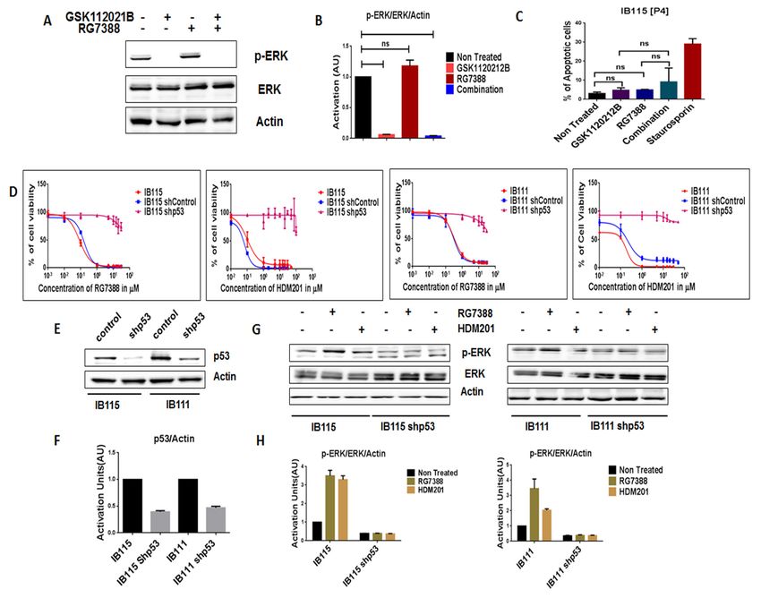
Cancers 2020, 12, 2253 2 of 15
p53 [6]. In addition, p53 binds to the promoter region of MDM2 and enhances its transcription,
thus creating an autoregulatory feedback loop that maintains the balance of p53 and MDM2 under
normal physiological conditions [7]. Activation of p53 exerts significant anticancer effects, inducing
apoptosis, controlling cell cycle progression, and promoting DNA repair and senescence [8]. In cells
with high expression of MDM2, p53 becomes inactivated, resulting in insufficient apoptosis and cell
cycle arrest [9]. Restoring the anticancer effects of p53 by blocking the MDM2–p53 interaction can be
an efficient targeted approach to treat DDLPS.
The cis-imidazolines (nutlin) compounds comprise the first class of anti-cancer agents specifically
designed to inhibit the interaction between MDM2 and p53, thereby stabilizing p53 and restoring
its anticancer effects [10,11]. These MDM2 antagonists bind to the p53-binding pocket of MDM2,
thereby blocking the MDM2–p53 interaction and increasing p53 levels [12]. More recently, additional
compounds have entered clinical development. For example, HDM201 is an imidazopyrrolidinone
scaffold-based inhibitor of the MDM2–p53 interaction with high in vitro activity/selectivity and
improved oral bioavailability and in vivo pharmacokinetic and pharmacodynamic profiles [13,14].
We and others have shown that only a minority of DDLPS patients treated with p53–MDM2 antagonists
experienced tumor shrinkage and that the median time to disease progression was only 6 months after
treatment onset. This points towards the existence of mechanisms that counterbalance the restoration
of the p53 pathway [12].
The mitogen-activated protein kinase/extracellular signal-regulated kinase (MAPK/ERK) pathway,
which is generally activated by growth factors, is known to play a crucial role in cellular proliferation,
differentiation, migration, and death [15]. It has previously been reported in human blood cancer,
colon cancer, and osteosarcoma that p53 stimulated the MAPK/ERK pathway, protecting the cancerous
cells from p53-dependent apoptosis [15,16]. Therefore, we decided to address the impact of MDM2
antagonists on the MAPK pathway in DDLPS and to determine the molecular mechanisms underlying
the potential synergy between these agents and a MAPK kinase (MEK) inhibitor.
2. Results
2.1. A MDM2 Antagonist Induces Significant Upregulation of Phosphorylated Erk in P53 Wild-Type, But Not
P53-Null, Sarcoma Cell Lines
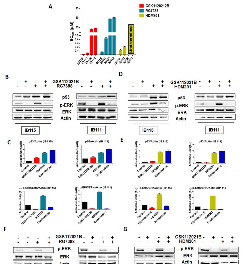
As expected, cell viability assays revealed that IB111 and IB115 cells were exquisitely sensitive to
MDM2 antagonists (RG7388 and HDM201), whereas p53-null (IB136 and IB112) cells were resistant,
with undetermined HDM201 EC50 values for IB136 and IB112 (Figure 1A and Table S1). We performed
immunoblot analysis to assess the levels of phosphorylated ERK (p-ERK) in p53 wild-type IB115
and IB111 cells and p53-null IB136 and IB112 treated with RG7388 and HDM201. We observed
that exposure of IB115 and IB111 cells to RG7388 or HDM201 induced a significant increase in the
expression of p-ERK, along with increase in the expression of p53 (Figure 1B–E). This increased
expression of p-ERK was inhibited by treatment with GSK112021B, a selective small-molecule and
adenosine triphosphate-noncompetitive inhibitor of the activation and kinase activity of MEK1 and
MEK2. In contrast, we did not observe any change in the expression of p-ERK in the p53-null IB136
and IB112 cells treated with RG7388 or HDM201 (Figure 1F–I). Collectively, these results suggested
that MDM2 antagonists induced a paradoxical activation of MEK/ERK signaling in DDLPS that could
be mediated by p53.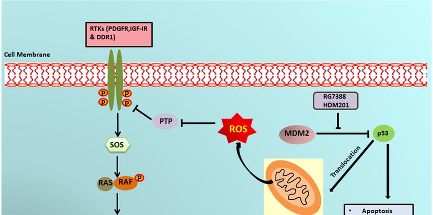
Cancers 2020, 12, 2253                                                                                                                 3 of 15
              Cancers 2020, 12, x                                                                                           3 of 15

                  Figure 1. Significant
      Figure 1. Significant                upregulation
                                 upregulation            of p-ERK
                                                    of p-ERK    bybyMDM2
                                                                      MDM2 antagonists
                                                                              antagonists  is observed  in p53 in
                                                                                               is observed      wild-type  cells.
                                                                                                                  p53 wild-type    cells.
                  (A) EC50 values for GSK112021B, RG7388, and HDM201 in p53 wild-type cells (IB115 and IB111) and
      (A) EC50 values for GSK112021B, RG7388, and HDM201 in p53 wild-type cells (IB115 and IB111) and
                  p53-null cells (IB136 and IB112). (B) Representative blots of p53 wild-type cells (IB115 and IB111)
      p53-null cells (IB136 and IB112). (B) Representative blots of p53 wild-type cells (IB115 and IB111) treated
                  treated with GSK112021B and RG7388, alone or in combination, at their EC50 values and
      with GSK112021B         and RG7388,
                  immunoblotted                   alone
                                      for p53, p-ERK,  andor  in combination,
                                                            ERK. (C) Quantificationatoftheir
                                                                                        blots ofEC 50 values
                                                                                                 IB115 and IB111 and   immunoblotted
                                                                                                                   treated with
                  GSK112021B
      for p53, p-ERK,     and ERK.and RG7388,    alone or in combination.
                                          (C) Quantification       of blots(D)
                                                                             of Representative
                                                                                 IB115 and IB111  blots oftreated
                                                                                                            p53 wild-type   cells
                                                                                                                    with GSK112021B
                  (IB115 and IB111) treated with GSK112021B and HDM201, alone or in combination, at their EC50
      and RG7388, alone or in combination. (D) Representative blots of p53 wild-type cells (IB115 and
                  values and immunoblotted for p53, p-ERK, and ERK. (E) Quantification of blots of IB115 and IB111
      IB111) treated   with GSK112021B and HDM201, alone or in combination, at their EC50 values and
                  treated with GSK112021B and HDM201, alone or in combination. (F) Representative blots of p53-null
      immunoblotted for p53, p-ERK, and ERK. (E) Quantification of blots of IB115 and IB111 treated with
            Cancers 2020, 12, x; doi:                                                                 www.mdpi.com/journal/cancers
      GSK112021B     and HDM201, alone or in combination. (F) Representative blots                            of p53-null cells (IB136
      and IB112) treated with GSK112021B and RG7388, alone or in combination, at their EC50 values and
      immunoblotted for p-ERK and ERK. (G) Quantification of blots of IB136 and IB112 cells treated with
      GSK112021B and RG7388, alone or in combination. (H) Representative blots of p53-null cells (IB136
      and IB112) treated with GSK112021B and HDM201, alone or in combination, at their EC50 values and
      immunoblotted for p-ERK and ERK. (I) Quantification of blots of IB136 and IB112 cells treated with
      GSK112021B and HDM201, alone or in combination. Immunoblot data shown represent the results of
      two independent experiments.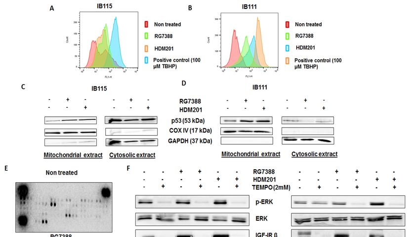
cells (IB136 and IB112) treated with GSK112021B and RG7388, alone or in combination, at their EC50
           values and immunoblotted for p-ERK and ERK. (G) Quantification of blots of IB136 and IB112 cells
           treated with GSK112021B and RG7388, alone or in combination. (H) Representative blots of p53-null
           cells (IB136 and IB112) treated with GSK112021B and HDM201, alone or in combination, at their EC50
Cancers 2020, 12, 2253
           values   and immunoblotted for p-ERK and ERK. (I) Quantification of blots of IB136 and IB112 cells   4 of 15
           treated with GSK112021B and HDM201, alone or in combination. Immunoblot data shown represent
           the results of two independent experiments.
2.2. MEK Inhibitor GSK1120212B and MDM2 Antagonists Synergistically Induce Apoptosis and G2/M Arrest
Cell Cycle Arrest
     2.2. MEK     in p53GSK1120212B
              Inhibitor  Wild-Type Cells
                                    and MDM2 Antagonists Synergistically Induce Apoptosis and G2/M
     Arrest Cell Cycle Arrest in p53 Wild-Type Cells
     Since high levels of p-ERK can affect the anti-survival and pro-apoptotic effects of MDM2
          Since
antagonists,  wehigh    levels of whether
                   investigated   p-ERK can     affect theofanti-survival
                                             inhibition                   and pro-apoptotic
                                                             p-ERK by GSK112021B        could effects
                                                                                               increaseof the
                                                                                                           MDM2
                                                                                                              response
     antagonists,
of DDLPS    cells towe   investigated
                      MDM2               whether
                                antagonists.    Weinhibition
                                                    found thatof MDM2
                                                                  p-ERK by    GSK112021B
                                                                           antagonists       could or
                                                                                         (RG7388    increase
                                                                                                       HDM201) the and
     response of DDLPS cells to MDM2 antagonists. We found that MDM2 antagonists (RG7388 or
GSK112021B synergistically inhibited the growth and induced apoptosis of p53 wild-type IB115 and
     HDM201) and GSK112021B synergistically inhibited the growth and induced apoptosis of p53 wild-
IB111 DDLPS cells (Figure 2A,B), but not p53-null IB112 and IB136 cells (Figure 2C). Moreover, cell cycle
     type IB115 and IB111 DDLPS cells (Figure 2A,B), but not p53-null IB112 and IB136 cells (Figure 2C).
analysis revealed
     Moreover,   cellthat  this
                       cycle    drug revealed
                             analysis combination     resulted
                                                that this       in significant
                                                          drug combination     G2/M arrest
                                                                             resulted         in p53 G2/M
                                                                                      in significant wild-type
                                                                                                            arrestIB115
and IB111  cells  (Figure    2D), but  not  p53-null   IB112  and  IB136  cells (Figure   2E). These  results
     in p53 wild-type IB115 and IB111 cells (Figure 2D), but not p53-null IB112 and IB136 cells (Figure 2E).   showed
the importance    of  the presence    of intact p53  for the effectiveness  of  MDM2    antagonists
     These results showed the importance of the presence of intact p53 for the effectiveness of MDM2  in  combination
with antagonists
     the MEK inhibitor       GSK112021B.
                  in combination    with the MEK inhibitor GSK112021B.

     Cancers 2020, 12, x; doi:                                                       www.mdpi.com/journal/cancers
     Figure   2. MDM2 antagonists synergize with a MEK inhibitor in p53 wild-type       cells. (A) Isobolograms
     showing the synergistic effect of RG7388 and HDM201 with GSK112021B in p53 wild-type cells (IB115
     and IB111). Effect of GSK112021B, RG7388, and HDM201, alone or in combination, on apoptosis in p53
     wild-type cells (IB115 and IB111) (B) and p53-null cells (IB136 and IB112) (C) Effect of GSK112021B,
     RG7388, and HDM201, alone or in combination, on cell cycle in p53 wild-type cells (IB115 and IB111)
     (D) and p53-null cells (IB136 and IB112) (E) * p ≤ 0.05, ** p ≤ 0.01, *** p ≤ 0.001, **** p ≤ 0.0001, and ns
     (non-significant). Data shown represent the results of three independent experiments.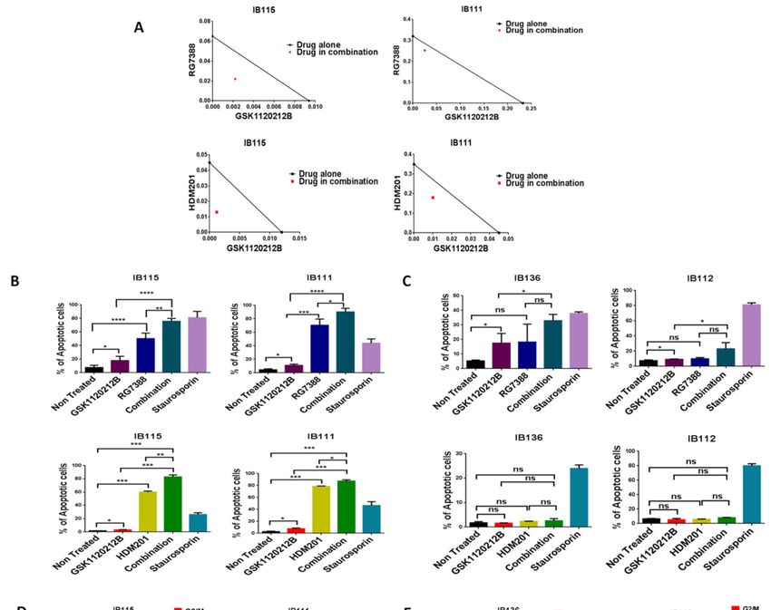
and p53-null cells (IB136 and IB112) (E) * p ≤ 0.05, ** p ≤ 0.01, *** p ≤ 0.001, **** p ≤ 0.0001, and ns (non-
         significant). Data shown represent the results of three independent experiments.

   2.3. Intact p53 Is Necessary for the MDM2 Antagonist-Induced Activation of MEK/ERK Signaling in DDLPS
         In order to further assess the importance of intact p53 for the MDM2 antagonist-induced
Cancers 2020, 12, 2253                                                                                             5 of 15
   activation of MEK/ERK signaling in DDLPS, we analyzed the expression of p-ERK in RG7388-
   resistant IB115 [P4] cells, which were derived from IB115 cells repeatedly exposed to RG7388 and
2.3.harboring
     Intact p53aIsTP53   mutation
                    Necessary       (12).
                               for the    As observed
                                       MDM2              via immunoblot
                                                Antagonist-Induced         analysis,
                                                                       Activation     treatment Signaling
                                                                                  of MEK/ERK      of IB115 [P4]  cells
                                                                                                             in DDLPS
   with RG7388 did not result in an increase in p-ERK expression (Figure 3A,B). Moreover, an apoptosis
      In order
   assay        to further
          revealed         assess the
                     that RG7388     andimportance
                                          GSK112021B  of were
                                                          intactnot
                                                                 p53synergistic
                                                                     for the MDM2    antagonist-induced
                                                                                (Figure   3C) in IB115 [P4].activation
                                                                                                               This is
of MEK/ERK       signaling  in  DDLPS,     we  analyzed     the expression   of p-ERK
   similar to the results observed in the p53-null cells (IB112 and IB136; Figure 2C).   in  RG7388-resistant IB115
[P4] cells,
         Wewhich     were derived
              then investigated    thefrom  IB115
                                        impact   of cells  repeatedly exposed
                                                    shRNA-mediated               to RG7388
                                                                         p53 silencing         and harboring
                                                                                         on IB115                  TP53
                                                                                                     and IB111 acells
mutation
   (Figure (12).
             3E,F).As  observed
                     Using a cell via  immunoblot
                                   viability  assay, weanalysis,  treatment
                                                           observed           of IB115 [P4]
                                                                     that p53-silenced    cellscells withresistant
                                                                                                became     RG7388to   did
notMDM2
     result in an  increase in  p-ERK    expression   (Figure  3A,B).  Moreover,   an  apoptosis
             antagonists (Figure 3D). Moreover, they did not induce any upregulation of p-ERK, as  assay revealed    that
RG7388    andin
   observed     GSK112021B      werecells
                  non-transfected      not (Figure
                                           synergistic
                                                    3G,H).(Figure
                                                             These3C)  in IB115
                                                                    results      [P4]. This
                                                                            confirmed    our is  similar tothat
                                                                                              prediction     theintact
                                                                                                                 results
   p53 is essential
observed              for MDM2
            in the p53-null  cellsantagonist-induced
                                     (IB112 and IB136;activation     of MEK/ERK signaling in DDLPS.
                                                           Figure 2C).

     Figure
        Figure    Intact
              3. 3.  Intactfunctional
                              functionalp53
                                          p53isisnecessary
                                                  necessary forfor MDM2     antagoniststo
                                                                   MDM2 antagonists       toactivate
                                                                                             activateMEK/ERK
                                                                                                      MEK/ERK     signaling
                                                                                                                signaling  in in
     dedifferentiated       liposarcoma.      (A)  Representative     blots   of IB115  [P4]  treated with
        dedifferentiated liposarcoma. (A) Representative blots of IB115 [P4] treated with GSK112021B and    GSK112021B      and
     RG7388,    alone    or  in combination,     at
        RG7388, alone or in combination, at the EC50the  EC     values  for IB115  and  immunoblotted     for p-ERK
                                                             50 values for IB115 and immunoblotted for p-ERK and ERK.and   ERK.
     (B)(B)
          Quantification
             Quantification    ofof
                                  blots
                                    blotsofofIB115
                                              IB115[P4]
                                                      [P4]cells
                                                           cellstreated
                                                                 treated with
                                                                          with GSK112021B       and/orRG7388.
                                                                                 GSK112021B and/or      RG7388.(C)(C) Effect
                                                                                                                    Effect of of
     GSK112021B
        GSK112021B     andand RG7388,
                                RG7388,alone
                                          aloneororinincombination,
                                                        combination, at at the
                                                                           the EC50  valuesfor
                                                                                  50 values  forIB115
                                                                                                 IB115ononapoptosis
                                                                                                           apoptosisinin IB115
                                                                                                                       IB115
     [P4] 2020,
   Cancers cells.12,(D) Efficacy of RG7388 and HDM201 in p53 wild-type and silenced
                     x; doi:                                                                            IB115 and IB111 cells
                                                                                                    www.mdpi.com/journal/cancers
     treated with increasing doses for 72 h. (E) Representative blots of p53 wild-type and silenced IB115 and
     IB111 immunoblotted for p53 to confirm p53 silencing. (F) Quantification of blots of p53 wild-type and
     silenced IB115 and IB111 cells immunoblotted for p53. (G) Representative blots of p53 wild-type and
     silenced IB115 and IB111 treated with RG7388 and HDM201 and immunoblotted for p-ERK and ERK.
     (H) Quantification of blots of p53 wild-type and silenced IB115 and IB111 cells treated with RG7388
     and HDM201 and immunoblotted for p-ERK and ERK. ns (non-significant). Data represent the results
     of two experiments. Immunoblot data represent the results of two independent experiments and
     apoptosis data represent the results of three independent experiments.

     We then investigated the impact of shRNA-mediated p53 silencing on IB115 and IB111 cells
(Figure 3E,F). Using a cell viability assay, we observed that p53-silenced cells became resistant to MDM2
antagonists (Figure 3D). Moreover, they did not induce any upregulation of p-ERK, as observed in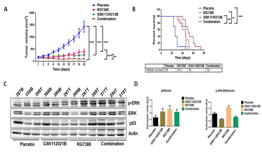
[P4] cells. (D) Efficacy of RG7388 and HDM201 in p53 wild-type and silenced IB115 and IB111 cells
       treated with increasing doses for 72 h. (E) Representative blots of p53 wild-type and silenced IB115
       and IB111 immunoblotted for p53 to confirm p53 silencing. (F) Quantification of blots of p53 wild-
       type and silenced IB115 and IB111 cells immunoblotted for p53. (G) Representative blots of p53 wild-
       type and silenced IB115 and IB111 treated with RG7388 and HDM201 and immunoblotted for p-ERK
Cancersand    12, 2253
        2020,ERK.  (H) Quantification of blots of p53 wild-type and silenced IB115 and IB111 cells treated with 6 of 15
       RG7388 and HDM201 and immunoblotted for p-ERK and ERK. ns (non-significant). Data represent
       the results of two experiments. Immunoblot data represent the results of two independent
non-transfected cells (Figure 3G,H). These results confirmed our prediction that intact p53 is essential
       experiments and apoptosis data represent the results of three independent experiments.
for MDM2 antagonist-induced activation of MEK/ERK signaling in DDLPS.
 2.4.Combination
2.4. CombinationTreatment
                  TreatmentofofGSK1120212B
                                GSK1120212Band
                                            andRG7388
                                                RG7388Resulted
                                                       resultedin
                                                                inDecreased
                                                                  DecreasedTumor
                                                                            TumorVolume
                                                                                  Volumeand
                                                                                         andIncreased
 Increased Survival
Survival of Mice    of Mice
       In order
      In   order to
                  to validate
                      validate our
                                our in
                                     in vitro
                                        vitro findings,
                                               findings, wewe treated
                                                               treated mice
                                                                        mice engrafted
                                                                              engrafted with
                                                                                          with IB115
                                                                                                IB115 cells
                                                                                                        cellswith
                                                                                                               with
  GSK1120212B     and  RG7388   as single  agents  and  in combination.  Through  immunoblot
GSK1120212B and RG7388 as single agents and in combination. Through immunoblot analysis,         analysis,  it was
itfound   that, that,
   was found    consistent   with with
                      consistent   the in
                                        thevitro   findings,
                                             in vitro         treatment
                                                      findings,  treatmentwith RG7388
                                                                            with RG7388induced
                                                                                           induceda asignificant
                                                                                                       significant
upregulation of MEK/ERK signaling (Figure 4C,D) that was not observed with the
  upregulation    of MEK/ERK      signaling   (Figure  4C,D)   that was   not observed   with   the combination
                                                                                                     combination
  treatment.Importantly,
treatment.    Importantly,thethecombination
                                 combinationtreatment
                                                treatmentsignificantly
                                                            significantly decreased
                                                                        decreased thethe tumor
                                                                                       tumor     volume
                                                                                             volume       (Figure
                                                                                                       (Figure  4A)
  4A)  and   increased   the  survival   of  mice   (Figure  4B)  compared    to treatment   with
and increased the survival of mice (Figure 4B) compared to treatment with single-agent GSK1120212B  single-agent
orGSK1120212B
    RG7388.       or RG7388.

     Figure 4. Combination treatment of GSK1120212B and RG7388 resulted in decreased tumor volume and
      Figure 4. Combination treatment of GSK1120212B and RG7388 resulted in decreased tumor volume
     increased survival of mice. (A) Effect of GSK112021B and RG7388, alone or in combination, on tumor
      and increased survival of mice. (A) Effect of GSK112021B and RG7388, alone or in combination, on
     growth. (B) Kaplan–Meier curve depicting overall survival of mice treated with GSK1120212B and/or
      tumor growth. (B) Kaplan–Meier curve depicting overall survival of mice treated with GSK1120212B
     RG7388. (C) Representative blots of tumor tissues from mice treated with GSK112021B, RG7388, or the
      and/or RG7388. (C) Representative blots of tumor tissues from mice treated with GSK112021B,
     combination and immunoblotted for p-ERK, ERK, and p53. (D) Quantification of blots of tumor
      RG7388, or the combination and immunoblotted for p-ERK, ERK, and p53. (D) Quantification of blots
     tissues from mice treated with GSK112021B, RG7388, or the combination. ** p ≤ 0.01, *** p ≤ 0.001,
      of tumor tissues from mice treated with GSK112021B, RG7388, or the combination. ** p ≤ 0.01, *** p ≤
     and **** p ≤ 0.0001.
      0.001, and **** p ≤ 0.0001.
2.5. MDM2 Antagonist-Induced Activation of MEK/ERK Signaling in DDLPS Is Associated with
 2.5. MDM2 Antagonist-Induced
Mitochondrial                     Activation
               Translocation of p53          of MEK/ERK
                                    and Generation           Signaling
                                                     of Reactive Oxygenin DDLPS
                                                                          Species Is Associated with
 Mitochondrial Translocation of p53 and Generation of Reactive Oxygen Species
      The results obtained led us to investigate whether MDM2 antagonist-induced activation
       The results
of MEK/ERK          obtained
                signaling  in led us towas
                               DDLPS     investigate
                                              related whether     MDM2
                                                        to a direct        antagonist-induced
                                                                     interaction  between p53activation
                                                                                                  and ERKofor
 MEK/ERK
another      signalinglinked
         mechanism      in DDLPS   was related and
                              the MDM2–p53       to a ERK/MAPK
                                                      direct interaction  between
                                                                     pathways.      p53 andwe
                                                                                 Therefore,   ERK   or another
                                                                                                 performed    an
 mechanism linked the
immunoprecipitation         MDM2–p53
                         analysis in IB115and
                                            cells,ERK/MAPK
                                                   which revealedpathways.   Therefore,
                                                                      no direct           we performed
                                                                                physical interaction         an
                                                                                                       between
 immunoprecipitation
p53                      analysis
     and p-ERK, as no p-ERK       in IB115
                                bands  werecells,  whichinrevealed
                                             observed                no direct physical samples
                                                             p53-immunoprecipitated      interaction  between
                                                                                                   (Figure S1).
 p53 Previous
      and p-ERK,
               studies suggested that p53 promotes the production of reactive oxygen species (ROS)S1).
                   as no p-ERK   bands were   observed    in p53-immunoprecipitated     samples   (Figure   [17],
and since ROS have been documented to activate ERK1/2 [18], we investigated the role played by
ROS   in 2020,
 Cancers  RG7388-     and HDM201-induced phosphorylation of ERK. The DCFDA
               12, x; doi:                                                 Cellular ROS Detection
                                                                        www.mdpi.com/journal/cancers
Assay revealed greater accumulation of ROS in IB115 and IB111 cells treated with RG7388 and
HDM201 than untreated cells (Figure 5A,B). It had been previously reported that p53 translocates
to mitochondria, the main site of ROS production [19]. This led us to investigate the connection
between ROS accumulation and mitochondrial translocation of p53 in IB115 and IB111 cells upon
treatment with RG7388 and HDM201. Immunoblot analysis of p53 in subcellular fractions showed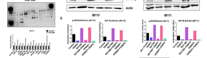
Previous studies suggested that p53 promotes the production of reactive oxygen species (ROS)
     [17], and since ROS have been documented to activate ERK1/2 [18], we investigated the role played
     by ROS in RG7388- and HDM201-induced phosphorylation of ERK. The DCFDA Cellular ROS
     Detection Assay revealed greater accumulation of ROS in IB115 and IB111 cells treated with RG7388
     and HDM201 than untreated cells (Figure 5A,B). It had been previously reported that p53 translocates
Cancers
     to 2020, 12, 2253
         mitochondria,    the main site of ROS production [19]. This led us to investigate the connection           7 of 15

     between ROS accumulation and mitochondrial translocation of p53 in IB115 and IB111 cells upon
     treatment with RG7388 and HDM201. Immunoblot analysis of p53 in subcellular fractions showed
greater accumulation of p53 in the mitochondrial extract of IB115 and IB111 cells upon treatment
     greater accumulation of p53 in the mitochondrial extract of IB115 and IB111 cells upon treatment with
with RG7388 and HDM201. COX IV was used as the loading control for the mitochondrial extract,
     RG7388 and HDM201. COX IV was used as the loading control for the mitochondrial extract, while
while   GAPDH
     GAPDH      was was  used
                       used as as
                               thethe   loading
                                     loading      control
                                               control for for cytosolic
                                                            cytosolic     extract
                                                                      extract      (Figure
                                                                               (Figure     5C,D).
                                                                                       5C,D).     Based
                                                                                              Based      on the
                                                                                                     on the       results
                                                                                                              results
obtained,
     obtained, we predicted that the mitochondrial translocation of p53 in IB115 and IB111 cells resulted in
            we    predicted  that  the   mitochondrial    translocation   of  p53  in IB115 and IB111  cells resulted
the induction     of ROSofand
     in the induction        ROSthatandthis increased
                                          that          accumulation
                                                this increased           of ROSofmight
                                                                accumulation             be involved
                                                                                    ROS might         in the induction
                                                                                                be involved    in the
of p-ERK.
     induction of p-ERK.

      Figure   5. The
           Figure   5. Themitochondrial
                             mitochondrialtranslocation
                                              translocation of
                                                             of p53 and
                                                                     and p53-induced
                                                                           p53-inducedproduction
                                                                                            production of of reactive
                                                                                                          reactive     oxygen
                                                                                                                   oxygen
      species   (ROS)
           species  (ROS)upon
                            upontreatment
                                    treatmentwith
                                               withMDM2
                                                     MDM2antagonists      ledto
                                                             antagonists led   tothe
                                                                                   thephosphorylation
                                                                                       phosphorylation     of ERK
                                                                                                        of ERK      in DDLPS
                                                                                                                in DDLPS
      cellscells through
            through     thethe  activationof
                              activation   of receptor
                                              receptor tyrosine
                                                         tyrosinekinases (RTKs).
                                                                   kinases   (RTKs).(A,B)(A,B)
                                                                                          MDM2    antagonists
                                                                                               MDM2           RG7388RG7388
                                                                                                        antagonists    and
           HDM201      induced    generation  of ROS   in IB115 and IB111   cells, which   were stained
      and HDM201 induced generation of ROS in IB115 and IB111 cells, which were stained with DCFDA      with  DCFDA    dye
           for 30  min  at 37  °C and
                                  ◦    treated for 4 h, and the intensities of intracellular DCF   were
      dye for 30 min at 37 C and treated for 4 h, and the intensities of intracellular DCF were analyzed analyzed  by flow
           cytometer.
      by flow            (C,D) MDM2
                 cytometer.              antagonist
                                (C,D) MDM2           (RG7388 and
                                                 antagonist        HDM201)-induced
                                                              (RG7388                     generation ofgeneration
                                                                          and HDM201)-induced           ROS is dependent
                                                                                                                     of ROS is
           on the mitochondrial translocation of p53. IB115 (C) and IB111 (D) were treated with RG7388 and
      dependent on the mitochondrial translocation of p53. IB115 (C) and IB111 (D) were treated with RG7388
           HDM201 for 24 h. After 24 h, the cells were subjected to subcellular fractionation into cytosolic and
      and HDM201 for 24 h. After 24 h, the cells were subjected to subcellular fractionation into cytosolic
           mitochondrial fractions for immunoblot analysis of p53. COX IV and GAPDH were used as
      and mitochondrial fractions for immunoblot analysis of p53. COX IV and GAPDH were used as
      mitochondrial
     Cancers 2020, 12, x;and
                          doi: cytosolic loading controls, respectively. (E) RG7388 treatment         on IB115 cells resulted
                                                                                                  www.mdpi.com/journal/cancers
      in an increased expression of Insulin R and PDGFR family RTKs through Phospho-RTK array profiling.
      Shown is a representative example of phosphoprotein array of control and RG7388-treated IB115 using
      the Proteome Profiler Human Phospho-RTK Array Kit. This platform allowed simultaneous screening
      of 49 different RTKs. (F) Effect of TEMPO on RG7388- and HDM201-induced phosphorylation of
      ERK. DDLPS IB115 and IB111 cells were treated with 2 mM TEMPO either alone or in combination
      with RG7388 or HDM201 for 24 h. Cells were subjected to immunoblot analysis against p-ERK, ERK,
      and IGF-1Rβ. Actin was used as the loading control. (G) Quantification of blots of IB115 and IB111 on
      treatment with TEMPO, RG7388, and HDM201 alone or in combination.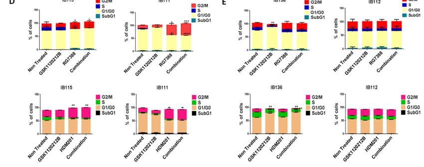
Cancers 2020, 12, 2253                                                                                8 of 15

2.6. Phosphorylation of Receptor Tyrosine Kinases by ROS Leads to the Activation of the ERK Pathway
      Our next question was how ROS were involved in the induction of p-ERK. It has been reported
previously that ROS can activate receptor tyrosine kinases (RTKs) by inhibiting protein tyrosine
phosphatases (PTPs). Activation of RTKs can cause phosphorylation of ERK [18]. To confirm this,
we performed Human Phospho-RTK Array profiling in untreated and RG7388-treated IB115 cells.
We observed upregulation of several RTKs in RG7388-treated IB115 cells, including IGF-1R, Insulin R,
DDR1, and the PDGFR family of RTKs (Figure 5E).
      To confirm that ROS were involved in the phosphorylation of ERK through the activation of RTKs,
we used a ROS scavenger, TEMPO. DDLPS IB115 and IB111 cells were treated with 2 mM TEMPO and
incubated in the presence or absence of RG7388 or HDM201 for 24 h. Immunoblot analysis revealed
an upregulation of p-ERK along with the RTK IGF-1Rβ upon treatment with RG7388 or HDM201,
with this upregulation of p-ERK and RTK IGF-1Rβ being significantly decreased in TEMPO-treated
cells (Figure 5F,G). These findings demonstrated that MDM2 antagonist-induced ROS production and
accumulation led to the phosphorylation of ERK through activation of RTKs like IGF-1Rβ.

3. Discussion
     The MDM2 gene has been found to be amplified in several human tumors, including in situ and
invasive breast adenocarcinomas [20], esophageal cancer [21], sarcomas (either common bone and soft
tissue forms) [22–24], and endometrial stromal tumors [25]. Importantly, MDM2 gene amplification
and TP53 mutation are usually exclusive events [24]. Therefore, several molecules have been developed
to inhibit the interaction between MDM2 and p53 and to restore the antitumor activity of p53. DDLPS
is one of the only tumor types (with intimal sarcomas) characterized by a consistent amplification of
MDM2 and is therefore an ideal tumor type in which to evaluate the clinical activity of such compounds.
However, all of the clinical trials conducted so far reported only modest antitumor activity, with tumor
shrinkage observed in only a minority of patients.
     The results we report here indicate that one explanation for the limited activity of MDM2
antagonists in DDLPS is the paradoxical activation of the MAPK pathway. The MAPK pathway is
known to transmit oncogenic signals, promoting cellular proliferation, differentiation, and migration
and inhibiting apoptosis [15,26]. Previous studies have suggested a link between p53 signaling and
the MAPK pathway. While p53 stimulated the ERK/MAPK pathway in several cancer types [15,16],
Pan et al. demonstrated a p53-mediated negative regulation of the ERK/MAPK pathway using RG7388
in acute myeloid leukemia [27]. Our results showed upregulation of p-ERK when DDLPS cells were
treated with two different MDM2 antagonists, RG7388 and HDM201, confirming that this event is not
related to a specific chemical class of MDM2–p53 interaction inhibitors. This increase in p-ERK levels
could explain the primary resistance to MDM2 antagonists observed in the majority of DDLPS patients
enrolled in previous clinical studies. Limited biomarker data are available from such studies. Twenty
patients with DDLPS were enrolled in a neoadjuvant biopsy-driven biomarker study investigating the
impact of the MDM2 antagonist RG7112 on the p53 pathway and proliferation through sequential
biopsies [10]. Interestingly, tumor biopsy samples from 6 of the 11 DDLPS patients with available
pre-treatment and end of protocol biopsies exhibited an increase in Ki-67-positive cells.
     We also elucidated the possible mechanism behind the upregulation of p-ERK induced by MDM2
antagonists. We first showed that intact p53 was indispensable for the stimulation of the MAPK/ERK
pathway. Since previous studies showed that upregulation of p-ERK may be related to p53-mediated
production of ROS, we investigated the role of this mechanism in DDLPS.
     It has previously been reported that a small portion of endogenous MDM2 can enter the
mitochondria and control mitochondrial dynamics and respiration independent of p53. Mitochondrial
MDM2 can enhance ROS production by repressing the transcription of the gene encoding
NADH-dehydrogenase 6 (MT-ND6) in vitro and in vivo, affecting respiratory complex I activity [28].
Apart from MDM2, p53 can trigger the transcription of pro-oxidant genes like quinone oxidoreductase
(NQO1, PIG3), proline oxidase (POX, PIG6), PUMA, BAX, and p66Shc and repress anti-oxidant genes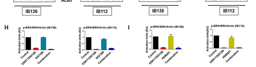
Cancers 2020, 12, 2253                                                                                             9 of 15

like manganese superoxide dismutase (MnSOD) [17], thereby causing an imbalance between ROS
production and ROS scavenging. This imbalance leads to accumulation of ROS. It has been seen that in
response to any kind of stress, there is an upregulation of p53 expression, and a fraction of this elevated
p53 (2%) translocates to mitochondria [17,29,30]. The p53 that translocates into the mitochondria
binds to MnSOD and inhibits its ROS scavenging activity, promoting ROS generation [19]. ROS can
increase phosphorylation of RTKs by inhibiting PTPs, which in turn activate Ras and, subsequently,
the MAPK/ERK pathway [18,31]. Our results showed that upregulation of p53 resulting from
treatment of DDLPS with MDM2 antagonists induced an upregulation of ROS through mitochondrial
translocation of p53. Interestingly, we observed that treatment of DDLPS with MDM2 antagonists
induced an upregulation of RTKs, including IGF-1R and PDGFRβ. These results are in line with
those of previous studies showing that nutlin increased activation of RTKs such as IGF-1R in other
tumor models [32]. Immunoblot analysis revealed increased expression of p-ERK along with IGF-1R
β in MDM2 inhibitor-treated IB115 and IB111 cells, which was not observed when these cells were
co-treated with the ROS scavenger TEMPO. Our results confirmed that MDM2 antagonists caused
accumulation of p53 that translocated inside the mitochondria and caused elevated generation of ROS.
This increased production of ROS resulted in phosphorylation of RTKs, perhaps through inhibition of
PTPs.  Activation of RTKs then led to the activation of the MAPK/ERK pathway (Figure 6). 10 of 15
    Cancers 2020, 12, x

          Figure
      Figure      6. Schematic
              6. Schematic     representationof
                            representation    ofMDM2
                                                MDM2 antagonist-induced
                                                        antagonist-inducedphosphorylation
                                                                              phosphorylation of ERK1/2  through
                                                                                                 of ERK1/2   through
          mitochondrial   p53. MDM2    antagonists activate p53, which  translocates into the mitochondria
      mitochondrial p53. MDM2 antagonists activate p53, which translocates into the mitochondria             and and
          causes  elevated production of reactive oxygen  species (ROS). The ROS  produced   inhibits the
      causes elevated production of reactive oxygen species (ROS). The ROS produced inhibits the protein  protein
          tyrosine phosphatases (PTPs), resulting in phosphorylation of receptor tyrosine kinases (RTKs) like
      tyrosine phosphatases (PTPs), resulting in phosphorylation of receptor tyrosine kinases (RTKs) like
          PDGFR, IGF-1R, and DDR1. The activated RTKs then activate the ERK pathway.
      PDGFR, IGF-1R, and DDR1. The activated RTKs then activate the ERK pathway.
     4. Materials and Methods
      These findings have important clinical implications. Indeed, identifying synergistic combinations
is crucial  to and
     4.1. Cells developing   successful MDM2 antagonist-based therapeutic strategies for liposarcoma.
                   Cell Culture
Previous studies have reported that the combination of nongenotoxic nutlin with genotoxic drugs
           The DDLPS (IB115 and IB111) and leiomyosarcoma (IB136 and IB112) cell lines used in this study
synergistically   activated p53 functions, paving the way for the possible use of nutlin in combinatorial
     were derived from human surgical specimens (Institut Bergonié, Bordeaux, France) after obtaining
     patient consent. The MDM2 antagonist-resistant IB115 [P4] cell line was derived from IB115 cells
     through repeated exposure to the nutlin compound RG7388, as previously described [12]. Cells were
     maintained in RPMI medium 1640 (Sigma Life Technologies, St. Louis, MO, USA) with 10% fetal calf
     serum (Dutscher, France) in a humidified incubator containing 5% CO2 maintained at 37 °C.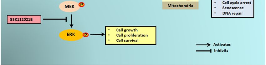
Cancers 2020, 12, 2253                                                                              10 of 15

drug therapy [3,33]. The effect of combination treatment of nutlin with cytotoxic drugs currently
in use (e.g., cisplatin, methotrexate, and doxorubicin) on sarcoma cells was investigated by
Ohnstad et al. [10,34]. Of all the drugs tested, doxorubicin is the only drug that is used in the treatment
of DDLPS. The authors observed a synergy that bolstered the development of this combination
in the clinic. Our group recently reported that, in a phase 1 study, combination therapy of the
nutlin compound RG7112 with doxorubicin potentiated p53 activation in a randomly selected patient
population with advanced soft tissue sarcoma [35]. However, the combination therapy led to a high rate
of grade 3 and 4 hematological toxicity, with 60% and 45% of patients experiencing acute neutropenia
or thrombocytopenia, respectively, blocking its future clinical development. Therefore, combination
therapy using nutlin and targeted nongenotoxic drugs may be a more appropriate approach. Our data
provide the first in vivo evidence that inhibition of MAPK signaling can augment anti-tumor activity
of a MDM2 antagonist in human tumor models.

4. Materials and Methods

4.1. Cells and Cell Culture
     The DDLPS (IB115 and IB111) and leiomyosarcoma (IB136 and IB112) cell lines used in this study
were derived from human surgical specimens (Institut Bergonié, Bordeaux, France) after obtaining
patient consent. The MDM2 antagonist-resistant IB115 [P4] cell line was derived from IB115 cells
through repeated exposure to the nutlin compound RG7388, as previously described [12]. Cells were
maintained in RPMI medium 1640 (Sigma Life Technologies, St. Louis, MO, USA) with 10% fetal calf
serum (Dutscher, France) in a humidified incubator containing 5% CO2 maintained at 37 ◦ C.

4.2. Reagents
     RG7388 was supplied by Roche (Roche Pharma Research & Early Development, Basel, Switzerland)
and was dissolved in the solution supplied by Roche to a concentration of 10 mM as a stock solution and
stored at −20 ◦ C. GSK1120212 (MEK inhibitor) and HDM201 were purchased from Selleck Chemicals
(Houston, TX, USA) and Novartis (Basel, Switzerland), respectively and were prepared as 10 mmol/L
stock solutions in DMSO and stored at −20 ◦ C. 2,2,6,6-Tetramethylpiperidine-1-oxyl (free radical;
(TEMPO) was purchased from Sigma-Aldrich.

4.3. Cell Viability and Synergy Assay
      The effects of GSK112021B, RG7388, and HDM201 on cell viability were investigated using the
3-(4, 5-dimethylthiazol-2-yl)-2, 5-diphenyl tetrazolium bromide (MTT) assay (Sigma-Aldrich Chimie,
Saint-Quentin-Fallavier, France). Briefly, cells were seeded at a density of 3000 cells/well in 96-well
plates and incubated for 24 h. Cells were then treated with increasing concentrations of the respective
compounds for 72 h at 37 ◦ C. After 72 h, MTT at a concentration of 0.5 mg/mL was added to each well
and was incubated for 3–4 h to allow the formation of formazan crystals. The crystals were dissolved in
DMSO and the absorbance of the colored solution was measured on a microplate-photometer (Bio-Tek
Instruments, Colmar, France) using a test wavelength of 570 nm and a reference wavelength of 630 nm.
The concentrations of substance required for 50% growth inhibition (EC50 ) were estimated using
GraphPad Prism software (GraphPad Software Inc., San Diego, CA, USA).
      To measure synergistic effects between two drugs, a diagonal constant ratio combination design
was used according to the protocol by Chou and Talalay [36]. Cells were incubated in a 2-fold
serial dilution of the two drugs at a constant ratio, with several concentrations above and below the
EC50 values of the drugs. After 72 h of incubation, MTT was immediately added to the wells and
the absorbance was measured. The analysis of synergy was performed using the isobologram and
combination index (CI) methods derived from the median effect principle of Chou and Talalay [36].
The combination effects of two compounds can be summarized as follows: CI < 1 (under the curve),
Cancers 2020, 12, 2253 11 of 15
CI = 1 (near the curve), and CI > 1 (above the curve) indicate synergistic, additive, and antagonistic
effects, respectively.
4.4. Apoptosis
For apoptosis analysis, 2 × 10⁵ cells/well were seeded in a 6-well plate. After 24 h, cells were
treated with the EC50 value of the compounds alone or in combination for 72 h at 37 ◦ C. After 72 h,
cells were exposed to FITC-Annexin V and propidium iodide (PI) according to the manufacturer’s
protocol (BD Biosciences, San Jose, CA, USA). Cells were analyzed by flow cytometry using FL1 for
Annexin V and FL2 for PI. The flow cytometry data (FACS Calibur; BD Biosciences) were analyzed
with FlowJo v.7.6.3 software (Version 10.2. Ashland, OR: Becton, Dickinson and Company; 2019.).
4.5. Cell Cycle Analysis
Cells were seeded at a density of 2 × 10⁵ cells/well followed by starvation for 6 h. The cells were
then treated with the EC50 values of the compounds alone or in combination for 48 h. After 48 h,
cells were harvested and centrifuged at 1500× g for 5 min and washed twice with phosphate-buffered
saline (PBS). Cells were then fixed and permeabilized with 70% ethanol at 4 ◦ C overnight. Ethanol
was removed and cells were washed twice with PBS. Next, 300 µL of a propidium iodide- and
ribonuclease-containing solution were added to cells, which were then incubated in the dark for 30 min,
followed by analysis by flow cytometry (FACS Calibur; BD Biosciences). The data obtained were
analyzed with FlowJo v.7.6.3. software and results were expressed as percentage of cells in a given
phase of the cell cycle.
4.6. Immunoblot
4.6.1. Protein Extraction from Cells
Non-treated and 24 h-treated cells were harvested in radioimmunoprecipitation assay lysis buffer
(150 mM NaCl, 50 mM Tris pH 7.5, 1% NP-40) containing a mix of protease inhibitors (Roche) and
phosphate inhibitors (1 mM NaO and 1 mM NaF). The lysate was centrifuged at 13,000 rpm for 10 min
at 4 ◦ C and the supernatant was collected and stored at −20 ◦ C.
4.6.2. Protein Extraction from Mouse Xenograft Tumors
Frozen tumor tissues stored in liquid nitrogen were broken into small pieces, and the piece to
be lysed was weighed. To the tissues, 5 µL/mg of lysis buffer (150 mM NaCl, 10 mM Tris pH 7.5,
1 mM EDTA, 1% NP-40, NaO, and 10 mM NaF containing a mix of protease inhibitors [Roche]) was
added. The tissues were homogenized in the lysis buffer using a tissue homogenizer. The lysates were
incubated in ice for 30 min followed by centrifugation at 13,000 rpm for 15 min at 4 ◦ C. The supernatant
was collected and stored at −20 ◦ C.
4.6.3. Mitochondrial/Cytosolic Fraction Isolation and Immunoblot Analysis
The mitochondrial/cytosolic fractions from IB115 and IB111 cells were isolated according to the
manufacturer’s protocol (Mitochondria/Cytosol Fractionation Kit, ab65320 (Abcam, Cambridge, MA,
USA). Next, 30 µg of protein extract was separated on SDS-polyacrylamide gel and transferred onto
PVDF membrane. The membrane was blocked with 5% blocking solution for 1 h at room temperature
(RT) and then probed with the primary antibody (1:1000) at 4 ◦ C overnight. The membrane was
washed with PBS with 0.1% Tween 20 and probed with horseradish peroxidase (HRP)-conjugated
secondary antibody (1:5000) for 2 h at RT. The blots were visualized on a Fusion FX7 imaging
system (Fisher Bioblock Scientific, Waltham, MA, USA) using the ImmobilonTM Western enhanced
chemiluminescence detection kit (Millipore Corporation, Billerica, MA, USA). The bands obtained
were analyzed and quantified using ImageJ® 1.49g software (National Institutes of Health, Bethesda,
MD, USA). The primary antibodies used were: anti-p53 (1:200, Santa Cruz sc-126); anti-p-ERK1/2Cancers 2020, 12, 2253 12 of 15
thr202/tyr204 (1:1000, CST 4370); anti-ERK1/2 (1:1000 ab17942); anti-IGF-1R (1:1000 CST 3027), anti-Actin
(1:1000 Sigma Aldrich A3853); anti-COX IV (1:1000, CST 4844) and anti-GAPDH (1:1000, Santa Cruz
sc-51907). HRP-conjugated secondary antibodies against rabbit or mouse IgG were obtained from
Santa Cruz. Glycine buffer (0.15 M, pH 2) was used to strip the membranes.
4.7. Cellular ROS Detection Assay
For this assay, 1.5 × 10⁴ cells/well were seeded in a 6-well plate. After 24 h, cells were trypsinized
and harvested. The harvested cells were stained according to the manufacturer’s protocol (DCFDA
Cellular ROS Detection Assay Kit, ab113851). Cells stained with 100 µM tert-butyl hydrogen peroxide
(TBHP) for 4 h before staining with DCFDA dye were the positive control.
4.8. Human Phospho-RTK Array
Non-treated and treated cells were solubilized in the lysis buffer provided in the kit (Human
Phospho-RTK Array Kit, R&D Systems, catalog number ARY001B). The lysates were centrifuged at
14,000× g for 5 min at 4 ◦ C and the supernatant was collected and stored at −80 ◦ C. The assay was
performed according to the manufacturer’s protocol.
4.9. Gene Silencing by Lentiviral Infection
The pLVTH-sip53 vector expressing green fluorescent protein (GFP) and a short hairpin RNA
targeting TP53 (plasmid 12239) and control vector pLVTH (plasmid 12262) were obtained from
Addgene. Viral particles were produced by calcium phosphate transfection of 293T cells. Infections
were performed at a multiplicity of 10 infectious units per cell. The efficacy of infection was checked
by assessing GFP expression via flow cytometry and p53 silencing was assessed by immunoblot.
4.10. Immunoprecipitation
Cell lysates were prepared using RIPA lysis buffer. The lysates were precleared at 4 ◦ C for 30 min
by adding 1 µg of IgG2a together with 20 µL of re-suspended volume of Protein A/G PLUS-Agarose.
The beads were pelleted at 2500 rpm for 5 min at 4 ◦ C and the precleared supernatant was collected.
Next, 1 µg of primary antibody was added to 500 µg of cellular protein and was incubated for 1 h at
4 ◦ C. Next, 20 µL of re-suspended volume of Protein A/G PLUS-Agarose was added and incubated
at 4 ◦ C on a rocker platform overnight. The immunoprecipitants were collected by centrifugation
at 2500 rpm for 5 min at 4 ◦ C. The supernatant was discarded carefully and the pellet obtained was
washed with PBS. After the final wash with PBS, the pellet was resuspended in electrophoresis buffer
and immunoblot was performed.
4.11. Animal Studies
All animal experiments were performed with the approval of the institutional animal use and care
committee under project license APAFIS#8415-2017010211442345 (University of Bordeaux). This study
followed the French and European Union guidelines for animal experimentation (RD 1201/05, RD
53/2013 and 86/609/CEE, respectively). IB115 cells (5 × 10⁶ cells/200 µL) were inoculated subcutaneously
into the right flank of RagΥ2C −/− mice (n = 10 per group). Once palpable, tumor volumes were
calculated using the following formula: length × width2 /2. Once the average size of the tumors
was 100 mm3 , animals were treated with RG7388 and GSK112021B via oral gavage. The mice
were randomized into four groups: vehicle, RG7388 alone (25 mg/kg, oral gavage five times per
week), GSK112021B alone (0.5 mg/kg oral gavage five times per week), and both drugs (RG7388 and
GSK112021B five times per week at 25 and 0.5 mg/kg, respectively). RG7388 was dissolved in a solution
supplied by Roche. GSK1120212 was prepared by dissolving in 1 volume of 1-methyl-2-pyrrolidone
(NMP) in a 100 ◦ C water bath followed by the addition of 9 volumes of PEG300 (Sigma-Aldrich,
St Quentin Fallavier, France). Mice in each group were treated for 3 weeks, after which treatment wasCancers 2020, 12, 2253 13 of 15
stopped and tumors were measured every 2–3 days with calipers and the diameters were recorded.
Mice were euthanized when the tumor volume reached 2000 mm3 . Tumor progression was analyzed
with GraphPad Prism software, and Kaplan–Meier curve analysis was used to compare the overall
survival. Log-rank (Mantel–Cox) tests were used to compare Kaplan–Meier curves, and p-values of
0.05 and below were considered statistically significant.
4.12. Statistical Analysis
GraphPad Prism software was used for statistical analysis. Two-way analysis of variance and
Student’s t-test was performed for more than two groups. All of the experiments were repeated in
duplicate or triplicate. Data are represented as mean ± standard deviation and significant differences
are indicated as * p ≤ 0.05, ** p ≤ 0.01, *** p ≤ 0.001, and **** p ≤ 0.0001, with ns indicating lack
of statistical significance. The analysis of progression-free survival was performed using log-rank
(Mantel–Cox) test.
5. Conclusions
Our data provide a rationale to assess the therapeutic potential of MEK inhibitors in enhancing the
effect of antagonists of MDM2–p53 in DDLPS. Due to the dismal prognosis of this disease, confirming
our data in the clinical setting would represent a strong accomplishment for the sarcoma community.
Supplementary Materials: The following are available online at http://www.mdpi.com/2072-6694/12/8/2253/s1,
Figure S1: Immunoprecipitation (IP) analysis revealed no direct physical interaction between p53 and p-ERK.
Full blots of all immunoblots, Table S1: EC50 values for GSK112021B, RG7388, and HDM201 in p53 wild-type cells
(IB115 and IB111) and p53-null cells (IB136 and IB112).
Author Contributions: A.I. and S.R. designed and coordinated the study. S.R., A.L.-C., S.V., and M.-A.D. carried
out the in vitro and in vivo experiments. A.I. and S.R. drafted the manuscript. All authors have read and agreed
to the published version of the manuscript.
Funding: This research was funded by SIRIC BRIO.
Conflicts of Interest: The authors declare no potential conflicts of interest.
References
1. Laroche-Clary, A.; Chaire, V.; Algeo, M.P.; Derieppe, M.A.; Loarer, F.L.; Italiano, A. Combined targeting
of MDM2 and CDK4 is synergistic in dedifferentiated liposarcomas. J. Hematol. Oncol. 2017, 10, 1–10.
[CrossRef] [PubMed]
2. Laroche, A.; Chaire, V.; Algeo, M.P.; Karanian, M.; Fourneaux, B.; Italiano, A. MDM2 antagonists
synergize with PI3K/mTOR inhibition in welldifferentiated/dedifferentiated liposarcomas. Oncotarget
2017, 8, 53968–53977. [CrossRef]
3. Italiano, A.; Toulmonde, M.; Cioffi, A.; Penel, N.; Isambert, N.; Bompas, E.; Duffaud, F.; Patrikidou, A.;
Lortal, B.; Le Cesne, A.; et al. Advanced well-differentiated/dedifferentiated liposarcomas: Role of
chemotherapy and survival. Ann. Oncol. 2012, 23, 1601–1607. [CrossRef] [PubMed]
4. Ou, W.-B.; Zhu, J.; Eilers, G.; Li, X.; Kuang, Y.; Liu, L.; Mariño-Enríquez, A.; Yan, Z.; Li, H.; Meng, F.; et al.
HDACi inhibits liposarcoma via targeting of the MDM2-p53 signaling axis and PTEN, irrespective of p53
mutational status. Oncotarget 2015, 6, 10510–10520. [CrossRef] [PubMed]
5. Thway, K.; Jones, R.L.; Noujaim, J.; Zaidi, S.; Miah, A.B.; Fisher, C. Dedifferentiated Liposarcoma: Updates on
Morphology, Genetics, and Therapeutic Strategies. Adv. Anat. Pathol. 2016, 23, 30–40. [CrossRef] [PubMed]
6. Tovar, C.; Rosinski, J.; Filipovic, Z.; Higgins, B.; Kolinsky, K.; Hilton, H.; Zhao, X.; Vu, B.T.; Qing, W.;
Packman, K.; et al. Small-molecule MDM2 antagonists reveal aberrant p53 signaling in cancer: Implications
for therapy. Proc. Natl. Acad. Sci. USA 2006, 103, 1888–1893. [CrossRef] [PubMed]
7. Yang, P.; Chen, W.; Li, X.; Eilers, G.; He, Q.; Liu, L.; Wu, Y.; Wu, Y.; Yu, W.; Fletcher, J.A.; et al. Downregulation
of cyclin D1 sensitizes cancer cells to MDM2 antagonist Nutlin-3. Oncotarget 2016, 7, 32652–32663. [CrossRef]
8. Lieschke, E.; Wang, Z.; Kelly, G.L.; Strasser, A. Discussion of some ‘knowns’ and some ‘unknowns’ about the
tumour suppressor p53. J. Mol. Cell Biol. 2019, 11, 212–223. [CrossRef]Cancers 2020, 12, 2253 14 of 15
9. Gupta, A.; Shah, K.; Oza, M.; Behl, T. Reactivation of p53 gene by MDM2 inhibitors: A novel therapy for
cancer treatment. Biomed. Pharmacother. 2019, 109, 484–492. [CrossRef]
10. Ray-Coquard, I.; Blay, J.Y.; Italiano, A.; Le Cesne, A.; Penel, N.; Zhi, J.; Heil, F.; Rueger, R.; Graves, B.;
Ding, M.; et al. Effect of the MDM2 antagonist RG7112 on the P53 pathway in patients with MDM2-amplified,
well-differentiated or dedifferentiated liposarcoma: An exploratory proof-of-mechanism study. Lancet Oncol.
2012, 13, 1133–1140. [CrossRef]
11. Vassilev, L.T.; Vu, B.T.; Graves, B.; Carvajal, D.; Podlaski, F.; Filipovic, Z.; Kong, N.; Kammlott, U.; Lukacs, C.;
Klein, C.; et al. In Vivo Activation of the p53 Pathway by Small-Molecule Antagonists of MDM2. Science
2004, 303, 844–848. [CrossRef]
12. Laroche, A.; Tran-Cong, K.; Chaire, V.; Lagarde, P.; Hostein, I.; Coindre, J.-M.; Chibon, F.; Neuville, A.;
Lesluyes, T.; Lucchesi, C.; et al. Heterogeneous Mechanisms of Secondary Resistance and Clonal Selection in
Sarcoma during Treatment with Nutlin. PLoS ONE 2015, 10, e0137794. [CrossRef] [PubMed]
13. Furet, P.; Masuya, K.; Kallen, J.; Stachyra-Valat, T.; Ruetz, S.; Guagnano, V.; Holzer, P.; Mah, R.; Stutz, S.;
Vaupel, A.; et al. Discovery of a novel class of highly potent inhibitors of the p53–MDM2 interaction
by structure-based design starting from a conformational argument. Bioorg. Med. Chem. Lett. 2016, 26,
4837–4841. [CrossRef] [PubMed]
14. Holzer, P.; Chène, P.; Ferretti, S.; Furet, P.; Gabriel, T.; Gruenenfelder, B.; Guagnano, V.; Hofmann, F.; Kallen, J.;
Mah, R.; et al. Abstract 4855: Discovery of NVP-HDM201-First disclosure of a Next-Generation Mdm2
inhibitor with superior characteristics. Cancer Chem. 2016, 76, 4855.
15. Lee, S.W.; Fang, L.; Igarashi, M.; Ouchi, T.; Lu, K.P.; Aaronson, S.A. Sustained activation of
Ras/Raf/mitogen-activated protein kinase cascade by the tumor suppressor p53. Proc. Natl. Acad. Sci. USA
2000, 97, 8302–8305. [CrossRef]
16. Lee, S.Y.; Shin, S.J.; Kim, H.S. ERK1/2 activation mediated by the nutlin-3-induced mitochondrial translocation
of p53. Int. J. Oncol. 2013, 42, 1027–1035. [CrossRef]
17. Liu, B.; Chen, Y.; St. Clair, D.K. ROS and p53: A versatile partnership. Free Radic. Biol. Med. 2008, 44,
1529–1535. [CrossRef]
18. Zhang, J.; Wang, X.; Vikash, V.; Ye, Q.; Wu, D.; Liu, Y.; Dong, W. ROS and ROS-Mediated Cellular Signaling.
Oxid. Med. Cell. Longev. 2016, 2016. [CrossRef]
19. Zhao, Y.; Chaiswing, L.; Velez, J.M.; Batinic-Haberle, I.; Colburn, N.H.; Oberley, T.D.; St. Clair, D.K.
P53 Translocation to Mitochondria Precedes Its Nuclear Translocation and Targets Mitochondrial Oxidative
Defense Protein-Manganese Superoxide Dismutase. Cancer Res. 2005, 65, 3745–3750. [CrossRef]
20. Baliou, E.; Nonni, A.; Keramopoulos, D.; Ragos, V.; Tsiambas, E.; Patsouris, E.; Pavlakis, K. Deregulation of
p53-MDM2 auto-regulatory pathway in breast carcinoma. J. BUON 2016, 21, 1099–1103.
21. Michalk, M.; Meinrath, J.; Künstlinger, H.; Koitzsch, U.; Drebber, U.; Merkelbach-Bruse, S.; Bollschweiler, E.;
Kloth, M.; Hartmann, W.; Hölscher, A.; et al. MDM2 gene amplification in esophageal carcinoma. Oncol. Rep.
2016, 35, 2223–2227. [CrossRef] [PubMed]
22. Panagopoulos, I.; Bjerkehagen, B.; Gorunova, L.; Berner, J.M.; Boye, K.; Heim, S. Several fusion genes
identified by whole transcriptome sequencing in a spindle cell sarcoma with rearrangements of chromosome
arm 12q and MDM2 amplification. Int. J. Oncol. 2014, 45, 1829–1836. [CrossRef] [PubMed]
23. Ware, P.L.; Snow, A.N.; Gvalani, M.; Pettenati, M.J.; Qasem, S.A. MDM2 copy numbers in well-differentiated
and dedifferentiated liposarcoma. Am. J. Clin. Pathol. 2014, 141, 334–341. [CrossRef] [PubMed]
24. Momand, J.; Jung, D.; Wilczynski, S.; Niland, J. The MDM2 gene amplification database. Nucleic Acids Res.
1998, 26, 3453–3459. [CrossRef]
25. Schoolmeester, J.K.; Sciallis, A.P.; Greipp, P.T.; Hodge, J.C.; Cin, P.D.; Keeney, G.L.; Nucci, M.R. Analysis of
MDM2 amplification in 43 endometrial stromal tumors: A potential diagnostic pitfall. Int. J. Gynecol. Pathol.
2015, 34, 576–583. [CrossRef]
26. Chandhanayingyong, C.; Kim, Y.; Staples, J.R.; Hahn, C.; Lee, F.Y. MAPK/ERK signaling in osteosarcomas,
Ewing sarcomas and chondrosarcomas: Therapeutic implications and future directions. Sarcoma 2012, 2012.
[CrossRef]
27. Pan, R.; Ruvolo, V.; Mu, H.; Leverson, J.D.; Nichols, G.; Reed, J.C.; Konopleva, M.; Andreeff, M. Synthetic
Lethality of Combined Bcl-2 Inhibition and p53 Activation in AML: Mechanisms and Superior Antileukemic
Efficacy. Cancer Cell 2017, 32, 748–760.e6. [CrossRef]Cancers 2020, 12, 2253 15 of 15
28. Arena, G.; Cissé, M.Y.; Pyrdziak, S.; Chatre, L.; Riscal, R.; Fuentes, M.; Arnold, J.J.; Kastner, M.; Gayte, L.;
Bertrand-Gaday, C.; et al. Mitochondrial MDM2 Regulates Respiratory Complex I Activity Independently of
p53. Mol. Cell 2018, 69, 594–609. [CrossRef]
29. Marchenko, N.D.; Zaika, A.; Moll, U.M. Death signal-induced localization of p53 protein to mitochondria:
A potential role in apoptotic signaling. J. Biol. Chem. 2000, 275, 16202–16212. [CrossRef]
30. Essmann, F.; Pohlmann, S.; Gillissen, B.; Daniel, P.T.; Schulze-Osthoff, K.; Jänicke, R.U. Irradiation-induced
translocation of p53 to mitochondria in the absence of apoptosis. J. Biol. Chem. 2005, 280, 37169–37177.
[CrossRef]
31. Son, Y.; Cheong, Y.-K.; Kim, N.-H.; Chung, H.-T.; Kang, D.G.; Pae, H.-O. Mitogen-Activated Protein Kinases
and Reactive Oxygen Species: How Can ROS Activate MAPK Pathways? J. Signal Transduct. 2011, 2011, 1–6.
[CrossRef] [PubMed]
32. Davaadelger, B.; Perez, R.E.; Zhou, Y.; Duan, L.; Gitelis, S.; Maki, C.G. The IGF-1R/AKT pathway has
opposing effects on Nutlin-3a-induced apoptosis. Cancer Biol. Ther. 2017, 18, 895–903. [CrossRef] [PubMed]
33. Coll-Mulet, L.; Iglesias-Serret, D.; Santidrián, A.F.; Cosialls, A.M.; De Frias, M.; Castaño, E.; Campàs, C.;
Barragán, M.; De Sevilla, A.F.; Domingo, A.; et al. MDM2 antagonists activate p53 and synergize with
genotoxic drugs in B-cell chronic lymphocytic leukemia cells. Blood 2006, 107, 4109–4114. [CrossRef]
[PubMed]
34. Ohnstad, H.O.; Paulsen, E.B.; Noordhuis, P.; Berg, M.; Lothe, R.A.; Vassilev, L.T.; Myklebost, O. MDM2
antagonist nutlin-3a potentiates antitumour activity of cytotoxic drugs in sarcoma cell lines. BMC Cancer
2011, 11, 211. [CrossRef] [PubMed]
35. Chawla, S.P.; Blay, J.-Y.; Italiano, A.; Gutierrez, M.; Le Cesne, A.; Gomez-Roca, C.A.; Gouw, L.G.;
von Mehren, M.; Wagner, A.; Maki, R.G.; et al. Phase Ib study of RG7112 with doxorubicin (D) in
advanced soft tissue sarcoma (ASTS). J. Clin. Oncol. 2013, 31, 10514. [CrossRef]
36. Chou, T.C.; Talalay, P. Quantitative analysis of dose-effect relationships: The combined effects of multiple
drugs or enzyme inhibitors. Adv. Enzyme Regul. 1984, 22, 27–55. [CrossRef]
© 2020 by the authors. Licensee MDPI, Basel, Switzerland. This article is an open access
article distributed under the terms and conditions of the Creative Commons Attribution
(CC BY) license (http://creativecommons.org/licenses/by/4.0/).You can also read